Embed Size (px)
Citation preview
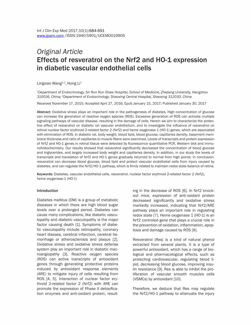
Int J Clin Exp Med 2017;10(1):684-691www.ijcem.com /ISSN:1940-5901/IJCEM0019905
Original Article
Effects of resveratrol on the Nrf2 and HO-1 expression in diabetic vascular endothelial cells
Lingxiao Wang1,2, Hong Li1
1Department of Endocrinology, Sir Run Run Shaw Hospital, School of Medicine, Zhejiang University, Hangzhou 310016, China; 2Department of Endocrinology, Shaoxing Central Hospital, Shaoxing 312030, China
Received November 17, 2015; Accepted April 27, 2016; Epub January 15, 2017; Published January 30, 2017
Abstract: Oxidative stress plays an important role in the pathogenesis of diabetes. High concentration of glucose can increase the generation of reactive oxygen species (ROS). Excessive generation of ROS can activate multiple signaling pathways of vascular disease, resulting in the damage of cells. Herein we aim to characterize the protec-tive effect of resveratrol on diabetic rat vascular endothelium, and to investigate the influence of resveratrol on retinal nuclear factor erythroid 2-related factor 2 (Nrf2) and heme oxygenase 1 (HO-1) genes, which are associated with elimination of ROS. In diabetic rat, body weight, blood lipid, blood glucose, capillaries density, basement mem-brane thickness and ratio of capillaries to muscle fibers were examined. Levels of transcripts and protein expression of Nrf2 and HO-1 genes in retinal tissue were detected by fluorescence quantitative PCR, Western blot and immu-nohistochemistry. Our results showed that resveratrol significantly decreased the concentration of blood glucose and triglycerides, and largely increased body weight and capillaries density. In addition, in our study the levels of transcripts and translation of Nrf2 and HO-1 genes gradually returned to normal from high points. In conclusion, resveratrol can decrease blood glucose, blood lipid and protect vascular endothelial cells from injury caused by diabetes, and can regulate the Nrf2/HO-1 pathway, which is firmly related to maintain redox state balance of retina.
Keywords: Diabetes, vascular endothelial cells, resveratrol, nuclear factor erythroid 2-related factor 2 (Nrf2), heme oxygenase-1 (HO-1)
Introduction
Diabetes mellitus (DM) is a group of metabolic diseases in which there are high blood sugar levels over a prolonged period. Diabetes can cause many complications, like diabetic vascu-lopathy and diabetic vasculopathy is the major factor causing death [1]. Symptoms of diabe- tic vasculopathy include retinopathy, coronary heart disease, cerebral infarction, cerebral he- morrhage or atherosclerosis and plaque [2]. Oxidative stress and oxidative stress defense system play an important role in diabetic mac-roangiopathy [3]. Reactive oxygen species (ROS) can active transcripts of antioxidant genes through generating protective proteins induced by antioxidant response elements (ARE) to mitigate injury of cells resulting from ROS [4, 5]. Interaction of nuclear factor ery-throid 2-related factor 2 (Nrf2) with ARE can promote the expression of Phase II detoxifica-tion enzymes and anti-oxidant protein, result-
ing in the decrease of ROS [6]. In Nrf2 knock- out mice, expression of anti-oxidant protein decreased significantly and oxidative stress markedly increased, indicating that Nrf2/ARE pathway plays an important role in regulating redox state [7]. Heme oxygenase 1 (HO-1) is an Nrf2 controled gene that plays a crucial role in the prevention of oxidation, inflammation, apop-tosis and damage caused by ROS [8].
Resveratrol (Res) is a kind of natural phenol extracted from several plants. It is a type of powerful antioxidant, which has a range of bio-logical and pharmacological effects, such as protecting cardiovascular, regulating blood li- pid, decreasing blood glucose, improving insu-lin resistance [9]. Res is able to inhibit the pro-liferation of vascular smooth muscles cells (VSMCs) by antioxidant [10].
Therefore, we deduce that Res may regulate the Nrf2/HO-1 pathway to attenuate the injury

Resveratrol on diabetic rats
685 Int J Clin Exp Med 2017;10(1):684-691
of diabetic vascular. Thus, in streptozotocin (STZ) induced type-I diabetes rats we charac-terized the effects of Res on the growth of dia-betes endothelial cells and the expression of retinal Nrf2/HO-1 genes, aiming to develop novel strategies of prevention and treatment of vascular complications of DM.
Materials and methods
Animals and treatments
A total of 60 healthy adult male and female Wistar rats were provided by the Experimental Animal Center of Luzhou Medical College. They were kept in standard conditions with free access to food and water. They were randomly divided into three groups: normal control group (n = 20), STZ group (n = 20) and Res group (n = 20). For the establishment of DM model, rats in STZ group were treated with a single intra-peritoneal injection of STZ at a dose of 55 mg/kg body weight. Blood glucose level was mea-sured at 72 h after STZ treatment and rats with blood glucose level > 16.7 mM were consid-ered as DM rats. Rats in Res group were treat-ed with STZ as rats of STZ groups and were then given Res at doses of 10 mg/kg, 20 mg/kg and 40 mg/kg body weight per day with gavages for 4 weeks. After 4 weeks, body weight and blood glucose of all rats were mea-sured. Then, all of the rats were sacrificed. Blood was collected by cardiac puncture and processed for biochemical estimations such as glucose, TC (total cholesterol), TG (total triglyc-eride), LDL-C (low density lipoprotein cholester-in) and HDL-C (high density lipoprotein choles-terol). Retinal tissues were collected for immu-nohistochemical analysis. Skeletal muscle tis-sues were collected from the left lower limb. All animal experiments were conducted according to the ethical guidelines of Zhejiang University.
Characterization of the neovascularization of rats
Skeletal muscle tissue was fixed in paraformal-dehyde, embedded in paraffin and cut into 4 μm tissue sections. Then the tissues were stained with haematoxylin and eosin. We ran-domly selected six microscope fields from each slice. The number of capillaries was counted at 200 × magnification with double-blind method. The thickness of capillary basement membrane was measured. The ratio of the number of capil-
laries to that of muscle fiber was calculated. Quantitative analysis of above measurements was performed using Image-Pro Plus 6.0 soft-ware (Media Cybernetics, Rockville, MD, USA).
Quantitative real time PCR (qRT-PCR)
Total RNA was isolated from retinal tissue fol-lowing the procedure described previously [11]. Then RNAs were transcribed into cDNA accord-ing to the instructions provided by the Prime- Script Kit (TAKARA BIOTECHNOLOGY (DALIAN) CO., LTD. Dalian, Liaoning, China). Then, qRT-PCR was performed. The forward and reverse primers for Nrf2 and HO-1 were synthesized at Shanghai Sangon Biological Engineering Tech- nology & Services, Shanghai, China. The for-ward and reverse primer sequences for Nrf2 were as follows: Nrf2 forward 5’CAAATCC- CACCTTGAACACA 3’, and, Nrf2 reverse 5’CG- ACTGACTAATGGCAGCAG 3’. The forward and reverse primers for HO-1 were HO-1 forward 5’CAGAGTTTCTTCGCCAGAGG 3’, and HO-1 re- verse 5’TGAGTGTGAGGACCCATCG 3’. PCR re- action were carried out in a final volume of 25 μl containing: 12.5 μl of SYBR Premix Ex Taq II (Qiagen, Hilden, Germany), 1.0 μl forward prim-er, 1.0 μl reverse primer, 2 μl template (about 120 ng) and 8.5 μl dH2O. The PCR procedure were initial denaturation step at 95°C for 30 s, followed by 40 cycles of 95°C for 5 s and 60°C for 30 s. GAPDH was used as an internal con-trol. The 2-ΔΔCt method was used to calculate the relative expression levels of Nrf2 and HO-1.
Western blot
Total proteins were extracted and separated by 12% SDS-PAGE. Then proteins were transferred onto nitrocellulose membrane. After blocking with non-fat milk, the membrane was incubat-ed with the primary antibodies of rabbit anti- rat polyclonal antibody anti-Nrf2 (Beijing Bio- synthesis Biotechnology, Co., LTD., Beijing, China) and rabbit anti-rat polyclonal antibody anti-HO-1 (Beijing Biosynthesis Biotechnology) at 4°C overnight. After washing, the membrane was then incubated with secondary antibody of goat anti-rabbit HRP conjugated antibody (Beijing Biosynthesis Biotechnology, Co., LTD., Beijing, China) at room temperature for 1 h. Finally, the membrane was developed by enhanced chemiluminescence plus reagent. The developed film was scanned using the AlphaImager gel imaging systems (AlphaIma-

Resveratrol on diabetic rats
686 Int J Clin Exp Med 2017;10(1):684-691
ger, Santa Clara, California, USA). And the Western blot images were analyzed using Quantity One software (Bio-Rad Laboratories, Hercules, CA, USA). β-actin was used as an internal control. The relative absorbance ratios of Nrf2 to β-actin and Ho-1 to β-actin were defined as the relative values of Nrf2 and HO-1, respectively.
Immunohistochemistry
Immunohistochemistry of Nrf2 and HO-1 was performed using the streptavidin-biotin com-plex (SABC) method as described previously [12]. Briefly, retinal tissues were fixed in formal-
dehyde and embedded in paraffin. Sections were dewaxed, and rehydrated in graded alco-hols. Then sections were incubated with 0.3% hydrogen peroxide to inactivate endogenous peroxidase activity. Antigen retrieval was achieved by incubating with sodium citrate (PH 6.0). After blocking, sections were incubated with the primary antibodies of rabbit anti-rat polyclonal antibody anti-Nrf2 (Beijing Biosyn- thesis Biotechnology, Co., LTD., Beijing, China) and rabbit anti-ratpolyclonal antibody anti- HO-1 (Beijing Biosynthesis Biotechnology) at 4°C overnight. Then, sections were incubated with secondary antibody of goat anti-rabbit (Beijing Biosynthesis Biotechnology, Co., LTD., Beijing, China) at room temperature for 40 min. After incubating with SABC at room tempera-ture for 40 min, sections were developed with DAB chromogenic reagent. Finally, sections were counterstained with haematoxylin. After mounting with neutral resin, sections were observed under microscopy (Leica DM4000B, Leica Microsystems, Wetzlar, Germany). Image-Pro Plus (Media Cybernetics, Inc., Rockville, MD, USA) was used to analyze the expression level of Nrf2 and HO-1.
Statistical analysis
Data were expressed as mean ± standard devi-ation. SPSS 17.0 was used for statistical analy-sis. One-way analysis of variance was per-formed. SNK test was conducted to compare differences between groups. Non-parame- trictest was applied to data that did not meet the homogeneity of variance. Results were con-sidered statistically significant when the P value was less than 0.05.
Results
Positive effects of Res on blood glucose, lipid and body weight in diabetes rats
To determine the effects of Res on blood glu-cose, lipid and body weight, daily treatment of Res on rats was performed. At day 1, blood glu-cose level was up to 29.01 mM in STZ group. And blood glucose level was 25.61 mM, 26.72 mM and 24.78 mM in Res group at doses of 10 mg/kg, 20 mg/kg and 40 mg/kg, respec-tively (Figure 1A). It indicated that we were suc-cessful in constructing DM rat models. After treatment with Res for 4 weeks, blood glucose level in Res group at three doses all decreased
Figure 1. Effects of Res treatment on blood glucose (A), blood lipid (B) and body weight (C) in diabetes rats. Control group: rats without STZ or Res treat-ment; STZ group: rats with STZ treatment; Res group: STZ group with Res treatment at doses of 10 mg/kg, 20 mg/kg and 40 mg/kg, respectively. The levels of blood glucose, blood lipid and body weight were measured at 1 day and 4 weeks of treatment. Com-pared with STZ group, *P < 0.05.

Resveratrol on diabetic rats
687 Int J Clin Exp Med 2017;10(1):684-691
to levels below 16 mM. Compared with STZ group, the decrease was significant (P < 0.05).
To determine the influence of Res on the capil-laries of DM rats, the characterization of capil-
Figure 2. Effects of Res treatment on density of capillaries (A), the repre-sentative pictures of H&E staining (Magnification 200 ×). (B) The ratio of capillaries to muscle fibers (C) and the basement membrane thickness (D) in diabetes rats examine by microscope. At 4 weeks of treatment, these mea-sures were performed. Compared with STZ group, *P < 0.05.
However, blood glucose level in Res group was still higher than that in normal control group.
Meanwhile, parameters, like blood lipid, TC and LDL, also presented the same trend with blood glucose (Figure 1B), especially after Res treat-ment of 20 and 40 mg/kg body weight per day. It was noteworthy that in STZ group, TG was up to 4.38 mM, which were 7 folds more than that of normal control group. But after 40 mg/kg Res treat-ment, the value of TG decre- ased to half of STZ group. In addition, quantification of blood lipid indicated that Res could decrease TC, TG and LDL, and increase HDL signifi-cantly (P < 0.05) compared with normal control group. In addition, the levels of TC, LDL and HDL in DM rats were simi-lar to those of normal control group after 40 mg/kg Res treatment.
As for body weight, even though there was no obvious change in body weight with 40 mg/kg Res treatment for 1 day, a significant increase (P < 0.05) appeared after 10 mg/kg Res treatment in dia-betes rats for 4 weeks com-pared with Res untreated dia-betes rats (Figure 1C). But there was not obvious differ-ence in Res group at doses of 10 mg/kg, 20 mg/kg and 40 mg/kg, and the body weight of Res treated rats was still lighter than that of normal control group. These results indicate that Res is able to improve the weight of DM rats significantly.
Influence of Res on capillar-ies of diabetes rats

Resveratrol on diabetic rats
688 Int J Clin Exp Med 2017;10(1):684-691
laries was analyzed. The representative pic-tures of H&E staining were shown in Figure 2A. Although there was no significant difference detected in diameter of capillaries in each group (data not shown), in STZ group both the number of capillaries and the ratio of capillar-ies to muscle fibers were only a quarter of that in normal control group. But when DM rats were treated with 10 mg/kg Res for 4 weeks, both of the number of capillaries and the ratio mentioned above increased by more than 2 folds (Figure 2B and 2C), which were almost close to the normal level. In addition, the thick-ness of capillaries basement membrane of STZ group had increased to more than twofold of normal control group. Compared with SZT group, thickness of basement membrane in
Res group dropped significantly (P < 0.05) to a lower position similar to normal level even with only 10 mg/kg body weight per day for 4 weeks (Figure 2D). These results indicate that Res can increase capillaries density and the ratio of capillaries to muscle fibers, and decrease the thickness of capillaries basement membrane.
Effects of Res on mRNA and protein levels of retinal Nrf2 and HO-1 genes
To determine the effect of Res on the mRNA and protein levels of retinal Nrf2 and HO-1, qRT-PCR and Western Blot were performed, respectively. As shown in Figures 3 and 4, the mRNA and protein levels of retinal Nrf2 and HO-1 were obviously higher in STZ group than
Figure 3. Effects of Res treatment on the levels of retinal Nrf2 and HO-1 in diabetes rats. Levels of retinal Nrf2 and HO-1 protein were detected with Western blot. β-actin was used as an internal control. A. Protein level of Nrf2. B. Protein level of HO-1. Using GAPDH as internal control, the mRNA levels of Nrf2 and HO-1 genes were tested through qRT-PCR. Total RNA was extracted from retinal tissue in diabetes rats after 4 weeks of treatment. C. mRNA level of Nrf2. D. mRNA level of HO-1. Compared with STZ group, *P < 0.05, **P < 0.01.

Resveratrol on diabetic rats
689 Int J Clin Exp Med 2017;10(1):684-691
those in normal control group. However, Res treatment on STZ group significantly decreased the mRNA and protein levels of Nrf2 and HO-1 in retinal tissue (P < 0.05) (Figures 3 and 4). In addition, at the dose of 40 mg/kg Res, the mRNA and protein levels of Nrf2 and HO-1 in Res group were similar to those in control group. These results indicate that Res can inhibit the expression of retinal Nrf-2 gene and HO-1 gene.
Discussion
The novel findings of the present study are: 1) parameters of diabetes rats including body weight, capillaries density, blood lipid and glu-cose came back to the levels similar to those of control rats after feeding them with Res, 2) Res downregulated expression of Nrf2 and HO-1 in retinal tissues of diabetes rats. Thus, we deduce that it is the Res-induced deduce of blood glucose and lipid that leads to the down-regulation of Nrf2, consequently followed by decrease of HO-1 in the retinal.
Diabetes is cogently associated with microvas-cular and macrovascular complications: cardi-
opathy, neuropathy, nephropathy, retinopathy, etc. [13-15]. High glucose promotes endothe- lial cell to die, resulting in damaging vascular endothelial [16]. Pathogenesis of microvascu-lar is associated with microcirculatory distur-bance, inflammation and abnormal oxygen-ation, retinal vascular dilation, increasing of advanced glycation end products (AGEs), changing of structure of blood vessels, thicken-ing of blood vessels basement membrane, and narrowing of capillary luminal [17-19].
This study showed increasing of body weight and decreasing of glucose in diabetes rats with the help of Res, suggesting that Res has posi-tive effect on diabetes. Further investigation on blood lipid showed that the concentrations of TC, TG and LDL decreased and HDL increased significantly. Meanwhile, there was no signifi-cant change in diameter of capillaries, while the density of capillaries and the ratio of capillaries to muscle fibers increased, but the basement membrane thickness decreased significantly. This indicates that Res has the function of vas-cular protection.
Figure 4. Effects of Res treatment on the protein levels of retinal Nrf2 and HO-1 in diabetes rats tested by immuno-histochemistry. At 4 weeks of treatment, immunohistochemistry was performed to detect Nrf2 and HO-1. A. Repre-sentative immunohistochemical staining results (Magnification: 40 ×). B. Relative level of Nrf2 protein. Compared with STZ group, *P < 0.05, **P < 0.01. C. Relative level of HO-1 protein. Compared with STZ group, *P < 0.05, **P < 0.01.

Resveratrol on diabetic rats
690 Int J Clin Exp Med 2017;10(1):684-691
In endothelial cell, high glucose can lead to peroxide, oxygen free radicals increasing, lipid peroxidation, macromolecules depositing on a base film, damage of tight junctions, increasing permeability, decreases of myocardial capillary and basement membrane thickening by incre- asing production of cytokines [20-23]. Nrf2 and HO-1 play an important role in anti-oxida-tive stress [24]. In diabetes rat, production of ROS has an increasing trend, following the increase of Nrf2 expression to resist the stimu-lation of ROS [25, 26]. In Nrf2 knockout mice, the expression of anti-oxidative genes decre- ased evidently, but oxidative-stress damage increased significantly, indicating that Nrf2/ARE pathway plays a key role in the balance of redox state [27]. Activation of GSK-3β/Nrf2 pathway can induce the upregulation of antioxi-dant protein [28].
It has been reported that Nrf2 expressed in many kinds of cells, and the production of ROS in Nrf2-/- diabetes rats is higher than in Nrf2+/+ [29]. Moreover, the expression of HO-1 is in type 2 diabetes mellitus, and it is positively associated with macroangiopathy [30]. Nrf2 can induce the transcripts of HO-1 gene to pro-duce antioxidant effect [31].
Our study characterized the effect of Res on the mRNA and protein levels of Nrf2 and HO-1, and the results showed that diabetes rats expressed less Nrf2 and HO-1 protein with the help of Res than control group. The previous study demonstrated that Res upregulated Nrf2 expression followed by up-regulation of HO-1 [32]. However, herein when the blood glucose decreased, levels of the Nrf2 and HO-1 expres-sion also came back to normal gradually. It sug-gests that Res can deduce the oxidation of dia-betes rats, following the decreasing of Nrf2 and HO-1 expression, and Nrf2/HO-1 pathway is also responsible for maintaining glucose ba- lance.
In conclusion, our study presents that Res is able to decrease glucose of diabetes rats and increase the number of capillaries, and reduce Nrf2/HO-1 expression, suggesting that Res may play a key role in the decreased expression of Nrf2 and HO-1, which provides a novel app- roach for prevention and treatment of dia- betes.
Acknowledgements
The authors wish to acknowledge Shou-Jie Wang (Department of Plastic Surgery, the First
Affiliated Hospital, College of Medicine, Zhe- jiang University) for his valuable help.
Disclosure of conflict of interest
None.
Address correspondence to: Hong Li, Department of Endocrinology, Sir Run Run Shaw Hospital, Sch- ool of Medicine, Zhejiang University, 3 East Qing- chun Road, Hangzhou 310016, China. Tel: +86- 571-86090073; Fax: +86-571-86044822; E-mail: [email protected]
References
[1] Sayin N, Kara N and Pekel G. Ocular complica-tions of diabetes mellitus. World J Diabetes 2015; 6: 92-108.
[2] Kowluru RA. Diabetic ret inopathy: mitochon-drial dysfunction and retinal capillary cell death. Antioxid Redox Signal 2005; 7: 1581-7.
[3] Hamed EA, Zakary MM, Abdelal RM and Abdel Moneim EM. Vasculopathy in type 2 diabetes mellitus: role of specific angiogenic modula-tors. J Physiol Biochem 2011; 67: 339-49.
[4] Wang X and Hai CX. ROS acts as a double-edged sword in the pathogenesis of type 2 dia-betes mellitus: is Nrf2 a potential target for the treatment? Mini Rev Med Chem 2011; 11: 1082-92.
[5] Nishikawa T and Araki E. Impact of mitochon-drial ROS production in the pathogenesis of diabetes mellitus and its complications. An- tioxid Redox Signal 2007; 9: 343-53.
[6] Uruno A, Yagishita Y and Yamamoto M. The Keap1-Nrf2 system and diabetes mellitus. Arch Biochem Biophys 2015; 566: 76-84.
[7] Cheng X, Chapple SJ, Patel B, Puszyk W, Sug- den D, Yin X, Mayr M, Siow RC and Mann GE. Gestational diabetes mellitus impairs Nrf2-mediated adaptive antioxidant defenses and redox signaling in fetal endothelial cells in ute-ro. Diabetes 2013; 62: 4088-97.
[8] Qiu C, Hevner K, Enquobahrie DA and Williams MA. Maternal serum heme-oxygenase-1 (HO-1) concentrations in early pregnancy and subse-quent risk of gestational diabetes mellitus. PLoS One 2012; 7: e48060.
[9] Shen LL, Wang XM and He BL. Advance of res-veratrol in treating diabetes mellitus. Zhongguo Zhong Xi Yi Jie He Za Zhi 2013; 33: 279-81.
[10] Guo R, Li W, Liu B, Li S, Zhang B and Xu Y. Resveratrol protects vascular smooth muscle cells against high glucose-induced oxidative stress and cell proliferation in vitro. Med Sci Monit Basic Res 2014; 20: 82-92.
[11] Siljo A, Bhat AI and Biju CN. Detection of Cardamom mosaic virus and Banana bract

Resveratrol on diabetic rats
691 Int J Clin Exp Med 2017;10(1):684-691
mosaic virus in cardamom using SYBR Green based reverse transcripts-quantitative PCR. Virusdisease 2014; 25: 137-41.
[12] Bolati D, Shimizu H, Higashiyama Y, Nishijima F and Niwa T. Indoxyl sulfate induces epitheli-al-to-mesenchymal transition in rat kidneys and human proximal tubular cells. Am J Nephrol 2011; 34: 318-23.
[13] Kowluru RA and Chan PS. Oxidative stress and diabetic retinopathy. Exp Diabetes Res 2007; 2007: 43603.
[14] Demirtas L, Degirmenci H, Akbas EM, Ozcicek A, Timuroglu A, Gurel A and Ozcicek F. Association of hematological indicies with dia-betes, impaired glucose regulation and micro-vascularcomplications of diabetes. Int J Clin Exp Med 2015; 8: 11420-7.
[15] Klen J, Goricar K, Janez A and Dolzan V. NLRP3 inflammasome polymorphism and macrovas-cular complications in type 2 diabetes pa-tients. J Diabetes Res 2015; 2015: 616747.
[16] Tabak O, Gelisgen R, Erman H, Erdenen F, Muderrisoglu C, Aral H and Uzun H. Oxidative lipid, protein, and DNA damage as oxidative stress markers in vascular complications ofdi-abetes mellitus. Clin Invest Med 2011; 34: E163-71.
[17] Singh R, Barsen A, Mori T and Beilin L. Advanced glycation end-products: a review. Diabetologia 2001; 44: 129-46.
[18] Penmetsa GS, Baddam S, Manyam R and Dwarakanath CD. Comparison of the number of gingival blood vessels between type 2 diabe-tes mellitus and chronic periodontitis patients: An immunohistological study. J Indian Soc Periodontol 2015; 19: 164-8.
[19] Hayden MR, Sowers JR and Tyagi SC. The cen-tral role of vascular extracellular matrix and basement membrane remodeling in metabolic syndrome and type 2 diabetes: the matrix pre-loaded. Cardiovasc Diabetol 2005; 4: 9.
[20] Qi Nan W, Ling Z and Bing C. The influence of the telomere-telomerase system on diabetes mellitus and its vascular complications. Expert Opin Ther Targets 2015; 19: 849-64.
[21] Song H, Wu F, Zhang Y, Zhang Y, Wang F, Jiang M, Wang Z, Zhang M, Li S, Yang L, Wang XL, Cui T and Tang D. Irisin promotes human umbilical vein endothelial cell proliferation through the ERK signaling pathway and partly suppresses high glucose-induced apoptosis. PLoS One 2014; 9: e110273.
[22] Terao Y, Satomi-Kobayashi S, Hirata K and Rikitake Y. Involvement of Rho-associated pro-tein kinase (ROCK) and bone morphogenetic protein-binding endothelial cell precursor-de-rived regulator (BMPER) in high glucose-in-creased alkaline phosphatase expression and activity in human coronary artery smooth mus-cle cells. Cardiovasc Diabetol 2015; 14: 104.
[23] Zhong Q, Mishra M and Kowluru RA. Trans- cription factor Nrf2-mediated antioxidant de-fense system in the development of diabetic retinopathy. Invest Ophthalmol Vis Sci 2013; 54: 3941-8.
[24] Fan J, Xu G, Jiang T and Qin Y. Pharmacologic induction of heme oxygenase-1 plays a protec-tive role in diabetic retinopathy in rats. Invest Ophthalmol Vis Sci 2012; 53: 6541-56.
[25] Mishra M, Zhong Q and Kowluru RA. Epigenetic modifications of Nrf2-mediated glutamate-cys-teine ligase: implications for the development of diabetic retinopathy and the metabolic memory phenomenon associated with its con-tinued progression. Free Radic Biol Med 2014; 75: 129-39.
[26] Xu Z, Wei Y, Gong J, Cho H, Park JK, Sung ER, Huang H, Wu L, Eberhart C, Handa JT, Du Y, Kern TS, Thimmulappa R, Barber AJ, Biswal S and Duh EJ. NRF2 plays a protective role in dia-betic retinopathy in mice. Diabetologia 2014; 57: 204-13.
[27] Ramprasath T and Selvam GS. Potential im-pact of genetic variants in Nrf2 regulated anti-oxidant genes and risk prediction of diabetes and associated cardiac complications. Curr Med Chem 2013; 20: 4680-93.
[28] Bitar MS. The GSK-3β/Fyn/Nrf2 pathway in fi-broblasts and wounds of type 2 diabetes: On the road to an evidence-based therapy of non-healing wounds. Adipocyte 2012; 1: 161-3.
[29] Peake BF, Nicholson CK, Lambert JP, Hood RL, Amin H, Amin S and Calvert JW. Hydrogen sul-fide preconditions the db/db diabetic mouse heart against ischemia-reperfusion injury by activating Nrf2 signaling in an Erk-dependent manner. Am J Physiol Heart Circ Physiol 2013; 304: H1215-1224.
[30] Mishra M, Zhong Q and Kowluru RA. Epigenetic modifications of Keap1 regulate its interaction with the protective factor Nrf2 in the develop-ment of diabetic retinopathy. Invest Ophthalmol Vis Sci 2014; 55: 7256-65.
[31] Uruno A, Furusawa Y, Yagishita Y, Fukutomi T, Muramatsu H, Negishi T, Sugawara A, Kensler TW and Yamamoto M. The Keap1-Nrf2 system prevents onset of diabetes mellitus. Mol Cell Biol 2013; 33: 2996-3010.
[32] Bolati D, Shimizu H, Yisireyili M, Nishijima F and Niwa T. Indoxyl sulfate, a uremic toxin, downregulates renal expression of Nrf2 thr- ough activation of NF-κB. BMC Nephrology 2013; 14: 56.