Embed Size (px)
Citation preview
PAPILLARY CYSTADENOCARCINOMA O F PANCREAS
CASE REPORT, WITH NOTES ON CLASSIFICATION OF MALIGNAN,T CYSTIU TUNORS OF PANCREAS
L. LIGHTENSTEIN, M.D.
(From the Laborutories of The Mount Sinai Hospital, New Pork)
The case here reported is interesting both because of, the rarity of the tumo~r and because the complete clinical record over a period of six years may throw possible light upon the pathogenesis of the neo- plasm. The latter was an encapsulated cystic tumor of the tail of the pancreas, the size of a child’s head, which, after an interval of about five years, had, in part, undergone carcinomatous change, invading the capsule and metastasizing to the peritoneum, omentum, and liver.
Large papillary cystadenooarcinomata of the pancreas of this type are admittedly rare (Kaufmann, 1; Ewing, 2), especially cases proved by adequate necropsy or biopsy data. In fact, there appears to be no authentic case report in the American literature. Malignant cystic tumors of the pancreas of all types have, of course, been described with increasing frequency within the past twenty years. Only a very small proportion of these, however, are cystsdenocaroinomata in the specific sense of the author’s case, as will be seen later from a brief survey of the literature.
REPORT OF CASE?
The patient, a female nurse, a Russian, aged forty-four, single, Arst came under observation a t the Mount Sinai Hospital in December 1928 (three years before necropsy), with a complaint of intermittent edema of the ankles of six years’ duration, pain in the left thigh, and a tendency to fatigue. I n 1919, a hemorrhoidectomy had been performed. I n November 1925, approximately six years before death, the patient had been observed for a period of two weeks a t another hospital in New York City, where an exploratory laparotomy was performed for suspected acute intestinal obstruction. At operation, marked distention of all the intestine, including the sigmoid colon, was found, as well as marked edema of the mesosigmoid and an appreciable quantity of free mrous fluid the abdomen. The uterus showed a large intramural flbromyoma. The adnexa and appendix appeared normal. No intestinal obstruction was found, and the cause of the distention and peritoneal effusion was not clear. The patient was discharged improved. Shortly afterwards, her “ spleen was felt to be enlarged.”
Physical examination a t Mt. Sinai Hospital revealed a palpable mass with a hard, round edge, extending below the left costal margin, interpreted as a moderately large. spleen. There were, in addition, a palpable right kidney, a uterine fibroid, and pretibial edema. The blood picture was normal. The etiology of the alleged splenomegaly oould not be determined, and the patient was discharged after one month’s observation. The diagnosis was splenomegaly of undetermined etiology, possibly sp&uc vein thrombosis.
1 This work was aided by a grant of the Eruaiiuel Libman Fellowship Fund. Accepted for publication in June 1933.
ZAcknowledgment is made to Dr. I3. S. Oppenheimer for permission to use the ohical record of this case.
642
PAPILLARY CYSTADENOCABCIKOBIA OF PANCREAS 543
In November 1931, three years later, the patient was readmitted, complaining of edema of the ankles and persistence of left-sided pain. She had otherwise enjoyed good health in the interval. The left upper quadrant mass had grown somewhat larger. An anemia had developed, which was treated with Blaud’s pills. Three weeks before ad- mission, anorexia and malaise set in, and two weeks later, progressive enlargement of the abdomen was observed. The abdomen was markedly distended, with shifting dullness and a fluid wave. An irregular mass was palpable in the left upper quadrant. Roentgen examination after induction of pneumoperitoneum showed the superior border of the liver to be Armly adherent to the right leaf of the diaphragm, while a large mass was outlined in the left abdomen, interpreted as probably the spleen.
The blood count showed hemoglobin 92 per cent; red blood cells 4,470,000; white blood cells 14,000 with 78 per cent polynuclear leukocytes and platelets 160,000. Blood pressure was 140 mm. Hg systolic and 80 diastolic. The Wassermann test was negative.
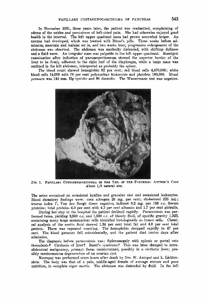
h o . 1. PAPILLARY CYSTADENOCARCINOMA OF THE TAIL OF THE PANCREAS: AUTHOR’S CASE About 1/3 natural eize.
The urine contained an oecasional hyaline and granular cast and occasional leukocytes. Blood chemistry Andings were: urea nitrogen 20 mg. per cent; cholesterol 225 mg.; icterus index 7; Van den Rergh dirert negative, indirect 0.2 mg. per 100 C.C. Serum proteins: total proteins 6.0 per cent with 4.3 per cent albumin and 1.7 per cent globulin.
Paracentesis was per- formed twice, yielding 3,500 C.C. ant1 1,600 ex. of hloody fluid, of specific gravity 1.023, containing many large mononucirar cells identified histologically as tumor cells. Chemi- cal analysis of the aseitic fluid showed 1.34 per cent total f a t and 4.8 per cent total protein. The hemoglobin dropped rapidly to 47 per cent. The blood pressure fell coincidentally, and the patient died twelve days after admission.
The diagnosis before paracentesis was : Splenomegaly with splenic or portal vein thrombosis? Cirrhosis of liver? Hanti’s qyndromr.9 This was later changed to intra- abdominal malignancy, primary forus undetermined, possibly in a cirrhotic liver; pos- sibly carcinomatous degeneration of an ovarian cyst.
Necropsy was performed seven hours after death by Drs. W. Antopol and L. Lichten- stein. The body was that of a pale, middle-aged female of average stature and poor nutrition, in complete rigor niortis. The abdomen was distended by fluid. I n the left
During her stay in the hospital the patient declined rapidly.
There was repeated vomiting.

544 L. LIOHTENSTEIN
upper quadrant there protruded a prominent, Arm, rounded mass, the size of a child’s head. There was slight pitting edema of the lower extremities. There was no jaundice or superficial lymphadenopathy.
Abdominal C a d ? / : The panniculus adiposus was poorly developed. The abdomen contained several liters of hemorrhagic fluid. The peritoneum was studded by firm, grayish, elevated nodules, 2-5 mm. in diameter, some of which were umbilicated. The intestinal loops were somewhat edematous but not distended. The liver WIM enlarged and likewise showed grayish carcinomatous nodules upon its surface. The diaphragm was elevated.
The tumor mass in the left upper quadrant was independent of the spleen, which WM normal in size, and was situated just inferior to it, contiguous to the lower pole (Fig. 1). It was tense, as though cystic, and surrounded by a thick, grayish, flbrous capsule. The tail of the pancreas was thinned out and merged with the tumor capsule anteriorly and posteriorly. The splenic artery and vein, on dissection, were seen to course over the surface of the cyst without being compressed. The pancreatic duct of Wirsung, of
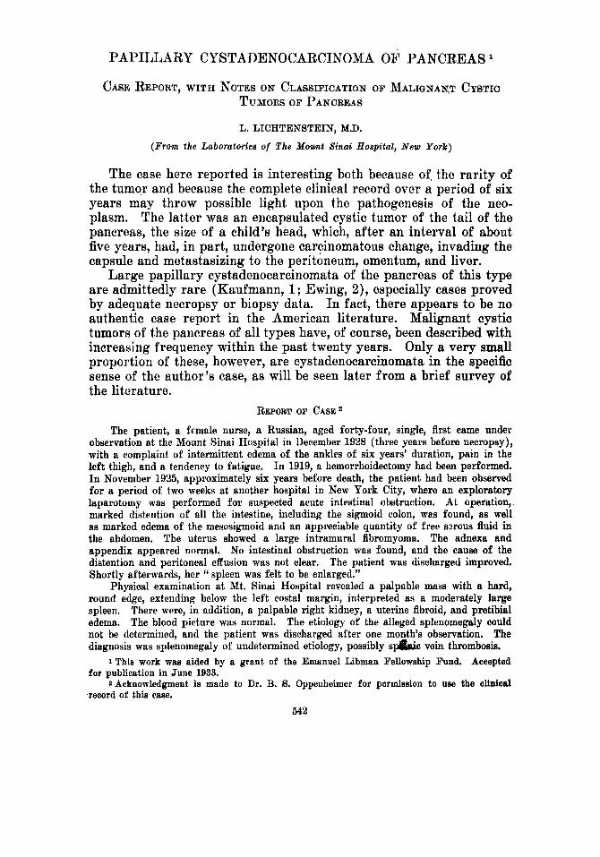
FIO. 2. BENION PORTION OF TVYOR LINED BY REGULAR COLUMNAR EPITHELIUM; CALCIUM. CONCREYENTS IN FIBROUS SEPTA
normal diameter, was readily traced up to the poidt on the capsule where it led into the cyst. The cyst, 011 section, was filled with about 500 C.C. of thick, grumous, choeolaty material, free of concretions and containing the enzyme diastase but not trypsin. The interior of the cyst was incompletely divided by ti thick, fibrous ridge into two oom- municating compartments, each of which was in turn loculated. Its walls were lined in places by a dirty, grayish-yellowish or brown necrotic material, in other places by small papillary branching growths attached by a broad base. The tumor tissue on close in- spection was seen a t one point to penetrate the capsule.
LuBgs: Both lungs showed apical fibrous adhesions and patches of atelectasis in the lower lobes. On section, the lungs were mottled dark red and exuded frothy fluid. There was a healed tuberculous process at the right apex with small, circumscribed, partially caseated nodules and grayish flbrous strands. The apical branch of the right upper lobe bronchus was dull, reddened, and showed m u s e dilatation of moderate extent. Pulmonary arteries and veins were without change.
The upper lobes were hypercrepitant.
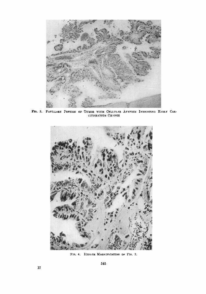
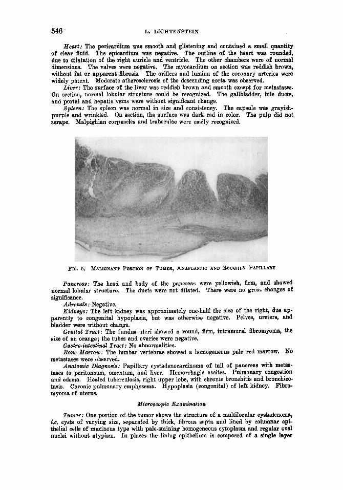
FIQ. 3. PAPILLABY PORTION OF TUMOR WITH CELLULAR ATYPISM INDICATINQ EARLY CAB- CIP*’OMATOUR CH-4NQE
27
FIG. 4. HIGIIER MAQNIFICATION OF FIG. 3.
545
546 L. LIOHTENSTEIN
Hemt: The pericardinm was smooth and glistening and contained a small quantity of clear fluid. The epicardium was negative. The outline of the heart was rounded, due to dilatation of the right auricle and ventricle. The other chambers were of normal dimensions. The valves were negative. The myocardium on section was reddish brown, without fat or apparent fibrosis. The orifices and lumina of the coronary arteriea were widely patent. Moderate atherosclerosis of the descending aorta was observed.
Liver: The surfaoe of the liver was reddish brown and smooth except for metastases. On section, normal lobular structure could be recognized. The gallbladder, bile ducts, and portal and hepatic veins were without significant change.
Spleen: The spleen was normal in size and consistency. The capsule was grayish- purple and wrinkled. On section, the surface was dark red in color. The pulp did not scrape. Malpighian corpuscles and trabeciilae were easily recognized.
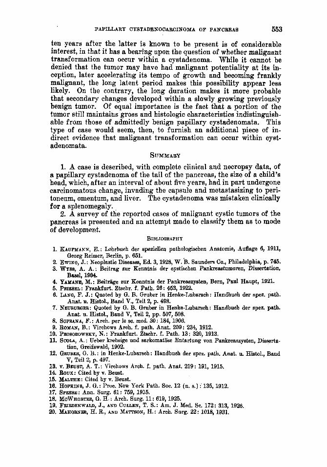
FIo. 5. MALIQNANT PORTION OF TUMOR, ANAPLASTIO AND RoUQHLY PAPILLABY
Palzcreas: The head and body of the pancreas were yellowish, flim, and showed There were no gross changes of normal lobular structure.
significance. The ducts were not dilated,
Adrenals: Negative. Kidmys: The left kidney was approximately one-half the size of the right, due ap-
parently to congenital hypoplasia, but was otherwise negative. Pelves, ureters, and bladder were without change.
GmUal Truct: The fundus uteri showed a round, firm, intramural fibromyoma, the size of an orange; the tubes and ovaries were negative.
Gastio-intestinal Truct : No abnormalities. Bone Yarrow: The lumbar vertebrae showed a homogeneous pale red marrow. No
metastases were observed. Allatomic Diugm8is: Papillary cystadenocarcinoma of tail of pancreas with metaa-
tases to peritoneum, omenturn, and liver. Hemorrhagic ascites. Pulmonary congestion and edema. Healed tuberculosis, right upper lobe, with chronic bronchitis and bronchiec- tasis. Chronic pulmonary emphysema. Hypoplasia (congenital) of left kidney. Fibro- myoma of uterus.
Nicroscopic Examination
Tumor: One portion of the tumor shows the structure of a multilocular cystudenoma, i.e. cysts of varying size, separated by thick, fibrous septa and lined by columnar epi- thelial cells of mucinous type with pale-staining homogeneous cytoplasm and regular oval nuclei without atypism. In places the lining epithelium is composed of a single layer

PAPILLARY OYSTADENOOAROINOMA OF PANOREAS 547
of cells, in others of several layers, with heaping u p of nuclei and transition to papillary excrescences with delicate fibrous stalks. The cyst wall and septa are composed largely of fibrous tissue, as demonstrated by Van Gieson stain. Elastica stain shows very few elastic fibers in the cyst wall o r septa. The latter also contain small focal calcium de- posits. Elsewhere the neoplasm shows a papillary type of growth made up of compact anaplastic cells with larger, irregular hyperchromatic nuclei, numerous mitoses, and giant cells. Widespread necrosis of tumor tissue i R conspicuous. There is evidence of recent hemorrhage. Dispersed through the tumor are many multinuclear wlls with pink- staining cytoplasm which simulate foreign-body giant cells. Sections through the thick, fibrous tumor capsule show occasional lobules of compressed pancreatic acinar and islet tissue, as well as penetration in places by nests of tumor cells, anaplastic for the most part, to the outermost layer, and infiltration of surrounding fa t and connective tissue.
FIQ. 6. CELLULAR, ANAPLASTIC CARCINOMA WITH N u ~ m o u s M I T ~ I C FIQ- (Higher magnification of Fig. 5) .
Heart: The heart shows congestion, perinuclear lipochrome pigment, and slight intimal thickening of the coronary arteries.
Lungs: There are congestion and edema of the lungs. Section through the healed tuberculous lesion shows an extensive area of flbrosis with concentration of pigment-laden phagocytes, foci of small round cells, and dilated bronchioles.
Liver: There are vacuolar degeneration of liver cells and congestion. The periportal fields show jnfiltration with polynuclear leukorytes. Section shows a round unencap- sulated area of tumor metastasis, the greater portion of which is completely necrotic. At the periphery is a narrow fringe of tumor cells with large, pale, oval nuclei showing numerous mitotic figures, hyperchromasia, and a tendency to form glandular structures. The tumor is surrounded by a zone of liver cells showing vacuolar degeneration, marked congestion, and infiltration by small round cells and polynuclear leukocytes.
Spleen: A t the lower pole the capsule is thickened by an irregular zone of Abrous and granulation tissue containing small nests of anaplastic tumor cells. The malpighian bodies are small, numerous, and distinct, with thickened and hyalinized vessels. The pulp shows many pigment-containing macrophages.
Kidney: The surface of the kidney shows very small scars without contraction. Glomeruli show insignificant changes. There is hyaline droplet degeneration of tubular epithelium. The arcuate artery branch shows organizing thrombus partially obliterating

548 L. LICHTENSTEIN
its lumen. leukocytes in the medullary stroma.
There are food accumulations of small round cells and a few polynuclear
Adrenut: Negative. Pancreas (Head a d Body): The pancreas shows moderate lipomatosis. The acini
and islets are of normal structure throughout. The ducts are not unusual.
SUMMARY OF CASE The case reported is that of a progressively enlarging encapsulated
cystic tumor mass as large as a child’s head, situated in the cauda of the pancreas, in a woman aged forty-seven, observed clinically for six years and making its appearance soon after an exploratory laparotorny for an acute abdominal condition of obscure nature. Clinically the tumor was mistaken for splenomegaly, as is not infrequently the case in cystic tumors of the pancreas situated in the cauda. Anatomically the neo- plasm was a papillary cystadenocarcinoma. From the complete clini- cal data and from the necropsy findings, one is justified in assuming that the tumor started as a benign cystadenoma, for it is highly im- probable that a patient free of symptoms of cancer, either local or con- stitutional, and without any anemia whatsoever, f o r at least five years, should have been the host of a malignant tumor during that period. The anatomic features of the tumor are likewise consistent with this interpretation of its pathogenesis. The physical attributes of this cystic tumor are those frequently encountered in the group of benign cystadenomata. Thus we know that the latter are most frequently situated in the cauda, that they are slow growing, that their size may be tremendous (some of them attaining the dimensions of a man’s head), that many of them are multilocular, that papillary excrescences from the cyst wall are common, and that hemorrhage into them also occurs frequently (hence the brown or reddish-brown contents). Histologic support for this concept is found, furthermore, in the fact that one por- tion of the tumor still shows the structure of a benign multilocular cystadenoma without cellular atypism. The tumor, however, after this initial phase of approximately five years, had in part undergone car- cinomatous change, so that nests of anaplastic tumor cells penetrated the fibrous capsule of the cyst and metastasized to the peritoneum, omentum, and liver. The rapid fall in hemoglobin from 90 to 40 per cent during the last weeks of observation suggests a large hemorrhage into the cystic tumor. This is borne out by the chocolaty, grumoua contents of the cyst and the evidence of recent hemorrhage found his- tologically.
COMMEXT It is known that cystadenomat,a of the pancreas may increase very
slowly in size and remain innocent for many years, producing symp toms only when there is hemorrhage into them, when mechanical acci- dents occur, such as rupture or torsion of the cyst, or when the latter becomes large enough to produce pressure symptoms. Eventually,

PAPILLARY CYSTADENOCARCINOMA OF PANCREAS 549
however, as in the author’s case, the epithelial portion may undergo malignant change, penetrating the capsule, infiltrating adjacent struc- tures, and even giving rise to distant metastases. A quite similar instance, apparently, is that of Kaufmann (l), who mentions the case of a fist-sized papillary cystadenoma of the tail of the pancreas which had in part undergone carcinomatous change, invading the spleen and metastasizing to the liver and omentum.
I t appears, however, that malignant change within these cystadenom- ata of the pancreas occurs but rarely. The benign tumors, though unusual, are reported not infrequently. A small group of benign cyst- adenomata was collected by Wyss (3) in 1904 in a widely-quoted dis- sertation, citing the cases of Thierf elder, Riondi, Neve, Cesaris-Demel, Poncet and BQrard, as well as his own. Since then an appreciable lit- erature has accumulated 011 the subject. Yamane (4) in 1921 could collect 37 cases of cystadenoma of the pancreas, to which Priesel (5) has added a number of cases, as have F. J. Lang (6) Neuburger (7), and, in recent years, many others. Proved cases of malignant cyst- adenoma or cystadenocarcinoma, on the other hand, are extremely rare. While the tumor reported here presents sufficient evidence that it may have originated in a benign cystic tumor, the literature reveals sur- prisingly meager evidence of this occurrence. Only Kaufmann ’s case is of this type. By contrast, in the ovary, papillary cystadenocar- cinoma resulting from malignant proliferation of the epithelium lining a benign cyst is not a t all unusual. In the pancreas this appears to occur but rarely.
Classification of Mal ipmat Cystic Tzirnors of the Pamreas There appear in the literature reports of malignant cystic tumors
of the pancreas which differ from the author’s case clinically as well as in gross and histologic appearance. Discussion of these tumors is simplified if three groups of malignant cystic tumors ’ or of so-called “carcinomatous cysts” are recognized, as follows : (1) essentially solid adenocarcinomata with epithelial-lined cysts ; ( 2 ) large epithelial cysts with carcinoma in the pancreas outside the cyst wall; (3) papillary cys tadenocarcinomat a.
1. Essentially Solid Adeiaocarcinoinata with Epithelial-lirzed Cys ts (Occasionally Papillary) : It is well known that pancreatic carcinomata not infrequently contain, in part or throughout, multiple epithelial-lined cysts of varying size, occasionally even macroscopic, resembling a Swiss-cheese pattern. Tumors of this type must be distinguished in a classification from the malignant tumors consisting of a single large encapsulated, perhaps loculated, cyst with papillary excrescences upon its inner wall. The cases of Soprana (8), Roman (9) , and Prosorowsky ( lo) , may be regarded as belonging to the first group. These cases, as well as several others of the same type, must be briefly considered, since
8 The unusual iiistaiiees of sarcomata of the paiicrcas of cystic nature will not be dis- cussed in this paper.

560 L. LIUHTENSTEIN
they are commonly referred to in the literature as instances of cyst- adenocarcinoma or malignant cystoma.
Scola ( l l ) , as early as 1902, in describing two cases of malignant cystic tumors, discussed krebsige Entartunny v o n Pancreasxysten. Soprana (8), under the title Adeno-cistoma-papitlifero del pancreas, described in 1906 an essentially solid tumor of the head, body, and part of the tail of the pancreas, with widespread metastases, which only histologically showed numerous epithelial-lined cysts, occasionally papillary. Soprana believed it to be derived from the islands of Langerhans, an interpretation subsequently refuted by Prosorowsky (lo), Yamane (4), and Gruber (12). This case, together with that of Roman (9) , was considered by Prosorowsky (1913) as essentially a solid adenomatous tumor with small cysts. Roman (1912) described a tumor of the body of the pancreas, the size of a hen's egg, with a tendency to infiltrate the surrounding pancreatic tissue, showing micro- scopically multiple cysts of varying size, lined by cuboidal epithelium, occasionalIy papillary and filled with fluid, often bloody. This case also is referred to in the literature as a malignant cystadenoma, but is classi- fied by Prosorowsky a8 a more or less solid adenoma (locally invasive). He also described a so-called adenoma of the head of the pancreas, 7 X 8 X 9 cm., essentially solid with only small cysts, and small nodules on the surface, histologically adenocarcinoma with liver metastases. He distinguished between the more or less solid adenomata of the pan- creas with cysts, and the true K y s t o m e n or Proliferationsxystelz (cystadenomata) , emphasizing, however, the tendency of either type of adenoma to become malignant. Beust (13), in 1915, cited as instances of malignant cystic tumors the e'pithe'liome kystique described by Roux (14) and the case of Malthe (15), who described eirz Pancreaskarxinnorn, das xystische Riiume enthielt. The latter tumors also appear to belong to this group of essentially solid adenocarcinomata.
2. Large Epithelial Cys ts with Carcinoma in the Pancreas Outside the Cys t Wall: An instance of a so-called carcinomatous cyst of this type is the case of Hopkins (16), who described a large thin-walled cyst, 7 cm. in diameter, with chocolate fluid contents, attached to the head of the pancreas. The cyst was lined by flat epithelial cells. Outside the cyst wall was scar tissue, containing tumor cells. The head of the pancreas showed fibrosis with carcinoma, which had obstructed the common bile-duct. The tumor was regarded by Hopkins as a car- cinoma of the head of the pancreas, possibly developing in the cyst wall.
In these instances one is apparently dealing with the coincidental occurrence of an epithelial-lined cyst and a scirrhous carcinoma in the pancreas, of the ordinary variety. The changes occurring within the pancreas which give rise to a retention cyst may at a later date predis- pose to neoplastic change elsewhere in the organ. This possibility must be considered, also, in interpreting the group of surgically treated cases of pancreatic cysts associated with malignant tumors which will be discussed in conjunction with the next group.
3. Papillary Cystadenocarcinomata: The author's case and that

PAPILLARY CYSTADENOCABCINOMA O F PANUREAS 551
mentioned by Kaufmann belong in this group of malignant tumors con- sisting of a single large, encapsulated, perhaps loculated cyst with papillary excrescences upon its wall, not unlike the neoplasms of similar nature seen much more frequently in the ovary.
It is possible also that into this group may fall some of the surgical cases of carcinomata developing either in the old sinus tract of a sur- gically drained pancreatic cyst or within the pancreas after the surgical drainage of a cyst. In recent years surgeons have reported a group of these tumors, as well as malignant cystic tumors without previous operation, observed at laparotomy and thought to originate in the pan- creag. Some of the carcinomata developed as late as seven to ten years after the original operation fo r pancreatic cyst drainage. For the most part, unfortunately, they are not proved cases, scarcely any of them being based on adequate or convincing biopsy or autopsy data. While these reports are of considerable clinical interest, nevertheless, they cannot be used for the purpose of classification. They are briefly enumerated, however, in order to bring together the recent literature on this subject and to evaluate this group of cases, since they have been classified by their respective authors somewhat loosely as malignant cystic tumors of the pancreas.
The first type, i s . carcinomata developing in the old sinus tract of a surgically drained cyst, is illustrated by the case of Speese (16), who in 1915 reported a scirrhous carcinoma developing in the skin of the ab- dominal wall and attached by a peritoneum-lined pedicle to the “abdominal organs” ten years after the drainage of a large pancreatic cyst, at the point of drainage. The original cyst showed a fibrous wall without epithelial lining. In the absence of an autopsy record it can- not be assumed that this is an instance of a malignant proliferating cystadenoma of the pancreas as the author intimates.
Among the surgical cases of pancreatic cysts associated with ma- lignant tumors are those compiled by McWhorter (18) in 1924 from the Chicago Surgical Society Proceedings. These are as follows : 1. Phemister’s Case : Pancreatic cyst aspirated and 2,000 C.C. of clear fluid removed.
Patient tlicld presmnahly of cnrcinoma of the head of thc pancreas. I t s relation to drained cyst not asccrtainetl. No autopsy record.
2. D. W. Graham’s Case: Small pancreatic cyst drained. Patient died presumably of carcinoma of head of pancreas. No autopsy record.
3. V. C. David’s Case: On laparotoniy there was found a cystic tumor containing 4,000 C.C. of hemorrhagic fluid, with nodular thick wall and filling the abdomen.
Necropsy showecl death from hemorrhage into cyst. No metastases. Section from wall of ryst showed degenerated tissue and smooth muscle.
From the scanty data given, it is impossible to determine whether this is a malignant tunior a t all, and, if so, what type it is and where i t arose. It may be noted that giant sarcomata of the stomach of the exogrtstric cystic type may present very similar clinical and pathological findings.
Other surgical cases in this group arc those cited (1926) by Frieden- wald and Cullen (19) as follows :
1. Two years after drainage of a cyst of the head of the pancreas containing light Icterus with chocolate viscid fluid, a tumor mass developed beneath the line of incision,

652 L. LIUHTENSTEIN
liver metastases suggested the diagnosis of carcinoma of the head of the pancmw. No autopsy record.
2. Carcinoma of sinus tract developing nearly seven years after operation for pan- creatic cyst, which had contained 1,000 C.C. of chocolate-like fluid. No autopsy data.
3. Case of secondary drainage of cyst eight years after original drainage operation, with changes in cyst wall “ suspicious of adenocaroinoma.” Section of cyst wall obtained at operation showed fibrous tissue with a lining of very high cylindrical epithelium. Saa found at operation contained 1,000 C.C. of glairy mucoid fluid. Death six months later with jaundice suggested the diagnosis of carcinoma of the pancreas. No autopsy record.
It cannot be assumed from such incomplete data that these are cases of carcinoma arising within a pancreatic cyst, as the authors suggest.
Recently (1931) Mahorner and Mattson (20) reported among 88 cases of pancreatic cysts treated surgically at the Mayo Clinic 4 cases of so-called “carcinomatous cysts” which may be briefly cited, a8 follow :
1. On exploration there was found a large cystic tumor with nodules on the surface and carcinomatous metastases in the liver, which the surgeon thought might have orig- inated in the pancreas. No autopsy,
2. At operation a cystic mass in the pancreas, measuring 26 X 25 cm., was Been. The part removed proved to be benign. Tissue removed from other cystic masses in the same pancreas was reported as cystadenocaroinoma. No follow-up or necropsy data given.
3. A large cystic tumor removed surgically from the tail of the pancreas contained four liters of fluid. “ One part of mass was solid with cyst next to it. Section showed carcinoma.” No follow-up or autopsy data.
4. Death following surgical drainage of cyst. Autopsy showed a cyst, 10 em. in diameter, filled with straw-colored fluid, in the lesser peritoneal sac and “ adherent to tho pancreas from head to tail,” ‘’ carcinoma was found in the pancreas.” No description k given of the cyst wall itself or of its relation to the tumor and to the pancreas.
From the inconclusive data presented, it can hardly be assumed that the tumors described in these four cases were pathogenetically related to the cysts. The attempt of the authors to link them etiologically is not warranted by the evidence.
This interesting group of surgically treated cases of pancreatic cysts associated with malignant tumors of the pancreas, so-called car- cinomatous cysts, cannot be classified at present because of incomplete data, and constitute an indeterminate group. Several of the aforemen- tioned cystic tumors observed at exploratory laparotomy may have originated in structures other than the pancreas. Others appear, how- ever, to be genuine malignant cystic pancreatic tumors. That some of these cases may possibly represent tumors of the papillary cystadeno- carcinoma group in one or another phase of development, cannot be denied. Still others may belong in the second group of the proposed classification, i.e. large epithelial cysts with coincident carcinoma in the pancreas outside the cyst wall. This possibility must be held in abey- ance. The proper classification of this indeterminate group of ma- lignant cystic tumors of the pancreas should be deferred until suoh time as more complete reports of similar cases are forthcoming.
An interesting aspect of oncology is suggested by the case here re- ported and perhaps by several of the aforementioned surgical cases. The fact that a carcinoma may develop within a pancreatic cyst six to
PAPILLARY CYSTADENOCAROINOMA OF PANCREAS 563
ten years after the latter is known to be present is of considerable interest, in that it has a bearing upon the question of whether malignant transformation can occur within a cystadenoma. While it cannot be denied that the tumor may have had malignant potentiality at its in- ception, later accelerating its tempo of growth and becoming frankly malignant, the long latent period makes this possibility appear less likely. On the contrary, the long duration makes it more probable that secondary changes developed within a slowly growing previously benign tumor. Of equal importance is the fact that a portion of the tumor still maintains gross and histologic characteristics indistinguish- able from those of admittedly benign papillary cystadenomata. This type of case would seem, then, to furnish an additional piece of in- direct evidence that malignant transformation can occur within cyst- adenomata.
SUM MARY
1. A case is described, with complete clinical and necropsy data, of a papillary cystadenoma of the tail of the pancreas, the size of a child’s head, which, after an interval of about five years, had in part undergone carcinomatous change, invading the capsule and metastasizing to peri- toneum, omentum, and liver. The cystadenoma was mistaken clinically for a Rplenomegaly.
2. A survey of the reported cases of malignant cystic tumors of the pancreas is presented and an attempt made to classify them as to mode of development.
BIBLIOQRAPHY
1. KAUFMANN, E.: Lehrbuch der speeiellen pathologisohen Anatomie, Auflage 6, 1911,
2. EWINQ, J. : Neoplastic Diseases, Ed. 3, 1928, W. B. Saunders Co., Philadelphia, p. 746. 3. WYEIB, A. A.: Beitrag zur Kenntnis der cystischen Pankreastumoren, Dissertation,
4. YAMANE, M.: Beitriige cur Kenntnis der Pankreaszysten, Bern, Paul Haupt, 1921. 6. PRIESEL : Frankfurt. Ztschr. f , Path. 26 : 463, 1922. 6. LANG, F. J,: Quoted by 0. B. Gruber in Henke-Lubamch: Handbuch der spez. path.
Anat. u. HLtol., Band V., Teil 2, p. 498. 7. NEUJKJRQEB : Quoted by 0. B. 0ruber in Henke-Lubarsch : Handbuch der spez. path.
Anat. u. Histol., Band V, Teil 2, pp. 607, 608. 8. ~OPRANA, F.: Arch. per le sc. med. 30: 184,1908. 9, ROMAN, B. : Virchows Arch. f. path. Anat. 209 : 234, 1912. 10. PBOEIOROW~KY, N.: Frankfurt. Ztschr. f . Path. 13: 320, 1913. 11. SCOLA, A. : Ueber krebsige und sarkomatose Entartung von Pankreaszg9ten, Disserta-
tion, Greifswald, 1902. 12. ORUBER, G. B. : in Benke-Lubarsch : Handbuch der spez. path. Anat. u. Histol., Band
V, Teil 2, p. 497. 13. v. BEUST, A. T. : Virchows Arch. f. path. Anat. 219 : 191, 1916. 14. Row: Cited by v. Beust. 16. MALTHE: Cited by v. Beust. 16. HOPICINS, J. G. : Proc. New York Path. SOC. 12 (n. 8 . ) : 136, 1912. 17. SPEE~E: Ann. Surg. 61: 759,1916. 18. MCWHORTER, 0. H.: Arch. Surg. 11: 619,1926. 19. FRIEDENWALD, J., AND CULLEN, T. S.: Am. J. Med. Sc. 172: 313, 1926. 20. E~RORNER, H. R., AND MATTSON, H.: Arch. Surg, 22: 1016,1931.
Gteorg Reimer, Berlin, p. 661.
Basel, 1904.