Embed Size (px)
Citation preview
Participation of Endomembrane Cation/H1 ExchangerAtCHX20 in Osmoregulation of Guard Cells1[W][OA]
Senthilkumar Padmanaban, Salil Chanroj, June M. Kwak, Xiyan Li, John M. Ward, and Heven Sze*
Department of Cell Biology and Molecular Genetics, University of Maryland, College Park, Maryland20742–5815 (S.P., S.C., J.M.K., X.L., H.S.); and Department Plant Biology, University of Minnesota,St. Paul, Minnesota 55108 (J.M.W.)
Guard cell movement is induced by environmental and hormonal signals that cause changes in turgor through changes inuptake or release of solutes and water. Several transporters mediating these fluxes at the plasma membrane have been char-acterized; however, less is known about transport at endomembranes. CHX20, a member of a poorly understood cation/H1
exchanger gene family in Arabidopsis (Arabidopsis thaliana), is preferentially and highly expressed in guard cells as shown bypromoterTb-glucuronidase activity and by whole-genome microarray. Interestingly, three independent homozygous mutantscarrying T-DNA insertions in CHX20 showed 35% reduction in light-induced stomatal opening compared to wild-type plants.To test the biochemical function of CHX20, cDNA was expressed in a yeast (Saccharomyces cerevisiae) mutant that lacksNa1(K1)/H1 antiporters (Dnhx1 Dnha1 Dkha1) and plasma membrane Na1 pumps (Dena1-4). Curiously, CHX20 did notenhance tolerance of mutants to moderate Na1 or high K1 stress. Instead, it restored growth of the mutant on medium withlow K1 at slightly alkaline pH, but had no effect on growth at acidic pH. Green fluorescent protein-tagged CHX20 expressed inmesophyll protoplasts was localized mainly to membranes of the endosomal system. Furthermore, light-induced stomatalopening of the Arabidopsis mutants was insensitive to external pH and was impaired at high KCl. The results are consistentwith the idea that, in exchanging K1 for H1, CHX20 maintains K1 homeostasis and influences pH under certain conditions.Together, these results provide genetic and biochemical evidence that one CHX protein plays a critical role in osmoregulationthrough K1 fluxes and possibly pH modulation of an active endomembrane system in guard cells.
One of the most fascinating processes in most landplants is the ability to regulate gas exchange andtranspiration by the opening and closing of the sto-matal aperture. The movement of a pair of specialepidermal cells, the guard cells, controls the size of thestomatal aperture and so determines the extent ofwater loss via transpiration and of CO2 uptake into theleaf for photosynthetic carbon fixation. At the begin-ning of the day, light stimulates the opening of thestomatal aperture of most plants by increasing soluteconcentration and decreasing water potential, thus at-tracting water into the guard cells (for review, seeAssmann, 1993; Schroeder et al., 2001; Roelfsema andHedrich, 2005). The increase in turgor pressure causesthe guard cells to swell and pushes the pair of cellsapart, increasing the aperture between the two cells.
At dusk, the aperture size decreases and becomesnearly closed at night, thus reducing transpiration andgas exchange. During drought, the amount of abscisicacid (ABA) reaching the guard cells can increase, trig-gering the efflux of ions and loss of water and turgorpressure, leading to closure of the stomatal aperture.ABA also prevents light-induced stomatal opening(Schroeder et al., 2001).
Although our knowledge of cellular signaling andosmoregulation in guard cells has advanced signifi-cantly in the last decade, the osmotic changes drivingguard cell movement have focused mainly on the rolesof plasma membrane (PM)-associated transportersand signaling elements regulating the transporters(Blatt, 2000; Fan et al., 2004; Roelfsema and Hedrich,2005). Advances have been triggered by the ability topatch guard cell PM, to study transport across thismembrane, and to analyze mutants. Light-inducedstomatal opening starts when light activates the PMH1-ATPase causing membrane hyperpolarization. K1
then enters via inward-rectifying channels, and anionsenter via predicted H1/Cl2 and H1/NO3
2 symporters.Ion, malate, and sugar accumulation decreases the waterpotential; thus, water is taken up, increasing turgorpressure. Several inward-rectifying K1 channels (e.g.KAT1, KAT2, AKT1) in stomatal opening have beenidentified at the molecular level (for review, see Veryand Sentenac, 2003; Fan et al., 2004). Nitrate is onecounterion that balances K1 uptake via an H1-coupledNO3
2 symporter (AtNRT1.1; Guo et al., 2003). Stoma-tal closing begins when the membrane depolarizes,
1 This work was supported in part by a National Science Foun-dation (NSF) Arabidopsis 2010 grant (no. IBN0209788 to H.S. and no.0209792 to J.M.W.), a Department of Energy grant (no. DE–FG02–95ER20200 to H.S.), and by an NSF grant (no. MCB–0614203 toJ.M.K.).
* Corresponding author; e-mail [email protected]; fax 301–314–9081.
The author responsible for distribution of materials integral to thefindings presented in this article in accordance with the policydescribed in the Instructions for Authors (www.plantphysiol.org) is:Heven Sze ([email protected]).
[W] The online version of this article contains Web-only data.[OA] Open Access articles can be viewed online without a sub-
scription.www.plantphysiol.org/cgi/doi/10.1104/pp.106.092155
82 Plant Physiology, May 2007, Vol. 144, pp. 82–93, www.plantphysiol.org � 2007 American Society of Plant Biologists
Dow
nloaded from https://academ
ic.oup.com/plphys/article/144/1/82/6106875 by guest on 16 February 2022
causing the opening of outward-rectifying K1 chan-nels. Dark-induced depolarization is caused by deac-tivation of the PM H1 extrusion pump and by openingof anion efflux channels. Loss of K1 and anions leadsto a decrease in solute concentration, water efflux,and loss of guard cell turgor. GORK is suggested to bethe major outward-rectifying K1 channel (Hosy et al.,2003); however, the molecular identity of PM R-typeand S-type anion channels is still unclear. Geneticevidence suggests that the AtMRP5 ABC (ATP-bindingcassette) transporter mediates anion efflux (Klein et al.,2003).
Less well understood are the changes of intracellu-lar compartments during guard cell movement. Asguard cells increase in volume, the size of vacuolesincreases considerably (Louget et al., 1990), indicatingthat the bulk of solutes entering guard cells accumu-late in the large vacuoles (MacRobbie, 1999), which isiso-osmotic with the cytosol. When stomata close,guard cells are filled with numerous relatively smallvacuoles. Many vacuolar transporters identified in plantcells are expressed in guard cells according to theAffymetrix 8K GeneChip results (Leonhardt et al., 2004).Endomembrane compartments, including vacuoles, areacidified by electrogenic H1-pumping vacuolar-typeATPases (V-ATPase) and H1-pumping pyrophospho-tases (Sze, 1985; Rea and Poole, 1993). Thus, it is verylikely that the vacuolar membrane potential (DCvac)slightly positive inside the lumen relative to the cyto-solic side and DpH acidic inside the lumen relative tothe cytosol could drive the accumulation of K1 into thelumen via H1/cation antiporters. Anions, includingCl2 and NO3
2, were predicted to enter vacuoles viaanion-specific channels because these anions rapidlydissipate the membrane potential generated by theV-ATPase of intracellular vesicles (Sze, 1985), althoughrecent evidence showed that NO3
2 enters vacuolesthrough a H1-coupled NO3
2 antiporter (ClC-a) at thevacuolar membrane (De Angeli et al., 2006). VK chan-nel activity previously characterized to function inK1 release from vacuoles in response to elevated cyto-solic Ca21 (Ward and Schroeder, 1994) is mediatedby TPK1/KCO1 (Bihler et al., 2005). FV channels areinhibited by elevated cytosolic Ca21 and may modu-late K1 uptake into vacuoles during stomatal opening(Pei et al., 1999). It is unclear how most of these trans-porters are integrated with guard cell movement.
Here, we provide genetic evidence for the role of anovel endomembrane transporter in guard cell move-ment. Arabidopsis (Arabidopsis thaliana) CHX20 be-longs to a large family of 28 cation/proton exchangerswhose functions are largely unknown (Sze et al., 2004).Functional expression of CHX20 in a salt-sensitiveyeast (Saccharomyces cerevisiae) strain suggests that ithas a role in pH regulation and K1 transport. Intrigu-ingly, CHX20 is preferentially expressed in guard cells,and chx20 null mutants showed a reduction in light-induced stomatal opening. Together, these results pro-vide evidence that a member of the CHX family playsa critical role in osmoregulation of guard cells.
RESULTS
AtCHX20 cDNA Isolation and Predicted Protein
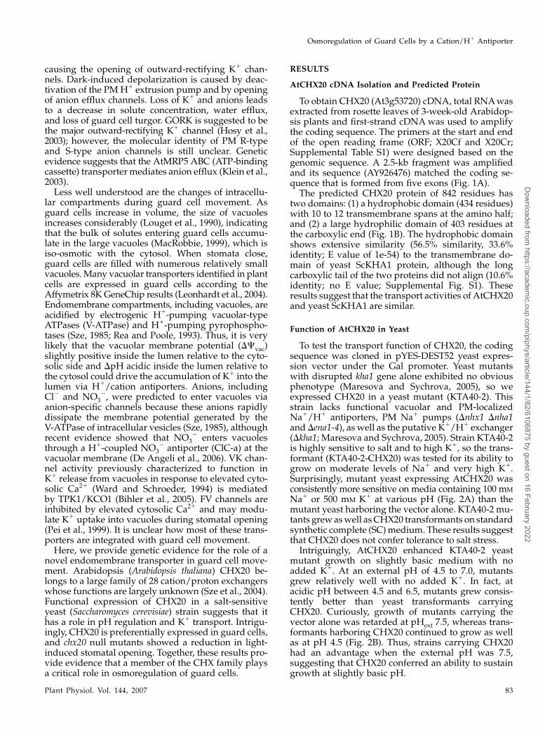
To obtain CHX20 (At3g53720) cDNA, total RNA wasextracted from rosette leaves of 3-week-old Arabidop-sis plants and first-strand cDNA was used to amplifythe coding sequence. The primers at the start and endof the open reading frame (ORF; X20Cf and X20Cr;Supplemental Table S1) were designed based on thegenomic sequence. A 2.5-kb fragment was amplifiedand its sequence (AY926476) matched the coding se-quence that is formed from five exons (Fig. 1A).
The predicted CHX20 protein of 842 residues hastwo domains: (1) a hydrophobic domain (434 residues)with 10 to 12 transmembrane spans at the amino half;and (2) a large hydrophilic domain of 403 residues atthe carboxylic end (Fig. 1B). The hydrophobic domainshows extensive similarity (56.5% similarity, 33.6%identity; E value of 1e-54) to the transmembrane do-main of yeast ScKHA1 protein, although the longcarboxylic tail of the two proteins did not align (10.6%identity; no E value; Supplemental Fig. S1). Theseresults suggest that the transport activities of AtCHX20and yeast ScKHA1 are similar.
Function of AtCHX20 in Yeast
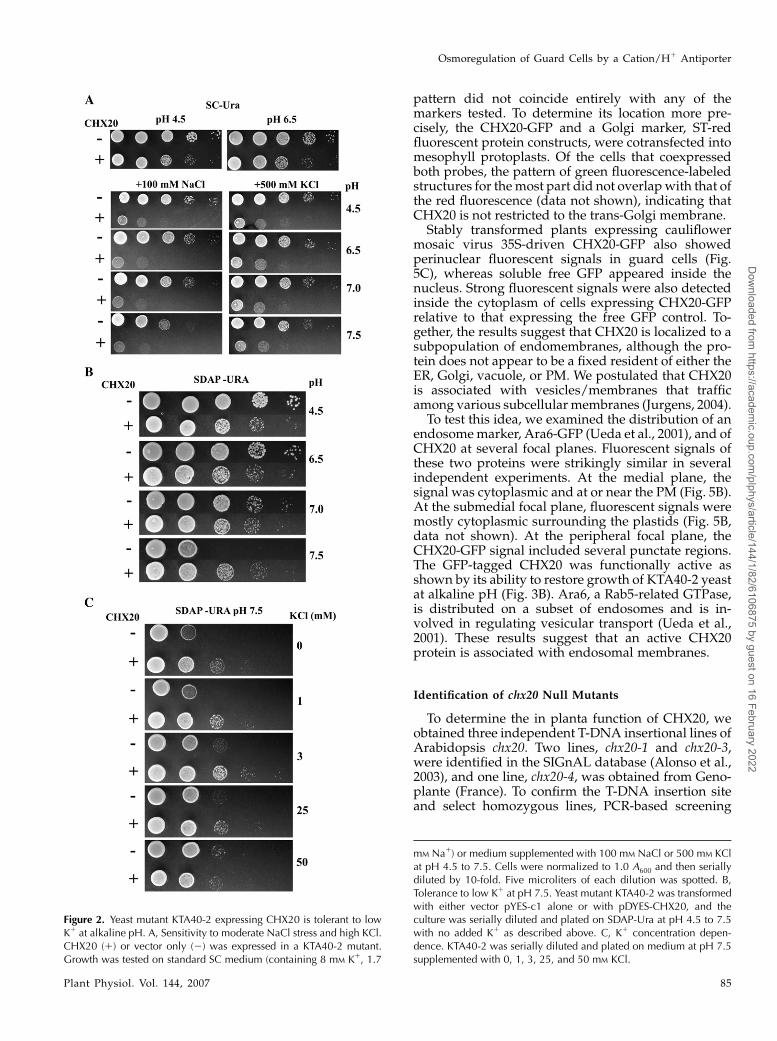
To test the transport function of CHX20, the codingsequence was cloned in pYES-DEST52 yeast expres-sion vector under the Gal promoter. Yeast mutantswith disrupted kha1 gene alone exhibited no obviousphenotype (Maresova and Sychrova, 2005), so weexpressed CHX20 in a yeast mutant (KTA40-2). Thisstrain lacks functional vacuolar and PM-localizedNa1/H1 antiporters, PM Na1 pumps (Dnhx1 Dnha1and Dena1-4), as well as the putative K1/H1 exchanger(Dkha1; Maresova and Sychrova, 2005). Strain KTA40-2is highly sensitive to salt and to high K1, so the trans-formant (KTA40-2-CHX20) was tested for its ability togrow on moderate levels of Na1 and very high K1.Surprisingly, mutant yeast expressing AtCHX20 wasconsistently more sensitive on media containing 100 mM
Na1 or 500 mM K1 at various pH (Fig. 2A) than themutant yeast harboring the vector alone. KTA40-2 mu-tants grew as well as CHX20 transformants on standardsynthetic complete (SC) medium. These results suggestthat CHX20 does not confer tolerance to salt stress.
Intriguingly, AtCHX20 enhanced KTA40-2 yeastmutant growth on slightly basic medium with noadded K1. At an external pH of 4.5 to 7.0, mutantsgrew relatively well with no added K1. In fact, atacidic pH between 4.5 and 6.5, mutants grew consis-tently better than yeast transformants carryingCHX20. Curiously, growth of mutants carrying thevector alone was retarded at pHext 7.5, whereas trans-formants harboring CHX20 continued to grow as wellas at pH 4.5 (Fig. 2B). Thus, strains carrying CHX20had an advantage when the external pH was 7.5,suggesting that CHX20 conferred an ability to sustaingrowth at slightly basic pH.
Osmoregulation of Guard Cells by a Cation/H1 Antiporter
Plant Physiol. Vol. 144, 2007 83
Dow
nloaded from https://academ
ic.oup.com/plphys/article/144/1/82/6106875 by guest on 16 February 2022
We tested the effect of external K1 concentration onyeast growth at pH 7.5. Transformants harboringCHX20 consistently grew better than KTA40-2 mu-tants as long as the K1 level was kept low, fromapproximately 0.4 to 3 mM (Fig. 2C). When no exog-enous K1 was added, the agar medium containedabout 0.4 mM K1. Increasing external KCl concentra-tion beyond 25 mM decreased the beneficial effect ofCHX20. Because K1 is required to sustain growth ofall cells, the enhanced growth of transformants at lowK1 levels would suggest that CHX20 has a role inacquiring K1 when the external pH is slightly alkalineor in maintaining suitable cellular homeostasis forgrowth. This idea is supported by nearly similar growthexhibited by yeast mutants carrying either vectoralone or CHX20 when K1 is raised to 50 mM.
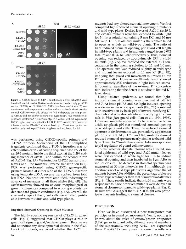
Yeast KHA1 was shown before to confer tolerance tohygromycin (Maresova and Sychrova, 2005) as con-firmed here in the AXT3 strain (Fig. 3A). This strainhas a functional wild-type KHA1 but lacks three Natransporters (ena1-4D nha1D nhx1D). However, al-though transformants expressing CHX20 grew wellat pH 5.5, they showed no growth in the presence of150 mM hygromycin B. CHX20 did promote growth ofmutants grown on yeast nitrogen base (YNB) mediumat pH 7.5 similar to yeast KHA1 (Fig. 3B). These resultssuggest that CHX20 and KHA1 share similar, but notidentical, activities.
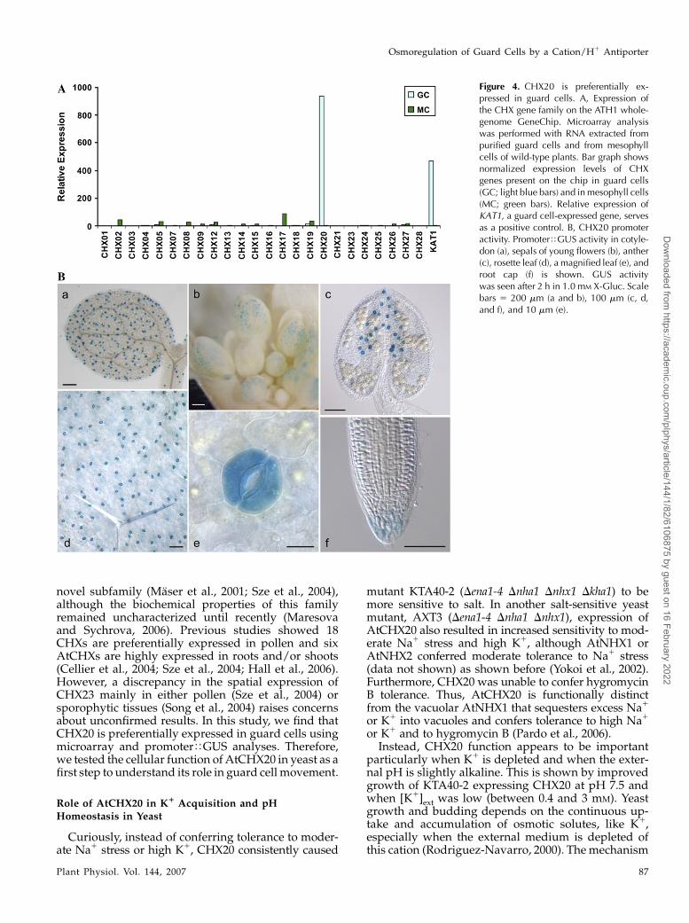
AtCHX20 Is Preferentially Expressed in Guard Cells
Analyses of a guard cell transcriptome (Leonhardtet al., 2004; J. Kwak, N. Leonhardt, and J.I. Schroeder,unpublished data) revealed that only one member ofthe CHX gene family was highly expressed in guardcells. CHX20 showed little or no expression in meso-phyll cells, whereas several other genes, such as CHX17,showed low to moderate expression (Fig. 4A). Fur-thermore, CHX20 expression is particularly strong inguard cells as shown by the 2-fold increase in normal-ized relative expression of CHX20 compared to that of
AtKAT1, a K1 channel preferentially expressed inguard cells (Nakamura et al., 1995).
To verify the microarray results, CHX20 promoter-driven GUS activity was determined. Arabidopsis(Columbia [Col]) plants were transformed with a con-struct containing a 2-kb region upstream of the CHX20ORF transcriptionally fused to the GUS reporter gene.T2 seeds were collected from six independent trans-genic lines and all six lines of CHX20TGUS analyzedgave similar expression patterns. Striking GUS activitywas observed in guard cells located in expanded cot-yledons and in hypocotyls of 1-week-old seedlings(Fig. 4B, a). Three-week-old rosette leaves (Fig. 4B, dand e) and cauline leaves also showed very high GUSstaining in guard cells. However, GUS staining wasnot detected in leaf pavement epidermal cells or inmesophyll cells. Interestingly, GUS activity was alsodetected in guard cells of floral organs, including thesepal, anther (Fig. 4B, b and c), and carpel (data notshown). GUS activity was not detected in the differen-tiated cells of roots, although CHX20 expression wasonly observed in the root cap of 1-week-old seedlings(Fig. 4B, f), consistent with the microarray results of rootcap cells (P. Benfey, personal communication). Thus,analyses of both CHX20 promoter-GUS expression andguard cell-specific transcriptome data clearly indicateselective expression of CHX20 in guard cells.
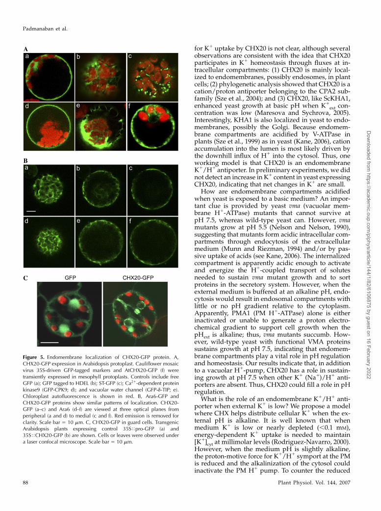
AtCHX20-GFP Is Localized to Endomembranes
When transiently expressed in Arabidopsis meso-phyll protoplasts, CHX20-GFP was visualized at theperiphery of the nucleus and in the cytosol (Fig. 5A, f),suggesting that it is localized at the endoplasmicreticulum (ER) or in endomembranes. The CHX20-GFP signal was compared with those from a solubleGFP, GFP tagged to an ER retention sequence (GFP-HDEL), or to markers such as sialyltransferase (ST)-GFP for trans-Golgi, GFP-CPK9 for PM, and GFP-dTIPfor vacuolar membrane (Fig. 5A). Although CHX20-GFP appeared to be localized to endomembranes, its
Figure 1. CHX20 gene organization and protein sequence. A, Genomic structure of CHX20 was confirmed by the cDNA(accession no. AY926476). The positions of three independent T-DNA insertional mutants are shown. The T-DNA sequences areshown in lowercase. Mutants chx20-1 and chx20-3 correspond to SALK lines SALK_031420 and SALK_011726, and chx20-4 wasobtained from Genoplante. B, Predicted topology of AtCHX20 in the membrane (see Supplemental Fig. S1 for protein sequence).
Padmanaban et al.
84 Plant Physiol. Vol. 144, 2007
Dow
nloaded from https://academ
ic.oup.com/plphys/article/144/1/82/6106875 by guest on 16 February 2022
pattern did not coincide entirely with any of themarkers tested. To determine its location more pre-cisely, the CHX20-GFP and a Golgi marker, ST-redfluorescent protein constructs, were cotransfected intomesophyll protoplasts. Of the cells that coexpressedboth probes, the pattern of green fluorescence-labeledstructures for the most part did not overlap with that ofthe red fluorescence (data not shown), indicating thatCHX20 is not restricted to the trans-Golgi membrane.
Stably transformed plants expressing cauliflowermosaic virus 35S-driven CHX20-GFP also showedperinuclear fluorescent signals in guard cells (Fig.5C), whereas soluble free GFP appeared inside thenucleus. Strong fluorescent signals were also detectedinside the cytoplasm of cells expressing CHX20-GFPrelative to that expressing the free GFP control. To-gether, the results suggest that CHX20 is localized to asubpopulation of endomembranes, although the pro-tein does not appear to be a fixed resident of either theER, Golgi, vacuole, or PM. We postulated that CHX20is associated with vesicles/membranes that trafficamong various subcellular membranes (Jurgens, 2004).
To test this idea, we examined the distribution of anendosome marker, Ara6-GFP (Ueda et al., 2001), and ofCHX20 at several focal planes. Fluorescent signals ofthese two proteins were strikingly similar in severalindependent experiments. At the medial plane, thesignal was cytoplasmic and at or near the PM (Fig. 5B).At the submedial focal plane, fluorescent signals weremostly cytoplasmic surrounding the plastids (Fig. 5B,data not shown). At the peripheral focal plane, theCHX20-GFP signal included several punctate regions.The GFP-tagged CHX20 was functionally active asshown by its ability to restore growth of KTA40-2 yeastat alkaline pH (Fig. 3B). Ara6, a Rab5-related GTPase,is distributed on a subset of endosomes and is in-volved in regulating vesicular transport (Ueda et al.,2001). These results suggest that an active CHX20protein is associated with endosomal membranes.
Identification of chx20 Null Mutants
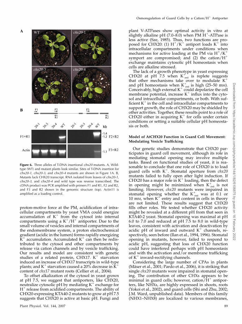
To determine the in planta function of CHX20, weobtained three independent T-DNA insertional lines ofArabidopsis chx20. Two lines, chx20-1 and chx20-3,were identified in the SIGnAL database (Alonso et al.,2003), and one line, chx20-4, was obtained from Geno-plante (France). To confirm the T-DNA insertion siteand select homozygous lines, PCR-based screening
Figure 2. Yeast mutant KTA40-2 expressing CHX20 is tolerant to lowK1 at alkaline pH. A, Sensitivity to moderate NaCl stress and high KCl.CHX20 (1) or vector only (2) was expressed in a KTA40-2 mutant.Growth was tested on standard SC medium (containing 8 mM K1, 1.7
mM Na1) or medium supplemented with 100 mM NaCl or 500 mM KClat pH 4.5 to 7.5. Cells were normalized to 1.0 A600 and then seriallydiluted by 10-fold. Five microliters of each dilution was spotted. B,Tolerance to low K1 at pH 7.5. Yeast mutant KTA40-2 was transformedwith either vector pYES-c1 alone or with pDYES-CHX20, and theculture was serially diluted and plated on SDAP-Ura at pH 4.5 to 7.5with no added K1 as described above. C, K1 concentration depen-dence. KTA40-2 was serially diluted and plated on medium at pH 7.5supplemented with 0, 1, 3, 25, and 50 mM KCl.
Osmoregulation of Guard Cells by a Cation/H1 Antiporter
Plant Physiol. Vol. 144, 2007 85
Dow
nloaded from https://academ
ic.oup.com/plphys/article/144/1/82/6106875 by guest on 16 February 2022
was performed using CHX20-specific primers andT-DNA primers. Sequencing of the PCR-amplifiedfragments confirmed that a T-DNA insertion was lo-cated within exon 2 at coding sequence base 477 of thechx20-1 mutant, inside the third exon at the 1,299 cod-ing sequence of chx20-3, and within the second intronof chx20-4 (Fig. 1A). We tested for CHX20 transcripts inleaves of all the mutants. Reverse transcription (RT)-PCR was performed using CHX20 gene-specificprimers located at either side of the T-DNA insertionusing template cDNA reverse transcribed from totalleaf RNA. No products were amplified, indicating anabsence of messages in all three alleles (Fig. 6B). Thechx20 mutants showed no obvious morphological orgrowth differences compared to wild-type plants un-der standard growth conditions (Fig. 6A). Overall, thesize and shape of the guard cells were indistinguish-able between mutants and wild-type plants.
Impaired Stomatal Opening in chx20 Mutants
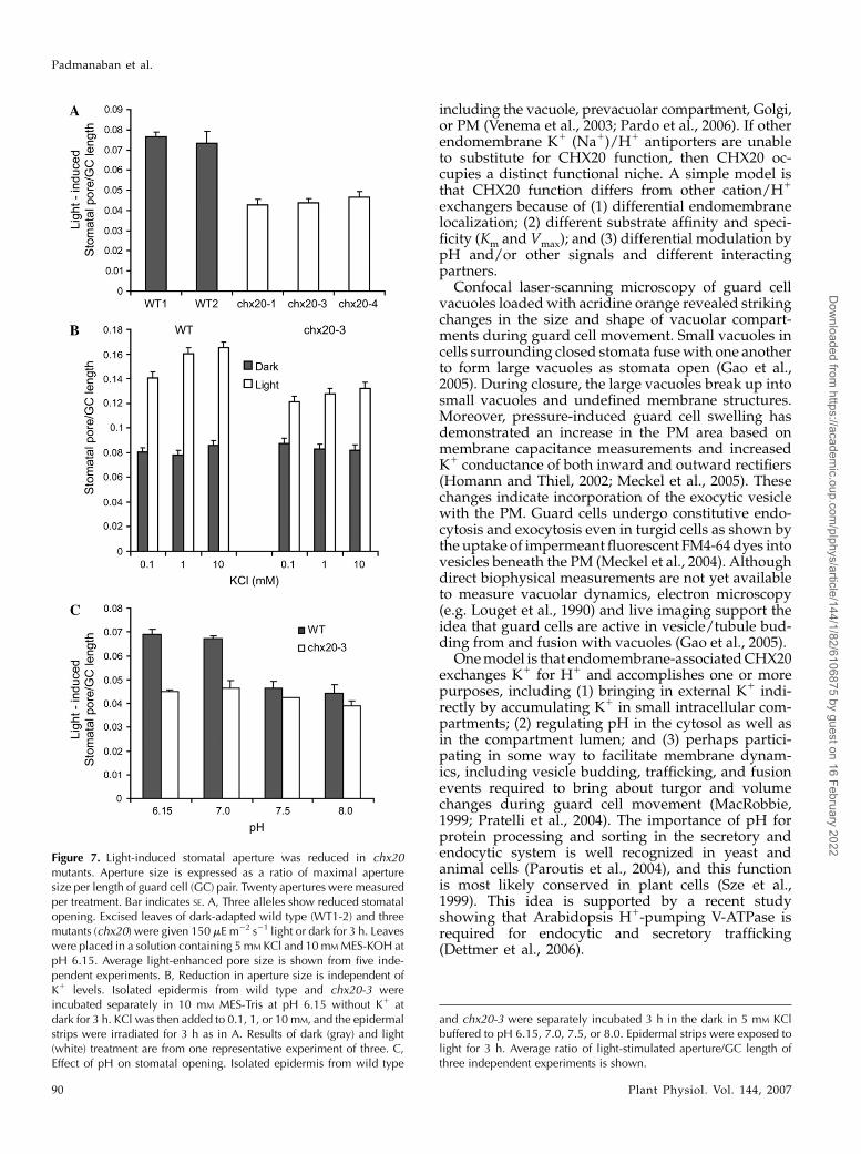
The highly specific expression of CHX20 in guardcells (Fig. 4) suggested that CHX20 plays a role inguard cell signaling and/or development. Because wedid not notice any developmental defects in the chx20knockout mutants, we tested whether the chx20 null
mutants had any altered stomatal movement. We firstcompared light-induced stomatal opening in mutantsand wild-type plants. Excised leaves of chx20-1, chx20-3,and chx20-4 mutants were first exposed to white lightfor 3 h in a solution containing 5 mM KCl and 10 mM
MES at pH 6.15. In all three mutants, the stomata failedto open as widely as wild-type plants. The ratio oflight-induced stomatal opening per guard cell lengthin wild-type plants and in mutants ranged from 0.072to 0.076 and 0.042 to 0.047, respectively. Thus, stomatalopening was reduced by approximately 35% in chx20mutants (Fig. 7A). We reduced the external KCl con-centration in the opening solution to 0.1 and 1.0 mM.The aperture size was reduced slightly in wild-typeand mutant leaves exposed to 0.1 mM K1 (Fig. 7B),implying that guard cell movement is limited at lowK1 concentration. However, chx20 mutants still showedapproximately 35% reduction in light-induced stoma-tal opening regardless of the external K1 concentra-tion, indicating that the defect is not due to limited K1
level alone.Using isolated epidermis, we found that light-
induced stomatal opening was maximal at pH 6.1and 7. At basic pH 7.5 and 8.0, light-induced openingwas decreased in wild-type plants (Fig. 7C) consistentwith inactivation by basic pH of inward-rectifying K1
channels and activation of outward-rectifying K1 chan-nels in Vicia fava guard cells (Ilan et al., 1994, 1996).However, mutants appeared to be insensitive to anacidic apoplastic pH that stimulated stomatal openingof wild-type guard cells. Thus, the reduced stomatalaperture of chx20 mutants was particularly apparent atpH 6.1 and 7.0. At pH 7.5 and 8.0, mutants showedreduced stomatal aperture nearly similar to that of wildtype. Thus, chx20 mutants appeared to be unresponsiveto pH regulation of guard cell movement.
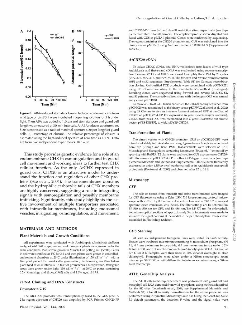
To test whether stomatal closure was affected, iso-lated epidermis of wild-type and chx20 mutant leaveswere first exposed to white light for 3 h to inducestomatal opening and then incubated in 1 mM ABA toinduce closure. The decrease in stomatal aperture wasmeasured at 30-min intervals for 3 h. Although theaperture size of wild-type plants was larger than that ofmutants before ABA addition, the percentage of closureof wild type was higher than that of mutants at all times(Fig. 8). These results indicate that chx20 mutants wereresponsive to ABA; however, mutants were delayed instomatal closure compared to wild-type plants (Fig. 8).Results would suggest that CHX20 might also partic-ipate in events leading to stomatal closure.
DISCUSSION
Here we have discovered a new transporter thatparticipates in guard cell movement. Nearly nothing isknown about the roles of cation/proton antiporter(CPA) genes in guard cells, although several membersof the superfamily, including NHX1, are expressedthere. The AtCHX family was uncovered recently as a
Figure 3. CHX20 fused to GFP is functionally active. KTA40-2 yeast(ena1-4D nha1D nhx1D kha1D) was transformed with empty pDR196vector, CHX20, or CHX20-GFP. AXT3 (ena1-4D nha1D nhx1D) wastransformed with empty vector and served as a native ScKHA1 positivecontrol. Cells were serially diluted 10-fold and spotted on YNB plates.A, CHX20 did not confer tolerance to hygromycin. Five microliters ofyeast was spotted on YNB medium at pH 5.5 with or without hygromycinB (150 mg/mL) and incubated for 4 d. B, CHX20 fused to GFP conferredtolerance to the KTA40-2 strain at basic pH. Yeast was spotted onmedium adjusted to pH 7.5 with Arg base and incubated for 2 d.
Padmanaban et al.
86 Plant Physiol. Vol. 144, 2007
Dow
nloaded from https://academ
ic.oup.com/plphys/article/144/1/82/6106875 by guest on 16 February 2022
novel subfamily (Maser et al., 2001; Sze et al., 2004),although the biochemical properties of this familyremained uncharacterized until recently (Maresovaand Sychrova, 2006). Previous studies showed 18CHXs are preferentially expressed in pollen and sixAtCHXs are highly expressed in roots and/or shoots(Cellier et al., 2004; Sze et al., 2004; Hall et al., 2006).However, a discrepancy in the spatial expression ofCHX23 mainly in either pollen (Sze et al., 2004) orsporophytic tissues (Song et al., 2004) raises concernsabout unconfirmed results. In this study, we find thatCHX20 is preferentially expressed in guard cells usingmicroarray and promoterTGUS analyses. Therefore,we tested the cellular function of AtCHX20 in yeast as afirst step to understand its role in guard cell movement.
Role of AtCHX20 in K1 Acquisition and pHHomeostasis in Yeast
Curiously, instead of conferring tolerance to moder-ate Na1 stress or high K1, CHX20 consistently caused
mutant KTA40-2 (Dena1-4 Dnha1 Dnhx1 Dkha1) to bemore sensitive to salt. In another salt-sensitive yeastmutant, AXT3 (Dena1-4 Dnha1 Dnhx1), expression ofAtCHX20 also resulted in increased sensitivity to mod-erate Na1 stress and high K1, although AtNHX1 orAtNHX2 conferred moderate tolerance to Na1 stress(data not shown) as shown before (Yokoi et al., 2002).Furthermore, CHX20 was unable to confer hygromycinB tolerance. Thus, AtCHX20 is functionally distinctfrom the vacuolar AtNHX1 that sequesters excess Na1
or K1 into vacuoles and confers tolerance to high Na1
or K1 and to hygromycin B (Pardo et al., 2006).Instead, CHX20 function appears to be important
particularly when K1 is depleted and when the exter-nal pH is slightly alkaline. This is shown by improvedgrowth of KTA40-2 expressing CHX20 at pH 7.5 andwhen [K1]ext was low (between 0.4 and 3 mM). Yeastgrowth and budding depends on the continuous up-take and accumulation of osmotic solutes, like K1,especially when the external medium is depleted ofthis cation (Rodriguez-Navarro, 2000). The mechanism
Figure 4. CHX20 is preferentially ex-pressed in guard cells. A, Expression ofthe CHX gene family on the ATH1 whole-genome GeneChip. Microarray analysiswas performed with RNA extracted frompurified guard cells and from mesophyllcells of wild-type plants. Bar graph showsnormalized expression levels of CHXgenes present on the chip in guard cells(GC; light blue bars) and in mesophyll cells(MC; green bars). Relative expression ofKAT1, a guard cell-expressed gene, servesas a positive control. B, CHX20 promoteractivity. PromoterTGUS activity in cotyle-don (a), sepals of young flowers (b), anther(c), rosette leaf (d), a magnified leaf (e), androot cap (f) is shown. GUS activitywas seen after 2 h in 1.0 mM X-Gluc. Scalebars 5 200 mm (a and b), 100 mm (c, d,and f), and 10 mm (e).
Osmoregulation of Guard Cells by a Cation/H1 Antiporter
Plant Physiol. Vol. 144, 2007 87
Dow
nloaded from https://academ
ic.oup.com/plphys/article/144/1/82/6106875 by guest on 16 February 2022
for K1 uptake by CHX20 is not clear, although severalobservations are consistent with the idea that CHX20participates in K1 homeostasis through fluxes at in-tracellular compartments: (1) CHX20 is mainly local-ized to endomembranes, possibly endosomes, in plantcells; (2) phylogenetic analysis showed that CHX20 is acation/proton antiporter belonging to the CPA2 sub-family (Sze et al., 2004); and (3) CHX20, like ScKHA1,enhanced yeast growth at basic pH when K1
ext con-centration was low (Maresova and Sychrova, 2005).Interestingly, KHA1 is also localized in yeast to endo-membranes, possibly the Golgi. Because endomem-brane compartments are acidified by V-ATPase inplants (Sze et al., 1999) as in yeast (Kane, 2006), cationaccumulation into the lumen is most likely driven bythe downhill influx of H1 into the cytosol. Thus, oneworking model is that CHX20 is an endomembraneK1/H1 antiporter. In preliminary experiments, we didnot detect an increase in K1 content in yeast expressingCHX20, indicating that net changes in K1 are small.
How are endomembrane compartments acidifiedwhen yeast is exposed to a basic medium? An impor-tant clue is provided by yeast vma (vacuolar mem-brane H1-ATPase) mutants that cannot survive atpH 7.5, whereas wild-type yeast can. However, vmamutants grow at pH 5.5 (Nelson and Nelson, 1990),suggesting that mutants form acidic intracellular com-partments through endocytosis of the extracellularmedium (Munn and Riezman, 1994) and/or by pas-sive uptake of acids (see Kane, 2006). The internalizedcompartment is apparently acidic enough to activateand energize the H1-coupled transport of solutesneeded to sustain vma mutant growth and to sortproteins in the secretory system. However, when theexternal medium is buffered at an alkaline pH, endo-cytosis would result in endosomal compartments withlittle or no pH gradient relative to the cytoplasm.Apparently, PMA1 (PM H1-ATPase) alone is eitherinactivated or unable to generate a proton electro-chemical gradient to support cell growth when thepHext is alkaline; thus, vma mutants succumb. How-ever, wild-type yeast with functional VMA proteinssustains growth at pH 7.5, indicating that endomem-brane compartments play a vital role in pH regulationand homeostasis. Our results indicate that, in additionto a vacuolar H1-pump, CHX20 has a role in sustain-ing growth at pH 7.5 when other K1 (Na1)/H1 anti-porters are absent. Thus, CHX20 could fill a role in pHregulation.
What is the role of an endomembrane K1/H1 anti-porter when external K1 is low? We propose a modelwhere CHX helps distribute cellular K1 when the ex-ternal pH is alkaline. It is well known that whenmedium K1 is low or nearly depleted (,0.1 mM),energy-dependent K1 uptake is needed to maintain[K1]cyt at millimolar levels (Rodriguez-Navarro, 2000).However, when the medium pH is slightly alkaline,the proton-motive force for K1/H1 symport at the PMis reduced and the alkalinization of the cytosol couldinactivate the PM H1 pump. To counter the reduced
Figure 5. Endomembrane localization of CHX20-GFP protein. A,CHX20-GFP expression in Arabidopsis protoplast. Cauliflower mosaicvirus 35S-driven GFP-tagged markers and AtCHX20-GFP (f) weretransiently expressed in mesophyll protoplasts. Controls include freeGFP (a); GFP tagged to HDEL (b); ST-GFP (c); Ca21-dependent proteinkinase9 (GFP-CPK9; d); and vacuolar water channel (GFP-d-TIP; e).Chloroplast autofluorescence is shown in red. B, Ara6-GFP andCHX20-GFP proteins show similar patterns of localization. CHX20-GFP (a–c) and Ara6 (d–f) are viewed at three optical planes fromperipheral (a and d) to medial (c and f). Red emission is removed forclarity. Scale bar 5 10 mm. C, CHX20-GFP in guard cells. TransgenicArabidopsis plants expressing control 35STpro-GFP (a) and35STCHX20-GFP (b) are shown. Cells or leaves were observed undera laser confocal microscope. Scale bar 5 10 mm.
Padmanaban et al.
88 Plant Physiol. Vol. 144, 2007
Dow
nloaded from https://academ
ic.oup.com/plphys/article/144/1/82/6106875 by guest on 16 February 2022
proton-motive force at the PM, acidification of intra-cellular compartments by yeast VMA could energizeaccumulation of K1 from the cytosol into internalcompartments using a K1/H1 antiporter. Due to thesmall volume of vesicles and internal compartments ofthe endomembrane system, a proton electrochemicalgradient (acidic in the lumen) forms rapidly energizingK1 accumulation. Accumulated K1 can then be redis-tributed to the cytosol and other compartments byrelease via cation channels and by vesicle trafficking.Our results and model are consistent with geneticstudies of a related protein, CHX17. K1 starvationinduced an increase of CHX17 transcripts in wild-typeplants; and K1 starvation caused a 20% decrease in K1
content of chx17 mutant roots (Cellier et al., 2004).To offset alkalization of the cytosol in yeast grown
at pH 7.5, we suggest that antiporters, like CHX20,neutralize cytosolic pH by mediating K1 exchange forH1 release from acidified compartments. The ability ofCHX20-expressing KTA40-2 mutants to grow at pH 7.5suggests that CHX20 is active at basic pH. Fungi and
plant V-ATPases show optimal activity in vitro atslightly alkaline pH (7.0–8.0) when PM H1-ATPase isless active (Sze, 1985). Thus, two functions are pro-posed for CHX20: (1) H1/K1 antiport loads K1 intointracellular compartments under conditions whenmechanisms for active loading at the PM via H1/K1
symport are compromised; and (2) the cation/H1
exchange maintains cytosolic pH homeostasis whencells are alkaline stressed.
The lack of a growth phenotype in yeast expressingCHX20 at pH 7.5 when K1
ext is replete suggeststhat other mechanisms take over to modulate K1
and pH homeostasis when K1ext is high (25–50 mM).
Conceivably, high external K1 could depolarize the cellmembrane potential, increase K1 influx into the cyto-sol and intracellular compartments, or both. With suf-ficient K1 in the cell and intracellular compartments tosupport growth, the role of CHX20 may be shielded byother activities. Together, these results point to a role ofCHX20 either in acquiring K1 for cells under certainconditions or setting a suitable cellular pH homeosta-sis or both.
Model of AtCHX20 Function in Guard Cell Movement:
Modulating Vesicle Trafficking
Our genetic studies demonstrate that CHX20 par-ticipates in guard cell movement, although its role inmediating stomatal opening may involve multipletasks. Based on functional studies of yeast, it is rea-sonable to conclude that one role of CHX20 is to loadguard cells with K1. Stomatal aperture from chx20mutants failed to fully open after light induction. IfCHX20 has a major role in K1 loading, then the defectin opening might be minimized when K1
ext is notlimiting. However, chx20 mutants were impaired instomatal opening whether the K1
ext was at 0.1 or10 mM, when K1 entry and content in cells in theoryare not limited. These results suggest that CHX20fills other roles. We tested whether CHX20 activitymight be revealed at a different pH from that seen inKTA40-2 yeast. Stomatal opening was maximal at pH6.0 to 7.0 and reduced at pH 7.5 to 8.0 in wild-typeleaves, consistent with activation and deactivation byacidic pH of inward and outward K1 channels, re-spectively, seen before (Ilan et al., 1994, 1996). Stomatalopening in mutants, however, failed to respond toacidic pH, suggesting that loss of CHX20 functioncould have interfered perhaps with pH homeostasisand with the activation and/or membrane traffickingof K1 inward-rectifying channels.
Considering the large number of CPAs in plants(Maser et al., 2001; Pardo et al., 2006), it is striking thatsingle chx20 mutants were impaired in stomatal open-ing. The contribution of other CHXs appears to beminimal in guard cells; however, cation/H1 antipor-ters, like NHXs, are highly expressed in shoots, roots(Yokoi et al., 2002), and guard cells (Shi and Zhu, 2002;J.M. Ward, unpublished data). Members of this family(NHX1–NHX8) are localized to various membranes,
Figure 6. Three alleles of T-DNA insertional chx20 mutants. A, Wild-type (WT) and mutant plants look similar. Sites of T-DNA insertion forchx20-1, chx20-3, and chx20-4 mutants are shown in Figure 1A. B,Mutants lack CHX20 transcript. RNA isolated from leaves of chx20-1,chx20-3, and chx20-4 and wild type was reverse transcribed. ThecDNA product was PCR amplified with primers F1 and R1, F2 and R2,and F3 and R2 shown in the genomic structure (top). Actin11 isamplified as a loading control.
Osmoregulation of Guard Cells by a Cation/H1 Antiporter
Plant Physiol. Vol. 144, 2007 89
Dow
nloaded from https://academ
ic.oup.com/plphys/article/144/1/82/6106875 by guest on 16 February 2022
including the vacuole, prevacuolar compartment, Golgi,or PM (Venema et al., 2003; Pardo et al., 2006). If otherendomembrane K1 (Na1)/H1 antiporters are unableto substitute for CHX20 function, then CHX20 oc-cupies a distinct functional niche. A simple model isthat CHX20 function differs from other cation/H1
exchangers because of (1) differential endomembranelocalization; (2) different substrate affinity and speci-ficity (Km and Vmax); and (3) differential modulation bypH and/or other signals and different interactingpartners.
Confocal laser-scanning microscopy of guard cellvacuoles loaded with acridine orange revealed strikingchanges in the size and shape of vacuolar compart-ments during guard cell movement. Small vacuoles incells surrounding closed stomata fuse with one anotherto form large vacuoles as stomata open (Gao et al.,2005). During closure, the large vacuoles break up intosmall vacuoles and undefined membrane structures.Moreover, pressure-induced guard cell swelling hasdemonstrated an increase in the PM area based onmembrane capacitance measurements and increasedK1 conductance of both inward and outward rectifiers(Homann and Thiel, 2002; Meckel et al., 2005). Thesechanges indicate incorporation of the exocytic vesiclewith the PM. Guard cells undergo constitutive endo-cytosis and exocytosis even in turgid cells as shown bythe uptake of impermeant fluorescent FM4-64 dyes intovesicles beneath the PM (Meckel et al., 2004). Althoughdirect biophysical measurements are not yet availableto measure vacuolar dynamics, electron microscopy(e.g. Louget et al., 1990) and live imaging support theidea that guard cells are active in vesicle/tubule bud-ding from and fusion with vacuoles (Gao et al., 2005).
One model is that endomembrane-associated CHX20exchanges K1 for H1 and accomplishes one or morepurposes, including (1) bringing in external K1 indi-rectly by accumulating K1 in small intracellular com-partments; (2) regulating pH in the cytosol as well asin the compartment lumen; and (3) perhaps partici-pating in some way to facilitate membrane dynam-ics, including vesicle budding, trafficking, and fusionevents required to bring about turgor and volumechanges during guard cell movement (MacRobbie,1999; Pratelli et al., 2004). The importance of pH forprotein processing and sorting in the secretory andendocytic system is well recognized in yeast andanimal cells (Paroutis et al., 2004), and this functionis most likely conserved in plant cells (Sze et al.,1999). This idea is supported by a recent studyshowing that Arabidopsis H1-pumping V-ATPase isrequired for endocytic and secretory trafficking(Dettmer et al., 2006).
Figure 7. Light-induced stomatal aperture was reduced in chx20mutants. Aperture size is expressed as a ratio of maximal aperturesize per length of guard cell (GC) pair. Twenty apertures were measuredper treatment. Bar indicates SE. A, Three alleles show reduced stomatalopening. Excised leaves of dark-adapted wild type (WT1-2) and threemutants (chx20) were given 150 mE m22 s21 light or dark for 3 h. Leaveswere placed in a solution containing 5 mM KCl and 10 mM MES-KOH atpH 6.15. Average light-enhanced pore size is shown from five inde-pendent experiments. B, Reduction in aperture size is independent ofK1 levels. Isolated epidermis from wild type and chx20-3 wereincubated separately in 10 mM MES-Tris at pH 6.15 without K1 atdark for 3 h. KCl was then added to 0.1, 1, or 10 mM, and the epidermalstrips were irradiated for 3 h as in A. Results of dark (gray) and light(white) treatment are from one representative experiment of three. C,Effect of pH on stomatal opening. Isolated epidermis from wild type
and chx20-3 were separately incubated 3 h in the dark in 5 mM KClbuffered to pH 6.15, 7.0, 7.5, or 8.0. Epidermal strips were exposed tolight for 3 h. Average ratio of light-stimulated aperture/GC length ofthree independent experiments is shown.
Padmanaban et al.
90 Plant Physiol. Vol. 144, 2007
Dow
nloaded from https://academ
ic.oup.com/plphys/article/144/1/82/6106875 by guest on 16 February 2022
This study provides genetic evidence for a role of anendomembrane CHX in osmoregulation and in guardcell movement and working ideas to further test CHXcellular function. As the only AtCHX expressed inguard cells, CHX20 is an attractive model to under-stand the function and regulation of other CHX pro-teins (Sze et al., 2004). The transmembrane domainsand the hydrophilic carboxylic tails of CHX membersare highly conserved, suggesting a role in integratingsignals with osmoregulation and possibly membranetrafficking. Significantly, this study highlights the ac-tive involvement of multiple transporters associatedwith intracellular membranes, including endosomalvesicles, in signaling, osmoregulation, and movement.
MATERIALS AND METHODS
Plant Materials and Growth Conditions
All experiments were conducted with Arabidopsis (Arabidopsis thaliana)
ecotype Col-0. Wild-type, mutant, and transgenic plants were grown under the
same conditions. Plants were grown in Miracle-Gro potting soil (Scotts). Seeds
in soil were stratified at 4�C for 3 d and then plants were grown in controlled-
environment chambers at 20�C under illumination of 150 mE m22 s21 with a
16-h photoperiod. Two weeks after germination, plants were given Miracle-Gro
plant food at 20-d intervals. To test for promoterTGUS expression, transgenic
seeds were grown under light (150 mE m22 s21) at 20�C on plates containing
0.53 Murashige and Skoog (1962) salts and 1.0% agar, pH 5.8.
cDNA Cloning and DNA Constructs
PromoterTGUS
The AtCHX20 promoter was transcriptionally fused to the GUS gene. A
2-kb region upstream of CHX20 was amplified by PCR. Primers CHX20-PF
and CHX20-PR have SalI and BamHI restriction sites, respectively (see Sup-
plemental Table S1 for all primers). The amplified products were digested and
fused with GUS in pRITA I plasmid. Clones were confirmed by sequencing.
The region containing the CHX20 promoter and GUS was subcloned into the
binary vector pMLBart using NotI and named CHX20TGUS (Supplemental
Table S2).
AtCHX20 cDNA
To isolate CHX20 cDNA, total RNA was isolated from leaves of wild-type
Arabidopsis and first-strand cDNA was synthesized using reverse transcrip-
tase. Primers X20Cf and X20Cr were used to amplify the cDNA by 25 cycles
(94�C 30 s, 55�C 30 s, and 72�C 90 s). The forward and reverse primers contain
attB1 and attB2 sequences (Supplemental Table S1) for Gateway recombina-
tion cloning. Gel-purified PCR products were recombined with pDONR221
using BP Clonase according to the manufacturer’s method (Invitrogen).
Resulting clones were sequenced using forward and reverse M13, S1, S2,
and S3 primers. The correctly spliced clone with the longest ORF was named
entry clone pECHX20.
To make a CHX20-GFP fusion construct, the CHX20 coding sequence from
pECHX20 was recombined to the binary vector pK7FWG2 (Karimi et al., 2002)
using LR Clonase to give an in-frame fusion of enhanced GFP at the C tail of
CHX20 or pDCHX20-GFP. For expression in yeast (Saccharomyces cerevisiae),
CHX20 from pECHX20 was recombined into a yeast-Escherichia coli shuttle
vector, pYES-DEST52, to yield pDYES-CHX20.
Transformation of Plants
The binary vectors with CHX20 promoterTGUS or pDCHX20-GFP were
introduced stably into Arabidopsis using Agrobacterium tumefaciens-mediated
floral dip (Clough and Bent, 1998). Transformants were selected on 0.53
Murashige and Skoog plates containing kanamycin (50 mg mL21) or on soil by
spraying with BASTA. T2 plants were analyzed for GUS expression or CHX20-
GFP fluorescence. pDCHX20-GFP or other GFP-tagged constructs (see Sup-
plemental Materials and Methods S1; Supplemental Table S2) were transiently
expressed in onion (Allium cepa) epidermal cell or in Arabidopsis mesophyll
protoplasts (Kovtun et al., 2000) and observed after 12 to 16 h.
Microscopy
GFP
The cells or tissues from transient and stable transformants were imaged
for GFP fluorescence using a Zeiss LSM 510 laser-scanning confocal micro-
scope with a 103 dry 0.8 numerical aperture lens and a 633 1.2 numerical
aperture water immersion lens (Zeiss). The filter settings are Ex 488 nm/Em
BP 510 to 530 nm for GFP, and Ex 488 nm/Em LP 570 nm for chlorophyll.
Sometimes optical sections of approximately 5-mm increments were made to
visualize the signal patterns at the medial to the peripheral plane. Images were
assembled in Photoshop (Adobe).
GUS Staining
At least six independent transgenic lines were tested for GUS activity.
Tissues were incubated in a mixture containing 84 mM sodium phosphate, pH
7.0, 0.5 mM potassium ferrocyanide, 0.5 mM potassium ferricyanide, 0.5%
Triton X-100, and 1.5 mM 5-bromo-4-chloro-3-indolyl-b-D-GlcUA (X-Gluc) at
37� C for 2 h. Samples were then fixed in 70% ethanol overnight to clear
chlorophyll. Photographs were taken under a Nikon stereoscopic zoom
microscope SMZ1000 or with differential interference contrast using a Nikon
E600 microscope.
ATH1 GeneChip Analysis
The ATH1 23K GeneChip experiment was performed with guard cell and
mesophyll cell RNA extracted from wild-type plants using methods described
for the 8K chip (Leonhardt et al., 2004; see Supplemental Materials and
Methods S1). Overall intensity normalization for the entire probe set was
performed using Affymetrix Microarray Suite 5.0. Using the GeneChip Suite
5.0 default parameters, the detection P value and the signal value were
Figure 8. ABA-induced stomatal closure. Isolated epidermal cells fromwild type or chx20-3 were incubated in opening solution for 3 h underlight. Then ABA was added to 1.0 mM and stomatal pore and guard celllength was measured at 30-min intervals. A, ABA reduces aperture size.Size is expressed as a ratio of maximal aperture size per length of guardcells. B, Percentage of closure. The relative percentage of closure isestimated using the light-induced aperture at zero time as 100%. Dataare from two independent experiments. Bar 5 SE.
Osmoregulation of Guard Cells by a Cation/H1 Antiporter
Plant Physiol. Vol. 144, 2007 91
Dow
nloaded from https://academ
ic.oup.com/plphys/article/144/1/82/6106875 by guest on 16 February 2022
calculated for each probe set from each independent guard cell and mesophyll
cell hybridization.
Yeast Strains and Growth Conditions
Yeast Strains
Yeast strains used in the study are (1) AXT3 (MATa his3-11 leu2-112 trp1-1
ade2-1 ura3-1 ena1DThis3Tena4D nha1DTleu2 nhx1DTtrp1 in W303-1B); (2)
KTA40-2 (MATa ade2-1 can1-100 his3-11,15 leu2-3,112 trp1-1 ura3-1 mall0
ena1DThis3Tena4D nha1DTlew2 nhx1DTtrp1 kha1DTkanMX); and (3) LMB
01 (MATa ade2-1 can1-100 his3-11,15 leu2-3,112 trp1-1 ura3-1 mall0 ena1DT
his3Tena4D nha1DTleu2 kha1DTkanMX) (Quintero et al., 2000; Maresova
and Sychrova, 2005). Yeast was transformed with plasmid DNA using the
lithium acetate method (Gietz et al., 1992) and the resulting transformants
were selected on SC medium minus Ura (0.67% YNB, 2% Glc, 2% drop-out
mix, 2% agar).
Determination of Growth
Fresh cells grown in liquid medium were washed and suspended in water
and then adjusted to OD600 of 1.0 (13). Ten-fold serial dilutions of the cells
were prepared with sterile water and 5 mL of each dilution was spotted on
plates containing appropriate SC minus Ura (0.67% YNB, 2% Glc or Gal, 2%
drop-out mix minus Ura, 2% agar) or SDAP minus Ura and adjusted to the
desired pH. To reduce K1 and NH41, modified SDAP medium was used. SDAP
minus Ura medium consisted of 10 mM Arg-HCl (or Arg base), 2% (w/v)
Glc or Gal, 2% drop-out mix minus Ura, 2 mM MgSO4, 0.9 mM CaCl2, trace
minerals, vitamins, and 2% agar. Medium containing Arg-HCl was adjusted to
pH 4.5 with tartaric acid and to pH 5.5 to 6.0 with 10 mM HEPES and Tris. For
pH 7.0 to 7.5, medium contained Arg base and 10 mM HEPES and was
adjusted to desired pH with tartaric acid or Tris. Plates were incubated at
30�C for 2 d and the relative growth of yeast was recorded using a Nikon
Coolpix995 digital camera.
For some experiments, 3-d yeast cells were cultured in liquid YNB medium
containing 0.67% YNB without amino acids, 2% Glc, 0.01% adenine, 0.01%
Trp, and 10 mM MES adjusted to pH 5.5 with Arg base and grown for 18 h at
30�C. One-milliliter cultures were diluted to 6 mL with YNB medium without
Glc and then starved for 18 h at 30�C. Starved cells were washed with 6 mL
water, pelleted, and suspended in water. Cell density was normalized to OD600
of 0.2 and subjected to 10-fold serial dilution. Five-microliter aliquots were
spotted on modified YNB plates at pH 7.5 or 5.5. The YNB medium also
contained 2% agar, 0.02% bromocresol purple (catalog no. 860891; Sigma), and
20 mM MES adjusted to either pH 5.5 or 7.5 with Arg base. Hygromycin B,
when added, was 150 mg/mL (catalog no. H7772; Sigma). Plates were
incubated at 30�C for 2 to 4 d.
Arabidopsis T-DNA Mutant Analyses
T-DNA insertional mutants of chx20 (Alonso et al., 2003) were detected in
the SALK database (http://signal.salk.edu/cgi-bin/tdnaexpress). Homozy-
gous mutants, SALK_031420 (chx20-1), and SALK_011726 (chx20-3) seeds were
first identified by PCR using LBa1 primer and CHX20-specific primers:
CHX20-1-LP, CHX20-1-RP, CHX20-3-LP, CHX20-3-RP, CHX20-4F-P1, and
CHX20-4F-P2 (Supplemental Table S1). The site of insertion was verified by
sequencing.
To detect CHX20 transcript, total RNA isolated from chx20-1, chx20-3, and
chx20-4 mutant and wild-type plants was reverse transcribed. Primer sets
(Supplemental Table S1) F1 (from 215 to 30 bp) and R1 (1,300–1,252 bp); F2
(1,252–1,300 bp) and reverse primer R2 (3#-untranslated region); and F3
(1,506–1,541 bp) and R2 are expected to amplify products of 1,230, 1,413, and
1,158 bp in wild-type plants, respectively.
Guard Cell Movement
To test light-induced stomatal opening (Kwak et al., 2001), leaves were
excised from 3-week-old wild-type and chx20 mutants. Leaves were separated
into two batches and placed in aluminum foil-covered containers in the
opening solution (5 mM KCl and 10 mM MES-KOH at pH 6.15) for 3 h. The
dark-adapted leaves were then exposed to white light (approximately 150 mE
m22 s21) or dark for 3 h at 20�C. Leaves were blended and filtered through
200-mm nylon mesh. Isolated epidermis was observed under a microscope
(Axiovert, 40 CFL; Zeiss), and 20 stomata were measured for each condition.
To study the effect of KCl concentration, isolated epidermal cells from wild
type or chx20-3 were incubated in 10 mM MES-Tris at pH 6.15 without K1 for
3 h in the dark and then KCl was added to a final concentration of 0.1, 1, and
10 mM before exposure to 3 h of light. To test pH, the pH 6.15 medium was
buffered with 10 mM MES-Tris and that of pH 7.0 to 8.0 was adjusted with
10 mM HEPES-Tris. To test ABA-induced stomatal closure, isolated epidermal
cells from wild type or chx20-3 were incubated in opening solution with light
for 3 h, then ABA was added to 1 mM and stomatal pore size and guard cell
length were measured at 30-min intervals using Scion image analysis. The
stomatal aperture was measured as the maximal width between the inner
cuticular lips.
Sequence data from this article can be found in the GenBank/EMBL data
libraries under accession number AY926476 (AGI no. At3g53720).
Supplemental Data
The following materials are available in the online version of this article.
Supplemental Figure S1. AtCHX20 and ScKHA1 proteins.
Supplemental Table S1. Primer sequences.
Supplemental Table S2. Vectors used.
Supplemental Materials and Methods S1. Plant transformation and
microarray analysis.
ACKNOWLEDGMENTS
We thank H. Sychrova (Institute of Physiology, Prague) for providing
yeast strains KTA40-2 and LMB01. Strain AXT3 was a gift from J.M. Pardo
(Instituto de Recursos Naturales y Agrobiologia). F. Cellier and Genoplante
provided mutant chx20-4. We gratefully acknowledge J.I. Schroeder and
N. Leonhardt for making the guard cell transcriptome available, and J.Y. Lee
(University of Delaware), Inhwan Hwang (Pohang University), N. Federoff
(Penn State University), and T. Ueda (RIKEN) for GFP-tagged markers (see
Supplemental Table S2). H.S. thanks J. Sheen (Harvard Medical School) for
introducing the protoplast model and R. Rao (Johns Hopkins University) and
Kendal Hirschi (Baylor College of Medicine) for suggestions.
LITERATURE CITED
Alonso JM, Stepanova AN, Leisse TJ, Kim CJ, Chen HM, Shinn P,
Stevenson DK, Zimmerman J, Barajas P, Cheuk R, et al (2003) Genome-
wide insertional mutagenesis of Arabidopsis thaliana. Science 301:
653–657
Assmann SM (1993) Signal transduction in guard cells. Annu Rev Cell Biol
9: 345–375
Bihler H, Eing C, Hebeisen S, Roller A, Czempinski K, Bertl A (2005)
TPK1 is a vacuolar ion channel different from the slow-vacuolar cation
channel. Plant Physiol 139: 417–424
Blatt MR (2000) Cellular signaling and volume control in stomatal move-
ments in plants. Annu Rev Cell Dev Biol 16: 221–241
Cellier F, Conejero G, Ricaud L, Luu DT, Lepetit M, Gosti F, Casse F (2004)
Characterization of AtCHX17, a member of the cation/H1 exchangers,
CHX family, from Arabidopsis thaliana suggests a role in K1 homeostasis.
Plant J 39: 834–846
Clough SJ, Bent AF (1998) Floral dip: a simplified method for
Agrobacterium-mediated transformation of Arabidopsis thaliana. Plant J
16: 735–743
De Angeli A, Monachello D, Ephritikhine G, Frachisse JM, Thomine S,
Gambale F, Barbier-Brygoo H (2006) The nitrate/proton antiporter
AtCLCa mediates nitrate accumulation in plant vacuoles. Nature 442:
939–942
Dettmer J, Hong-Hermesdorf A, Stierhof YD, Schumacher K (2006)
Vacuolar H1-ATPase activity is required for endocytic and secretory
trafficking in Arabidopsis. Plant Cell 18: 715–730
Padmanaban et al.
92 Plant Physiol. Vol. 144, 2007
Dow
nloaded from https://academ
ic.oup.com/plphys/article/144/1/82/6106875 by guest on 16 February 2022
Fan LM, Zhao Z, Assmann SM (2004) Guard cells: a dynamic signaling
model. Curr Opin Plant Biol 7: 537–546
Gao XQ, Li CG, Wei PC, Zhang XY, Chen J, Wang XC (2005) The dynamic
changes of tonoplasts in guard cells are important for stomatal move-
ment in Vicia faba. Plant Physiol 139: 1207–1216
Gietz D, Jean AS, Woods RA, Schiestl RH (1992) Improved method for
high efficiency transformation of intact yeast. Nucleic Acids Res 20: 1425
Guo FQ, Young J, Crawford NM (2003) The nitrate transporter AtNRT1.1
(CHL1) functions in stomatal opening and contributes to drought
susceptibility in Arabidopsis. Plant Cell 15: 107–117
Hall D, Evans AR, Newbury HJ, Pritchard J (2006) Functional analysis of
CHX21: a putative sodium transporter in Arabidopsis. J Exp Bot 57:
1201–1210
Homann U, Thiel G (2002) The number of K1 channels in the plasma
membrane of guard cell protoplasts changes in parallel with the surface
area. Proc Natl Acad Sci USA 99: 10215–10220
Hosy E, Vavasseur A, Mouline K, Dreyer I, Gaymard F, Poree F, Boucherez
J, Lebaudy A, Bouchez D, Very AA, et al (2003) The Arabidopsis
outward K1 channel GORK is involved in regulation of stomatal
movements and plant transpiration. Proc Natl Acad Sci USA 100:
5549–5554
Ilan N, Schwartz A, Moran N (1994) External pH effects on the depolarization-
activated K1 channels in guard cell protoplasts of Vicia faba. J Gen Physiol
103: 807–831
Ilan N, Schwartz A, Moran N (1996) External protons enhance the activity
of the hyperpolarization-activated K1 channels in guard cell protoplasts
of Vicia faba. J Membr Biol 154: 169–181
Jurgens G (2004) Membrane trafficking in plants. Annu Rev Cell Dev Biol
20: 481–504
Kane PM (2006) The where, when and how of organelle acidification by the
yeast vacuolar H1-ATPase. Microbiol Mol Biol Rev 70: 177–191
Karimi M, Inze D, Depicker A (2002) GATEWAY vectors for Agrobacterium-
mediated plant transformation. Trends Plant Sci 7: 193–195
Klein M, Perfus-Barbeoch L, Frelet A, Gaedeke N, Reinhardt D, Mueller-
Roeber B, Martinoia E, Forestier C (2003) The plant multidrug resis-
tance ABC transporter AtMRP5 is involved in guard cell hormonal
signaling and water use. Plant J 33: 119–129
Kovtun Y, Chiu W-L, Tena G, Sheen J (2000) Functional analysis of
oxidative stress-activated MAPK cascade in plants. Proc Natl Acad Sci
USA 97: 2940–2945
Kwak JM, Murata Y, Baizabal-Aguirre VM, Merrill J, Wang M, Kemper A,
Hawke SD, Tallman G, Schroeder JI (2001) Dominant negative guard
cell K1 channel mutants reduce inward-rectifying K1 currents and light-
induced stomatal opening in Arabidopsis. Plant Physiol 127: 473–485
Leonhardt N, Kwak JM, Robert N, Waner D, Leonhardt G, Schroeder JI
(2004) Microarray expression analyses of Arabidopsis guard cells and
isolation of a recessive abscisic acid hypersensitive protein phosphatase
2C mutant. Plant Cell 16: 596–615
Louget P, Coudret A, Couot-Gastelier J, Lasceve G (1990) Structure and
ultrastructure of stomata. Biochem Physiol Pflanz 186: 273–279
MacRobbie EAC (1999) Vesicle trafficking: a role in trans-tonoplast ion
movements? J Exp Bot 50: 925–934
Maresova L, Sychrova H (2005) Physiological characterization of Saccha-
romyces cerevisiae kha1 deletion mutants. Mol Microbiol 55: 588–600
Maresova L, Sychrova H (2006) Arabidopsis thaliana CHX17 gene com-
plements the kha1 deletion phenotypes in Saccharomyces cerevisiae.
Yeast 16: 1167–1171
Maser P, Thomine S, Schroeder JI, Ward JM, Hirschi K, Sze H, Talke IN,
Antmann A, Maathius FL, Sanders D, et al (2001) Phylogenetic rela-
tionships within cation-transporter families of Arabidopsis thaliana. Plant
Physiol 126: 1646–1667
Meckel T, Hurst AC, Thiel G, Homann U (2004) Endocytosis against high
turgor: intact guard cells of Vicia faba constitutively endocytose fluo-
rescently labeled plasma membrane and GFP-tagged K1-channel KAT1.
Plant J 39: 182–193
Meckel T, Hurst AC, Thiel G, Homann U (2005) Guard cells undergo
constitutive and pressure-driven membrane turnover. Protoplasma 226:
23–29
Munn AL, Riezman H (1994) Endocytosis is required for the growth of
vacuolar H1-ATPase-defective yeast: identification of six new END
genes. J Cell Biol 127: 373–386
Murashige T, Skoog F (1962) A revised medium for rapid growth and
bioassays with tobacco tissue cultures. Physiol Plant 15: 473–497
Nakamura RL, McKendree WL Jr, Hirsch RE, Sedbrook JC, Gaber RF,
Sussman MR (1995) Expression of an Arabidopsis potassium channel
gene in guard cells. Plant Physiol 109: 371–374
Nelson H, Nelson N (1990) Disruption of genes encoding subunits of yeast
vacuolar H1-ATPase causes conditional lethality. Proc Natl Acad Sci
USA 87: 3503–3507
Pardo JM, Cubero B, Leidi EO, Quintero FJ (2006) Alkali cation ex-
changers: roles in cellular homeostasis and stress tolerance. J Exp Bot 57:
1181–1199
Paroutis P, Touret N, Grinstein S (2004) The pH of the secretory pathway:
measurement, determinants and regulation. Physiology (Bethesda) 19:
207–215
Pei ZM, Ward JM, Schroeder JI (1999) Magnesium sensitizes slow vacuolar
channels to physiological cytosolic calcium and inhibits fast vacuolar
channels in fava bean guard cell vacuoles. Plant Physiol 121: 977–986
Pratelli R, Sutter JU, Blatt MR (2004) A new catch in the SNARE. Trends
Plant Sci 9: 187–195
Quintero FJ, Blatt MR, Pardo JM (2000) Functional conservation between
yeast and plant endosomal Na1/H1 antiporters. FEBS Lett 471: 224–228
Rea PA, Poole RJ (1993) Vacuolar H1-translocating pyrophosphatase.
Annu Rev Plant Physiol Plant Mol Biol 44: 157–180
Rodriguez-Navarro A (2000) Potassium transport in fungi and plants.
Biochim Biophys Acta 1469: 1–30
Roelfsema MR, Hedrich R (2005) In the light of stomatal opening: new
insights into ‘the Watergate’. New Phytol 167: 665–691
Schroeder JI, Allen GJ, Hugouvieux V, Kwak JM, Waner D (2001)
Cell signal transduction. Annu Rev Plant Physiol Plant Mol Biol 52:
627–658
Shi H, Zhu JK (2002) Regulation of expression of the vacuolar Na1/H1
antiporter gene AtNHX1 by salt stress and abscisic acid. Plant Mol Biol
50: 543–550
Song CP, Guo Y, Qiu Q, Lambert G, Galbraith DW, Jagendorf A, Zhu JK
(2004) A probable Na1(K1)/H1 exchanger on the chloroplast envelope
functions in pH homeostasis and chloroplast development in Arabi-
dopsis thaliana. Proc Natl Acad Sci USA 101: 10211–10216
Sze H (1985) H1-translocating ATPase: advances using membrane vesicles.
Annu Rev Plant Physiol 36: 175–208
Sze H, Li X, Palmgren MG (1999) Energization of plant cell membrane
H1-pumping ATPase: regulation and biosynthesis. Plant Cell 11:
677–689
Sze H, Padmanaban S, Cellier F, Honys D, Cheng NH, Bock KW, Conejero
G, Li X, Twell D, Ward J, et al (2004) Expression pattern of a novel gene
family AtCHX highlights their potential roles in osmotic adjustment and
K1 homeostasis in pollen biology. Plant Physiol 136: 2532–2547
Ueda T, Yamaguchi M, Uchimiya H, Nakano A (2001) Ara6, a plant-unique
novel type Rab GTPase, functions in the endocytic pathway of Arabi-
dopsis thaliana. EMBO J 20: 4730–4741
Venema K, Belver A, Marın-Manzano MC, Rodrıguez-Rosales MP,
Donaire JP (2003) A novel intracellular K1/H1 antiporter related to
Na1/H1 antiporters is important for K1 ion homeostasis in plants. J Biol
Chem 278: 22453–22459
Very AA, Sentenac H (2003) Molecular mechanisms and regulation of K1
transport in higher plants. Annu Rev Plant Biol 54: 575–603
Ward JM, Schroeder JI (1994) Calcium-activated K1 channels and calcium-
induced calcium release by slow vacuolar ion channels in guard cell
vacuoles implicated in the control of stomatal closure. Plant Cell 6:
669–683
Yokoi S, Quintero FJ, Cubero B, Ruiz MT, Bressan RA, Hasegawa PM,
Pardo JM (2002) Differential expression and function of Arabidopsis
thaliana NHX Na1/H1 antiporters in the salt stress response. Plant J 30:
529–539
Osmoregulation of Guard Cells by a Cation/H1 Antiporter
Plant Physiol. Vol. 144, 2007 93
Dow
nloaded from https://academ
ic.oup.com/plphys/article/144/1/82/6106875 by guest on 16 February 2022