Embed Size (px)
Citation preview
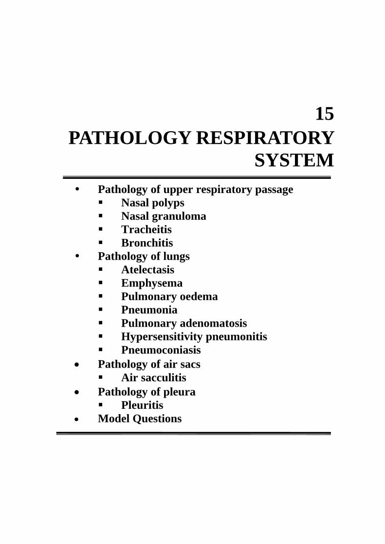
15
PATHOLOGY RESPIRATORY
SYSTEM
Pathology of upper respiratory passage
Nasal polyps
Nasal granuloma
Tracheitis
Bronchitis
Pathology of lungs
Atelectasis
Emphysema
Pulmonary oedema
Pneumonia
Pulmonary adenomatosis
Hypersensitivity pneumonitis
Pneumoconiasis
Pathology of air sacs
Air sacculitis
Pathology of pleura
Pleuritis
Model Questions
159
PATHOLOGY OF UPPER RESPIRATORY
TRACT
In many infectious diseases, there is inflammation
of mucosa of upper respiratory passage leading to
nasal discharge which is catarrhal, purulent or
fibrinous depending on the type of infection. The
infection may extend to lower parts of respiratory
tract and reach in lungs causing pathological
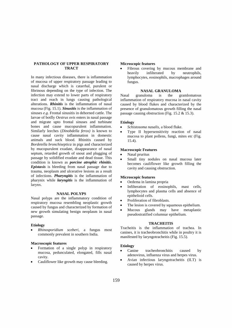
alterations. Rhinitis is the inflammation of nasal
mucosa (Fig. 15.1). Sinusitis is the inflammation of
sinuses e.g. Frontal sinusitis in dehorned cattle. The
larvae of botfly Oestrus ovis enters in nasal passage
and migrate upto frontal sinuses and turbinate
bones and cause mucopurulent inflammation.
Similarly leeches (Dinobdella ferox) is known to
cause nasal cavity inflammation in domestic
animals and suck blood. Rhinitis caused by
Bordetella bronchiseptica in pigs and characterized
by mucopurulent exudate, disappearance of nasal
septum, retarded growth of snout and plugging of
passage by solidified exudate and dead tissue. This
condition is known as porcine atrophic rhinitis.
Epistaxis is bleeding from nasal passage due to
trauma, neoplasm and ulcerative lesions as a result
of infections. Pharyngitis is the inflammation of
pharynix while laryngitis is the inflammation of
larynx.
NASAL POLYPS
Nasal polyps are the inflammatory condition of
respiratory mucosa resembling neoplastic growth
caused by fungus and characterized by formation of
new growth simulating benign neoplasm in nasal
passage.
Etiology
Rhinosporidium sceberi, a fungus most
commonly prevalent in southern India.
Macroscopic features
Formation of a single polyp in respiratory
mucosa, pedunculated, elongated, fills nasal
cavity.
Cauliflower like growth may cause bleeding.
Microscopic features
Fibrous covering by mucous membrane and
heavily infilterated by neutrophils,
lymphocytes, eosinophils, macrophages around
fungus.
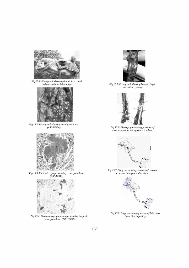
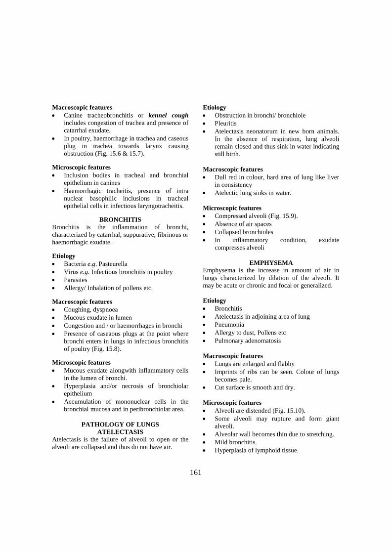
NASAL GRANULOMA
Nasal granuloma is the gramlomatous
inflammation of respiratory mucosa in nasal cavity
caused by blood flukes and characterized by the
presence of granulomatous growth filling the nasal
passage causing obstruction (Fig. 15.2 & 15.3).
Etiology
Schistosoma nasalis, a blood fluke.
Type II hypersensitivity reaction of nasal
mucosa to plant pollens, fungi, mites etc (Fig.
15.4).
Macroscopic Features
Nasal pruritus
Small tiny nodules on nasal mucosa later
becomes cauliflower like growth filling the
cavity and causing obstruction.
Microscopic features
Oedema in lamina propria
Infilteration of eosinophils, mast cells,
lymphocytes and plasma cells and absence of
epithelioid cells.
Proliferation of fibroblasts.
The lesion is covered by squamous epithelium.
Mucous glands may have metaplastic
pseudostratified columnar epithelium.
TRACHEITIS
Tracheitis is the inflammation of trachea. In
canines, it is tracheobronchitis while in poultry it is
manifested by laryngotracheitis (Fig. 15.5).
Etiology
Canine tracheobronchitis caused by
adenovirus, influenza virus and herpes virus.
Avian infectious laryngotracheitis (ILT) is
caused by herpes virus.
160
Fig.15.4. Photomicrograph showing causative fungus in
nasal granuloma (ARS/USDA)
Fig.15.3. Photomicrograph showing nasal granuloma
(ARS/USDA)
Fig.15.2. Photograph showing nasal granuloma
(ARS/USDA)
Fig.15.1. Photograph showing rhinitis in a camel
and catrrhal nasal discharge Fig.15.5. Photograph showing haemorrhagic
trachetis in poultry
Fig.15.8. Diagram showing lesions of Infectious
bronchitis in poultry
Fig.15.7. Diagram showing presence of caseous
exudates in largnx and trachea.
Fig.15.6. Photograph showing presence of
caseous exudate in largnx and trachea.
161
Macroscopic features
Canine tracheobronchitis or kennel cough
includes congestion of trachea and presence of
catarrhal exudate.
In poultry, haemorrhage in trachea and caseous
plug in trachea towards larynx causing
obstruction (Fig. 15.6 & 15.7).
Microscopic features
Inclusion bodies in tracheal and bronchial
epithelium in canines
Haemorrhagic tracheitis, presence of intra
nuclear basophilic inclusions in tracheal
epithelial cells in infectious laryngotracheitis.
BRONCHITIS
Bronchitis is the inflammation of bronchi,
characterized by catarrhal, suppurative, fibrinous or
haemorrhagic exudate.
Etiology
Bacteria e.g. Pasteurella
Virus e.g. Infectious bronchitis in poultry
Parasites
Allergy/ Inhalation of pollens etc.
Macroscopic features
Coughing, dyspnoea
Mucous exudate in lumen
Congestion and / or haemorrhages in bronchi
Presence of caseaous plugs at the point where
bronchi enters in lungs in infectious bronchitis
of poultry (Fig. 15.8).
Microscopic features
Mucous exudate alongwith inflammatory cells
in the lumen of bronchi.
Hyperplasia and/or necrosis of bronchiolar
epithelium
Accumulation of mononuclear cells in the
bronchial mucosa and in peribronchiolar area.
PATHOLOGY OF LUNGS
ATELECTASIS Atelectasis is the failure of alveoli to open or the
alveoli are collapsed and thus do not have air.
Etiology
Obstruction in bronchi/ bronchiole
Pleuritis
Atelectasis neonatorum in new born animals.
In the absence of respiration, lung alveoli
remain closed and thus sink in water indicating
still birth.
Macroscopic features
Dull red in colour, hard area of lung like liver
in consistency
Atelectic lung sinks in water.
Microscopic features
Compressed alveoli (Fig. 15.9).
Absence of air spaces
Collapsed bronchioles
In inflammatory condition, exudate
compresses alveoli
EMPHYSEMA
Emphysema is the increase in amount of air in
lungs characterized by dilation of the alveoli. It
may be acute or chronic and focal or generalized.
Etiology
Bronchitis
Atelectasis in adjoining area of lung
Pneumonia
Allergy to dust, Pollens etc
Pulmonary adenomatosis
Macroscopic features
Lungs are enlarged and flabby
Imprints of ribs can be seen. Colour of lungs
becomes pale.
Cut surface is smooth and dry.
Microscopic features
Alveoli are distended (Fig. 15.10).
Some alveoli may rupture and form giant
alveoli.
Alveolar wall becomes thin due to stretching.
Mild bronchitis.
Hyperplasia of lymphoid tissue.
162
Fig.15.12. Photomicrograph of lung showing
oedematous fluid in alveoli.
Fig-15.14. Diagram showing bronchogenous
spread of causal agent in lung.
Fig.15.13. Photograph of lamb showing signs of
pneumonia.
Fig.15.9. Photomicrograph of lung showing
atelectasis.
Fig.15.10. Photomicrograph lung showing
emphysema.
Fig.15.11. Photograph of lung showing
odema
Fig.15.15. Photomicrograph showing
bronchopneumonia.
Fig.15.16. Diagram showing hematogenous
spread of causal agent in lung.

163
PULMONARY OEDEMA
In pulmonary oedema, there is accumulation of
serous fluid in alveoli of lungs (Fig. 15.11 &
15.12).
Etiology
Bacteria
Virus
Allergy
Macroscopic features
Lungs become enlarged
Weight of lungs increases
Cut surface releases fluid and frothy exudate in
trachea and/or bronchi.
Microscopic features
Serous fluid accumulation in alveoli of lungs
Fluid may also be seen in some bronchi/
bronchioles.
Infilteration of inflammatory cells.
Congestion of lungs.
PNEUMONIA
Pneumonia is the inflammation of lungs
characterized by congestion and consolidation of
lungs. The pathological lesions in lungs are
produced in a similar way irrespective of the type
of etiological agent and includes various stages like
congestion, red hepatization, grey hepatization and
resolution.
Stage of congestion: This stage of lung is
characterized by active hyperemia and pulmonary
oedema. The capillaries are distended with
engorged blood and alveoli are filled with watery
serous exudate. This requires 2 minutes to few
hours to initiate the congestion.
Stage of red hepatization: This stage of lung is
characterized by the consolidation of lungs due to
accumulation of blood in blood vessels
(congestion). The consolidated lungs are firm and
looking like liver and hence the name “red
hepatization”. Such affected lung always sinks in
water. Alveoli are filled with serous or
serofibrinous exudate giving hardness to lungs. In
inflammatory condition, the neutrophils,
macrophages and lymphocytes along with
erythrocytes infilterate the affected area of lungs.
This stage of red hepatization takes 2 days for
development of firmness of lung.
Stage of grey hepatization: The lung remains hard
but due to lysis and removal of erythrocytes, it
becomes grey or less red in colour. Firmness/
hardness of lung remains same and thus, the name
grey hepatization. There is increase in infilteration
of inflammatory cells like macrophages,
lymphocytes, epithelioid cells depending on the
virulence of etiological agents.
Stage of resolution: After a week, the recovery
starts in the form of resorption of fluid; autolized
cells and debris is removed by phagocytic cells.
The causative organism is neutralized or removed
from the lungs through immunity of body. After
few days the lung parenchyma becomes normal and
starts functioning. If the causative agent is more
virulent, it may cause death of animal due to
respiratory failure or may cause permanent lesions
like formation of scar, carnification, granuloma etc.
There are various types of pneumonia caused by
bacteria, virus, fungi, parasites, allergens,
chemicals and all such affections of lungs are
classified as under.
BRONCHOPNEUMONIA
Bronchopneumonia is the inflammation of lungs
involving bronchi or bronchioles along with
alveoli. It is thought to be spread through
bronchogenous route and is the common type of
pneumonia in animals (Fig. 15.14 & 15.15).
Etiology
Virus
Bacteria
Chemicals
Mycoplasma
Chlamydia
Parasites
Fungus
Mainly through bronchogenous route
164
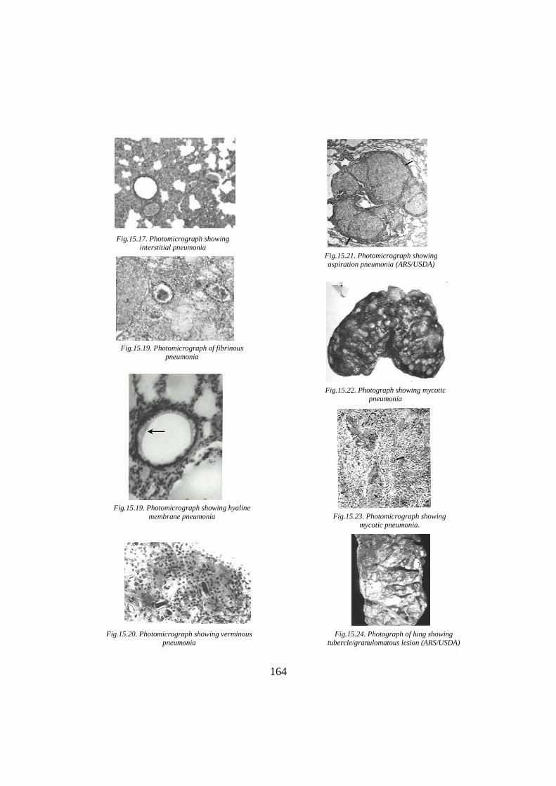
Fig.15.17. Photomicrograph showing
interstitial pneumonia
Fig.15.19. Photomicrograph of fibrinous
pneumonia
Fig.15.19. Photomicrograph showing hyaline
membrane pneumonia
Fig.15.20. Photomicrograph showing verminous
pneumonia
Fig.15.21. Photomicrograph showing
aspiration pneumonia (ARS/USDA)
Fig.15.24. Photograph of lung showing
tubercle/granulomatous lesion (ARS/USDA)
Fig.15.22. Photograph showing mycotic
pneumonia
Fig.15.23. Photomicrograph showing
mycotic pneumonia.
165
Macroscopic features
Congestion and consolidation of anterior and
ventral parts of lungs (Lobular pneumonia).
Patchy lesions on one or several lobes and
adjacent area shows emphysema.
Mediastinal lymphnodes are swollen.
Microscopic features
Congestion, oedema or haemorrhage in lung.
Infilteration of neutrophils, mononuclear cells
in and around bronchioles/ bronchi.
Catarrhal inflammation of bronchi.
Proliferation of bronchiolar epithelium
INTERSTITIAL PNEUMONIA
Interstitial pneumonia is the inflammation of the
lungs characterized by thickening of alveolar septa
due to serous/fibrinous exudate alongwith
infilteration of neutrophils and/or mononuclear
cells and proliferation of fibroblasts. It is also
known as lobar pneumonia (Fig. 15.16 & 15.17).
Etiology
Bacteria
Virus
Chlamydia
Parasites
Mainly through hematogenous route
Macroscopic features
Lungs are pale or dark red in colour.
Oedema, dripping of fluid from cut surface
Microscopic features
Alveoli may have serous or fibrinous exudate.
Thickening of alveolar septa due to
accumulation of exudate, inflammatory cells
and in chronic cases, proliferation of fibrous
tissue.
Infilteration of mononuclear cells in alveolar
septa.
FIBRINOUS PNEUMONIA
Fibrinous pneumonia is the inflammation of lungs
characterized by the presence of fibrin in alveoli or
bronchioles and may give rise to hyaline membrane
formation over the surface of alveoli or bronchiole.
Etiology
Bacteria
Virus
Parasites
Toxin/ Poisons
Macroscopic features
Antero-ventral portion of lung is congested
and consolidation.
Colour of lungs become deep red due to
congestion
Surface of lungs is covered by fibrin sheet.
Interlobular septa are prominent due to
accumulation of plasma and fibrin.
Microscopic features
Principal exudate is fibrin, fills alveoli,
bronchioles and bronchi (Fig. 15.18).
Congestion and/or haemorrhages
Infilteration of neutrophils, macrophages and
giant cells
Formation of eosinophilic false membrane of
fibrin over the surface of alveoli and
bronchiole and then known as “hyaline
membrane pneumonia” (Fig. 15.19).
VERMINOUS PNEUMONIA
Verminous pneumonia is caused by parasites and
characterized by the presence of lesions of
broncho-pneumonia along with parasites or their
larva (Fig. 15.20).
Etiology
Metastrongylus apri in pig.
Dictyocaulus filariae in sheep and goat.
D. viviparus in cattle and buffaloes.
Capillaria aerophila in dogs and cats.
D. arnfieldi in horse and donkeys.
Macroscopic features
Multiple petechial haemorrhage in lungs at the
site of parasite penetration.
166
Fig.15.28. Photograph showing pulmonary
adenomatosis (ARS/USDA)
Fig.15.32. Photograph showing air
sacculitis in poultry.
Fig.15.25. Photomicrograph of lung showing
tubercle.
Fig.15.26. Photomicrograph of lung showing
granulomatous lesions.
Fig.15.27. Photomicrograph of lung showing
granulomatous lesions and giant cells
Fig.15.29. Photomicrograph showing pulmonary
adenomatosis (ARS/USDA)
Fig.15.30. Photograph showing deposition of carbon
particles in trachea in chicks.
Fig.15.31. Photomicrograph showing pneumoconiasis
167
Mature worms in alveoli, bronchioles and
bronchi.
Mucopurulent exudate in alveoli/bronchi.
Pulmonary oedema, emphysema.
Microscopic features
Dilation of bronchiole/ bronchi
Lesions of chronic suppurative bronchiolitis
Focal areas of inflammation in the vicinity of
parasites and around bronchioles.
Hyperplasia of bronchiolar epithelium.
Infilteration of eosinophils and lymphocytes.
ASPIRATION PNEUMONIA
Aspiration pneumonia is caused by faulty
medication through drenching which reaches in
lungs instead of target place (digestive track) and
characterized by necrosis and gangrene of lung
paranchyma.
Etiology
Drugs, food, foreign body and oil drench
which reaches in lungs through trachea.
Paresis of throat predisposes the animal for
aspiration pneumonia.
Macroscopic features
Congestion and consolidation of anterior and
ventral portion of lung.
Affected part becomes green/ black in colour,
moist gangrene.
Affected lungs are often foul smelling.
Presence of foreign body like heads of wheats,
parts of corn, oil, milk etc.
Microscopic features
Thrombosis of blood vessels.
Necrosis in lungs.
Presence of saprophytes, leucocytes and
bacteria cause liquefaction and gangrene.
Gangrenous lesions surrounded by intense
inflammation (Fig. 15.21).
Congestion
MYCOTIC PNEUMONIA
Mycotic pneumonia is caused by a variety of fungi
and characterized by the presence of chronic
granulomatous lesions in lungs (Fig. 15.22 &
15.23).
Etiology
Aspergillus fumigatus
Blastomyces sp.
Cryptococcus sp.
Coccidioidomyces immitis
Macroscopic features
Nodules in lungs
On cut, cheese like caseative mass comes out
from nodules.
Caseation involves both bronchiole and
alveoli.
Such lesions may also present in trachea,
bronchi and air sacs.
Microscopic features
Presence of granulomatus lesions i.e. caseative
necrosis, macrophages, epithelioid cells,
lymphocytes, giant cells, fibroblasts etc.
Presence of branched hyphae of fungi in the
necrosed area.
TUBERCULOUS PNEUMONIA
Tuberculous pneumonia is caused by
Mycobacterium sp. and characterized by the
presence of chronic granulomatous lesions in the
lungs (Fig. 15.24 to 15.27).
Etiology
Mycobacterium tuberculosis
M. bovis
Macroscopic features
Grey, white or light yellowish nodules in
lungs.
Nodules are hard, painful and/or calcified.
Animal carcass is cachectic, weak or
emaciated.
On cut, the cheesy material comes out from the
nodules.
168
Microscopic features
Presence of tubercle/granuloma in lungs which
comprises a central necrosed area surrounded
by macrophages, epithelioid cells,
lymphocytes, Langhan’s giant cells and
covered by fibrous covering.
Acid-fast rod shaped bacteria may present in
necrosed area.
Central area may be calcified.
PULMONARY ADENOMATOSIS
Pulmonary adenomatosis is a slow viral disease of
sheep and characterized by metaplasia of alveolar
squamous epithelium to cuboidal and /or columnar
epithelium leading to glandular appearance of
alveoli (Fig. 15.28 & 15.29).
Etiology
Retrovirus
■ Pulmonary adenomatosis virus
Macroscopic features
Multiple focal areas of consolidation in lungs.
Imprint of ribs on lungs.
Congestion and hardening of mediastinal
lymphnodes.
Microscopic features
Metaplasia of alveolar epithelium leading to
formation of glandular structures in alveoli.
Metaplasia of simple squamous epithelium to
cuboidal or columnar epithelium which gives
alveoli a gland like look.
Mild inflammatory reaction.
Proliferation of fibrous tissue.
HYPERSENSITIVITY PNEUMONITIS
Hypersensitivity pneumonitis is the inflammation
of lung caused by an allergic reaction of antigen
(allergen) and characterized by interstitial
pneumonia, emphysema, hyaline membrane
formation and hyperplasia of alveolar epithelium.
Etiology
Allergens
Parasites – Dictyocaulus viviparous
Moldy hay
Fungus- Aspergillus sp.
Macroscopic features
Lobes may contain small grey foci
Presence of yellow and dense mucus in lumen
of bronchi
Excessive accumulation of air in lungs due to
emphysema
Presence of worms/larvae.
Microscopic features
Extensive infilteration of lymphocytes,
monocytes and eosinophils around the bronchi
and bronchioles.
Accumulation of catarrhal exudate in bronchi/
bronchiole.
Emphysema as a result of widening of alveoli.
Hyperplasia of bronchiolar musculature.
Inflammatory cells in interalveolar septa may
form small granulomas.
Formation of hyaline membrane over alveolar
and bronchiolar epithelium.
PNEUMOCONIASIS
Pneumoconiasis is the granulomatous inflammation
of lungs caused by aerogenous dust particles of
sand, silica, beryllium, carbon or asbestos. It is also
known as anthracosis (Fig. 15.30 & 15.31).
Etiology
Silica
Asbestos
Beryllium
Bauxite
Graphite
Carbon
Bronchogenous/aerogenous administration of
particles inhaled with air, mostly around
mines/factories.
Generator smoke.
Macroscopic features
Dense fibrous nodules in lungs.
Presence of carbon particles in trachea/bronchi
mixed with mucous exudate.

169
Microscopic features
Granuloma formation around the particles of
silica/ asbestos infilterated by macrophages,
lymphocytes and giant cells
Silica produces cellular reaction ‘Silicosis’.
Beryllium granuloma looks like tubercule
without caseation.
Asbestosis is characterized by the presence of
club shaped filaments bearing cells in lesion.
PATHOLOGY OF AIR SACS
AIR SACCULITIS
Air sacculitis is inflammation of air sacs caused by
E.coli, Mycoplasma, reovirus etc. and characterized
by thickening of the wall of air sacs and presence
of cheesy exudate (Fig. 15.32).
Etiology
Escherichia coli
Mycoplasma gallisepticum
Avian reovirus
Macroscopic features
Thickening of the air sac wall, which becomes
dirty and cloudy.
Presence of cheesy exudate in air sacs,
congestion of lungs.
Fibrinous pericarditis
Liver is covered with thin fibrinous membrane.
Microscopic features
Oedema and infilteration of neutrophils and
lymphocytes in air sacs
Caseous exudate in lungs and air sacs.
PATHOLOGY OF PLEURA
PLEURITIS
Pleuritis is the inflammation of pleura character-
-ized by serous, fibrinous or purulent exudate. It is
also known as pleurisy.
Etiology
Mycobacterium tuberculosis
Mycoplasma mycoides
Haemophilus suis
Organisms responsible for pneumonia/
traumatic pericarditis may also cause pleuritis.
Macroscopic features
Congestion of pleura
Serous, fibrinous or purulent exudate.
Accumulation of clear fluid in pleura/thoracic
cavity is called as hydrothorax.
Presence of blood in thoracic cavity is known
as Hemothorax.
Suppurative exudate in thoracic cavity is
known as pyothorax.
Presence of air in pleural cavity is termed as
pneumothorax, while presence of lymph in
pleural cavity is called as chylothorax.
Tuberculous pleuritis is characterized by small
nodules on pleura and is known as “pearly
disease”.
In chronic cases, development of fibrous tissue
causes adhesions and is known as adhesive
pleuritis.
Microscopic features
Congestion of blood vessels
Infilteration of neutrophils and lymphocytes.
Thickening of pleura due to oedema
Proliferation of fibroblasts producing adhesive
lesions.
MODEL QUESTIONS
Q. 1. Fill in the gaps with suitable word(s).
1. ................. is the inflammation of lungs characterized by ................. and ................. of lungs.
2. Lobar pneumonia is characterized by ................. of interalveolar septa.
3. Fibrinous pneumonia is characterized by the presence of ................. exudate in alveoli and may
give rise to.............formation which is............of fibrin over the surface of.............and ............
170
4. Aspiration pneumonia is caused by ................. of drugs/ milk and characterized by .................
and ................. formation in the lungs.
5. Mycobacterium tuberculosis produces ................. pneumonia in lungs characterized by
................. formation consisting of ................. central area surrounded by .................,
................., ................., ................., and covered by ................. capsule.
6. Pulmonary adenomatosis is caused by ................. and characterized by ................. of alveolar
squamous epithelium to ................. or ................. leading to ................. appearance of alveoli.
7. Allergic reaction due to ................. may cause ................. characterized by .................,
................., ................. and ................. of alveolar epithelium.
8. Pneumoconiasis is ................. inflammation of lungs caused by aerogenous................. of
................., ................. or ................. and it is also known as .................
9. Inflammation of air sacs in poultry is known as.................caused by ................., .................
and................. and characterized by................. and .................
10. ................. pleuritis is also known as................. while the presence of lymph in pleural cavity is
termed as.................
Q. 2. Write true or false against each statement and correct the false statements.
1. ...........Bronchopneumonia is the inflammation of lungs characterized by thickening of
interalveolar septa.
2. ...........Verminous pneumonia is caused by Bordetella bronchiseptica.
3. ...........Gangrenous pneumonia occurs due to faulty drenching of medicines.
4. ...........Mycotic pneumonia is caused by E. coli.
5. ...........Granulomatous pneumonia is produced by Blastomyces sp.
6. ...........Pearly disease is caused by Mycoplasma myoides.
7. ...........Atelectic lung floats in water.
8. ...........Oestrus ovis is the cause of nasal granuloma is sheep.
9. ...........Metaplasia of alveolar epithelium occurs in hypersensitivity pneumonitis.
10. ...........Air sacculitis is caused by E. coli.
Q.3. Define the followings.
1. Rhinitis 14. Tracheobronchitis
2. Sinusitis 15. Pneumothorax
3. Laryngitis 16. Red hepatization
4. Pharyngitis 17. Carnification
5. Hydrothorax 18. Lung worms
6. Pyothorax 19. Atelectasis neonatorum
7. Epistaxis 20. Bronchiolitis
8. Hyaline membrane 21. Beryllium granuloma
9. Silicosis 22. Peribronchitis
10. Asbestosis 23. Hemothorax
11. Pleurisy 24. Alveolitis
12. Chylothorax 25. Pearly disease
13. Adhesive pleuritis
171
Q. 4. Write short notes on.
1. Porcine atrophic rhinitis 9. Infectious laryngotracheitis
2. Nasal polyps 10. Emphysema
3. Nasal granuloma 11. Pulmonary adenomatosis
4. Atelectasis 12. Bronchopneumonia
5. Pathogenesis of pneumonia 13. Mycotic pneumonia
6. Lobar pneumonia 14. Granulomatous pneumonia
7. Hyaline membrane pneumonia 15. Air sacculitis
8. Gangrenous pneumonia
Q. 5. Match the word(s) from four options given against each statement.
1. Nasal polyps are caused by ...........
(a) Schistosoma nasalis (b) Rhinosporidium sceberi (c) E. coli (d) Mycoplasma mycoides
2. Canine tracheobronchitis is caused by...........
(a) Adenovirus (b) Influenza virus (c) Herpes virus (d)All of the above
3. Presence of caseous plugs in bronchi at the point of entrance in lungs in characteristic lesions
of ...........
(a) Infectious bronchitis (b) Infectious laryngotracheitis (c) Air sacculitis (d) Pleuritis
4. This is not the pathologic lesion of pneumonia...........
(a) Congestion (b) Red hepatization (c) Yellow hepatization (d) Resolution
5. Infection through aerogenous route may cause ...........pneumonia
(a) Lobar (b) Lobular (c) Hypersensitivity (d) Fibrinous
6. Verminous pneumonia is caused by ...........
(a) Mycoplasma (b) Chlamydia (c) Dictayocaulus sp. (d) E.coli
7. Langhan’s type giant cell is characteristic feature of ...........pneumonia
(a) Tuberculous (b) Verminous (c) Broncho (d) Pulmonary adenomatosis
8. Atelectasis neonatorum is characteristic features of ...........
(a) Premature birth (b) Aborted foetus (c) Still birth (d) None
9. Hypersensitivity pneumonitis is caused by ...........
(a) Allergens (b) Parasites (c) Moldy hay (d)All of the above
10. Pneumoconiasis is characterized by ...........lesions in lungs
(a) Serus (b) Fibrinous (c) Haemorrhagic (d) Granulomatous
![Respiratory system roadmap.pptx [Repaired] - Loginanatomical-sciences.health.wits.ac.za/roadmaps/Respiratory system... · DIVISION OF THE RESPIRATORY SYSTEM CONDUCTING PORTION Nasal](https://img.pdfslide.net/doc/110x75/5a78c3d87f8b9ae6228c9db0/respiratory-system-repaired-loginanatomical-scienceshealthwitsaczaroadmapsrespiratory.jpg)