Embed Size (px)
Citation preview
Asian Journal of Urology (2016) 3, 99e102
Available online at www.sciencedirect.com
ScienceDirect
journal homepage: www.elsevier .com/locate/ajur
CASE REPORT
Pediatric fibrous pseudotumor of the tunicavaginalis testis
Ryan Yu a,*, Jefferson Terry a, Mutaz Alnassar b, Jorge Demaria c
a Department of Pathology and Molecular Medicine, McMaster University, Hamilton, Ontario, Canadab Department of Radiology, McMaster University, Hamilton, Ontario, Canadac Department of Surgery, Division of Urology, McMaster University, Hamilton, Ontario, Canada
Received 9 September 2015; received in revised form 19 January 2016; accepted 19 January 2016Available online 2 March 2016
KEYWORDSAdolescent;Testis;Ultrasonography;Pathology
* Corresponding author.E-mail address: ryan.yu@medportaPeer review under responsibility
University.
http://dx.doi.org/10.1016/j.ajur.20162214-3882/ª 2016 Editorial Office of Athe CC BY-NC-ND license (http://crea
Abstract We describe a 16-year-old male with ultrasound evidence of a 1.3 cm right parates-ticular nodule, which was managed by intraoperative frozen section and excisional biopsy. Thepathologic findings were consistent with benign fibrous pseudotumor of the tunica vaginalistestis, which is a very rare lesion in the pediatric population. Consideration of fibrous pseudo-tumor in the differential diagnosis of pediatric paratesticular masses may help prevent unnec-essarily aggressive therapy.ª 2016 Editorial Office of Asian Journal of Urology. Production and hosting by Elsevier B.V. Thisis an open access article under the CC BY-NC-ND license (http://creativecommons.org/licenses/by-nc-nd/4.0/).
1. Introduction
Paratesticular fibrous pseudotumor is a rare, non-neoplastic, fibroproliferative lesion that arises mostcommonly from the tunica vaginalis, occasionally from theepididymis, and rarely from the spermatic cord and tunicaalbuginea [1]. It has been variably described in the litera-ture as fibroma, nodular fibropseudotumor, inflammatorypseudotumor, fibrous mesothelioma, non-specific
l.ca (R. Yu).of Second Military Medical
.02.003sian Journal of Urology. Productitivecommons.org/licenses/by-nc-
peritesticular fibrosis, nodular fibrous periorchitis, chronicproliferative periorchitis, reactive periorchitis, pseudofi-brous periorchitis, and peritesticular fibromatosis. Thewide variety of terms reflects its presentation, which iseither that of a grayewhite nodule (i.e., pseudotumor) or athick, fibrotic band that encases the testis (i.e., peri-orchitis). Although fibrous pseudotumor is benign, it isclinically important because it may mimic malignant tu-mors, such as rhabdomyosarcoma, leiomyosarcoma, anddesmoplastic small round cell tumor for which radical or-chiectomy is indicated. Occurrence of this lesion in thepediatric population is exceedingly rare and it may not beconsidered in the clinical differential diagnosis, leading tounnecessary treatment. We describe a teenage patientwith a fibrous pseudotumor of the tunica vaginalis testis.
on and hosting by Elsevier B.V. This is an open access article undernd/4.0/).

100 R. Yu et al.
2. Case report
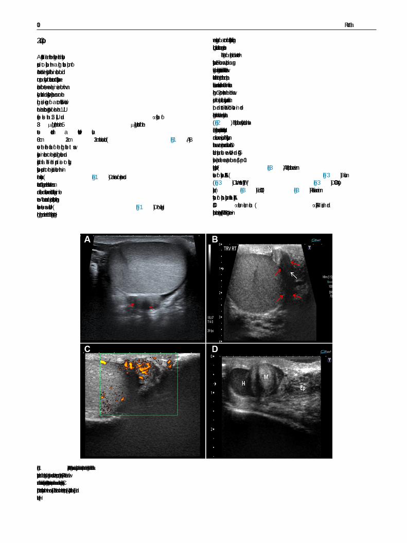
A 16-year-old male without significant medical historypresented to hospital with a right testicular lump of1-month duration. He played football in school, but didnot report a history of testicular trauma. On physical ex-amination, both testes were equal in size, but with aneasily visualized and superficially palpable mass on theright side, concerning for a tumor. Bloodwork showedtotal human chorionic gonadotropin of less than 1 IU/L(reference: less than 2.5 IU/L) and a-fetoprotein of3.8 mg/L (reference: less than 5 mg/L). Ultrasound of thetestes demonstrated a well-defined, oval,0.8 cm � 1.2 cm � 1.3 cm soft tissue nodule (Fig. 1A, B)over the inferior surface of the right testicle. It wasexophytic in relation to the adjacent right testicle andepididymal tail. The nodule appeared iso- to slightlyhypoechoic compared to the adjacent testicle with in-ternal vascularity (Fig. 1C) and areas of posterior soundattenuation. Acute angles were formed between the massand testicle and vessels were identified traversing in be-tween. A small amount of adjacent hydrocele containinglow-level echoes was identified (Fig. 1D). No enlargedright inguinal nodes were found. The sonographic findings
Figure 1 (A) Transverse sonogram shows an oval, iso-hypoechoicpoorly-demarcated areas of distal shadowing in keeping with densedemarcated from the testicle (white arrows). Shadowing fibrous comDoppler demonstrates vascularity within the mass. (D) Paratestichematocele (H).
were suggestive of a tumor of tunical/epididymal origin,including adenomatoid tumor among others.
The presence of a paratesticular nodule was confirmedintraoperatively. Frozen section was interpreted as sug-gestive of a benign connective tissue lesion. The nodule waswell-circumscribed from the adjacent testicular and para-testicular tissue and was excised with a 0.5 cm rim of tunicaalbuginea. On microscopic examination, the nodule wascomprised of spindle cells, lymphoplasmacytic inflamma-tion, and scattered thin-walled blood vessels in densecollagenous matrix with occasional less dense myxoid areas(Fig. 2). The spindle cells were focally clustered with ovalnuclei, single nucleoli, and open chromatin. Mitotic activityand necrosis were inconspicuous. The lymphoplasmacyticinfiltrate was most prominent around vessels. Occasionalmultinucleated plasma cells were identified and IgG4-positive plasma cells were present but rare (16 per 10high-power fields) (Fig. 3A). The spindle cells were immu-noreactive for cytokeratin AE1/AE3 (Fig. 3B), vimentin(Fig. 3C), Wilms tumor-1 (WT-1) (Fig. 3D), CD99 (cyto-plasmic) (Fig. 3E) and CD31 (Fig. 3F). There was no immu-noreactivity for anaplastic lymphoma kinase-1 (ALK-1),CD34, a-smooth muscle actin (a-SMA), desmin, andepithelial membrane antigen (EMA). The findings were in
soft tissue nodule posterior to the right testicle. The nodule hasfibrous stromal component (red arrows). (B) The mass is well-ponent obscures part of the mass and testicle (red arrows). (C)
ular mass with whorled pattern (M), epididymal tail (Ep) and

Figure 2 (A) Well-circumscribed tumor (H&E, 40�).(B) Abundant collagen (Trichrome, 40�). (C) Spindle cells,blood vessels, lymphocytes, and plasma cells in dense collag-enous stroma (H&E, 200�). (D) H&E, 400�.
Pediatric fibrous pseudotumor of the tunica vaginalis testis 101
keeping with a fibrous pseudotumor. No evidence ofrecurrence was found at 2 months follow-up.
3. Discussion
Besides fibrous pseudotumor, the differential diagnosis of aparatesticular mass in children includes other benign le-sions (such as lipoma, adenomatoid tumor, leiomyoma,hemangioma, and lymphangioma) and malignant lesions(most commonly rhabdomyosarcoma, rarely leiomyo-sarcoma and fibrosarcoma). Fibrous pseudotumor is rare,but represents the third most common tumor of the para-testicular tissues, after lipoma and adenomatoid tumor [2].The etiology is unclear but is thought to be related to prior
Figure 3 (A) Occasional IgG4-positive plasma cells (200�). (Bvimentin-positive spindle cells (200�). (D) Nuclear WT1-positive ce(F) CD31-positive spindle cells (200�).
trauma, although some have recently included this entity inthe spectrum of IgG4-related sclerosing disease [3]. It oc-curs most frequently in adults in the 3rd decade of life. It isvery rare in the pediatric population, with only few casesdescribed in the literature [4e11] including one of bilateralfibrous pseudotumors in an adolescent African Americanwith a history of medulloblastoma and meningioma [11].The clinical and pathologic features of pediatric fibrouspseudotumor overlap with those found in adult cases. Pa-tients present with scrotal swelling that is painless or ofmild discomfort. History of preceding trauma or infection iscommon, but not invariable. A single or sometimes multiplesmall nodules may be found on palpation. Transilluminationis negative. Serum b-human chorionic gonadotropin and a-fetoprotein are within normal limits, which helps todifferentiate this tumor from germ cell tumors.
Preoperative characterization of pediatric scrotalmasses is best accomplished with ultrasound because itprovides exceptional anatomic detail and distinguishesintratesticular from extratesticular lesions. In the diffusetype of fibrous pseudotumor, a band of tissue involving thetunica vaginalis surrounds the testis. Indentation of thetunica albuginea and displacement of the testis in thescrotal sac may be found. In the nodular type, one or morenodular masses are seen adjacent to the testis, which maybe indented or partially obscured by the nodules. Thenodules may appear well-marginated, lobulated, or poorly-defined. They may appear hyperechoic or hypoechoiccompared with the adjacent testicle, depending on theproportion of collagen, cells, and presence of calcification.Posterior acoustic shadowing can occur in the absence ofcalcification owing to dense stromal collagen. The nodulesusually exhibit a small to moderate amount of vascularityby color flow Doppler, but may be avascular. Detachment ofthe nodules produces floaters or scrotal pearls in the tun-ical space [12]. Hydrocele is an associated finding in about50% of cases. Occasionally, diffuse low-level echoes sug-gestive of hematocele or proteinaceous debris may beseen. The tunica albuginea may appear normal or focally
) AE1/AE3-positive spindle cells (200�). (C) Strong, diffusells (200�). (E) Cytoplasmic CD99-positive spindle cells (200�).

102 R. Yu et al.
thickened. The underlying testicular parenchyma usuallyappears normal or with mass effect related to adjacenttumor. When ultrasound evaluation of scrotal lesions isinconclusive, magnetic resonance (MR) imaging may beuseful as an adjunctive modality for further tissue charac-terization. Fibrous pseudotumor is expected to demon-strate low signal intensity on both T1- and T2-weightedimages because of the presence of fibrosis [13].
Intraoperative consultation is helpful in guiding themost appropriate management at the time of surgery.However, definitive diagnosis of paratesticular fibrouspseudotumor at the time of frozen section is challengingand often only a descriptive diagnosis is rendered [14].Historically, the diagnosis of fibrous pseudotumor wasusually established after radical orchiectomy. However,the nodular type of fibrous pseudotumor is frequentlyamenable to excision with testicle sparing. Radical orchi-ectomy may be unavoidable for the diffuse type. Micro-scopic examination and immunophenotyping usuallyconfirm the diagnosis, but genetic studies may be helpfulin situations where spindle cell lesions with diagnostically-useful cytogenetic findings, such as inflammatory myofi-broblastic tumor, desmoplastic small round cell tumor, andsynovial sarcoma, are being considered in the differentialdiagnosis. Electron microscopy may also be diagnosticallyuseful in establishing the fibroblastic/myofibroblastic na-ture of fibrous pseudotumor.
4. Conclusion
The urologist should be aware of the occurrence of para-testicular fibrous pseudotumors in pediatric patients as itsconsideration in the preoperative diagnosis may help avoidunnecessary radical orchiectomies.
Conflicts of interest
The authors declare no conflict of interest.
References
[1] Parker PM, Pugliese JM, Allen Jr RC. Benign fibrous pseudo-tumor of tunica vaginalis testis. Urology 2006;68.427.e17ee19.
[2] Germaine P, Simerman LP. Fibrous pseudotumor of thescrotum. J Ultrasound Med 2007;26:133e8.
[3] Bosmuller H, von Weyhern CH, Adam P, Alibegovic V, Mikuz G,Fend F. Paratesticular fibrous pseudotumor--an IgG4-relateddisorder? Virchows Arch 2011;458:109e13.
[4] Corcione N, Mancini P, Cecchi M, Pingitore R. Fibrous pseu-dotumor of tunica vaginalis. Report of a case. Pathologica1988;80:723e7.
[5] Vates TS, Ruemmler-Fisch C, Smilow PC, Fleisher MH. Benignfibrous testicular pseudotumors in children. J Urol 1993;150:1886e8.
[6] Atahan O, Atahan S, Kayigil O, Metin A. Fibrous pseudotumourof tunica vaginalis testis in childhood. Br J Urol 1995;75:795.
[7] Sonmez K, Turkyilmaz Z, Boyacio�glu M, Edali MN, Ozen O,Bas‚aklar AC, et al. Diffuse fibrous proliferation of tunicavaginalis associated with testicular infarction: a case report. JPediatr Surg 2001;36:1057e8.
[8] Zannoud M, Ghadouane M, Alami M, Benissa L, Amil T,Abbar M. Intrascrotal inflammatory pseudotumor (a casereport). Ann Urol (Paris) 2002;36:322e5.
[9] Pohl HG, Shukla AR, Metcalf PD, Cilento BG, Retik AB,Bagli DJ, et al. Prepubertal testis tumors: actual prevalencerate of histological types. J Urol 2004;172:2370e2.
[10] Zenker I, Schutz A, Sorge I, Trobs RB. Nodular periorchitismasquerading as a malignant parafunicular tumor in anadolescent. J Pediatr Surg 2006;41:e33e5.
[11] Kern SQ, McMann LP. Bilateral fibrous pseudotumors of thetunica albuginea in a pediatric patient. J Pediatr Urol 2012;8:e1e3.
[12] Yang DM, Kim HC, Lim SJ. Sonographic findings of fibrouspseudotumor of the tunica vaginalis. J Clin Ultrasound 2012;40:252e4.
[13] Cassidy FH, Ishioka KM, McMahon CJ, Chu P, Sakamoto K,Lee KS, et al. MR imaging of scrotal tumors and pseudotumors.Radiographics 2010;30:665e83.
[14] Gordetsky J, Findeis-Hosey J, Erturk E, Messing EM, Yao JL,Miyamoto H. Role of frozen section analysis of testicu-lar/paratesticular fibrous pseudotumours: a five-case experi-ence. Can Urol Assoc J 2011;5:E47e51.