Embed Size (px)
Citation preview
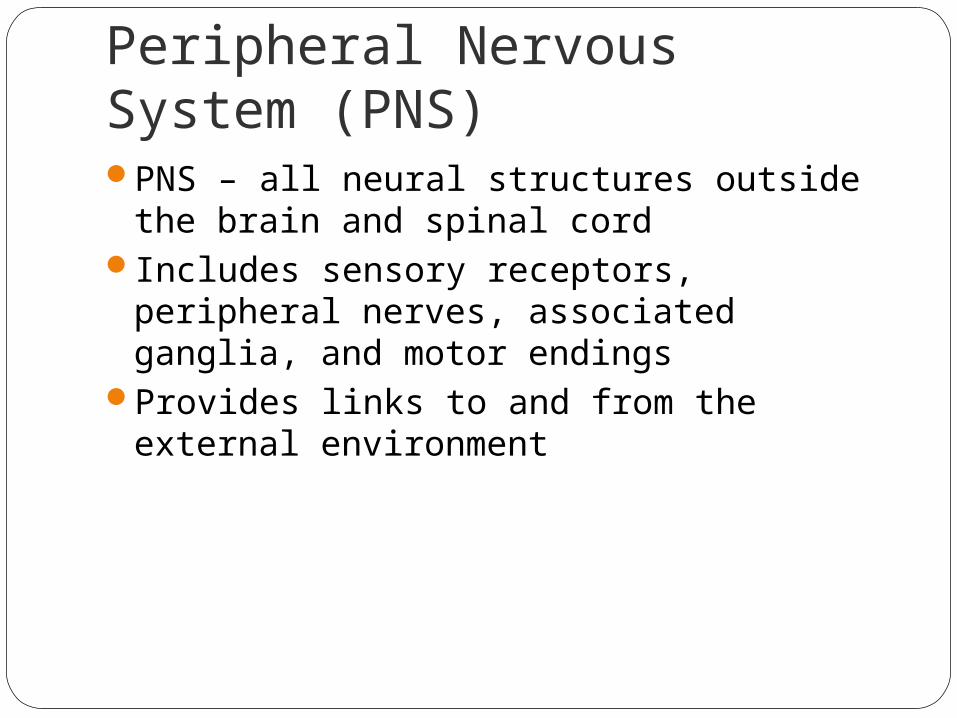
Peripheral Nervous System (PNS)PNS – all neural structures outside the brain
and spinal cordIncludes sensory receptors, peripheral
nerves, associated ganglia, and motor endings
Provides links to and from the external environment

PNS in the Nervous System
Figure 13.1

Sensory ReceptorsStructures specialized to respond to stimuliActivation of sensory receptors results in
depolarizations that trigger impulses to the CNS
The realization of these stimuli, sensation and perception, occur in the brain

Receptor Classification by Stimulus TypeMechanoreceptors – respond to touch,
pressure, vibration, stretch, and itchThermoreceptors – sensitive to changes in
temperaturePhotoreceptors – respond to light energy
(e.g., retina)Chemoreceptors – respond to chemicals
(e.g., smell, taste, changes in blood chemistry)
Nociceptors – sensitive to pain-causing stimuli

Receptor Class by Location: ExteroceptorsRespond to stimuli arising outside the bodyFound near the body surfaceSensitive to touch, pressure, pain, and
temperatureInclude the special sense organs

Receptor Class by Location: InteroceptorsRespond to stimuli arising within the bodyFound in internal viscera and blood vesselsSensitive to chemical changes, stretch, and
temperature changes

Receptor Class by Location: ProprioceptorsRespond to degree of stretch of the organs
they occupyFound in skeletal muscles, tendons, joints,
ligaments, and connective tissue coverings of bones and muscles
Constantly “advise” the brain of one’s movements

Receptor Classification by Structural Complexity
Receptors are structurally classified as either simple or complex
Most receptors are simple and include encapsulated and unencapsulated varieties
Complex receptors are special sense organs

Simple Receptors: Unencapsulated
Free dendritic nerve endingsRespond chiefly to temperature and pain
Merkel (tactile) discsHair follicle receptors

Simple Receptors: EncapsulatedMeissner’s corpuscles (tactile corpuscles) Pacinian corpuscles (lamellated corpuscles)Muscle spindles, Golgi tendon organs, and
Ruffini’s corpusclesJoint kinesthetic receptors

Simple Receptors: Unencapsulated
Table 13.1.1

Simple Receptors: Encapsulated
Table 13.1.2

From Sensation to PerceptionSurvival depends upon sensation and
perceptionSensation is the awareness of changes in
the internal and external environmentPerception is the conscious interpretation
of those stimuli

Organization of the Somatosensory SystemInput comes from exteroceptors,
proprioceptors, and interoceptorsThe three main levels of neural integration
in the somatosensory system are:Receptor level – the sensor receptorsCircuit level – ascending pathwaysPerceptual level – neuronal circuits in the
cerebral cortex

Processing at the Receptor LeverThe receptor must have specificity for the
stimulus energy The receptor’s receptive field must be
stimulatedStimulus energy must be converted into a
graded potentialA generator potential in the associated
sensory neuron must reach threshold

Adaptation of Sensory ReceptorsAdaptation occurs when sensory receptors
are subjected to an unchanging stimulusReceptor membranes become less
responsiveReceptor potentials decline in frequency or
stop
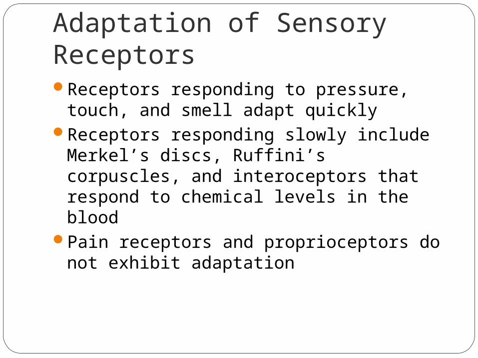
Adaptation of Sensory ReceptorsReceptors responding to pressure, touch,
and smell adapt quicklyReceptors responding slowly include
Merkel’s discs, Ruffini’s corpuscles, and interoceptors that respond to chemical levels in the blood
Pain receptors and proprioceptors do not exhibit adaptation

Processing at the Circuit Level
Chains of three neurons conduct sensory impulses upward to the brain
First-order neurons – soma reside in dorsal root or cranial ganglia, and conduct impulses from the skin to the spinal cord or brain stem
Second-order neurons – soma reside in the dorsal horn of the spinal cord or medullary nuclei and transmit impulses to the thalamus or cerebellum
Third-order neurons – located in the thalamus and conduct impulses to the somatosensory cortex of the cerebrum

Processing at the Perceptual LevelThe thalamus projects fibers to:
The somatosensory cortexSensory association areas
First one modality is sent, then those considering more than one
The result is an internal, conscious image of the stimulus

Main Aspects of Sensory PerceptionPerceptual detection – detecting that a
stimulus has occurred and requires summation
Magnitude estimation – how much of a stimulus is acting
Spatial discrimination – identifying the site or pattern of the stimulus

Main Aspects of Sensory PerceptionFeature abstraction – used to identify a
substance that has specific texture or shape
Quality discrimination – the ability to identify submodalities of a sensation (e.g., sweet or sour tastes)
Pattern recognition – ability to recognize patterns in stimuli (e.g., melody, familiar face)

Structure of a NerveNerve – cordlike organ of the PNS
consisting of peripheral axons enclosed by connective tissue
Connective tissue coverings include:Endoneurium – loose connective tissue that
surrounds axonsPerineurium – coarse connective tissue that
bundles fibers into fasciclesEpineurium – tough fibrous sheath around a
nerve

Structure of a Nerve
Figure 13.3b

Classification of NervesSensory and motor divisionsSensory (afferent) – carry impulse to the
CNSMotor (efferent) – carry impulses from CNSMixed – sensory and motor fibers carry
impulses to and from CNS; most common type of nerve
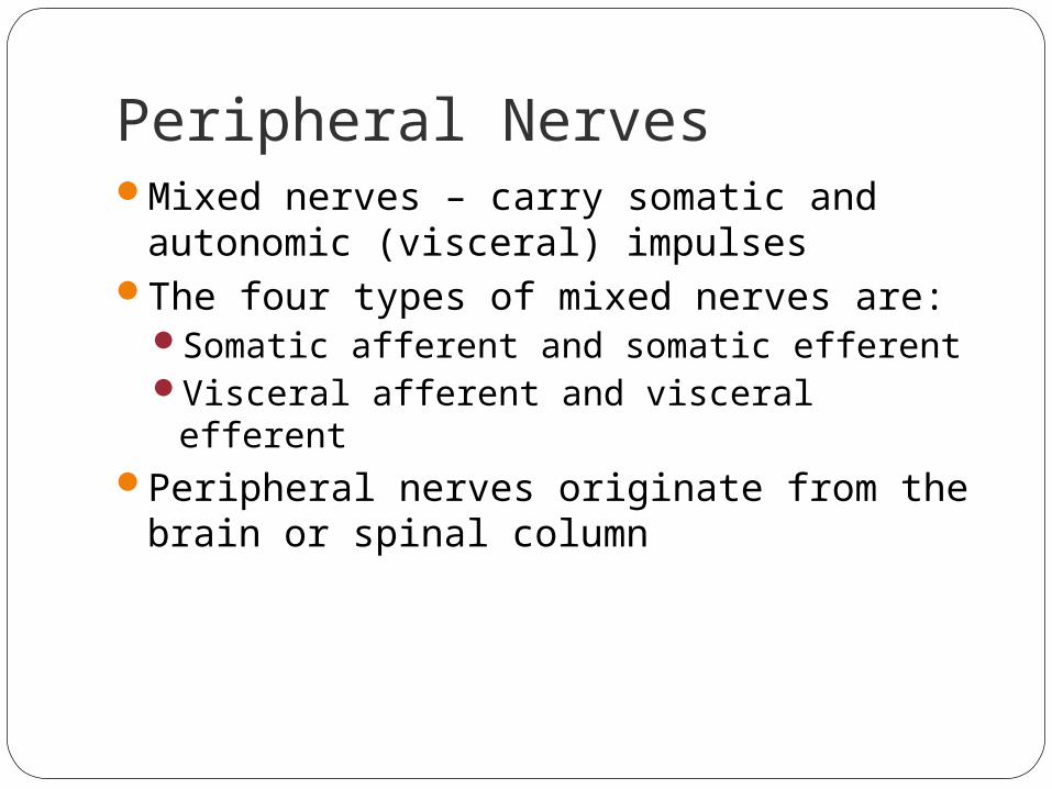
Peripheral NervesMixed nerves – carry somatic and
autonomic (visceral) impulsesThe four types of mixed nerves are:
Somatic afferent and somatic efferentVisceral afferent and visceral efferent
Peripheral nerves originate from the brain or spinal column

Regeneration of Nerve FibersDamage to nerve tissue is serious because
mature neurons are amitoticIf the soma of a damaged nerve remains
intact, damage can be repaired Regeneration involves coordinated activity
among:Macrophages – remove debrisSchwann cells – form regeneration tube and
secrete growth factorsAxons – regenerate damaged part

Regeneration of Nerve Fibers
Figure 13.4

Regeneration of Nerve Fibers
Figure 13.4

Cranial NervesTwelve pairs of cranial nerves arise from
the brain They have sensory, motor, or both sensory
and motor functionsEach nerve is identified by a number (I
through XII) and a nameFour cranial nerves carry parasympathetic
fibers that serve muscles and glands

Cranial Nerves
Figure 13.5a

Summary of Function of Cranial Nerves
Figure 13.5b

Cranial Nerve I: OlfactoryArises from the olfactory epitheliumPasses through the cribriform plate of the
ethmoid boneFibers run through the olfactory bulb and
terminate in the primary olfactory cortexFunctions solely by carrying afferent
impulses for the sense of smell

Cranial Nerve I: Olfactory
Figure I from Table 13.2

Cranial Nerve II: OpticArises from the retina of the eyeOptic nerves pass through the optic canals
and converge at the optic chiasmThey continue to the thalamus where they
synapseFrom there, the optic radiation fibers run to
the visual cortexFunctions solely by carrying afferent
impulses for vision

Cranial Nerve II: Optic
Figure II from Table 13.2

Cranial Nerve III: OculomotorFibers extend from the ventral midbrain,
pass through the superior orbital fissure, and go to the extrinsic eye muscles
Functions in raising the eyelid, directing the eyeball, constricting the iris, and controlling lens shape
Parasympathetic cell bodies are in the ciliary ganglia

Cranial Nerve III: Oculomotor
Figure III from Table 13.2

Cranial Nerve IV: TrochlearFibers emerge from the dorsal midbrain
and enter the orbits via the superior orbital fissures; innervate the superior oblique muscle
Primarily a motor nerve that directs the eyeball

Cranial Nerve IV: Trochlear
Figure IV from Table 13.2

Cranial Nerve V: TrigeminalThree divisions: ophthalmic (V1), maxillary
(V2), and mandibular (V3)Fibers run from the face to the pons via the
superior orbital fissure (V1), the foramen rotundum (V2), and the foramen ovale (V3)
Conveys sensory impulses from various areas of the face (V1) and (V2), and supplies motor fibers (V3) for mastication

Cranial Nerve V: Trigeminal
Figure V from Table 13.2

Cranial Nerve VI: Abducens
Fibers leave the inferior pons and enter the orbit via the superior orbital fissure
Primarily a motor nerve innervating the lateral rectus muscle
Figure VI from Table 13.2

Cranial Nerve VII: FacialFibers leave the pons, travel through the
internal acoustic meatus, and emerge through the stylomastoid foramen to the lateral aspect of the face
Mixed nerve with five major branchesMotor functions include facial expression, and
the transmittal of autonomic impulses to lacrimal and salivary glands
Sensory function is taste from the anterior two-thirds of the tongue
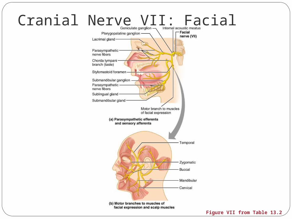
Cranial Nerve VII: Facial
Figure VII from Table 13.2

Cranial Nerve VIII: VestibulocochlearFibers arise from the hearing and
equilibrium apparatus of the inner ear, pass through the internal acoustic meatus, and enter the brainstem at the pons-medulla border
Two divisions – cochlear (hearing) and vestibular (balance)
Functions are solely sensory – equilibrium and hearing
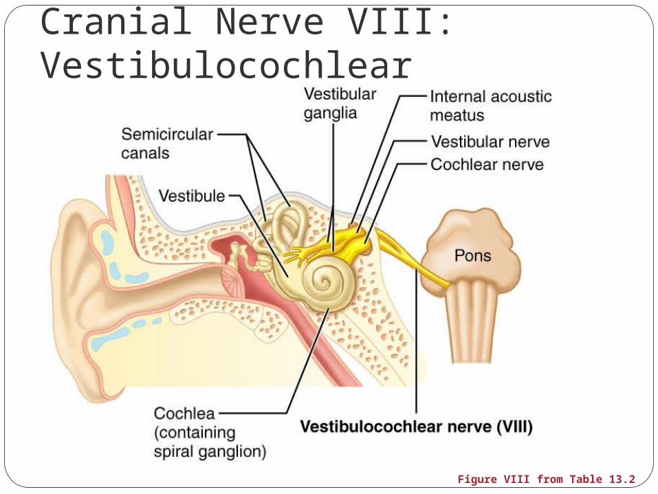
Cranial Nerve VIII: Vestibulocochlear
Figure VIII from Table 13.2

Cranial Nerve IX: GlossopharyngealFibers emerge from the medulla, leave the
skull via the jugular foramen, and run to the throat
Nerve IX is a mixed nerve with motor and sensory functions
Motor – innervates part of the tongue and pharynx, and provides motor fibers to the parotid salivary gland
Sensory – fibers conduct taste and general sensory impulses from the tongue and pharynx

Cranial Nerve IX: Glossopharyngeal

Cranial Nerve X: VagusThe only cranial nerve that extends beyond
the head and neckFibers emerge from the medulla via the
jugular foramenThe vagus is a mixed nerveMost motor fibers are parasympathetic
fibers to the heart, lungs, and visceral organs
Its sensory function is in taste

Cranial Nerve X: Vagus
Figure X from Table 13.2

Cranial Nerve XI: AccessoryFormed from a cranial root emerging from
the medulla and a spinal root arising from the superior region of the spinal cord
The spinal root passes upward into the cranium via the foramen magnum
The accessory nerve leaves the cranium via the jugular foramen

Cranial Nerve XI: AccessoryPrimarily a motor nerve
Supplies fibers to the larynx, pharynx, and soft palate
Innervates the trapezius and sternocleidomastoid, which move the head and neck

Cranial Nerve XI: Accessory
Figure XI from Table 13.2

Cranial Nerve XII: HypoglossalFibers arise from the medulla and exit the
skull via the hypoglossal canalInnervates both extrinsic and intrinsic
muscles of the tongue, which contribute to swallowing and speech

Cranial Nerve XII: Hypoglossal
Figure XII from Table 13.2

Spinal NervesThirty-one pairs of mixed nerves arise from
the spinal cord and supply all parts of the body except the head
They are named according to their point of issue8 cervical (C1-C8)12 thoracic (T1-T12)5 Lumbar (L1-L5)5 Sacral (S1-S5)1 Coccygeal (C0)

Spinal Nerves
Figure 13.6

Spinal Nerves: RootsEach spinal nerve connects to the spinal
cord via two medial rootsEach root forms a series of rootlets that
attach to the spinal cord Ventral roots arise from the anterior horn
and contain motor (efferent) fibersDorsal roots arise from sensory neurons in
the dorsal root ganglion and contain sensory (afferent) fibers

Spinal Nerves: Roots
Figure 13.7a

Spinal Nerves: RamiThe short spinal nerves branch into three or
four mixed, distal ramiSmall dorsal ramusLarger ventral ramusTiny meningeal branchRami communicantes at the base of the
ventral rami in the thoracic region

Nerve PlexusesAll ventral rami except T2-T12 form
interlacing nerve networks called plexusesPlexuses are found in the cervical, brachial,
lumbar, and sacral regionsEach resulting branch of a plexus contains
fibers from several spinal nerves

Nerve PlexusesFibers travel to the periphery via several
different routesEach muscle receives a nerve supply from
more than one spinal nerveDamage to one spinal segment cannot
completely paralyze a muscle

The back is innervated by dorsal rami via several branches
The thorax is innervated by ventral rami T1-T12 as intercostal nerves
Intercostal nerves supply muscles of the ribs, anterolateral thorax, and abdominal wall
Spinal Nerve Innervation: Back, Anterolateral Thorax, and Abdominal Wall

Spinal Nerve Innervation: Back, Anterolateral Thorax, and Abdominal Wall
Figure 13.7b

Cervical PlexusThe cervical plexus is formed by ventral
rami of C1-C4
Most branches are cutaneous nerves of the neck, ear, back of head, and shoulders
The most important nerve of this plexus is the phrenic nerve
The phrenic nerve is the major motor and sensory nerve of the diaphragm

Cervical Plexus
Figure 13.8

Brachial PlexusFormed by C5-C8 and T1 (C4 and T2 may also
contribute to this plexus)It gives rise to the nerves that innervate
the upper limb

Brachial PlexusThere are four major branches of this
plexus Roots – five ventral rami (C5-T1)Trunks – upper, middle, and lower, which
form divisionsDivisions – anterior and posterior serve the
front and back of the limbCords – lateral, medial, and posterior fiber
bundles

Brachial Plexus
Figure 13.9a

Brachial Plexus: NervesAxillary – innervates the deltoid and teres
minorMusculocutaneous – sends fibers to the
biceps brachii and brachialisMedian – branches to most of the flexor
muscles of armUlnar – supplies the flexor carpi ulnaris and
part of the flexor digitorum profundusRadial – innervates essentially all extensor
muscles

Brachial Plexus: Distribution of Nerves
Figure 13.9c

Brachial Plexus: Nerves
Figure 13.9b

Lumbar PlexusArises from L1-L4 and innervates the thigh,
abdominal wall, and psoas muscleThe major nerves are the femoral and the
obturator

Lumbar Plexus
Figure 13.10
Sacral PlexusArises from L4-S4 and serves the buttock,
lower limb, pelvic structures, and the perineum
The major nerve is the sciatic, the longest and thickest nerve of the body
The sciatic is actually composed of two nerves: the tibial and the common fibular (peroneal) nerves

DermatomesA dermatome is the area of skin innervated
by the cutaneous branches of a single spinal nerve
All spinal nerves except C1 participate in dermatomes

Dermatomes
Figure 13.12

Innervation of JointsHilton’s law: any nerve serving a muscle
that produces movement at a joint also innervates the joint itself and the skin over the joint

Motor EndingsPNS elements that activate effectors by
releasing neurotransmitters at:Neuromuscular junctions Varicosities at smooth muscle and glands

Innervation of Skeletal MuscleTakes place at a neuromusclular junctionAcetylcholine is the neurotransmitter that
diffuses across the synaptic cleftACh binds to receptors resulting in:
Movement of Na+ and K+ across the membrane
Depolarization of the interior of the muscle cell
An end-plate potential that triggers an action potential

Innervation of Visceral Muscle and GlandsAutonomic motor endings and visceral
effectors are simpler than somatic junctionsBranches form synapses en passant via
varicositiesAcetylcholine and norepinephrine are used
as neurotransmittersVisceral responses are slower than somatic
responses

Levels of Motor ControlThe three levels of motor control are
Segmental levelProjection levelPrecommand level

Hierarchy of Motor Control
Figure 13.13

Segmental LevelThe segmental level is the lowest level of
motor hierarchyIt consists of segmental circuits of the
spinal cordIts circuits control locomotion and specific,
oft-repeated motor activityThese circuits are called central pattern
generators (CPGs)

Projection LevelThe projection level consists of:
Cortical motor areas that produce the direct (pyramidal) system
Brain stem motor areas that oversee the indirect (multineuronal) system
Helps control reflex and fixed-pattern activity and houses command neurons that modify the segmental apparatus

Precommand LevelCerebellar and basal nuclei systems that:
Regulate motor activityPrecisely start or stop movementsCoordinate movements with postureBlock unwanted movementsMonitor muscle tone

ReflexesA reflex is a rapid, predictable motor
response to a stimulusReflexes may:
Be inborn (intrinsic) or learned (acquired)Involve only peripheral nerves and the spinal
cord Involve higher brain centers as well
Reflex ArcThere are five components of a reflex arc
Receptor – site of stimulusSensory neuron – transmits the afferent impulse
to the CNSIntegration center – either monosynaptic or
polysynaptic region within the CNSMotor neuron – conducts efferent impulses from
the integration center to an effectorEffector – muscle fiber or gland that responds to
the efferent impulse

Reflex Arc
Figure 13.14

Stretch and Deep Tendon ReflexesFor skeletal muscles to perform normally:
The Golgi tendon organs (proprioceptors) must constantly inform the brain as to the state of the muscle
Stretch reflexes initiated by muscle spindles must maintain healthy muscle tone

Muscle SpindlesAre composed of 3-10 intrafusal muscle fibers
that lack myofilaments in their central regions, are noncontractile, and serve as receptive surfaces
Muscle spindles are wrapped with two types of afferent endings: primary sensory endings of type Ia fibers and secondary sensory endings of type II fibers
These regions are innervated by gamma () efferent fibers
Note: contractile muscle fibers are extrafusal fibers and are innervated by alpha () efferent fibers

Muscle Spindles
Figure 13.15

Operation of the Muscle SpindlesStretching the muscles activates the
muscle spindleThere is an increased rate of action potential
in Ia fibersContracting the muscle reduces tension on
the muscle spindleThere is a decreased rate of action potential
on Ia fibers

Operation of the Muscle Spindle
Figure 13.17

Stretch ReflexStretching the muscle activates the muscle
spindleExcited motor neurons of the spindle cause
the stretched muscle to contractAfferent impulses from the spindle result in
inhibition of the antagonistExample: patellar reflex
Tapping the patellar tendon stretches the quadriceps and starts the reflex action
The quadriceps contract and the antagonistic hamstrings relax

Stretch Reflex
Figure 13.16

Golgi Tendon ReflexThe opposite of the stretch reflexContracting the muscle activates the Golgi
tendon organs Afferent Golgi tendon neurons are
stimulated, neurons inhibit the contracting muscle, and the antagonistic muscle is activated
As a result, the contracting muscle relaxes and the antagonist contracts

Golgi Tendon Reflex
Figure 13.18

Flexor and Crossed Extensor ReflexesThe flexor reflex is initiated by a painful
stimulus (actual or perceived) that causes automatic withdrawal of the threatened body part
The crossed extensor reflex has two partsThe stimulated side is withdrawnThe contralateral side is extended

Afferentfiber
Efferentfibers
Extensorinhibited
Flexorstimulated
Right arm(site of stimulus)
Left arm (site ofreciprocal activation)
Arm movements
Interneurons
Key:+ Excitatory synapse– Inhibitory synapse
Efferentfibers
Flexorinhibited
Extensorstimulated
+
–+
–
+
+
Flexes
Extends
Figure 13.19
Crossed Extensor Reflex

Superficial ReflexesInitiated by gentle cutaneous stimulationExample:
Plantar reflex is initiated by stimulating the lateral aspect of the sole of the foot
The response is downward flexion of the toesIndirectly tests for proper corticospinal tract
functioningBabinski’s sign: abnormal plantar reflex
indicating corticospinal damage where the great toe dorsiflexes and the smaller toes fan laterally

Developmental Aspects of the PNS
Spinal nerves branch from the developing spinal cord and neural crest cellsSupply motor and sensory function to
developing musclesCranial nerves innervate muscles of the
head

Developmental Aspects of the PNS
Distribution and growth of spinal nerves correlate with the segmented body plan
Sensory receptors atrophy with age and muscle tone lessens
Peripheral nerves remain viable throughout life unless subjected to trauma


![The Nervous System. Divisions of the Nervous System Central Nervous System [CNS] = Spinal Cord Brain Peripheral Nervous System [PNS]= Spinal Nerves](https://img.pdfslide.net/doc/110x75/56649d6c5503460f94a4c71d/the-nervous-system-divisions-of-the-nervous-system-central-nervous-system.jpg)