Embed Size (px)
Citation preview

PHD THESIS
CHARACTERIZATION OF SECRETED ASPARTYL
PROTEASES IN CANDIDA PARAPSILOSIS
DHIRENDRA KUMAR SINGH
SUPERVISOR:
PROF. DR. ATTILA GÁCSER
DOCTORAL SCHOOL OF BIOLOGY
DEPARTMENT OF MICROBIOLOGY
FACULTY OF SCIENCE AND INFORMATICS
UNIVERSITY OF SZEGED
2019
SZEGED

2
TABLE OF CONTENTS
LIST OF ABBREVIATIONS .................................................................................................. 6
1. INTRODUCTION ................................................................................................................ 8
1.1 Aspartyl proteases ............................................................................................................ 8
1.1.1 Human aspartyl proteases .......................................................................................... 9
1.1.2 Aspartyl proteases in fungi ...................................................................................... 10
1.1.3 Synthesis and processing of fungal aspartyl proteases ............................................ 10
1.1.4 Inhibitors of aspartyl proteases ................................................................................ 11
1.1.5 Regulation of aspartyl proteases .............................................................................. 12
1.2 Introduction of genus Candida....................................................................................... 13
1.2.1 Genral characterstics of Candida parapsilosis genome and morphology ............... 14
1.2.2 Biofilm formation .................................................................................................... 17
1.3 Candida parapsilosis: commensal to pathogen ............................................................. 18
1.3.1 Adherence and invasion .......................................................................................... 19
1.3.2 Virulence factors ..................................................................................................... 21
1.3.3 Hydrolytic enzymes of pathogenic Candida species and their role in virulence .... 21
1.3.4 Secreted aspartyl proteases of Candida parapsilosis .............................................. 22
1.4 Immune recognition and response against Candida species .......................................... 24
1.5 Overview of complement system and its activation ...................................................... 25
1.5.1 Complement system and regulation ........................................................................ 29
1.5.2 The FHL/FHR protein family .................................................................................. 29
1.5.3 Complement receptors ............................................................................................. 30
1.6 Interaction of complement cascade proteins with Candida species ............................... 31
1.7 Regulation of host complement cascade by Candida species ........................................ 32
1.7.1 Binding to complement regulators .......................................................................... 32
1.7.2 Regulation of crucial complement factors by aspartyl proteases ............................ 33
1.8 Sap mediated modulation of macrophage and neutrophil responses ............................. 34
1.9 Vaginal Candidiasis ....................................................................................................... 35

3
2. Objective of the present work ............................................................................................ 37
3. Material and methods ......................................................................................................... 38
3.1 Media used in this study ................................................................................................. 38
3.2 C. parapsilosis and E. coli strains used in the study and cultivation conditions ........... 38
3.3 Generation of sapp1/2/3−/−
and reintegrant mutant strains ............................................ 39
3.4 Validation of reintegrant mutant strain .......................................................................... 40
3.4.1 Fast track DNA isolation ......................................................................................... 41
3.4.2 PCR conditions ........................................................................................................ 41
3.4.3 Detection of PCR products ...................................................................................... 41
3.5 Southern blot analysis .................................................................................................... 41
3.5.1 Hybridization of the DIG-labeled probe to DNA .................................................... 41
3.5.2 Detection of the hybridized DIG-labeled probe ...................................................... 42
3.6 RNA extraction .............................................................................................................. 42
3.6.1 cDNA synthesis ....................................................................................................... 42
3.6.2 Quantitative real-time PCR ..................................................................................... 43
3.7 Phenotypic characterization of mutants by protease assay ............................................ 43
3.8 Growth curve analysis .................................................................................................... 43
3.9 Serum sensitivity assay .................................................................................................. 44
3.10 Biofilm formation assay ............................................................................................... 44
3.11 Pseudohyphae growth assay ......................................................................................... 44
3.12 Analysis of cell wall composition with microscopy imaging technique ...................... 44
3.13 Cultivation conditions of TR146 and A-431 cell lines ................................................ 45
3.14 Adhesion assay ............................................................................................................. 45
3.15 Isolation and differentiation of PBMCs ....................................................................... 46
3.16 Cell damage assay ........................................................................................................ 46
3.17 Macrophage killing assay ............................................................................................. 46
3.18 Whole blood killing assay ............................................................................................ 47
3.19 Phagocytosis assay ....................................................................................................... 47
3.20 Phagosome-lysosome fusion ........................................................................................ 47
3.21 Cytokine measurements ............................................................................................... 48

4
3.22 Detection of proteolytic activity of Sapp1p and Sapp2p ............................................. 48
3.23 Staining of macrophage CD11b/CD11c receptors ....................................................... 49
3.24 Galleria mellonella fungal burden and survival .......................................................... 49
3.25 Drosophila melanogaster infection .............................................................................. 50
3.26 Statistical analyses ....................................................................................................... 50
3.27 Ethical statement .......................................................................................................... 50
4. Results .................................................................................................................................. 52
4.1 Reintegration of the SAPP1, SAPP2, and SAPP3 genes C. parapsilosis
sapp1/2/3−/−
strain ................................................................................................................ 52
4.2 Relative transcription of SAPP genes in reintegrant C. parapsilosis strains ................. 53
4.3 Detection of protease activities of Sapp in WT and mutant strains of C.
parapsilosis .......................................................................................................................... 54
4.4 C. parapsilosis WT, sapp1/2/3−/−
, and RI_SAPP strains grow at similar rate ............... 55
4.5 C. parapsilosis SAPP3 reintegrant and sapp1/2/3−/−
mutant strains are more
sensitive to normal human serum ......................................................................................... 56
4.6 Deletion or reintegration of SAPP genes did not alter the pseudohyphae formation
abilities of C. parapsilosis.................................................................................................... 57
4.7 Deletion and reintegration of SAPP genes did not alter the biofilm formation in C.
parapsilosis .......................................................................................................................... 60
4.8 Secreted aspartyl proteases of C. parapsilosis affect its adhesion ................................. 61
4.9 SAPP null mutant C. parapsilosis is less capable of causing host-cell damage
whereas reintegration of SAPP1 and SAPP2 genes restored cell damage causing
capabilities ............................................................................................................................ 63
4.10 Macrophages phagocytosed sapp1/2/3−/−
mutant C. parapsilosis more efficiently
than WT, RI_SAPP1, RI_SAPP2, and RI_SAPP3 cells ....................................................... 64
4.11 SAPP2 influences the phagosome-lysosome fusion .................................................... 66
4.12 SAPP null mutant and RI_SAPP3 C. parapsilosis cells are killed more efficiently
by PBMC-DMs and in human whole blood compared to RI_SAPP1/2 strains ................... 67
4.13 SAPPs regulate the cytokine response of host macrophages ....................................... 68
4.14 Sapp1 and Sapp2 have differential cleavage capacity against human complement
proteins ................................................................................................................................. 69
4.15 Sapp2p but not Sapp1p can cleave FHR-5 ................................................................... 70
4.16 Sapps can not alter the expression of CR3 and CR4 on macrophage surface .............. 72

5
4.17 Galleria mellonella infection ....................................................................................... 73
4.18 Survival of Drosophila melanogaster after C. parapsilosis infection ......................... 75
5. Discussion ............................................................................................................................ 77
6. Summary .............................................................................................................................. 83
7. Hungarian summary ........................................................................................................... 86
8. Financial support ................................................................................................................ 88
9. Acknowledgement ............................................................................................................... 89
10. References .......................................................................................................................... 91
11. Supplementary Information .......................................................................................... 109

6
LIST OF ABBREVIATIONS
ALS Agglutinin-like sequence
AP Alternative pathway
BSA Bovine serum albumin
CFU Colony forming unit
CLR C type lectin receptor
CP Classical pathway
CR Complement receptor
CW Calcofluor white
DIG Digoxigenin
DMSO Dimethyl sulfoxide
DTT Dithiothreitol
EDTA Ethylene diamine tetra-acetic acid
ELISA Enzyme-linked immunosorbent assay
EtBr Ethidium bromide
FBS Fetal bovine serum
FH Factor H
FHL Factor H like
FHR Factor H related
FITC Fluorescein isothiocyanate
HiS Heat inactivated serum
IL Interleukin
LDH Lactate dehydrogenase
LiAc Lithium acetate
LP Lectin pathway
mt Mitochondria
NAT Nourseothricin N-acetyltransferase
NCAC Non Candida albicans Candida

7
NHS Normal human serum
NLRP Nucleotide-binding oligomerization domain, Leucine rich repeat
and Pyrin domain containing
ORF Open reading frame
PBS Phosphate buffered saline
PCR Polymerase chain reaction
PAGE Polyacrylamide gel electrophoresis
PBMC-DM Peripheral blood mono-nuclear cells derived macrophages
PS Penicillin-Streptomycin
PTX Pentraxin
qRT-PCR Quantitative real time polymerase chain reaction
RPMI Roswell Park Memorial Institute medium
RT Room temperature
SAP Secreted aspartyl protease
SDS Sodium dodecyl sulphate
SSC Saline-sodium citrate
TAE Tris-acetate-EDTA
TDH3 Glyceraldehyde-3-phosphate dehydrogenase 3
TF Transcription factor
TLR Toll like receptor
TMB Tetramethylbenzidine
TNF Tumor necrosis factor
TRIS Tris- (hydroxymethyl) aminomethane
UV Ultra Violet
WGA-TRITC Wheat germ agglutinin conjugated to tetramethylrhodamine
isothiocyanate-dextran
XTT
2-methoxy-4-nitro-5-sulfophenyl)-5-[(phenylamino)-carbonyl]-2H
tetrazolium
YCB Yeast carbon base
YNB Yeast nitrogen base

8
1. INTRODUCTION
1.1 Aspartyl proteases
Aspartyl proteases[1]
are also named as aspartic-, aspartate-, or acid proteases (enzyme
class: 3.4.23). They are a family of proteolytic enzymes that usually functional at acidic
pH (pH 1.9–4.0). Aspartyl proteases belong to endopeptidase family, share common
catalytic apparatus and cleave dipeptide bonds between two hydrophobic amino acid
residues. Aspartyl proteases have a conserved sequence Asp-Gly-Thr (DTG) and/or
Asp-Ser-Gly (DSG) at the active site and highly susceptible to pepstatin (pentapeptides
produced by various species of Actinomyces)1,2
. These proteases are present in diverse
range of organisms including protozoa, viruses, bacteria, plants, and higher vertebrates
such as humans2–6
. Some of these important proteases are described in table 1. In higher
organisms, aspartyl proteases play range of important roles including protein processing
(renin, cathepsin D, gastric enzymes) and degradation, whereas, viral, bacterial and
fungal secreted aspartyl proteases are crucial for their nutrient acquisition and
virulence7,8
. Mostly, aspartyl proteases are synthesized as single chain proenzymes or
zymogens (inactive precursor of enzymes) having molecular weight (MW)
approximately 40,000 Da. These proenzymes are further processed to form active
enzyme (approx. MW 35,000 Da) by removing 45 residues from the N-terminal peptide
residues. One of the oldest studied aspartyl proteases, pepsin has substrate-binding cleft
with two catalytically competent aspartic (Asp) residues (Fig. 1). These aspartic
residues are present in the center of the cleft formed by two domains and large enough
to accommodate a substrate or inhibitor with seven amino acid residues9. Hypothesis of
sharing common catalytic site by all aspartyl proteases also supported by the fact that
they are universally inhibited by pepstatin (exception includes secreted aspartic protease
from Candida albicans: Sap7)10,11
. Catalytic mechanism of aspartyl proteases which
generally involves the hydrolysis of amide bond through an active site water molecule
named as “push-pull” mechanism is now generally accepted and described elsewhere in
detail3,12
.
[1]
In this thesis term aspartyl protease is used uniformly for aspartic and aspartate proteases.

9
1.1.1 Human aspartyl proteases
There are several well studied human aspartyl proteases including pepsin, gastricsin,
cathepsins D and E. Pepsin is one of the predominant aspartyl protease produced by
fundus (upper part of the human stomach). Pepsin is secreted in its inactive form called
pepsinogen which further processed by hydrochloric acid released by stomach parietal
cells to form active form of
pepsin. In the acidic
environment of the stomach
(pH 1.5-2.5) pepsin
hydrolyzes the peptide
bonds of most of the
proteins, breaking them
down into smaller
polypeptide fragments13
.
Role of pepsin in
inflammation has also been
investigated in some
clinical studies14,15
. One
such study highlights the inflammatory role of pepsin in otitis media, a group of
inflammatory diseases of the middle ear. This study clearly showed that the presence of
higher concentration of pepsin A in nasopharynx correlates with higher level of
inflammatory cytokines, such as tumor necrosis factor-α (TNFα), interleukin-6 (IL-6),
and interleukin-8 (IL-8). Increased level of pro-inflammatory cytokines aggravates
preexisting disease condition of otitis media15
. Being a soluble lysosomal aspartyl
endopeptidase, cathepsin D (CD) is synthesized as preprocathepsin D and transported to
intracellular compartments such as to lysosomes, endosomes, phagosomes after removal
of the signal peptide. Removal of 44 amino acids (aa) N-terminal from propeptide in
acidic endosomal and lysosomal compartment results in a 48 kDa single-chain
intermediate active enzyme16
. CD plays many crucial roles in degradation and
activation of polypeptide hormones and growth factors, activation and processing of
Figure 1: Roles of human aspartyl proteases. Human
aspartyl proteases play various roles in cellular processes.

10
enzymes, antigen processing and regulation of caspase-dependent cell death (Fig.1)16
.
Role of human CD in the metastasis of breast cancer and in Alzheimer‟s disease is
under extensive investigation16–18
. Renin is also an important factor in body‟s
homeostasis. Secreted mainly by the kidney and some other tissues such as brain and
the adrenal gland, renin plays an important role in controlling the blood pressure and
maintaining body homeostasis, bone marrow differentiation, and regulating defense
mechanism against injuries19–21
. These studies show the important roles of aspartyl
proteases in human physiology and disease which signifies their importance as an
alternative target to treat disease.
1.1.2 Aspartyl proteases in fungi
Several fungi from order Mucorales and some species of genus Aspergillus are the
source of commercially important aspartyl proteases. Members of genera Mucor,
Rhizomucor, Absidia, and Cunninghamella are commonly used in industry as microbial
sources of renins and other aspartyl protease22,23
. Endothia parastica, a member of
Ascomycota is also reported to produce renin24
.
Aspergillopepsin produced by different Aspergillus species; mucorpepsin produced by
Rhizomucor miehei; penicillopepsin by Penicillium janthinellum; rhizopuspepsin by
Rhizopus microsporus; endothiapepsin from Cryphonectria parasitica;
trichodermapepsin from Trichoderma reesei; saccharopepsin/proteinase A/Yapsins
from Saccharomyces cerevisiae, candidapepsin (Sap1-10) from C. albicans;
candidaparapepsin (Sapp1-3) from C. parapsilosis; canditropsin (Sapt1-8) from C.
tropicalis are known25–27
.
1.1.3 Synthesis and processing of fungal aspartyl proteases
Secretory pathway of fungal aspartyl proteases is best studied in C. albicans. In C.
albicans newly synthesized mRNA is transported to the cell cytosol, where it binds to
the ribosome and gets translated into the pre-proenzyme and enters to rough
endoplasmic reticulum (ER) through 16–18 residues of signal sequence (N-terminal
hydrophobic peptide) mediated entry. In ER, N-terminal signal peptide is removed by

11
specific peptidase. As the journey progresses the proenzyme is transferred to the Golgi
apparatus which leads it further processing by a Kexin-like proteinases (Kex2
proteinase). Kex2 recognizes Lys-Arg sequence and removes N-terminal extension.
This N-terminal sequence also known as pro-region or pro-part or pro-peptide and
serves as a stabilizer of inactive form of protein. It also play role in proper folding of the
domains and further keeping zymogens inactive28
. Once packed into the secretory
vesicles, the enzymes are transported to the plasma membrane, where it may later be
incorporated into the cell wall or released to the extracellular micro-niche of the host.
1.1.4 Inhibitors of aspartyl proteases
Role of aspartyl proteases received enormous attention because of their significance in
human diseases, such as renin in hypertension, cathepsin D in cancer, and the HIV
proteases in acquired immune deficiency syndrome (AIDS). Therefore, designing of
inhibitor drugs against aspartyl proteases could be an effective and medically relevant
therapy. Scientists have successfully designed inhibitors of aspartyl proteases by
mimicking the transition state formed during amide hydrolysis of a protein substrate by
an aspartyl protease29
.
Aspartyl proteases are universally inhibited by pepstatin, a peptide produced by various
species of Streptomyces30,31
.
Pepstatin also inhibits the
activities of cathepsin D,
cathepsin E, renin,
pseudorenin and aspartyl
proteases produced by
microorganisms. Recently,
one study showed that C.
albicans aspartyl protease 7
(Sap7) is insensitive to
pepstatin11
. 1,2-epoxy-3-(p-
nitrophenoxy) propane
Figure 2: Inhibitory activity of HIV-1 protease against
secreted aspartyl protease from pathogenic Candida
species. HIV protease inhibitor ritonavir and saquinavir
inhibit the activity of aspartyl proteases secreted by
different Candida species.
12
(EPNP) and diazoacetyl-DL-norleucine methyl ester (DAN) are also known to inhibit
aspartyl proteases by binding to their active site22,32
. Low molecular weight aspartyl
protease inhibitor (API) isolated from a thermotolerant Bacillus licheniformis show
higher inhibiting affinity against pepsin whereas, they show very weak inhibitory
activity against other aspartyl proteases. Several HIV-1 protease inhibitors such as
saquinavir, indinavir, nelfinavir, and ritonavir are used to improve the clinical outcome
of HIV patients33,34
. Inhibitory activity of HIV-1 protease inhibitors was also tested
against aspartyl proteases from Candida spp. It has been shown that aspartyl proteases
secreted from C. tropicalis, C. parapsilosis, and C. lusitaniae are inhibited by ritonavir
and saquinavir but not by other HIV protease inhibitors (Fig. 2)35
.
1.1.5 Regulation of aspartyl proteases
In general, pH is the principal factor behind the regulation of the gene expression of
fungal aspartyl proteases. Proteolysis of PacC (transcription factor) at alkaline pH
generates active form of PacC protein, which, negatively regulate the genes expressed
under acidic conditions, e.g. genes encoding aspartyl proteases36,37
. With this, in
pathogenic fungi, such as in Candida spp. expression of aspartyl protease encoding
genes are also associated with several environmental as well as morphological
attributes, including white to opaque switching, yeast to hyphae transition, change in
temperature, presence of nitrogen and carbon sources, and with the host response (Fig.
3)38,39
.

13
Figure 3: Number of environmental factors affecting fungal secretom. Several factors are
reported to activate transcription factors which regulate the expression of genes responsible for
aspartyl protease production.
1.2 Introduction of genus Candida
Christine Marie Berkhout proposed genus “Candida” in 1923 for a group of “thrush”
fungi which was originally inaccurately classified as Monilia40
. In 1954, the name C.
albicans was adopted as nomen conservandum (i.e., conserved name). Species C.
parapsilosis was described first time in 1928 by B. Ashford in samples originated from
Puerto Rico, and classified as Monilia parapsilosis41,42
. In 1932, this species has been
reclassified as Candida by M. Langernon and R. Talice43,44
.
Unlike other fungal representative species, Candida belongs to „CTG-Ser clade‟ (except
C. glabrata and C. krusei) in which CTG codes amino acid serine instead of leucine45
.
Members of CTG clade are highly diverse in both genotype and phenotype, such as C.
guilliermondii, C. lusitaniae, and Debaryomyces hansenii are haploid, whereas, the
other members are diploid. Majority of clinically important Candida species are part of
this „CTG-Ser clade‟ (Fig. 4). Series of studies using murine model have shown that C.
albicans is the most virulent member of this genus, followed by C. tropicalis, C.
parapsilosis, C. glabrata, C. krusei, and C. guilliermondii46–49
.

14
Figure 4: Phylogenetic tree representing closely related Candida species belonging to CTG
clade46.
1.2.1 General characteristics of Candida parapsilosis genome and morphology
A ubiquitous presence in nature and existence as free-living saprobes, easy transition
from being a commensal to a pathogen of humans or animals makes fungus one of the
prevalent cause of disease. Members of genus Candida reside in healthy hosts without
causing any notable damage. Being a commensal pathogen, C. albicans easily and
quickly adapts to host‟s environmental changes50,51
. These ordinarily harmless
commensal microorganisms may cause a variety of infections in humans, most
commonly called candidiasis. Members of opportunistic pathogenic Candida species
generally colonize on mucosal surfaces (oral, vaginal or gastrointestinal tract) and
disseminate into the organs of the human host. C. albicans fits within all six classes of
the „damage-response‟ framework (DRF) of microbial pathogenesis which mainly
defines microbial pathogenesis as an outcome of the interaction between a host and a
microorganism52–54
. Out of approximately 150 known Candida species, 95% of
infections are caused by only five species: C. albicans, C. tropicalis, C. parapsilosis, C.
glabrata and recently included C. auris55–57
. The most important representative of
pathogenic Candida spp. is C. albicans which is studied more extensively than any
other Candida spp. Nonetheless, the clinical significance of non Candida albicans

15
Candida (NCAC) species increased over the past years and more attention is being paid
to species like C. tropicalis, C. parapsilosis, C. auris, and C. glabrata.
C. parapsilosis belongs to a
heterogeneous taxon, also
referred as C. parapsilosis sensu
lato. Previously, members of
this species are divided into
three distinct groups designated
I to III but later they were
reclassified in three separate
“psilosis” species. C.
parapsilosis (clause sensu
stricto) is classified as a member
of group I, C. orthopsilosis in
group II and C. metapsilosis in
group III58
(Fig. 5).
In several clinical studies C. parapsilosis sensu stricto remained the more frequently
isolated (approximately 90%
of all isolates) “psilosis”
species followed by C.
orthopsilosis (nearly 10%)
and C. metapsilosis59,60
.
Recently, it has been shown
that C. orthopsilosis isolates
are capable of causing
damage to epithelial cells and
epidermal tissues, whereas
C. metapsilosis is less
effective61
. C. parapsilosis
Figure 5: Phylogeny of C. parapsilosis. Phylogenetic
tree representing relationship of C. parapsilosis with
other closely related species70.
Figure 6: Cell morphology of C. parapsilosis (GA1).
Bold red-pseudohyphae; and light blue arrows-budding
cells. Scale bar 10μm.
16
classified as a diploid organism and forms small chain of budding cells or
pseudohyphae (Fig. 6)41
. In pseudohyphal form of C. parapsilosis elongated mother and
daughter cells are separated by septa and form chain like structures.
C. albicans was one of the first eukaryotic pathogens to have its genome sequenced and
immediately after that followed by other NCAC species. C. parapsilosis possess 13.1
Mb nuclear genome which is smaller than that of C. albicans (16 Mb)62
. C. parapsilosis
reported to possess 8 chromosomes and 30.9 kb long linear double-stranded
mitochondrial (mt) DNA terminating with of a 738 bp of tandem repeats. mt DNA of C.
parapsilosis is highly compact with more than 90% corresponds to coding sequences
for respiratory enzymes, ribosomal RNA, transfer RNA, and other proteins important
for mitochondrial functions63,64
. C. parapsilosis also lacks sexual cycle which is present
in some Candida members such as C. guilliermondii and C. lusitaniae, which is
justified by the presence of mating-type locus (MTLα) as a pseduogene63
. In contrast to
C. albicans which forms true hyphae, C. parapsilosis forms pseudohyphae (Fig. 6).
Hyphal/pseudohyphal form of pathogenic members of Candida genus is directly related
to its virulence and can be induced by specific environmental and cellular signals.
Hyphal/pseudohyphal forms of C. albicans and C. parapsilosis possesses ability to
adhere (ALS: agglutinin-like sequence expressed on hyphal form and critical for
adhesion) and penetrate more efficiently in host tissues65–67
.
C. parapsilosis forms white and
creamy colonies with variable
morphology. Laffey and
coworker described four stable
and heritable colony phenotypes
including concentric, smooth,
crater and most common crepe
phenotype in C. parapsilosis
CLIB21468
. These colony
phenotypes are also closely
related to cell morphology as crepe and concentric phenotypes are mostly associated
Figure 7: Colony morphology of C. parapsilosis in
spider media.
17
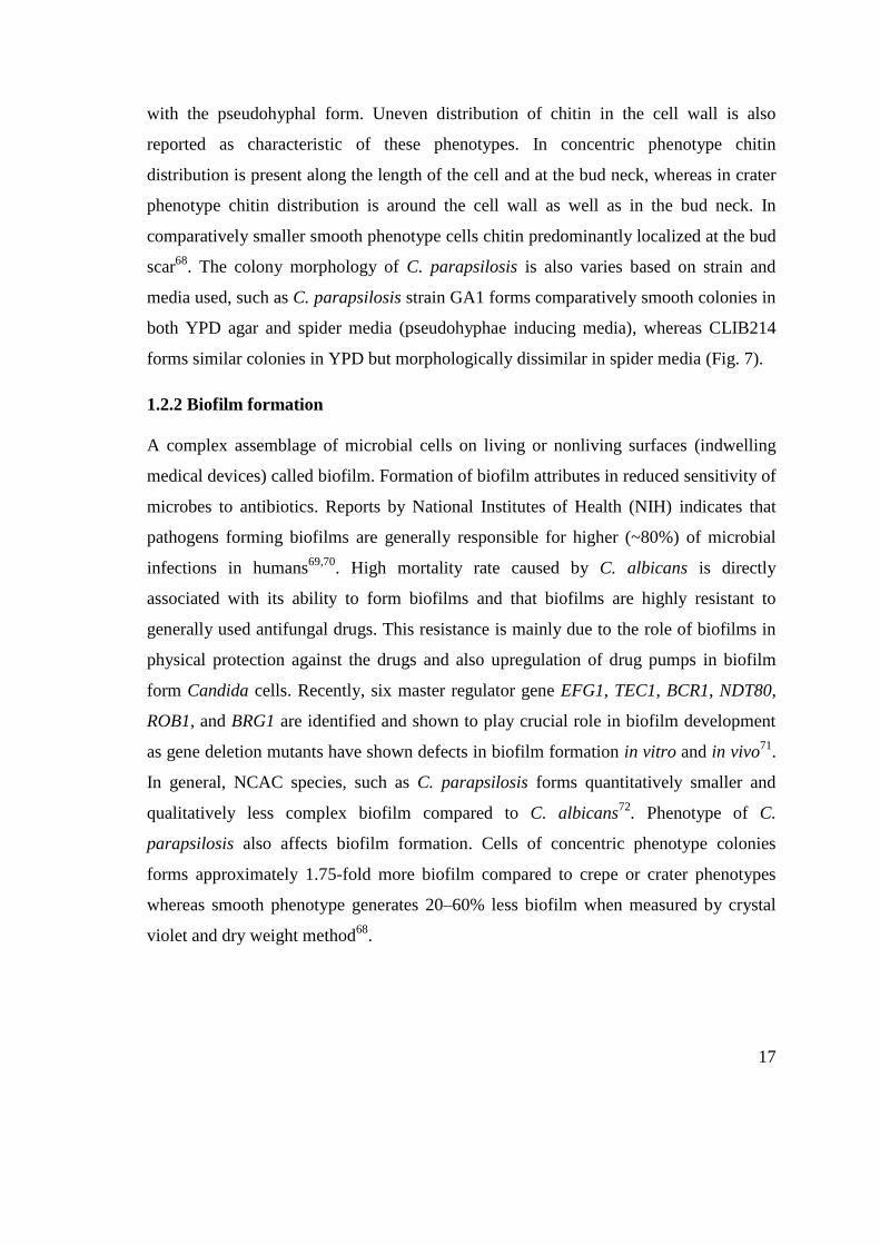
with the pseudohyphal form. Uneven distribution of chitin in the cell wall is also
reported as characteristic of these phenotypes. In concentric phenotype chitin
distribution is present along the length of the cell and at the bud neck, whereas in crater
phenotype chitin distribution is around the cell wall as well as in the bud neck. In
comparatively smaller smooth phenotype cells chitin predominantly localized at the bud
scar68
. The colony morphology of C. parapsilosis is also varies based on strain and
media used, such as C. parapsilosis strain GA1 forms comparatively smooth colonies in
both YPD agar and spider media (pseudohyphae inducing media), whereas CLIB214
forms similar colonies in YPD but morphologically dissimilar in spider media (Fig. 7).
1.2.2 Biofilm formation
A complex assemblage of microbial cells on living or nonliving surfaces (indwelling
medical devices) called biofilm. Formation of biofilm attributes in reduced sensitivity of
microbes to antibiotics. Reports by National Institutes of Health (NIH) indicates that
pathogens forming biofilms are generally responsible for higher (~80%) of microbial
infections in humans69,70
. High mortality rate caused by C. albicans is directly
associated with its ability to form biofilms and that biofilms are highly resistant to
generally used antifungal drugs. This resistance is mainly due to the role of biofilms in
physical protection against the drugs and also upregulation of drug pumps in biofilm
form Candida cells. Recently, six master regulator gene EFG1, TEC1, BCR1, NDT80,
ROB1, and BRG1 are identified and shown to play crucial role in biofilm development
as gene deletion mutants have shown defects in biofilm formation in vitro and in vivo71
.
In general, NCAC species, such as C. parapsilosis forms quantitatively smaller and
qualitatively less complex biofilm compared to C. albicans72
. Phenotype of C.
parapsilosis also affects biofilm formation. Cells of concentric phenotype colonies
forms approximately 1.75-fold more biofilm compared to crepe or crater phenotypes
whereas smooth phenotype generates 20–60% less biofilm when measured by crystal
violet and dry weight method68
.

18
1.3 Candida parapsilosis: commensal to pathogen
C. parapsilosis is a ubiquitous microorganism in natural environment, such as soil,
seawater, plants, insects, and domestic animals. C. parapsilosis can easily be isolated
from mucosal surfaces, skin, and nails of human and mammalian hosts where it resides
asymptomatically as a commensal host. Being a skin commensal and its unique ability
to adhere on physical surfaces makes it a frequent cause of nosocomial infections in the
hospitals. It is also transmitted from mothers to neonates73
. Among non-albicans
Candida species, infections caused by C. parapsilosis increasing worldwide and it
became the second or third most common yeast isolated from hospitals of Asian and
American countries whereas, the increasing incidence in European countries is also
reported74–76
(Fig. 8). In European hospitals, C. parapsilosis causes approximately 25%
of total Candida infections and in South America the incidence of infections caused by
this species increased from 12% to 29% in the last few years77
. It is now the second-
most commonly isolated Candida species from blood cultures in Europe, Canada, Latin
America, moreover in some European hospitals it even outranks C. albicans, such as
Spain has shown highest prevalence of this species in last few years78
. C. parapsilosis is
also second most causative agent of onychomycosis, whereas in some studies it
outranked C. albicans. Pathogenicity of C. parapsilosis is associated with low birth
weight (≤1,500 grams) neonates, hospitalized immunocompromised patients and
disease such as fungemia, endocarditis, endophthalmitis, arthritis, peritonitis, all of
which usually occur in association with receiving parenteral nutrition, invasive surgical
intervention and use of intravascular devices44,74,79
. Despite its clinical significance and
higher pathogenicity, less is known about the mechanism of C. parapsilosis
pathogenicity.

19
Figure 8: Temporal trend of occurrence of C. albicans and other NCAC species during 11-year
period (2002-2013). Graph shows the proportion of different Candida species isolated in some
Europian countries and USA. While C. albicans remain most frequently isolated fungi from
invasive candidiasis patients, C. parapsilosis is the second or third most isolated species77
.
1.3.1 Adherence and invasion
Successful adhesion of Candida spp. is proportional to its successful colonization and
pathogenicity to host cells. Hyphal cells express higher hyphal-specific cell wall
proteins (CWPs) thus show stronger adherence ability with host tissues and with abiotic
materials. Several Candida adhesion proteins, such as Eap1p (enhanced adhesions to
polystyrene), Iff4p, Hwp1p, Int1p, Als3p, GlcNAc (N-acetylglucosamine)-binding
protein, and fimbrial adhesin mediate Candida binding with epithelial cells80
. Absence
of ALS (agglutinin-like sequence) gene family in nonpathogenic species such as in
Saccharomyces cerevisiae and presence in pathogenic species indicates their roles in
20
pathogenesis. C. parapsilosis possess 5 possible homologs of CaALS genes63
. Recent
study by Bertini et al. highlighted the role of C. parapsilosis CPAR2_404800 (an
ortholog of C. albicans ALS7 gene), in determining its phenotyping traits and adhesion
to human buccal epithelial cells65,81
. Study shows deletion of CPAR2_404800 did not
alter the growth, morphology and stress tolerance ability of C. parapsilosis whereas it
did affect its cell adhesion and virulence potential: in knock out mutants significantly
reduced C. parapsilosis adhesion with human buccal epithelial cells and reduced
virulence were detected in in vivo urinary candidiasis model. During C. albicans
infection adhesion of cells is followed by invasion by induced endocytosis, where
fungal hyphal associated proteins interacts with host epithelial surface proteins,
triggering fungal engulfment into the cell. Detailed molecular events behind the
invasion of C. albicans are not well known but studies have shown that proteases
secreted by C. albicans play important role in degradation of hosts epithelial cell
junction proteins, thus help Candida to easily evade first line of immune surveillance.
Recently identified candidalysin (encoded by ECE1) from C. albicans shown crucial for
damage to epithelial cells as ECE1 null mutant shows attenuated virulence82
. C.
parapsilosis is not reported to produce candidalysin.
Using three-dimensional model of the human oral mucosa, Villar et al., have shown that
protease secreted by C. albicans are responsible for E-cadherin degradation which
further attributes in its pathogenicity and infection83
. In contrast, use of pepstatin A
(protease inhibitor) completely inhibits the E-cadherin degradation. sap1–3Δ/Δ and
sap4–6Δ/Δ triple SAP deleted mutants show poor invasive capacity signifies the roles of
C. albicans secreted aspartyl proteases in its invasion. Candida secreted proteases have
also been shown to degrade several host defense and extracellular proteins, such as
mucin (muc 2) and endothelial cell basement membrane proteins83–85
. However, a clear
role and mechanism behind invasion mediated by C. parapsilosis secreted aspartyl
proteases are not well studied yet.

21
1.3.2 Virulence factors
Similar to other opportunistic pathogens, Candida species pathogenesis starts with host
cell surface recognition and adhesion, change in morphology to facilitate further
attachment and invasion in host cells, avoiding the host immune recognition by
degrading several host defense proteins by secreting various hydrolytic enzymes and
further adopting survival defense strategies inside the host cells86
.
1.3.3 Hydrolytic enzymes of pathogenic Candida species and their role in virulence
Saprophytic microorganisms secrete proteases to decompose complex nutrients in to the
simple materials which can easily and readily be available to the cell. However,
pathogenic microorganisms adopted this strategy to combat various host defense
mechanism and to fulfill other specialized functions during infection87
. Bacterial
hydrolytic enzymes such as pesticin (Yersinia pestis), botulinum B, and tetanus toxins
have long been proven as an important player in pathogenesis88
. Secreted hydrolytic
enzymes such as lipases and proteases has been associated with the virulence of
bacteria, parasites, and fungi89
.
Role of lipases and proteases in Candida caused virulence is extensively studied.
Secreted aspartyl proteases from different C. albicans are reported to play important
role in its pathogenesis. Nevertheless, knowledge regarding the virulence properties of
NCAC member species including C. tropicalis, C. parapsilosis, and C. dubliniensis
secreted proteases limited till date. Role of C. parapsilosis associated lipases in
virulence is studied in our laboratory. Recent study by Adél Tóth et al. shows that lipase
mutants cells (lip−/−
) are killed more efficiently by primary human peripheral blood
mononuclear cells (PBMCs) when compared with the WT strain of C. parapsilosis.
Interestingly, this study also showed that lip−/−
mutant more effectively elicit secretion
of proinflammatory cytokines from PBMCs which further highlights their role in
inflammation and C. parapsilosis mediated virulence90,91
. Deletion of lipase encoding
CpLIP1 and CpLIP2 genes also showed attenuated virulence in in vitro infection
models92
.

22
Secreted aspartyl proteases (Saps) represent potential virulence factors of Candida spp.
The role of Saps in yeast pathogenicity has been widely studied in C. albicans87
. It has
been anticipated in several studies that secreted proteases from NCAC spp. employ
virulence determinants similar to C. albicans, although they might possess some
specific and unique attributes which differ from C. albicans. The numbers of SAP gene
present in genome of genus Candida are variabe among species. C. albicans genome
contains ten genes encoding Saps, denominated as SAP1-10, and protein coded by these
genes is also named as candidapepsin. Genome of C. tropicalis encompasses four genes,
SAPT1-4, whereas, C. parapsilosis genome has three known SAPP genes designated as
SAPP1, SAPP2, and SAPP3. Some of the proteins encoded by these genes, such as
Sapp3 from C. parapsilosis and Sap7 from C. albicans, have not been comprehensively
characterized yet, thus their roles in virulence and properties are still remains enigmatic.
In general, secreted aspartyl proteases from C. albicans are divided into two groups
based on their secretion: into the first group Sap1-Sap8 and into the second Sap9-Sap10
proteins belongs. Proteinases Sap1-Sap8 are secreted into the extracellular space (except
for Sap7 whose exact localization remains unknown) whereas, Sap9 and Sap10 are GPI-
anchored plasma membrane proteins and active near neutral pH. Sap1-Sap3 are
optimally active at pH 3-5 and are mostly associated with mucosal infections whereas,
Sap4-Sap6 are active between pH 5 and 7 and associated with systemic infections87
.
Role of Sap7 and Sap8 have not been very well studied as compare to the other C.
albicans Saps. All Saps have characteristic of broad substrate specificity but they
preferably hydrolyze bonds between hydrophobic amino acid residues.
1.3.4 Secreted aspartyl proteases of Candida parapsilosis
C. parapsilosis genome possesses three genes named SAPP1, SAPP2, and SAPP3
encoding aspartyl acid protein designated as secreted aspartyl protease 1 (Sapp1),
secreted aspartyl protease 2 (Sapp2) and secreted aspartyl protease 3 (Sapp3),
respectively. These proteases are also collectively named as „candidaparapsin‟. Presence
of 14 SAPP genes are predicted in C. parapsilosis genome by phylogenetic studies
whereas, protein level studies are available only for Sapp1p and Sapp2p26
. Originally,
SAPP1 and SAPP2 genes were named as ACPR (acid proteinase-related gene) and

23
ACPL (acid proteinase-like gene)93
. SAPP1 encodes a major secreted protease Sapp1p.
Previous work by Horvath et al. revealed two identical copies of SAPP1 gene (SAPP1a
and SAPP1b) in the genome of C. parapsilosis94
. In the same work, to further explore
the virulence attributes of proteins encoded by these two homologous genes, C.
parapsilosis ∆/∆sapp1a-∆/∆sapp1b were generated. These mutants lacking SAPP1 and
SAPP1b genes for aspartyl proteases were more susceptible to human serum, have
attenuated capacity to damage host-effector cells, were relatively more prone to immune
cells attack, phagocytosed and killed more effectively by PBMCs and PBMC-derived
macrophages (PBMC-DMs) compared to WT94
. This study clearly highlighted the role
of SAPPs in C. parapsilosis mediated virulence. In another work, it is demonstrated that
the addition of protease inhibitor pepstatin A in culture of C. parapsilosis significantly
inhibits its epidermal and epithelial damage causing efficiency compared to untreated
control cells95
. Transcriptional study showed that induced expression of SAPP1 gene
can only observe in the presence of exogenous protein as the sole nitrogen source. Even
though, Sapp2 requires an alternative source of nitrogen in growth medium for
sufficient production of protein for purification96
. Recently, two homologous sequences
with high similarity of SAPP2 gene were reported in C. parapsilosis97
. Crystal
structures and substrate specific properties of Sapp1p and Sapp2p were studied but
functional characterization of SAPP3 needs more scientific attention and for time being
it is consider as pseudogene98,99
. Sapp1p and Sapp2p shares 53.55% sequence identity,
which is the highest structural similarity in the whole group of C. parapsilosis secreted
aspartyl proteases. Sapp1p and Sapp2p both are secreted as soluble enzymes; however,
Sapp1p have also been suggested to be attached to the cell wall97
.
Recombinant aspartyl proteases protein Sapp1p and Sapp2p show nearly identical
molecular mass of 37 kDa. An optimal pH range for candidaparapepsin proteolytic
activity is pH 3-5 which is nearly similar to most of the Saps. However, some
candidapepsin (Sap4/6/7/9/10) show optimal activity at less acidic pH compared to
candidaparapepsin and remains active in neutral pH as well. C. parapsilosis Sapp1p
exhibits broad range of substrate specificity in compare to Sapp2p. Broad range of
substrate specificity (including immunoglobulin A: IgA; albumin, cell junction proteins,

24
complement cascade proteins, blood coagulation proteins) of secreted C. albicans
aspartyl proteases have been reported, however, till now little is known about secreted
aspartyl proteases from NCAC species. C. parapsilosis Sapp1p can also cleave IgA,
which is resistant to several bacterial proteases. C. albicans Saps have been shown to
activate blood coagulating proteins such as factor XII, factor X and prothrombin,
however, C. parapsilosis Sapp1p also shown to activate factor X and prothrombin in
vitro100
. Sapp1p hydrolyzes bonds with Leu at the P1 position unlike Sapp2p, which
cleaves peptide bonds formed by polar residues. Study using reconstituted human oral
epithelium (RHOE) showed that C. parapsilosis causes significant tissue damage
however, damage is reduced in the presence of pepstatin (Sap inhibitor) strongly
indicating the involvement of Saps in tissue damage101
.
1.4 Immune recognition and response against Candida species
The cell wall of the pathogens is the first point of contact with the host epithelial cells,
which is the first-line defense, and immune cells. Recognition of PAMPs (pathogen
associated molecular patterns) by cell surface receptors associated with phagocytes
(granulocytes, monocytes/macrophages, dendritic cells) as well as non-immune cells
including epithelial cells elicit activation of intracellular signaling pathways, thereby,
stimulating production of inflammatory mediators including chemokines and cytokines.
Cell wall of C. albicans made up of outer and inner layers harboring several PAMP‟s
which are effectively recognized by host PRR‟s (pattern recognition receptors) present
on immune and epithelial cells. Highly glycosylated (with N- and O-linked glycans)
mannoproteins form an outer layer of cell wall of the C. albicans, while inner cell wall
layer composed of chitin and β-1,6- and β-1,3-glucans. By previous studies it is
established that N- and O-linked glycans are recognized by the mannose receptor (MR)
and Toll-like receptor 4 (TLR4), respectively, while the phospholipomannan is
recognized by TLR2, whereas CLR receptor galectin-3 recognizes pathogen associated
β-1,2-mannose residues102
. β-1,3-glucan, the most abundant sugar polymer in the inner
layer of the cell wall is recognized by dectin-1 and TLR2. Although, C. albicans
associated mannan illicit pro-inflammatory responses but recognition of β-1,3-glucan by

25
PRRs induce the strongest fungal recognition signal103
. Variability in the composition of
mannan and mannoprotein in yeast and hyphal cell also lead to significant differences in
secreted cytokines by macrophages and epithelial cells104
. Yeast form of C. albicans
shown to induce higher level of interferon-γ (IFNγ) release from human PBMCs
compared to hyphal form in TLR-4 dependent mechanism. In contrast, hyphal cells
induce higher IL-10 production by a mechanism involving TLR2104
. Beside TLRs and
CLRs, hyphae have also been shown to induce NLRP3 inflammasome activation thus
promote higher IL-1β secretion105
. Study have shown that members of Candida genus
interact differentially with host epithelial cells thus, clearly implicate that different
species may use different mechanisms to avoid recognition61
. Recently identified novel
epithelial receptor, EphA2 (ephrin type-A receptor 2), binds to β–glucan of fungal cell
wall and thereby respond with antifungal signals106
. Mucosal surface IL-17 mediated
immunity plays important role against fungal infections. Upon C. albicans challenge to
skin, intestinal cells or oropharynx infection host innate lymphoid cells (ILCs), Th17,
and γδ T cells produce IL-17 which intern induces production of antimicrobial peptides
such as β-defensin 3 (DEFB3) from epithelial cells. β-defensin 3 have been shown to
play a critical role against C. albicans infection as Defb3–/–
(defensin beta 3) mice are
reported to be susceptible to oropharyngeal candidiasis. C. albicans associated β-glucan
also induces cytokine responses from murine Ly6Chi
monocytes and human CD14+
monocytes and induces epigenetic and metabolic changes107–109
. Adaptive immune
response against Candida species rely on antigen presenting cells (APC) mainly
dendritic cells (DCs)110
. Upon phagocytosis of C. albicans yeast cells DCs produce
interleukin-12 (IL-12), which drives the polarization to the Th1 subset. In addition, C.
albicans cells penetrated in the epidermis are sensed by dermal CD11b– CD103
+ DCs,
which intern drives differentiation of Th1 cells. Fungal mediated induction of IL-1β
production also drives differentiation of naive CD4+ T-cells into Th17 phenotype which
is further extended by IL-6.
1.5 Overview of complement system and its activation
The complement system is made up of a more than fifty distinct circulating or
membrane bound plasma proteins. These proteins opsonize pathogens to facilitate
26
phagocytosis by host immune cells, induce a series of inflammatory responses that help
to fight infection or kill pathogen111
. Initiation of complement cascade leads to
formation of pore also known as membrane attack complex (MAC) assembly on the cell
surface of pathogens resulting in the lysis of target cell by freely diffusing metabolites
and small proteins.
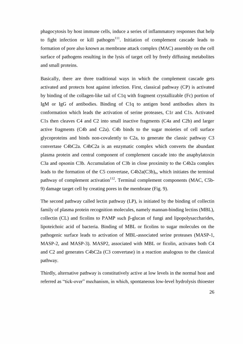
Basically, there are three traditional ways in which the complement cascade gets
activated and protects host against infection. First, classical pathway (CP) is activated
by binding of the collagen-like tail of C1q with fragment crystallizable (Fc) portion of
IgM or IgG of antibodies. Binding of C1q to antigen bond antibodies alters its
conformation which leads the activation of serine proteases, C1r and C1s. Activated
C1s then cleaves C4 and C2 into small inactive fragments (C4a and C2b) and larger
active fragments (C4b and C2a). C4b binds to the sugar moieties of cell surface
glycoproteins and binds non-covalently to C2a, to generate the classic pathway C3
convertase C4bC2a. C4bC2a is an enzymatic complex which converts the abundant
plasma protein and central component of complement cascade into the anaphylatoxin
C3a and opsonin C3b. Accumulation of C3b in close proximity to the C4b2a complex
leads to the formation of the C5 convertase, C4b2a(C3b)n, which initiates the terminal
pathway of complement activation112
. Terminal complement components (MAC, C5b-
9) damage target cell by creating pores in the membrane (Fig. 9).
The second pathway called lectin pathway (LP), is initiated by the binding of collectin
family of plasma protein recognition molecules, namely mannan-binding lectins (MBL),
collectin (CL) and ficolins to PAMP such β-glucan of fungi and lipopolysaccharides,
lipoteichoic acid of bacteria. Binding of MBL or ficolins to sugar molecules on the
pathogenic surface leads to activation of MBL-associated serine proteases (MASP-1,
MASP-2, and MASP-3). MASP2, associated with MBL or ficolin, activates both C4
and C2 and generates C4bC2a (C3 convertase) in a reaction analogous to the classical
pathway.
Thirdly, alternative pathway is constitutively active at low levels in the normal host and
referred as “tick-over” mechanism, in which, spontaneous low-level hydrolysis thioester

27
bond with rate (~1–2%/h) of complement C3 by water leads to the formation of
C3(H2O) that functions quite similar to C3b with regard to its ability to bind factor B
(CFB). Binding of CFB acts as a substrate for the serine protease factor D (CFD).
Cleavage of CFB by CFD results in the formation of the AP C3 convertase
[C3(H2O)Bb] which act similar to the classic C3 convertase C4bC2a and cleave C3 into
C3a and C3b. Generation of C3 convertase leads to the formation of C5 convertase
(C3bBbP) in appropriate circumstances which further progresses to MAC formation on
a foreign cell surface, similar to that of the CP. The binding of P to C3bBb on a
microbial (or protected) surface stabilizes and protect the convertase from inactivation
by regulatory proteins, thereby enhancing the convertase activity. In all pathways upon
activation C3 convertase cleaves α-chain of C3 to generate exposed and highly reactive
internal thiolester group.

28
Figure 9: General overview of complement cascade. Complement cascades start with three
distinct pathways classical pathway, lectin, and alternative ultimately leading to death of
pathogens by the formation of membrane attack complex (MAC) on their cell wall113
.
Intermediate proteins generated by complement cascade can also bind with the cell surface of
pathogens which leads to activation of immune cells by binding with complement receptors.
Exposed thiolester group binds covalently to biological surfaces exposing hydroxyl or
amino groups and leads to its deposition on the cell surface114
. Deposition of C3b on the
pathogen surface further leads to the opsonization for phagocytosis by
polymorphonuclear cells and macrophages. Complement receptor 3 (CR3) and CR4
present on macrophages and neutrophils also facilitate the ingestion and degradation of

29
pathogens115
. Mannan specific IgG present in serum also play role and facilitate the
binding of C3b to the Candida cell wall. To escape the complement attack pathogens
adopted several strategies including recruiting the complement regulatory factors (RCA)
or cleaving the main complement protein by secretory proteases.
1.5.1 Complement system and regulation
Activated complement components C4b, C3b, and C5b67 can attach to any nearby cell
surfaces including host cells and therefore activate complement cascade. To inhibit the
unwanted activation of the complement cascade, regulatory mechanisms have evolved.
Complement cascade regulation is mainly mediated by several inhibitors of complement
pathway called „regulators of complement activation‟ (RCA). Two of the main
complement inhibitors Factor H (FH) and Factor I (FI) had great attention due to their
ability to inhibit alternative complement pathway (“bystander” effect whereby C3b
generated in the vicinity becomes attached to a host cell). FH binds through sialic acid,
heparin, and sulfated glycosaminoglycans (GAGs) of the host surface thus distinguish
between self and non-self cells and prevent complement activation on host surfaces.
There are six other proteins related to FH: the product of complement factor H (CFH)
via alternative splicing, complement factor H-like protein (FHL-1), and CFH-related
proteins (CFHRs : FHR1 to FHR5)115
.
C4-binding protein (C4BP) is a fluid-phase regulator of the CP and LP. C4BP is both a
decay-accelerating factor dissociating C2a from the CP C3 convertase and a cofactor,
promoting FI-mediated cleavage of C4b into iC4b and further to C4c and C4d. Some
other RCA such as CD46, CD55/DAF, CR1 or CD35 and CR2 or CD21 are also
important players in complement activation regulation.
1.5.2 The FHL/FHR protein family
The FH like protein (FHL-1) is derived from CFH gene via alternative splicing whereas
FHR-1, FHR-2, FHR-3, FHR-4, and FHR-5 proteins encoded by the CFHR1–5 genes
present downstream of the CFH gene. Similar to FH, FHR proteins are also composed
of SCR/CCP domains (Fig. 10)116
. Biological functions of FHL-1 and FHR proteins are

30
under investigation however, scientific evidence till date suggests that they compete
with FH to bind C3b and/or glycosaminoglycans (GAGs). Recent evidence also
suggests that they are positive regulators of the alternative pathway of complement
system. The exact roles of these proteins against pathogens and in several diseases still
remain questionable117–120
.
Figure 10: Graphical representation of FH and FH family proteins. FH composed of 20
SCR regions, first three SCR (1, 2 and 3) bind C3b and therefore crucial for complement
regulation whereas SCR 19 and 20 bind with pathogens and surface proteins. Complement
regulatory regions are not conserved in FH family proteins. The N terminal SCR of FH family
proteins share homologies with SCR 6-9, SCR 10-14 and SCR 18-20 of FH. Based on formation
of homodimers and heterodimers these proteins are divided in two groups. Group 1 forms the
homodimers with each other through N terminal SCR domains whereas group 2 proteins lack
this feature. Colors of SCR domains reflect the similarities with SCR domain with FH.
1.5.3 Complement receptors
Immune cells harbor membrane-associated receptors interacting specifically with the
complement proteins, named as complement receptors. Binding of complement proteins
to these receptors kick-start the signaling and thus modulate the behavior of

31
complement receptor bearing cells. Based on their binding specificity to the
complement proteins, these receptors can be classified in three broad categories. (i)
receptors binding to the anaphylatoxins C3a, C5a, and C5a desArg (C3aR, C5aR), (ii)
receptors binding to C3 fragment, C3b and its degradation products, iC3b and C3dg
(CR1, CR2, CR3, CR4) and (iii) receptors for C1q and related collagenous lectins
(cC1qR, C1qRp, gC1qR)121
. These receptors play important roles in complement-
mediated phagocytosis, NO synthesis, leucocyte chemo-attraction, degranulation, and
B-cell proliferation122
. Innate immune cells such as macrophages, neutrophils, and
dendritic cells recognize pathogens either by pattern recognition receptors or indirectly
by opsonic receptors. Among all complement receptors, CR3 (CD11b/CD18) and CR4
(CD11c/CD18) are the major opsonic receptors. CR3 and CR4 belong to β2 integrin
family and possess high affinity towards C3b cleavage product iC3b123,124
. CR3 can also
directly bind to the β-glucan and pH-regulated antigen 1 of C. albicans. Interestingly,
recently it has been shown that binding CR3/CR4 with the FH coated pathogens
presumably facilitate the phagocytosis of pathogens and antifungal response of
macrophages. In contrast, C. albicans secreted Sap2 can down-regulate the CR3 and
CR4 expression on the cell surface of macrophages, therefore, alter the complement-
mediated phagocytosis by macrophage125
.
1.6 Interaction of complement cascade proteins with Candida species
Pathogenic members of Candida species, especially C. albicans is strong inducer of all
three complement activation pathways resulting in rapid assembly of C3 convertase.
Formation of C3 convertase leads to the generation of chemotactic cleavage fragments
such as anaphylatoxin C3a, C5a opsonizing fragments C3b which facilitates
phagocytosis. Components of complement activation cascade C3a and C5a126
, are
crucial against fungal infections, as mice lacking the C3a and C5a are reported to highly
susceptible to invasive C. albicans infection. Moreover, during fungal infections C5
deficiency is also associated with increased production of pro-inflammatory cytokines,
including TNFα and IL-6, and rapid fungal reproduction in many organs. Importance of
C5a during C. albicans infection is appreciated as C5a is critical for activation of human

32
monocytes to produce pro-inflammatory cytokines, e.g. IL-6 and IL-1β127
. Aside from
C5a, C3a also exerts antimicrobial effects against C. albicans presumably by binding to
its cell surface and inducing membrane perturbations to release extracellular material.
C. albicans isolates can bind with complement regulatory proteins such as complement
factor H and other members. CFHR-1, CFHL-1, CHR-4 can enhance CR3 mediated
antimicrobial activity such as the release of antimicrobial protease lactoferrin from
neutrophils and enhance generation of reactive oxygen species by binding to the cell
surface of C. albicans128
.
1.7 Regulation of host complement cascade by Candida species
1.7.1 Binding to complement regulators
C. albicans and NCAC species, such as C. parapsilosis, C. tropicalis, and C. lusitanae
reported to possess several cell wall associated proteins having binding affinity towards
host complement factors. Affinity of IgG towards mannan present on the cell surface of
fungi and deposition of C3 on the cell surface of pathogens activate classical, alternative
and MBL pathways and also facilitate phagocytosis mediated killing of pathogens by
host immune cells. Available literature suggests that C. albicans mainly uses two
mechanisms to evade the complement attack: first by recruiting complement regulators
on cell surface and second by secreting proteases to degrade complement proteins125,129
.
Presence of Factor H binding surface protein phosphoglycerate mutase (Gpm1), high-
affinity glucose transporter 1 (Hgtp1), glycerol-3-phosphate dehydrogenase 2 (Gpd2),
and C3, FH, C4bp, FHL-1 binding pH-regulated antigen 1 (Pra1) of C. albicans allow
this fungus to efficiently regulate complement system events130–132
. In addition, C.
albicans expresses a protein called αvβ3 integrin-like protein which is similar to human
αvβ3 integrin. This integrin-like protein acquires a terminal complement pathway
inhibitor, vitronectin to the yeast cell surface. Acquired vitronectin inhibits terminal
complement complex (TCC) formation and thus presumably help Candida to avoid
complement attack133
.
In the case of C. parapsilosis little is known about complement binding cell surface
proteins. With in silico experiments we found C. albicans orthologous of complement

33
binding proteins in C. parapsilosis genome (Table 1). Function of these proteins in
context with binding affinity with host complement proteins is not studied yet. Indeed,
higher protein identity can be correlated with function but research need to be done to
define exact roles of these proteins.
Table 1: C. albicans cell surface proteins important for binding with host complement proteins
and percent similarity with their orthologus in C. parapsilosis.
C.
albicans
Bind with host
complement
proteins
Orthologous in C.
parapsilosis
Known functions in C.
albicans
Amino acid
similarity
(%)
Gmp1/
CRASP1
FH, FHL-1, and
plasminogen
CPAR2_211810 gluconeogenesis,
glycolytic process,
interaction with host
85%
Pra1/
CRASP2
C3, C4BP, factor
H, FHL-1, and
plasminogen
No orthologous
match found
--------- ----------
Hgt1P FH CPAR2_108370 glucose transmembrane
transporter activity
81%
Gpd2 FH CPAR2_601770 [NAD+] activity and
cell surface localization
80%
1.7.2 Regulation of crucial complement factors by aspartyl proteases
In addition to recruitment of complement regulators on cell surface, C. albicans cleave
complement proteins by secreting aspartyl proteases. Activation of all three complement
pathway leads to the formation of C3 convertase named C4b2b in classical/lectin
pathway and C3bBb in alternative pathway. A recent study has shown the high level of
Saps specially Sap1, Sap2, and Sap3 blocks classical as well alternative complement
pathways by cleaving complement component C3b, C4b, C5, and also inhibit terminal
complement complex, whereas other Saps such as Sap9 lacks complement cleavage
activity, but cleaves antimicrobial peptide such as histatin 5 in C. albicans129
. C5 plays
role in all three complement pathways and cleaved by C5 convertase enzyme to form
C5a and C5b. Studies have shown that C5 deficient mice are more susceptible to

34
Candida infections106
. C5b is larger fragment further forms complex with C5b-9 to form
MAC, which attached to the cell wall of pathogens (especially to gram negative
bacteria) to form pores on the cell wall. C5a, the smaller fragment, is a vasodilator,
chemotactic, and anaphylatoxin that mediates inflammatory responses at the site of
injury by stimulating neutrophils, eosinophils, phagocytes, and endothelial cells134
.
Studies have shown that Saps in the culture supernatant of C. albicans as well as
recombinant Saps (Sap1p, Sap2p, and Sap3p) degrade host complement component C5
and thus inhibit MAC formation129
. C. albicans secreted Sap2p also cleaves
complement inhibitor FH which enhances the antifungal activity of human neutrophils
via binding to complement receptor type 3 (CR3)125
.
Even though, researchers have clearly illustrated indispensable role of C. albicans
secreted aspartyl proteases in evasion of host complement attack. To our knowledge not
even a single study has been done till date to find out the role of aspartyl proteases from
NCAC species in complement evasion135
.
1.8 Sap mediated modulation of macrophage and neutrophil responses
Macrophages and neutrophils are the main phagocytic cell types of the vertebrate innate
immune system136,137
. Ubiquitous presence of macrophages in the tissues makes them
one of the most important warriors against invading pathogens. Macrophages control
the infection by inflammatory responses and phagocytosis106
. Role of macrophages in
fungal clearance is described in a recently published article125
, but only few studies deal
with the mechanisms behind modulation of macrophage behavior by C. albicans
secreted aspartyl proateases. Sap1p, Sap2p, Sap3p, and Sap6p from C. albicans reported
to induce secretion of pro-inflammatory cytokines, such as IL-1β, TNFα, and IL-6, by
human monocytes. Additionally, Saps are also able to modulate physiology of
monocytes by inducing higher Ca2+
influx138
.
Neutrophils kill pathogens either by oxidative stress (reactive oxygen species generated
by NADPH oxidase) or by the release of NET (neutrophil extracellular trap)139
. NETs
are consisting of extracellular nucleic acids, histones, and granular proteins, such as
calprotectin and pentraxin-3 (PTX3), and crucial for fungal cell killing which are too

35
large for phago-lysosome mediated killing. Recently, it has been shown that C. albicans
strains lacking Sap1-Sap3, Sap4-Sap6, Sap9-Sap10, induces significant less NET
response compared to WT strain indicating the roles of Saps in regulation of NET
formation from neutrophils138
. Saps (especially Sap2) have reported to exhibit
chemoattractant activity towards neutrophils140
. Saps also stimulate epithelial cells to
produce chemokine IL-8 which is a strong chemoattractant for neutrophils. Sap2p and
Sap6p are reported to involved in internalization via a clathrin dependent mechanism
and therefore possibly stimulate inflammatory process141
. Despite several studies about
the role of C. albicans secretory aspartyl proteases in its virulence, not much known
about immune-modulatory roles of candidaparapepsins.
1.9 Vaginal Candidiasis
Vaginitis is an acute inflammatory disease, affecting three out of four women
worldwide at least once during their fertility life and more than 5% (approximately 150
million) experience subsequent recurrence142,143
. Although a number of predisposing
factors such as oral contraceptive usage, changes in estrogen and progesterone levels
during pregnancy, uncontrolled diabetes mellitus, and long-term broad-spectrum
antibiotic treatment as well as changes in the composition of the vaginal microbiota
have been identified to increase the risk of vaginitis; still mechanism of pathogenesis
behind Candida caused vaginitis (vaginal candidiasis) is not well studied144,145
.
Similar to the skin and oral cavity, vagina harbours various microbes. These microbes
help to maintain the adequate pH, hinder the growth of pathogens, stimulate the local
inflammation and decrease pregnancy complications146,147
. A slight change in the
composition of resident microbial community and host defense encourages the
emergence of opportunistic infections caused by C. albicans which is highly abundant
in vaginal mycobiome during vaginitis148
. Studies have been done to investigate on the
mechanisms of pathogenesis of C. albicans caused vaginitis by elucidating the roles of
fungal virulence factors, such as change in morphogenesis, secreted factors, and biofilm
formation as well as host immune responses against Candida during infection149,150
.

36
Secreted aspartyl proteases of Candida species have long been reported as a virulence-
associated trait of these pathogenic fungi and higher secretion of these proteases are
reported in vaginitis151,152
. Various studies have shown that members of C. albicans Sap
family have variable abilities to induce pro-inflammatory cytokine secretion mediated
by Akt/NF-κB activation153,154
. A recent study shows that C. albicans Saps, particularly
Sap2, Sap6, and chemo-attractive chemokines, such as IL-8 and MIP-2 released from
Sap treated vaginal epithelium, have ability to mediate neutrophil chemotaxis150
. Even
though significant studies have shown higher production of Saps in vaginal infection
and their role in vaginal inflammation, no clear scientific evidence has been shown for
the mechanisms involved. Additionally the role of proteases secreted from NCAC
species in vaginal candidiasis is also not well studied.

37
2. Objective of the present work
Aims
The specific aims of the present work were to generate the SAPP reintegrant mutant
strains (in sapp1/2/3−/−
background) in order to understand their role in C. parapsilosis
caused infection, additionally, to understand the regulation of hosts complement
cascade by C. parapsilosis secreted aspartyl proteases Sapp1p and Sapp2p.
Experimental strategies
Generation of SAPP reintegrant mutant strains.
Delineation of the role of C. parapsilosis secreted aspartyl proteases Sapp1,
Sapp2 and Sapp3 in C. parapsilosis biofilm formation, cell wall stability, and
epithelial adhesion and damage.
Determination of the role of individual aspartyl proteases of C. parapsilosis in
modulation of macrophage inflammatory responses.
An in depth study to understand the role of C. parapsilosis secreted aspartyl
proteases in the evasion of host complement attack.

38
3. Material and methods
3.1 Media used in this study
Liquid and solid media were prepared according to the following protocols:
LB: 1% (w/v) tryptone, 0.5% (w/v) yeast extract, 1% (w/v) NaCl in ddH2O. For
selection of bacterial colonies antibiotics were added to the media after autoclaving and
cooling: ampicillin (100 µg/ml), kanamycin (50 µg/ml) or chloramphenicol (20 µg/ml)
depending on the selection marker gene harbors by the plasmid.
YPD: 0.5% (w/v) yeast extract, 1% (w/v) peptone, 1% (w/v) glucose in ddH2O.
Nourseothricin (NTC) in 100 µg/ml concentration was added for selection of NTC
resistant colonies.
YNB: 0.67% (w/v) yeast nitrogen base (without amino acids, with (NH4)2SO4), 2%
(w/v) glucose in ddH2O.
YCB: 2.34% yeast carbon base (YCB), 0.2% BSA (pH 4.0) in ddH2O.
For the preparation of plates, the described LB and YCB media broths were
supplemented with 1.5% (w/v) agar and yeast media with 2% (w/v) agar.
Spider medium: 1% peptone, 1% yeast extract, 1% mannitol, 0.5% NaCl and 0.2%
K2HPO4.
3.2 C. parapsilosis and E. coli strains used in the study and cultivation conditions
List of C. parapsilosis strains used in this study are described in supplementary table 1.
Yeast strains used in the present study were maintained on YPD plates supplemented
with 1% 100 unit/ml penicillin-streptomycin solution (Sigma-Aldrich) at 4 °C. One day
prior to the experiments C. parapsilosis wild-type GA1 strain155
, reintegrant mutant
strains of aspartyl protease coding genes (RI_SAPP1, RI_SAPP2, and RI_SAPP3) and
sapp null mutant (sapp1/2/3−/−
) were grown in YPD or in yeast carbon base (YCB)
medium supplemented with 0.2% BSA at 30 °C on an orbital shaker at 180 rpm for 18-
20 h. Cells were harvested by centrifugation at 900 g for 5 min, washed twice with
sterile 1x PBS (phosphate-buffered saline; 137 mM NaCl, 2.7 mM KCl, 10 mM
Na2HPO4, 2 mM KH2PO4), further diluted in 1x PBS and counted in a Bürker chamber
chamber and diluted to the appropriate concentration prior to the experiments. Unless

39
otherwise stated, all experiments were performed using living C. parapsilosis cells and
without serum treatment.
E. coli 2T1 cells were stored on LB agar plates in 4 °C and routinely cultured at 37 °C
in LB liquid medium before competent cell preparation.
3.3 Generation of sapp1/2/3−/−
and reintegrant mutant strains
SAPP mutant strains C. parapsilosis sapp1/2/3−/−
and C. parapsilosis RI_SAPP were
generated as described previously with minor modifications94,135
. Briefly, to generate C.
parapsilosis sapp1/2/3−/−
, 500 bp upstream and downstream regions
of SAPP2 and SAPP3 were PCR amplified and cloned into the pSFS2a plasmid with a
recyclable NAT cassette. Further, the SAPP2 deletion cassette was introduced in
the ΔΔsapp1a ΔΔsapp1b deletion mutant C. parapsilosis strain to generate ΔΔsapp1a
ΔΔsapp1b ΔΔsapp2 mutants. Finally, the SAPP3 deletion cassette was generated
similarly to SAPP2, and C. parapsilosis ΔΔsapp1a ΔΔsapp1b ΔΔsapp2 mutant strain
was transformed with the construct to generate the sapp1/2/3−/−
strain as described
before92
.
Mutant C. parapsilosis strains expressing the individual SAPP genes were generated in
the sapp1/2/3−/−
mutant background. Solely SAPP1, SAPP2, and SAPP3 expressing
mutants were established broadly using Gateway cloning technology (Invitrogen) which
takes advantage of the site-specific recombination reactions and rely essentially on the
BP and LR clonase reactions (Fig. 11). In brief, the SAPP genes from WT (GA1) C.
parapsilosis strain were amplified with the help of attB tagged primer pairs. The attB
containing amplified SAPP genes were cloned in to a donor vector pDONR221 includes
attP sites resulting in the formation of an entry clones. The resulting entry clone
containing the SAPP genes flanked by attL sites further recombined with attR
containing destination vector to form expression clones containing SAPP genes under
CaTDH3 promoter. Fragement containing CaTDH3 and SAPP genes were digested and
cloned in pNRVL vector. Finally, pNRVL vetor was linearised using stuI restriction
enzyme and transformed in to the sapp1/2/3−/−
C. parapsilosis strain targeting the

40
neutral locus, CpNEUT5L (large intergenic region facilitates the integration and
expression of ectopic genes)156
.
Figure 11: Schematic overview of Gateway cloning strategy for reintegration of SAPP
genes. (A) SAPP genes were amplified by using attB primers and the amplified fragments were
used to generate expression vectors containing SAPP genes under the control of CaTDH3
promoter. (B) Expression vectors were further double digested and fragment containing
CaTDH3_SAPP1/2/3 were ligated into pNRVL vector. pNRVL vectors were linearized and
transfected into C. parapsilosis sapp1/2/3−/−
. Colonies grown on NTC containing agar plates
were checked using colony PCR, Southern blot, and phenotypic analysis.
3.4 Validation of reintegrant mutant strain
The successful reintegrations were identified by PCR screening and Southern blot
analysis.

41
3.4.1 Fast track DNA isolation
For fast track DNA extraction, a small amount of C. parapsilosis cells were picked by
sterile tooth pick and mixed in 1.5 ml centrifuge tube with 80 µl of lysis buffer (1%
SDS, 200 mM LiAc). Tubes were gently vortexed and incubated at 70 °C for 5 min. 240
µl of 96% ethanol was subsequently added to the mixture, vortexed and centrifuged for
5 min at 16,200 g in microcentrifuge. The pellet was washed with 70 % ethanol above
mentioned g force. Pellet was air dried and DNA was resuspended in 50 µl Milli-Q
water. Samples were stored at -20 °C for further analysis.
3.4.2 PCR conditions
For PCR amplification, each reaction contained 1x PCR buffer, 1 unit of Taq DNA
polymerase (Thermo Scientific), 10 µM of each forward and reverse primers (Table
S2), 2.5 mM of each deoxynucleoside triphosphate and 1 µl of DNA sample up to a
total reaction volume of 15 µl. The following PCR conditions were used to amplify C.
parapsilosis SAPP1/2/3: 94 oC for 1 min, followed by 35 cycles of 15 s at 94 °C, 20 s at
52 °C, and 2.5 min at 72 °C with a final extension of 5 min at 72 °C.
3.4.3 Detection of PCR products
2 µl of amplicons were analysed with agarose gel electrophoresis [0.8% agarose gel
containing EtBr dissolved in 1xTAE buffer (40mM Tris, 20mM Acetate and 1mM
EDTA)] and visualized under UV irradiation.
3.5 Southern blot analysis
Southern blot analysis was performed to verify integration of the SAPP containing
cassette to the proper genomic region (at CpNEUT5L locus) as desribed155
.
3.5.1 Hybridization of the DIG-labeled probe to DNA
Hybridisation was performed using the DIG-probe prepared from downstream sequence
of the C. parapsilosis neutral locus (CpNEUT5L). 10 μl (250 ng) of the DIG-probe was
diluted in 5 ml of hybridization buffer (5x SSC, 0.1% N-lauryl-sarcosine, 0.02% SDS,

42
1% Blocking reagent) and boiled in water bath for 10 min and immediately chilled on
ice for 5 min. The mixture was added to the nitrocellulose membrane followed by
incubation at 65 °C for 18-24 h. The membrane was washed twice with 100 ml pre-
warmed washing solution 1 (20xSSC, 10% SDS) for 5 min at 65 °C. The third and
fourth washing was done for 15 min at 65 °C with 100 ml pre-heated washing solution 2
(2x SSC, 2% SDS).
3.5.2 Detection of the hybridized DIG-labeled probe
A peroxidase labeled anti-digoxigenin-AP Fab fragments (Roche) was added to the
membrane to detect the DIG labeled probes for 30 min at RT. Afterwards the membrane
was washed with 1x washing buffer 2 (3M NaCl, 0.2M NaH2PO4-H2O, 0.02M EDTA,
1% SDS) for 2 times 15 min each at RT and once for 2 min with buffer 3 (0.1M Tris,
0.1M NaCl, 50mM MgCl2). NBT-BCIP substrate was prepared by mixing 120-150 µl
of NBT-BCIP (nitro blue tetrazolium/5-bromo-4-chloro-3-indolyl phosphate, Roche)
substrate in 10 ml of buffer 3. The solution was vortexed, poured in the transparent
polyethylene bag contain nitrocellulose membrane and kept in dark until bands
appeared. With probes used a 2,800bp fragment for SAPP1, 2791bp fragment for
SAPP2 and SAPP3 were detected in overexpression C. parapsilosis strains, whereas
these bands were absent in WT and sapp1/2/3−/−
strains.
3.6 RNA extraction
To extract RNA from C. parapsilosis WT, RI_SAPP and sapp1/2/3−/−
strains grown in
YCB+0.2% BSA medium we used Ribopure Yeast RNA isolation Kit (Ambion) and
followed the standared protocol provided by manufacturer. Purity of RNA samples was
also checked by real-time PCR.
3.6.1 cDNA synthesis
cDNA synthesis from RNA samples was performed with RevertAid First Strand cDNA
synthesis kit (Thermo Scientific) according to the protocol provided by the
manufacturer.

43
3.6.2 Quantitative real-time PCR
Real-time qPCR amplification and detection were carried out in 96-well optical plates
on a BioRad thermocycler with CFX96 detection system. Reaction mixture waswas set
up as follows: 1x SYBR Green qPCR Master Mix (Applied Biosystems), 0.4 μM
concentration of each primer, and 1 µl cDNA in a final volume of 20 μl per reaction.
The amplification conditions were as follows: denaturation for 3 min at 95 °C, followed
by 49 cycles of denaturation at 95 °C for 10 s, annealing at 60
°C for 30 s, and
elongation at 65 °C for 30 s, with a final extension step at 72
°C for 30 s. TUB4 gene
was used as a housekeeping gene for an internal control. The primers used in real-time
qPCR experiments are given in Table S2.
3.7 Phenotypic characterization of mutants by protease assay
For semiquantitative detection of proteinase production, 18 h culture of C. parapsilosis
WT, sapp1/2/3−/−
, and RI_SAPP strains were grown in YPD. Cells were collected,
washed two times with 1x PBS and resuspended in 500 µl of 1x PBS. 106
cells in 10 µl
1x PBS were spotted onto YCB+BSA containing plates and incubated for 3 days at
30 °C. After cultivation plates were stained with Amido Black for 3-4 min and washed
with methanol-acetic acid-water (3:1:6) solution. The experiments were repeated 4
times. The diameter of the clearing zones around the colonies was determined with
ImageJ software.
3.8 Growth curve analysis
C. parapsilosis strains grown in YPD and YCB media for 18 h were washed two times
with 1x PBS. Concentrations were adjusted to 5×10⁵ cells/ml diluting either in YPD or
YCB + 0.2% BSA medium. 100 µl of suspension was plated onto 96-well flat-bottom
microtitration plates (Biologix). The plates were incubated at 30 °C for 24 h in a plate
reader (SPECTROstar Nano, BMG labtech). The optical density (OD) at 600 nm was
recorded for each well automatically after every 30 min and post shaking. O.D.
measurements were performed assuming that O.D. values at 600 nm obtained are
proportional to the fungal cell numbers.

44
3.9 Serum sensitivity assay
Serum sensitivity assay of C. parapsilosis WT, sapp1/2/3−/−
, and RI_SAPP strains was
performed based on previously published protocol with minor modifications94
. Briefly,
1×10⁵ cells of C. parapsilosis were cultured into 1 ml YPD medium supplemented with
20% intact or heat-inactivated human serum and incubated at 30 °C with continuous
shaking. Aliquots were collected at 0,6,24 and 48 h postinoculation and plated to YPD
plates to determine colony forming units (CFU).
3.10 Biofilm formation assay
Biofilms were developed on commercially available pre-sterilized, polystyrene, flat-
bottom 96-well microtiter plates. Wells were coated with heat inactivated human serum
one day before the experiment. Thereafter wells were washed three times with sterile 1x
PBS. 106
C. parapsilosis cells in 100 μl RPMI or YNB media was transferred into each
well of a microtiter plates. Plates were sealed with parafilm and incubated for 48 h at
37 °C to allow yeasts to develop biofilm. After biofilm formation, medium was
aspirated carefully and wells were washed three times with 200 μl 1x PBS. Level of
biofilm formed was measured by XTT reduction assay and CV staining as discribed135
.
3.11 Pseudohyphae growth assay
Effect of SAPP gene deletion and reintegration on pseudohyphae formation was also
investigated. For that, C. parapsilosis cells were either stained with wheat germ
agglutinin-tetramethylrhodamine (WGA-TRITC) and visualized with Olympus light
microscope, examined with Amnis Flow Sight (Luminex) and colony morphology
investigated after spotting the C. parapsilosis cells on pseudohyphae inducing media
(YPD+10% fetal bovine serum and spider medium) as described before135
.
3.12 Analysis of cell wall composition with microscopy imaging technique
For cell wall content analysis C. parapsilosis cells were fluorescently labeled with
CCW fluorescent dye mix [8 μl 2.5 mg/ml Canavalia A-fluorescein isothiocyanate
conjugated (ConA-FITC), 1 μl 1 mg/ml Calcofluor White (CW), 1 μl 1mg/ml WGA-
45
TRITC, 90 μl 1% BSA] as described before135
. The fluorescently labeled cells were
washed three times with 1x PBS and resuspended in 100 μl of 1x PBS and visualized
with fluorescence microscope (Zeiss).
3.13 Cultivation conditions of TR146 and A-431 cell lines
The human buccal epithelial squamous cell carcinoma line TR146 was kindly provided
by Dr. Julian Naglik, Kings College London, UK and cultured as described157
.
The human A-431 vaginal epithelial cell line, obtained from ATCC (LGC Standards)
were grown in Dulbecco's modified Eagle's medium (DMEM) supplemented with 10%
heat inactivated (HI) foetal calf serum and 100 unit/ml penicillium/streptomycin
(Sigma-Aldrich) at 37°C as described151
.
3.14 Adhesion assay
The cell adhesion assay was performed by slightly modifying the protocol published
previously158
. 1x10⁷
C. parapsilosis cells were allowed to adhere in 96-well flat
bottomed, non-tissue culture plates (Biologix) in 200 µl RPMI medium (Gibco) at
37 °C, for 120 min. Non-adherent cells were removed by washing the wells two times
with 200 µl 1xPBS. The attached C. parapsilosis cells were then vigorously
resuspended in water, diluted (1,000 fold) and plated onto YPD plates. Colonies were
counted after two days of incubation at 30°C and data was represented based on CFU158
.
Experiment was performed three times in triplicates.
Adhesion to epithelial cells was performed as described previously159
. 3×10⁵ TR146 or
A-431 cells were grown to confluence in 24-well plates for 24 h in DMEM-F12 (Gibco)
medium. After washing with 1x PBS three times, 1 ml serum-free DMEM containing C.
parapsilosis cells in MOI 5 (TR146 cells:C. parapsilosis = 1:5) or MOI 10 (A-431
cells:C. parapsilosis = 1:10) was added to the epithelial cells and incubated for 90 min
at 37 °C with 5% CO2 in cell culture incubator. The non-adherent Candida cells were
aspirated and wells were washed three times with 1xPBS. Wells were filled with 1 ml of
double distilled water containing 0.1% triton x-100 at 37 °C for 1 h until epithelial cells
were lysed. Candida cells were collected, diluted, plated onto YPD plates and incubated

46
at 30 °C for 2 days. After incubation colonies were counted. Three independent
experiments were carried out in triplicates
3.15 Isolation and differentiation of PBMCs
Human peripheral blood mononuclear cells (PBMCs) were isolated from buffy coats of
healthy donors by Ficoll Paque PLUS (GE Healthcare) density gradient centrifugation
as described previously90
. The isolated cells were cultured for 7 days in the presence of
X-VIVO 15 medium (Lonza) supplemented with 100 U/ml penicillin, 100 mg/ml
streptomycin and 10 ng/ml recombinant human granulocyte-macrophage colony-
stimulating factor (GM-CSF, Sigma-Aldrich).
3.16 Cell damage assay
LDH activity in cell culture supernatants was measured by using the Cytotoxicity
Detection Kit (LDH; Roche) according to the manufacturer‟s instructions. Human
peripheral blood mononuclear cells derived macrophages (PBMC-DMs) were
stimulated with C. parapsilosis cells at a PBMC-DMs:C. parapsilosis ratio of 1:5 for 24
or 48 h, while A-431 cells were stimulated at a ratio of 1:10 (A-431: C. parapsilosis) or
left unstimulated (negative control). During analysis, the LDH activity measured in
cultures containing yeast cells alone was subtracted from the values measured in
stimulated samples. LDH released from lysed PBMC-DMs were used as a positive
control. Experiments were performed three times in triplicates.
3.17 Macrophage killing assay
PBMC-DMs were coincubated in 24-well plastic cell culture plates with C. parapsilosis
WT, RI_SAPP, and sapp1/2/3−/−
strains at MOI of 5 (PBMC-DM cells:C. parapsilosis
= 1:5). As a control, the same numbers of yeast cells were incubated in the appropriate
cell culture medium without macrophages. After 3 h of incubation, macrophages were
lysed using 0.1% triton x-100 in PBS. The lysates were then serially diluted, plated onto
YPD agar plates and incubated at 30 °C for 2 days. The number of CFU was determined
and multiplied by the dilution factor to calculate the original living cell number. The

47
killing percentage was calculated as follows: [(number of living Candida cells in
control plates – number of living Candida cells in cocultures) / number of living
Candida cells in control wells] x 100. Macrophages derived from 4 healthy human
blood donars used for killing assay.
3.18 Whole blood killing assay
For whole blood killing assay, fresh heparinized human blood from healthy donar was
mixed with 107 C. parapsilosis cells (total volume of 1 ml, 900 μl of blood and 100 μl
of Candida cells). The mixture was incubated at 37 °C for 24 h. 50 µl of 1000 times
diluted suspension was plated onto YPD agar plates in triplicates and incubated at 30 °C
for 2 days. Number of surviving fungal cells was counted and the killing efficiency of
whole blood was calculated as described above. Experiments were repeated three times
in triplicates.
3.19 Phagocytosis assay
Phagocytic activity was examined as previously described160
, with minor modifications.
C. parapsilosis cells were labeled with Alexa Fluor 488 carboxylic acid succinimidyl
ester (Invitrogen) and co-cultured with PBMC-DMs at MOI 5 for 3 h in 12-well tissue
culture plates (VWR). Cells were then washed two times with 1x PBS, gently
resuspended to a single cell suspension by pipetting, transferred to 1.5 ml
microcentrifuge tubes, harvested by centrifugation and resuspended in 200 μl 1x PBS.
Samples were measured using a Amnis Flow Sight (Luminex) and analysed using the
IDEAS software.
3.20 Phagosome-lysosome fusion
To examine the phagosome-lysosome fusion, C. parapsilosis cells were labeled with
fluorescent dye pHrodo® (Invitrogen). For labeling, first the yeast cell suspension (109
cells in 100 µl 1x PBS buffer) was treated with 11 μl of 1M Na2CO3 (pH=10) and then
2 μl pHrodo (1 mg/ml in DMSO) was added, and was incubated for for 1 h at room
temperature in dark. Next, cells were washed 4 times with 1x PBS and adjusted to the

48
proper concentration. Human PBMC-DMs were infected with labeled Candida cells at
1:5 ratio (PBMC-DMs: Candida) and kept for 2 h at static condition at 37 °C to allow
phagocytosis. Then the non-phagocytosed Candida cells were washed with 1x PBS and
macrophages were detached from cell culture plates by TrypLETM
Express solution
(Gibco). Finally, the macrophages were collected in FACS buffer (0.5% FBS in 1x
PBS), and suspended in 1x PBS, followed by measurement. Data were analyzed by the
IDEAS Software (Amnis).
3.21 Cytokine measurements
PBMC-DMs were infected with 2.5×10⁶
C. parapsilosis WT, RI_SAPP, and
sapp1/2/3−/−
cells and coincubated for 24 h at 37 °C. Concentration of IL-1β, Il-6,
TNFα and IL-8 was determined from supernatant by commercial ELISA kits (R&D
Systems) in according to the manufacturer‟s instructions. Concentration of cytokines
represented in pg/ml. The experiments were performed with macrophages derived from
PBMCs from at least six healthy donors.
3.22 Detection of proteolytic activity of Sapp1p and Sapp2p
The proteolytic activity of purified Sapp1p and Sapp2p (1 μg each) was assayed by
incubating them with purified human complement proteins C3b, C4b, and factor H (FH)
(Merck) or with recombinant factor H-like protein 1 (FHL-1) (expressed and purified as
described previously)161
or FHR-1 or FHR-5 (Novoprotein) for 3 h or 15 h at 37 °C.
Aliquots were taken at the indicated time points, separated by SDS-PAGE, and
analyzed by Western blotting. C3b was identified by the use of polyclonal goat anti-
human C3 (Calbiochem, Quidel) in combination with a horseradish peroxidase (HRP)-
conjugated goat antibody (DAKOCytomation). C4b was detected with a monoclonal
anti-C4c antibody (Quidel) and with HRP-conjugated goat anti-mouse Ig (Dako). To
detect cleavage of FH, FHL-1, FHR-1, and FHR-5, polyclonal goat anti-FH
(Calbiochem, Merck), mouse monoclonal anti-FH (A254; from Quidel), and polyclonal
goat anti-FHR-5 (R&D System) and the corresponding HRP-conjugated secondary
antibodies rabbit anti-goat Ig and goat anti-mouse Ig (Dako) were used. In addition,
cleavage of C3b and C4b by the natural, complement-specific protease factor I in the

49
presence of the cofactors FH and C4BP (Hyphen Biomed) was assayed to compare with
the cleavage patterns generated by the Sapp proteases.
3.23 Staining of macrophage CD11b/CD11c receptors
Isolation of CD14+ monocytes and macrophage differentiation was carried out as
described previously162
. To investigate the effect of Sapp proteins on cell surface
receptors, 1×10⁶ PBMC-DMs were incubated with Sapp1p or Sapp2p (5 µg each) for
30 minutes at 37 °C. In control experiments PBMC-DMs were incubated in 1x PBS
buffer without addition of any proteins. PBMC-DMs were washed with 1x PBS
containing 1% FCS and 0.1% NaN3. 100 µg/ml monoclonal antibodies were added to
the cells (against CD11b monoclonal mIgG1 clone ICRF44 (Biolegend), against CD11c
monoclonal mIgG1 clone BU15 (ImmunoTools GmbH) and against CD18 monoclonal
mIgG1 clone TS1/18 (Biolegend) and isotype control (mouse IgG1 clone MOPC-21,
Biolegend) for 20 min at 4 °C followed by the addition of goat anti-mouse IgG-Alexa
Flour 488 secondary antibody (1:200, Molecular Probes) for 20 min at 4 °C. 20,000
cells were measured by Cytoflex flow cytometer (Beckman Coulter) using CytExpert
software for data acquisition and Kaluza Analysis software for data analysis.
3.24 Galleria mellonella fungal burden and survival
Galleria mellonella larvae were purchased from TruLarv™ (Biosystems, Netherlands).
Upon arrival, the larvae were stored at 4 °C in dark and subsequently used within a
maximum of 7 days. Before infection, larvae were taken from cold storage, kept nearly
20 min at room temperature and proleg was sterilized gently with cotton swab dipped in
70% ethanol.
Initially the sub-lethal C. parapsilosis inoculum concentration was determined by
injecting larvae with serial dilutions of C. parapsilosis WT cells: 10 µl of cell
suspension containing 108, 5x10
7, 2.5x10
7 cells/ml were injected into G. mellonella by
sub cutaneous micro-injection by inserting needle of Hamilton syringe into the proleg of
larvae. In survival experiments larvae were infected with 10 µl of cell suspension
containing 108 Candida cells /ml). As a control groups, non-injected larvae and injected

50
with 10 μl of sterile 1x PBS were included. Infected larvae were incubated at 30 °C for
7 days and monitored daily for survival. Groups of twenty larvae were used per strain
with two experimental replicates.
For CFU determination, 0.25-0.30 gram larvae were infected with 10 µl of cell
suspension containing 107 C. parapsilosis cells /ml, sacrificed at 24 h post-infection and
fungal burden of each individual larvae was determined following the next protocol:
larvae was homogenized in 5 ml 1x PBS in 50 ml plastic canonical tube with
homogenizer at 3000 rpm for two cycles of 1 min. 50 μl of homogenate was plated onto
YPD plates containing penicillin and streptomycin. Plates were incubated at 30 °C for 2
days and colonies were counted.
3.25 Drosophila melanogaster infection
For the infection of flies, pricking method was used. Pricking method of infection is
proven to be more reliable and triggers comparatively lower induction of damage
caused antimicrobial proteins compared to other infection modes163
.
OregonR (OrR) flies (WT) and MyD88 mutant strain (females or males, 15 per
experimental group) were infected with a thin sterile needle dipped in the suspension of
C. parapsilosis cells (2×10⁷ cells/ml diluted in sterile 1x PBS). Flies were than
incubated at 25 °C on regular fly medium. 25 flies were infected with each mutant
strains and experiment was at least twice
3.26 Statistical analyses
Statistical analyses were performed either by t-test or by one way ANOVA using the
software GraphPad Prism 6 (GraphPad Software, Inc.). Mantel-Cox (Log-rank) tests
were used for survival data evaluation. Differences were considered statistically
significant at p ≤ 0.05 (*p≤0.05; **p≤0.01; ***p≤0.001)
3.27 Ethical statement
For isolation of PBMCs, blood samples were taken from healthy donors. This procedure
and the respective consent documents were approved by the Institutional Human

51
Medical Biological Research Ethics Committee of the University of Szeged. All healthy
donors provided written informed consent. All experiments were performed in
accordance with guidelines and regulations of the Ethics Committee of University of
Szeged, and experimental protocols were approved by institutional committee.

52
4. Results
4.1 Reintegration of the SAPP1, SAPP2, and SAPP3 genes C. parapsilosis
sapp1/2/3−/−
strain
Previous work from our laboratory clearly demonstrated that C. parapsilosis
SAPP deletion mutants, i.e. C. parapsilosis Δ/Δsapp1a-Δ/Δsapp1b and Δ/Δsapp1a-
Δ/Δsapp1b-Δ/Δsapp2 strains are more sensitive to human serum and in vitro less
virulent compared to the donor strain (WT)94
. To further analyze the role of SAPP
genes, we overexpressed SAPP1, SAPP2, and SAPP3 genes individually in C.
parapsilosis sapp1/2/3−/−
strains. pNRVL vector (Fig. S1) containing the SAPP1,
SAPP2, and SAPP3 genes under the control of constitutive CaTDH3 promoter was
linearized by enzyme StuI and transfected into C. parapsilosis sapp1/2/3−/−
. Colonies
were selected on NTC plates. Further we performed colony PCR (Fig. 12A) and
Southern blot to identify mutant strains and to validate the proper integration.
For Southern blot, C. parapsilosis WT, sapp1/2/3−/−
, and RI_SAPP strains genomic
DNA was digested with HindIII restriction endonuclease and transferred onto nylon
membrane. Hybridisation was performed with high stringency using the probe prepared
from downstream sequence of the C. parapsilosis neutral locus (CpNeut5l). Only one
band was detected in case of C. parapsilosis WT and sapp1/2/3−/−
strains whereas
mutant having correct integration of SAPP gene resulted 2 bands indicating that SAPP
genes are integrated to the correct locus (Fig. 12B).
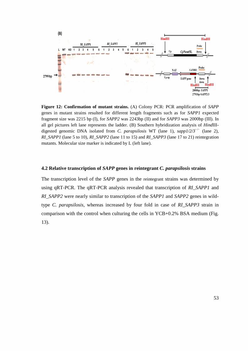
53
Figure 12: Confirmation of mutant strains. (A) Colony PCR: PCR amplification of SAPP
genes in mutant strains resulted for different length fragments such as for SAPP1 expected
fragment size was 2215 bp (I), for SAPP2 was 2243bp (II) and for SAPP3 was 2000bp (III). In
all gel pictures left lane represents the ladder. (B) Southern hybridization analysis of HindIII-
digested genomic DNA isolated from C. parapsilosis WT (lane 1), sapp1/2/3−/−
(lane 2),
RI_SAPP1 (lane 5 to 10), RI_SAPP2 (lane 11 to 15) and RI_SAPP3 (lane 17 to 21) reintegration
mutants. Molecular size marker is indicated by L (left lane).
4.2 Relative transcription of SAPP genes in reintegrant C. parapsilosis strains
The transcription level of the SAPP genes in the reintegrant strains was determined by
using qRT-PCR. The qRT-PCR analysis revealed that transcription of RI_SAPP1 and
RI_SAPP2 were nearly similar to transcription of the SAPP1 and SAPP2 genes in wild-
type C. parapsilosis, whereas increased by four fold in case of RI_SAPP3 strain in
comparison with the control when culturing the cells in YCB+0.2% BSA medium (Fig.
13).

54
Figure 13: Transcription of SAPP genes in the mutant C. parapsilosis RI_SAPP1,
RI_SAPP2 and RI_SAPP3 strains. Fold change of SAPP gene transcription in RI_SAPP
mutants compared to the wild-type strain corresponding genes after growth in YCB + 0.2%
BSA medium. Transcription of SAPP1 and SAPP2 genes in reintegrant strains RI_SAPP1 and
RI_SAPP2 was restored while SAPP3 gene‟s transcription was upregulated in the RI_SAPP3
strain by ≥4 fold. The experiment was performed in triplicates.
4.3 Detection of protease activities of Sapp in WT and mutant strains of C.
parapsilosis
Candida secreted aspartyl proteases can hydrolyze BSA. To test if reintegrant strains
restored protease production capability, WT, sapp1/2/3−/−
, and RI_SAPP1, RI_SAPP2,
RI_SAPP3 C. parapsilosis strains were spotted onto YCB + 0.2% BSA containing
plates and plates were stained with amido black. The widths diameter of clearance
zones were measured after three days of incubation. Wild-type C. parapsilosis showed a
clear hallow zone (7.3 mm) in BSA containing plates whereas, RI_SAPP1 (5.78 mm)
and RI_SAPP2 (5.76 mm) also showed proteolytic activity. In contrast, RI_SAPP3 and
sapp1/2/3−/−
C. parapsilosis strains did not show any proteolytic activity, although the
growth of the colonies was nearly similar to WT (Fig. 14A and B).

55
Figure 14: Protease activity of WT and SAPP mutant strains were detected by BSA
degradation assay. (A) 10 µl of 18 h WT and mutant C. parapsilosis cultures were spotted onto
agar plates containing YCB + 0.2% BSA. Halo zone around the colonies of wild-type,
RI_SAPP1, and RI_SAPP2 depicts protease activity. sapp1/2/3−/−
and RI_SAPP3 mutant strain
did not form halo zone around colonies indicating no protease secretion. Images are
representative of 4 independent repeats. (B) The radius of clear white zone was measured and
plotted in the graph.
4.4 C. parapsilosis WT, sapp1/2/3−/−
, and RI_SAPP strains grow at similar rate
Estimation of fungal growth by continuous monitoring the change in the OD has been
used to check the growth of WT and mutant C. parapsilosis strains. To monitor the
effect the deletion and reintegration of sapp genes in growth rate of C. parapsilosis first,
we examined growth of WT, sapp1/2/3−/−
, and RI_SAPP mutant strains in normal YPD
and YCB+0.2% BSA growth medium for 24 h. Deletion or reintegration of SAPP genes
had no effect on the growth of C. parapsilosis at 30 °C either in YPD or in YCB+BSA
medium (Fig. 15A and B). Growth curve results indicate that SAPP gene in C.
parapsilosis do not influence its growth.

56
Figure 15: Kinetic growth curve analysis. Growth of C. parapsilosis WT, sapp1/2/3−/−
, and
RI_SAPP strains (5×10⁵ cells/ml as initial concentration) was examined in YPD (A), and YCB
(B) at every 30 min time interval by measuring OD at 600 nm. WT, sapp1/2/3−/−
, and RI_SAPP
strains grown in YPD and YCB media with a similar growth rate at 30 °C. Experiments were
performed two times in triplicates.
4.5 C. parapsilosis SAPP3 reintegrant and sapp1/2/3−/−
mutant strains are more
sensitive to normal human serum
C. albicans Saps are reported to cleave human complement proteins and other
microbicidal plasma proteins thus providing additional advantage of avoiding host
immune response. To investigate the effect of the fungicidal effect of human serum,
yeast cells were cultivated in 20% normal human serum and heat inactivated serum;
then, CFU determinations were performed at different time intervals. Whereas all C.
parapsilosis strains grown similarly in 20% heat-inactivated serum, the viability of the
RI_SAPP3 and sapp1/2/3−/−
C. parapsilosis strains in intact serum was reduced
significantly after 18 and 24 h incubation, respectively (Fig. 16A and B). There were no
significant differences between the growth of WT, RI_SAPP, and sapp1/2/3−/−
mutant
strains in heat inactivated human serum at any time point of treatment.

57
Figure 16: Plasma sensitivity assay. (A) Growth of C. parapsilosis WT, sapp1/2/3−/−
, and
RI_SAPP strains in 20% normal human serum (NHS) and heat inactivated (HiS) examined by
CFU determination at 0, 6, 24 and 48 h. Results show that NHS significantly inhibits the growth
of sapp1/2/3−/−
strain at 16 and 24 h when compared with WT. (B) HiS did not show any
sensitivity against sapp1/2/3−/−
compared to WT.
4.6 Deletion or reintegration of SAPP genes did not alter the pseudohyphae
formation abilities of C. parapsilosis
Hyphae formation play important role in pathogenesis of C. albicans. Rather than
forming true hyphae C. parapsilosis forms pseudohyphae. It has been reported that
pseudohyphal form of C. parapsilosis express more adhesion proteins than yeast form,

58
thus it is more virulent. Therefore, we examined that deletion of SAPP genes has any
effect on pseudohyphal formation of C. parapsilosis. Interestingly, deletion of the SAPP
genes and reintegration of the genes did not result any significant change in
pseudohyphae formation either on solid or in liquid media compared to WT (Fig. 17A-
D). Effect of gene deletion and reintegration on phenotypic variation was also examined
by observing the wrinkling and peripheral filamentation. C. parapsilosis WT, RI_SAPP,
and sapp1/2/3−/−
mutants exhibited indistinguishable colony morphologies at 30 and 37
°C (Fig. S2). Additionally, to investigate on the effect of deletion and reintegration of
SAPP genes on cell wall composition, differential staining with Concanavalin A (dye
for mannoproteins, Calcofluor white (dye for chitin content), and Wheat germ
agglutinin (fluorescence dye for β glucan) was used. No differences in cell wall
composition was observed in WT, RI_SAPP, and sapp1/2/3−/−
C. parapsilosis strains
(Fig. S3).
Further, we also investigated stress tolerance of WT and sapp1/2/3−/−
C. parapsilosis
strains at several conditions (Table S3). Surprisingly, we did not found any differences
in stress tolerance of sapp1/2/3−/−
C. parapsilosis strain compared to WT (Fig. S4).
Thus, results indicate that SAPP did not play significant role in pseudophyphae
formation, stress tolerance, and cell wall composition in C. parapsilosis.

59
Figure 17: Pseudohyphae formation of C. parapsilosis WT and mutant strains. C.
parapsilosis WT and mutant strains were grown in pseudohyphae inducing conditions (YPD or
RPMI 1640 + FBS, 37 °C in CO2 incubator). Number of pseudohyphae forming cells was
counted and percentage of the pseudohyphae forming cells was calculated by dividing
pseudohyphae forming cells by the total number of cells. (A) Representative picture of C.
parapsilosis WT and mutant strains stained with concanavalin A to visualize the pseudohyphae.
(B) Percentage of pseudohyphae forming cells was calculated from three independent
microscopic pictures by dividing number of pseudohypha forming cells to total number of cells
and multiplied by 100. (C) Percentage of pseudohyphae forming cells analyzed by flow
cytometer. (D) Representative image shows yeast cell and pseudohyphal cell. Scale bar 10µm.

60
4.7 Deletion and reintegration of SAPP genes did not alter the biofilm formation in
C. parapsilosis
Previous study has shown a significant reduction in biofilm formation capabilities of the
sap5Δ/Δ, sap6Δ/Δ, and sap5/6 ΔΔ/ΔΔ deletion strains of C. albicans compared to the
WT reference strain164
. Thus to further understand whether deletion and reintgration of
SAPP genes affect biofilm formation of C. parapsilosis strains were inoculated in YNB
and RPMI media and allowed to form biofilm for 48 h at 37 °C; next MTT reduction
assay and crystal violet (CV) staining method was used to determine the biofilm
formation level. Growth assay was performed previously to rule out the possibility of
variation in the biofilm level due to the difference growth rate. No significant
differences were observed in biofilm formation of WT and mutant C. parapsilosis
strains (Fig. 18A and B).
Figure 18: Biofilm formation by different C. parapsilosis SAPP mutant strains. Biofilm was
quantified by crystal violet stain (A) and MTT colorimetric assay (B). C. parapsilosis WT,
RI_SAPP, and sapp1/2/3−/−
form similar biofilm either grown in RPMI or in YNB media.

61
4.8 Secreted aspartyl proteases of C. parapsilosis affect its adhesion
In C. albicans SAP (primarily SAP5 and SAP6) deletion mutants have pronounced
reduction in the number of adhered cells164
. Therefore, we examined weather
reintegration or deletion of SAPP genes in C. parapsilosis affect adhesion to artificial
surface and epithelial cells. Results have clearly shown that all three reintegrant C.
parapsilosis strains exhibited significant reduction in number of cells (CFU) adhered to
the wells when compared to the WT. Highest reduction in adhesion was observed in the
case of SAPP null mutant C. parapsilosis strains (nearly 60% of total cells were not
adhered), whereas lowest reduction was observed in C. parapsilosis RI_SAPP1 mutant
strain (20%) followed by RI_SAPP2 and RI_SAPP3 (25-25%). However, comparison of
the adhesion efficiency of reintegrant strains clearly showed that C. parapsilosis
RI_SAPP1 exhibited highest adhesion efficiency when compared with the RI_SAPP2
and RI_SAPP3 reintegrant C. parapsilosis mutant strains (Fig. 19A). A significant
reduction in adhesion to oral epithelial cells (TR146 cells) of RI_SAPP3, and
moderately lower adhesion in case of sapp1/2/3−/−
and RI_SAPP2 C. parapsilosis
strains were also observed. At the same time, RI_SAPP1 C. parapsilosis strain restored
adhesion capacity to epithelial cells nearly to WT (Fig. 19B).
We also monitored the adhesion efficiencies of WT and SAPP mutant C. parapsilosis
strains to vaginal epithelial cells (A-431 cells). Our result suggest that C. parapsilosis
WT and RI_SAPP1 adhere significantly more to A-431 cells as compared to RI_SAPP2,
RI_SAPP3 although without reaching significant difference to sapp1/2/3−/−
C.
parapsilosis strain (Fig. 19C). These finding clearly indicate the role of SAPP1 and
SAPP2 in adhesion of C. parapsilosis to epithelial cells.

62
Figure 19: In vitro cell adhesion assay. WT, sapp1/2/3−/−
, and RI_SAPP strains were allowed
to adhere either solid surface (A), to oral epithelial cells (B), or to the vaginal epithelial cells
(C), for 90 min. All experiments are repeated three times in triplicate and a significant
difference is considered in compare to WT.

63
4.9 SAPP null mutant C. parapsilosis is less capable of causing host-cell damage
whereas reintegration of SAPP1 and SAPP2 genes restored cell damage causing
capabilities
Damage of PBMC-DM cells in the presence of WT, RI_SAPP, and sapp1/2/3−/−
C.
parapsilosis strains was monitored with lactate dehydrogenase (LDH) release assay
after 24 and 48 h coincubation. WT, RI_SAPP1, and RI_SAPP2 C. parapsilosis strains
cause nearly similar damage to those seen with PBMC-DMs (7.779% ± 1.001% and
6.807% ± 1.642%, respectively) whereas, RI_SAPP3 and sapp1/2/3−/−
caused
significantly less damage (5.843% ± 0.5715% and 6.862% ± 1.340%, respectively) than
the wild-type strain (9.944% ± 0.6143%) after 24 h coincubation with PBMC-DMs.
Differences between the examined strains became more evident following 48 h of
coincubation. Host cell damage was least severe in macrophages infected with the
RI_SAPP3 and sapp1/2/3−/−
strains (11.28% ± 0.8304% and 13.95% ± 1.153%,
respectively), followed by RI_SAPP2 (19.98% ± 1.238%) and RI_SAPP1
(23.04% ± 1.661), compared to that seen with the wild-type strain (40.36% ± 0.6912%)
(Fig. 20A). Additionaly, in the case of PBMC-DMs, C. parapsilosis WT reported to
cause significantly higher damage at both 24 h and 48 h (Fig. 20B). LDH released by A-
431 cells infected with C. parapsilosis WT (9.774 ± 1.618) was significantly higher
compared to RI_SAPP2 (4.937 ± 0.8536), RI_SAPP3 (4.109 ± 0.9633), and sapp1/2/3−/−
(3.243 ± 0.3529) strains indicating that mainly SAPP1 and SAPP2 are crucial in C.
parapsilosis to cause host cell damage.
(A)

64
(B)
Figure 20: Host cell damage by WT and mutant strains: Host cell damage was measured by
the amount of lactate dehydrogenase (LDH) released by human PBMC-DM (A), and A-431
cells (B). Cells were stimulated with C. parapsilosis WT, sapp1/2/3−/−
, and RI_SAPP strains
with MOI 5 in case of PBMC-DMs and MOI 10 in case of A-431 cells. LDH released was
measured after 24 h and 48 h of post infection and expressed as % of positive control. Results
clearly showed that WT causes more damage either to PBMC-DM or A-431 cells compared to
sapp1/2/3−/−
. In A-431 cells WT and RI_SAPP1 strains causes similar damage however all
reintegrant mutant strains showed attenuated damage causing capacities to PBMC-DMs.
4.10 Macrophages phagocytosed sapp1/2/3−/−
mutant C. parapsilosis more
efficiently than WT, RI_SAPP1, RI_SAPP2, and RI_SAPP3 cells
Human PBMC-DMs were used to characterize the immune modulatory properties of
SAPP genes in C. parapsilosis. We first examined the phagocytic activity of PBMC-
DMs by fluorescence-activated cell sorting (FACS). C. parapsilosis yeast cells were
labeled with the fluorescent dye Alexa Fluor 488 (succimidyl ester), washed, and then
coincubated with PBMC-DMs for 2 h. Our results indicated that PBMC-DMs ingested
RI_SAPP3 (60.63 ± 5.430) mutant strains more efficiently compared to the wild-type
(46.70 ± 2.041) C. parapsilosis (Fig. 21), highlighting the importance of SAPPs in
modulating phagocytotic behavior of macrophages. Additionaly, sapp1/2/3−/−
(59.03 ±
2.675) was also significantly more phagocytosed by PBMC-DMs compared to WT C.
parapsilosis.

65
(B)
Figure 21: Phagocytosis of C. parapsilosis strains by human macrophages. Alexa Fluor 488
labeled yeast cells were co-incubated with PBMC-DMs. (A) Representative dot plots (gated
cells) show the PBMC-DMs phagocytosed Candida Ch1: brightfield image, Ch2: fluorescence
lebelled Candida, Ch1/Ch2: merge image. (B) Graph shows the difference between
phagocytosis in case of the wild-type and the mutant strains infected PBMC-DMs.

66
4.11 SAPP2 influences the phagosome-lysosome fusion
Next, we examined whether lack or reintegration of SAPP genes into C. parapsilosis
genome has any effect on phagosome-lysosome fusion in human PBMC-DMs. We
examined co-localization of phagosome-lysosome after coincubation with pHrodo
(Invitrogen) stained Candida cells with PBMC-DMs for 2 h. WT (16.66% ± 0.5732%)
and RI_SAPP2 (13.78% ± 1.216%) infected PBMC-DMs showed less phagolysosome
formation compared to RI_SAPP1, RI_SAPP3 and sapp1/2/3−/−
(20.76% ± 0.7194%,
29.52% ± 2.719%, 28.70% ± 2.025% respectively) C. parapsilosis strains, suggesting
that similar to C. albicans SAP2, C. parapsilosis SAPP2 gene‟s influence on phagosome
maturation (Fig. 22A and B).
(B)
Figure 22: Phagosome-
lysosome fusion in response to
WT and SAPP mutant C.
parapsilosis. Phagosome-
lysosome co-localization in
human PBMC-DMs following
the phagocytosis of pHrodo
labeled Candida cells was
investigated. (A)
Representative plots of a
phagocytosing macrophage
during quantitative imaging
analysis. Ch1: brightfield
image, Ch3: green fluorescence
channel, Ch1/Ch3: merge
image. (B) Graph shows the
difference between the extent of
phago-lysosome fusion in case
of the wild-type and the mutant
strains.

67
4.12 SAPP null mutant and RI_SAPP3 C. parapsilosis cells are killed more
efficiently by PBMC-DMs and in human whole blood compared to RI_SAPP1/2
strains
Phagocytosis of Candida cells by macrophages and subsequent formation of
phagolysosome in general leads to the killing of pathogen. C. albicans mediated
alteration of phagosome-lysosome fusion is well reported165
. In our above mentioned
results we observed that several C. parapsilosis mutant strains (RI_SAPP1, RI_SAPP3,
and sapp1/2/3−/−
) alter phagolysosome fusion compared to WT strain. Therefore, to
further examine if alteration of phagolysosome formation is related with yeast cell
killing efficiency of PBMC-DMs we compared fungal killing by recovered fungal CFUs
after coincubation PBMC-DMs with WT and mutant C. parapsilosis cells. Our data
showe that PBMC-DM cells were able to kill C. parapsilosis RI_SAPP3 mutant cells
with significantly higher efficiency compared to WT cells. Notably, PBMC-DM killed
significantly more [32% WT vs 53.9% sapp1/2/3−/−
and 50.39% RI_SAPP3] (Fig. 23A).
However, killing efficiency of macrophages was nearly similar against WT, RI_SAPP1,
and RI_SAPP2 C. parapsilosis strains indicating significant contribution of SAPP1 and
SAPP2 in modulation of killing capacity of macrophages. In addition to PBMC-DMs,
we also examined the killing efficiency of whole human blood. Our results show more
efficient killing of C. parapsilosis RI_SAPP3 and sapp1/2/3−/−
by human whole blood
indicating protective roles of SAPP1 and SAPP2 (Fig. 23B).
(A)

68
(B)
Figure 23: Killing efficiencies of human PBMC-DMs and whole blood. The efficiency of
killing by human macrophages (A), and human whole blood (B) was analyzed by CFU
determination. Experiments were performed in triplicates. The obtained data represents the
killing efficiency of macrophages gained from 5 healthy donors. Blood from three different
donors was used.
4.13 SAPPs regulate the cytokine response of host macrophages
Aspartyl proteases of Candida albicans effectively regulate inflammatory
responses140,141
. Recent studies have shown that sap2 and sap6 effectively induce the
activation of NLRP3 inflammasome and thus leading to the secretion of interleukin-1β
(IL-1β) and IL-18 via caspase-1 activation141
. These candidapepsins also induce
secretion of chemoattractive chemokine‟s such as IL-8 and MIP-2 by vaginal epithelial
cells140
. But, role of candidaparapesins in modulating the immune responses is still
enigmatic thus, in order to examine the immunological responses triggered by RI_SAPP
and sapp1/2/3−/−
mutant C. parapsilosis, we stimulated human PBMC-DMs for 24 h
with each strain and measured IL-1β, TNFα, IL-6 cytokine and IL-8 chemokine release.
The obtained results indicate that PBMC-DMs stimulated with either the wild-type or
RI_SAPP1 and RI_SAPP2 strains produced similar IL-1β, IL-8 and TNFα levels. In
contrast, macrophages stimulated with C. parapsilosis sapp1/2/3−/−
released
significantly less IL-1β and IL-6, and moderately but not significantly less IL-8

69
compared to the WT (Fig. 24). PBMC-DMs stimulated with RI_SAPP3 mutant C.
parapsilosis cells produced significantly lower IL-8 and moderately low IL-6, however,
no significant differences were observed in the secretion of IL-1β and TNFα compared
to WT.
Figure 24: Cytokine secretion of human PBMC-DMs in response to WT and SAPP mutant
C. parapsilosis strains. IL-1β, TNFα, IL6, and IL-8 levels were measured by ELISA after
stimulation of PBMC-DMs with WT or SAPP mutant C. parapsilosis strains for 24 h. Data
represent cytokine production ± SEM from 5 donors.
4.14 Sapp1 and Sapp2 have differential cleavage capacity against human
complement proteins
Fungal proteases are known to inactivate various host factors including complement
proteins such as C3b and C4b thus provide fungus an alternative mechanism to avoid
host complement attack. Therefore, to test if C. parapsilosis Sapp proteins can also
cleave human complement proteins we incubated human complement proteins C3b and
C4b with the purified Sapp proteins96,166
. Result indicated that Sapp1p and Sapp2p can
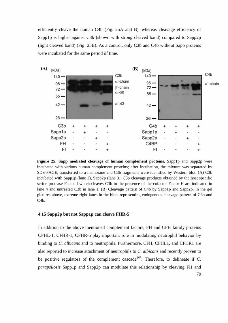
70
efficiently cleave the human C4b (Fig. 25A and B), whereas cleavage efficiency of
Sapp1p is higher against C3b (shown with strong cleaved band) compared to Sapp2p
(light cleaved band) (Fig. 25B). As a control, only C3b and C4b without Sapp proteins
were incubated for the same period of time.
4.15 Sapp2p but not Sapp1p can cleave FHR-5
In addition to the above mentioned complement factors, FH and CFH family proteins
CFHL-1, CFHR-1, CFHR-5 play important role in modulating neutrophil behavior by
binding to C. albicans and to neutrophils. Furthermore, CFH, CFHL1, and CFHR1 are
also reported to increase attachment of neutrophils to C. albicans and recently proven to
be positive regulators of the complement cascade167
. Therefore, to delineate if C.
parapsilosis Sapp1p and Sapp2p can modulate this relationship by cleaving FH and
Figure 25: Sapp mediated cleavage of human complement proteins. Sapp1p and Sapp2p were
incubated with various human complement proteins; after incubation, the mixture was separated by
SDS-PAGE, transferred to a membrane and C3b fragments were identified by Western blot. (A) C3b
incubated with Sapp1p (lane 2), Sapp2p (lane 3). C3b cleavage products obtained by the host specific
serine protease Factor I which cleaves C3b in the presence of the cofactor Factor H are indicated in
lane 4 and untreated C3b in lane 1. (B) Cleavage pattern of C4b by Sapp1p and Sapp2p. In the gel
pictures above, extreme right lanes in the blots representing endogenous cleavage pattern of C3b and
C4b.

71
CFH family protein we measured the capacity of Sapp1p and Sapp2p to degrade FH,
CFHL1, CFHR1, and CFHR5. Sapp1p and Sapp2p both cleaved FH after 15 h but not 3
h of incubation (Fig. 26A). Interestingly, Sapp2p but not Sapp1p cleaved FHR5,
indicating differential proteases activities of C. parapsilosis Sapps against human
complement proteins (Fig. 26B). Coincubation of Sapp1p or Sapp2p with FHL-1 or
FHR-1 revealed that the proteases cannot cleave these human complement proteins as
visualized by Western blot (Fig. 26C and D).
Figure 26: Sapp mediated cleavage of human complement like or related proteins. (A)
Cleavage of FH by Sapp1p and Sapp2p. (B) Cleavage of FHR5 by Sapp2p. (C&D) Sapp1p and
Sapp2p cannot cleave FHL1 and FHR1 either after 3 h or after 15 h of coincubation.

72
4.16 Sapps cannot alter the expression of CR3 and CR4 on macrophage surface
CR3 and CR4 are membrane bound receptors with multiple functions. CR3
(CD11b/CD18) plays important role in phagocytosis of opsonized pathogens, activates
NK cells, acts as adhesion molecule for leukocytes and induces NETosis from
neutrophils. Several bacterial proteases and C. albicans Saps are shown to modulate
their expression on macrophages125
. Thus, to understand the effect of C. parapsilosis
Sapp1p and Sapp2p proteins on the cell surface expression of CR3 and CR4, PBMC-
DMs were treated with these proteases. Unexpectedly, we did not found any substantial
differences in the CR3 and CR4 expression on macrophages either after Sapp1p or
Sapp2p treatment indicating in contrast to C. albicans, C. parapsilosis Sapp proteins
cannot alter CR3 and CR4 expression on macrophages (Fig. 27 A-C).
(A)
(B)
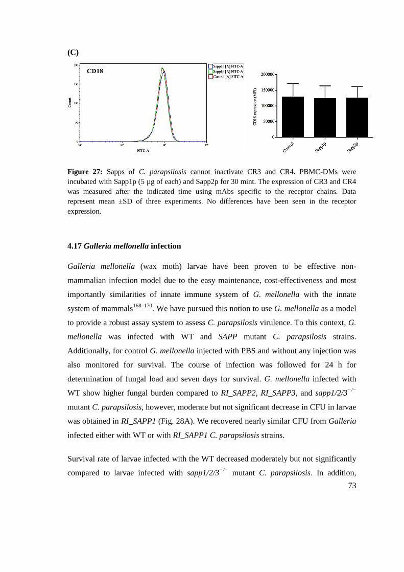
73
(C)
Figure 27: Sapps of C. parapsilosis cannot inactivate CR3 and CR4. PBMC-DMs were
incubated with Sapp1p (5 µg of each) and Sapp2p for 30 mint. The expression of CR3 and CR4
was measured after the indicated time using mAbs specific to the receptor chains. Data
represent mean ±SD of three experiments. No differences have been seen in the receptor
expression.
4.17 Galleria mellonella infection
Galleria mellonella (wax moth) larvae have been proven to be effective non-
mammalian infection model due to the easy maintenance, cost-effectiveness and most
importantly similarities of innate immune system of G. mellonella with the innate
system of mammals168–170
. We have pursued this notion to use G. mellonella as a model
to provide a robust assay system to assess C. parapsilosis virulence. To this context, G.
mellonella was infected with WT and SAPP mutant C. parapsilosis strains.
Additionally, for control G. mellonella injected with PBS and without any injection was
also monitored for survival. The course of infection was followed for 24 h for
determination of fungal load and seven days for survival. G. mellonella infected with
WT show higher fungal burden compared to RI_SAPP2, RI_SAPP3, and sapp1/2/3−/−
mutant C. parapsilosis, however, moderate but not significant decrease in CFU in larvae
was obtained in RI_SAPP1 (Fig. 28A). We recovered nearly similar CFU from Galleria
infected either with WT or with RI_SAPP1 C. parapsilosis strains.
Survival rate of larvae infected with the WT decreased moderately but not significantly
compared to larvae infected with sapp1/2/3−/−
mutant C. parapsilosis. In addition,

74
larvae infected with RI_SAPP mutant C. parapsilosis strains showed no any significant
difference in survival compared to WT (Fig. 28B).
Figure 28: Fungal burden and survival of Galleria mellonella post infection. (A) Fungal
burden WT, sapp1/2/3−/−
mutant strain, and RI_SAPP strains were determined using G.
mellonella larvae. For each individual larvae, 106 Candida cells were injected and the fungal
burden was determined by number of CFU. Each mutant was tested four times using at least
five larvae each time. The mean and SD of each data is represented by horizontal and vertical
bars, respectively. (B) Survival curves of G. mellonella infected with C. parapsilosis WT,
RI_SAPP1, RI_SAPP2, RI_SAPP3, and sapp1/2/3−/−
mutant with 106 Candida cells/larvae. No
significant difference in survival of larvae was observed after infection with different C.
parapsilosis strains.

75
4.18 Survival of Drosophila melanogaster after C. parapsilosis infection
The host innate immune response appears to be of major player of host defense
against invasive Candida infections in mice171
. Consequently, D. melanogaster, which
depend exclusively on an innate immune response to protect against pathogens, have
been considered appropriate models to study Candida infections172
. Therefore, we used
Drosophila as a model to provide a robust assay system to assess C. parapsilosis
virulence. We did not find any significant difference in survival either in wild-type
Oregon R-S (OreR) flies or in MyD88 (an adapter protein involved in the Toll-like
receptor and IL-1 receptor signaling pathway) knock out flies (MyD88−/−) mutant flies
infected with C. parapsillosis WT, sapp1/2/3−/−
, and RI_SAPP cells. Additionally,
comparing the survival of OreR and MyD88−/− flies clearly showed that MyD88−/−
flies
were approximately two times more susceptible to the infection of WT C. parapsilosis
during the first day than the wild-type flies (Fig. 29A and B). Moreover, killing of
MyD88 mutant flies by C. parapsilosis was more efficient at whole examination period
than killing of OreR flies, which clearly indicates that MyD88 signaling pathway plays
important role in protection against C. parapsilosis infection in D. melanogaster.

76
Figure 29: Estimated survival curves for OreR and MyD88−/−
flies infected with C.
parapsilosis WT and mutant strains. OreR (A), and MyD88−/−
(B), flies (n=40) infected with
WT and different mutant strains. For control, flies were injected with PBS to monitor the
physical effects of the injection per se. Flies were checked on every day after post infection over
7 days of period. No significant difference was observed in the survival of the flies infected
either WT or mutant C. parapsilosis strains.

77
5. Discussion
Aspartyl proteases are present in diverse range of microorganisms. These proteases play
crucial role in nutrition and pathogenesis. Presence of aspartyl protease gene family in
pathogenic non Candida albicans Candida species (NCAC) and absence in some non-
pathogenic fungal species such as in Saccharomyces cerevisiae suggests its role in
pathogenicity. The human fungal pathogen C. parapsilosis genome contains three
aspartyl protease genes, named SAPP1, SAPP2, and SAPP3 respectively. SAPP1 can be
found in duplicate (SAPP1a, SAPP1b) whereas, some sequences with high similarity
with SAPP2 was also identified in C. parapsilosis genome. The proteins level similarity
among the C. parapsilosis Sapps and also with C. albicans Saps is quite low, thus, we
presumed functional differences among Sapps in C. parapsilosis. Previously, in our
laboratory SAPP null mutant C. parapsilosis strains (∆/∆sapp1a, ∆/∆sapp1b,
∆/∆sapp1a/b) were generated and it had been shown that sapp mutant strains are less
virulent compared to wild-type C. parapsilosis. However, the study did not clearly
highlight the role of individual SAPP genes in C. parapsilosis virulence. Thereof, to
identify the roles of each aspartyl acid proteases in its virulence, first we generated
mutant strain lacking all aspartyl protease coding genes (C. parapsilosis sapp1/2/3−/−
)
and then integrated individual genes under the control of a strong constitutive C.
albicans (Ca) TDH3 promoter (CaTDH3) to CpNeut5L locus. Further, we analyzed the
virulence properties of wild-type, RI_SAPP, and sapp1/2/3−/−
C. parapsilosis strains.
Further, we also analyzed the protease activity of C. parapsilosis Sapp1p and Sapp2p
against human complement proteins.
Cell wall composition of Candida spp. and their associated proteins play important role
in determination of its morphology and also its recognition by host immune cells. Sapp
proteins, such as Sap9 and Sap10 are associated with cell wall of C. albicans and
reported to play role in cell wall integrity, biofilm formation, and interactions with hosts
immune cells173
. Therefore, first we tested role of SAPP genes in C. parapsilosis
pseudohyphae, biofilm formation, and cell wall composition. Surprisingly deletion or
reintegration of SAPP genes did not affect these properties of C. parapsilosis. We also

78
tested cell wall integrity of C. parapsilosis mutant strains by providing cell wall stress
our results have shown that even providing higher amount of cell wall stress
sapp1/2/3−/−
grow similar to WT, indicating SAPP in C. parapsilosis did not play role in
its stress tolerant abilities.
Efficient microbial adhesion to the epithelial cells is a crucial step for mucosal
colonization and infection. Several C. albicans and C. parapsilosis cell wall associated
proteins are engaged in attachment of C. albicans yeast to the host epithelium174
. Once
attached to epithelium pathogenic Candida spp. such as C. albicans, C. dubliniensis,
and C. tropicalis undergo morphological switching from yeast to hyphae, which is a
major virulence trait. C. parapsilosis and C. glabrata do not form true hyphae but still
adhere to epithelial cells however, the attachment of non-hyphae/pseudohyphae forming
cells are not as efficient as in the case of C. albicans. Apart from adhesion proteins such
as Hwp1p (hyphal wall protein 1, Als (agglutinin-like sequence) proteins, Hyr1p
(hyphal regulated protein, Eap1p (enhanced adherence to polystyrene), Sap4-6p bind to
host integrins, Sap6p presumably serve as ligands for several cell surfaces receptors
independently of its proteolytic activities in C. albicans. Interestingly, Sap9p and
Sap10p have contrary roles in epithelial adhesion as deletion of the SAP9 gene from C.
albicans increases adhesion, while deletion of the SAP10 gene reduces adhesion173
.
Thus, to understand the roles of C. parapsilosis Sapp proteins in adhesion to human
epithelial cells we performed adhesion experiment using human TR146 and A-431 cell
lines. Our results highlighted that deletion of all aspartyl protease genes of C.
parapsilosis moderately, but not significantly reduce the adhesion of C. parapsilosis to
epithelial cells. Of note, highest reduction was noted in case of RI_SAPP3 strain
whereas, reintegration of SAPP1 and SAPP2 genes restored the adhesion efficiencies of
sapp1/2/3−/−
strain. These results indicate the involvement of SAPP1 and SAPP2 in
attachment of C. parapsilosis to oral and vaginal epithelial cells.
We also investigated the interactions of WT and mutant strains with PBMC-DMs.
Macrophages are central players of the immune system, connecting the innate and
adaptive arms of the host‟s immune responses. Immune cells respond against pathogens

79
either by phagocytosis mediated killing or activating partner cells by secreting
cytokines. Phagocytosis starts by ingeston of pathogens. Internalized Candida cells get
enclosed in phagosomes, which further fused with lysosome. Fusion leads to
discharge of the lysosomal enzymes into the resulting phagolysosomes. Notably,
some bacterial pathogen such as Mycobacterium tuberculosis, Toxoplasma gondii
and fungus C. albicans and C. glabrata evade phagolysosome mediated killing by
altering phagosomal pH, hyphae mediated rupturing of phagosomal wall and by
utilizing amino acids inside the phagosomes175
. Of note, acidic pH (4.5-5) of
phagolysosome also favors the activity of secreted aspartyl proteases. Expression of
Sap genes of C. albicans and C. tropicalis after ingestion by immune cells suggest their
role in fungal adaptation to the intracellular environment176
. Presumably, acidic
environment of phagosome-lysosome intensifies the activity of acid pH induced
pathogenic factors, including secreted aspartyl proteases which in turn benefit microbial
intracellular survival. Here, in present study interestingly we also reported less
phagolysosome fusion in macrophages infected with C. parapsilosis WT compared to
sapp null mutant strain.
Flow cytometric analysis revealed that primary human monocyte-derived macrophages
efficiently ingest and kill RI_SAPP3 and sapp1/2/3−/−
mutant C. parapsilosis strains
compared to WT in vitro. In addition to the intracellular killing of microbes, the
secretion of inflammatory mediators by macrophages is essential for the efficient
eradication of pathogens. TNFα, IL-1β, IL-6, and IL-8 are the potent inflammatory
cytokines with pleiotropic effects, including the enhancement of antimicrobial capacity
of neutrophils and macrophages, modification of vascular permeability, and recruitment
of leukocytes to the site of inflammation, whereas the chemokine IL-8 is the most
potent activator of neutrophil recruitment. Release of several inflammatory mediators
were analysed in macrophages in response to WT and mutant strains of C. parapsilosis.
We found that macrophages stimulated with wild-type and RI_SAPP1 or RI_SAPP2
strains of C. parapsilosis produced more IL-1β and IL-6 and mild but not significantly
higher IL-8 and TNFα after 24 h compared to RI_SAPP3 and sapp1/2/3−/−
mutant
strains. Interestingly, we also observed that Sapp1 and Sapp2 play important role in
80
adhesion and cause damage to vaginal epithelial cells (A-431), indicating important role
of C. parapsilosis Sapps in vaginitis. However, further studies are required to get clear
understanding of C. parapsilosis aspartyl proteases role in vaginitis.
With nearly 50 serum and cell membrane-anchored proteins complement system plays
an essential humoral part of innate immunity. These complement proteins act in a
systemic myriad to opsonize the microbes and facilitate their phagocytosis, activate
cellular responses, initiate inflammation, or directly lyse certain microbes by forming
the pores in their cell wall177
. C3, as a crucial component of complement pathways and
plays a central role in activation of all three complement pathways. Factor H (FH)
which is composed of 20 individually folding complement control proteins (CCP) or
short complement regulatory (SCR) domains, act as regulator of complement pathway
by binding with C3b through its N-terminal CCP1–4 domains, C3b/C3d, pentraxins,
and sialic acid/ glycosaminoglycans through C-terminal CCP19–20. Thus binding of FH
on host surfaces under complement attack restricts complement activation. FH is
reported to induce secretion of IL-1β from human monocytes and chemokine IL-8 from
neutrophils125
. Recently, FH family proteins FHR and FHL are also reported to be
crucial for complement cascade regulation but exact roles of these proteins are
uncertain178
. FHR-1 was reported to inhibit C5 and the terminal pathway whereas FHR-
2 inhibits the alternative pathway and activation of the terminal pathway and FHR-3 and
FHR-4 enhances the cofactor activity of FH. FHR-5 was to display weak cofactor
activity and inhibit the C3 convertase in fluid phase. In general, FHR proteins appear to
lack significant complement inhibitory activity, but further studies are needed to clarify
if any of the FHRs possess some form of such activity128
. Prevailing evidence suggest
that bacterial and fungal pathogens can effectively bypass or alter host complement
attack either by recruiting complement regulator protein on their cell surface or by
cleaving them by secreting aspartyl proteases.
We evaluated the growth inhibitory efficiency of human serum against C. parapsilosis
wild-type and mutant strains. Congruent with the previous study, we found that
RI_SAPP3 and sapp1/2/3−/−
mutant strains are hypersensitive towards human serum

81
compared to wild-type, RI_SAPP1, and RI_SAPP2 C. parapsilosis strains. Heat
inactivation of serum did not show any growth inhibitory effect either on wild-type or
mutant strains. Heat inactivation of serum reported to inhibit the activity of complement
component C2 and factor B (FB) of complement system. C2 is the crucial component in
the activation of classical and lectin pathways, whereas FB is important for alternative
pathway179
. Inactivation of C2 and FB cannot lead activation of complement pathways
and that can be one of the possible reasons why heat inactivated plasma did not alter the
growth of C. parapsilosis wild-type or mutant strains.
To further determine whether C. parapsilosis Sapps can cleave of these complement
proteins and regulators, we incubated human complement proteins with Sapp1p and
Sapp2p for different period of time. Interestingly, our results demonstrated divergent
cleavage activity of Sapp1p and Sapp2p on complement proteins. Sapp1p and Sapp2p
both can cleave C3b, C4b, and FH whereas, Sapp2p but not Sapp1p cleaved FHR-5.
Interestingly, both of the protease cannot cleave FHL-1 or FHR-1. The cleavage of
FHR-5 but not FHL-1 and FHR-1 clearly demonstrates that the protease activity of
Sapp2p presumably lies somewhere near to SCR 3,4,5,6 or 7 SCR domains which are
absent in FHL-1 and FHR-1 but present in FHR-5 and FH, however, further detailed
studies are needed to confirm the Sapp cleavage sites in complement proteins. Taken
together, here we demonstrated for the first time that Sapp1p, Sapp2p both can cleave
complement proteins C3b, C4b and FH but not FHR-1 and FHL-1 whereas only Sapp2p
can cleave FHR-5. With this, the roles of these proteases on complement receptors such
as CR3 and CR4 were largely unknown thus, we aim to characterize the expression of
these receptors mainly on Sapp1p and Sapp2p treated human macrophages.
Unexpectedly, we did not find any change in expression of either on CR3 or CR4
receptor on Sapp treated macrophages. However, this does not exclude the possibility
that CR3 and CR4 functions are not affected by these proteases as they can efficiently
bind with complement proteins C3b, iC3b and FH which are efficiently cleaved by
these proteases.

82
In conclusion, our data show that secreted aspartyl proteases of C. parapsilosis
promotes the survival of fungal cells in macrophages and mitigate the inflammatory
response of the host, thereby interfering with the efficient clearing of the pathogen. As
per our best knowledge in present study first time we have reported that Sapp1p and
Sapp2p have differential cleavage activities against human complement proteins.
Even though, the finding of present study made a considerable progress to understand
the role of C. parapsilosis aspartyl proteases in modulation of the behavior of
macrophages and host complement cascade further understanding of the signalling
cascades induced by C. parapsilosis aspartyl proteases in immune cells undoubtedly
provide a great insight into the mechanisms that underpin during its virulence and could
lead us towards new therapeutic interventions against C. parapsilosis caused
pathogenesis.

83
6. Summary
Candida parapsilosis is one of the most frequently isolated, non Candida albicans
Candida (NCAC) species from preterm infants and neonates, skin and nails of
healthcare personnel. Being a skin commensal it has unique abilities to adhere on
physical surface and possess potent abilities to form biofilm on indwelling devices, such
as central venous catheters. Among NCAC species, infections caused by C. parapsilosis
increasing worldwide and it became the second or third most common yeast isolated
from hospitals of Asian and American countries whereas, the increasing incidence in
European countries is also reported. Several factors adhesion to host surfaces, switching
from yeast to pseudohyphal form, biofilm formation and secretion of hydrolytic
enzymes such as lipases, phospholipases and aspartyl proteases are considered as
crucial virulence factors. Historically C. parapsilosis are considered to possess three
aspartyl protease genes designated as SAPP1, SAPP2, and SAPP3 but recently 14 more
potential aspartyl protease encoding genes were identified. Proteins coded by these
aspartyl protease genes considered important for nutrient acquisition and critical for
penetration of host epithelial barrier. Crystal structures of Sapp1p and Sapp2p are
known but their virulence properties remain enigmatic. Therefore, present study was
designed to get deeper insight of virulence properties of C. parapsilosis Sapps.
To identify the virulence properties of individual Sapp isoenzymes in present study we
generated reintegrate strains of SAPP1, SAPP2, and SAPP3 under a strong promoter in a
SAPP null (sapp1/2/3−/−
) mutant of C. parapsilosis. Further to demonstrate if
reintegration of SAPP genes alter the physical attributes of C. parapsilosis, growth rate,
pseudohyphae formation, stress tolerance, alteration in cell wall composition, and
biofilm formation of mutant strains were examined. Interestingly, these mutant strains
did not show any difference either in cell wall composition or in biofilm formation
however sapp null (sapp1/2/3−/−
) and RI_SAPP3 mutant C. parapsilosis showed
reduced adhesion to human epithelial cells compared to the wild-type strain. Further,
virulence properties of the mutant strains were also examined using in vitro and in vivo
models. The sapp null (sapp1/2/3−/−
) and RI_SAPP3 mutant C. parapsilosis caused less
damage to human PBMC-DMs, phagocytosed and killed more efficiently compared to

84
wild-type. In addition, sapp1/2/3−/−
mutant strain was less capable to induce
inflammatory cytokine release, mainly IL-1β, compared to the wild-type C.
parapsilosis. More colony forming units (CFU) was recovered from Galleria mellonella
infected with either wild-type or RI_SAPP1 strain compared to other mutant strains,
which clearly indicate that SAPP1 play important role in C. parapsilosis virulence.
C. parapsilosis activate all three pathways of complement system. Activated
complement system act as host weapon against pathogens. Thus, to avoid complement
attack pathogen either cleaves complement proteins or regulates their activation by
recruiting complement regulator proteins on their cell surface. Till today, role of C.
parapsilosis secreted aspartyl proteases in regulation of complement cascade is largely
unknown. Here, using purified Sapp1p and Sapp2p we showed that these proteases can
efficiently cleave human complement proteins C3b, C4b. Complement regulators
control the spontaneously activated complement cascade and thus prevent unwanted
self-tissue destruction and autoimmune disease. Interestingly, factor H (FH), a potent
complement regulator of alternative complement pathway is also cleaved by both
Sapp1p and Sapp2p. Complement factor H-related/like proteins (FHRs/FHLs) are
recently proven to be positive complement regulators. Surprisingly, FHR-5 is cleaved
by Sapp2p but not by Sapp1p whereas Sapp1p and Sapp2p cannot cleave FHR-1 and
FHL-1.
Taken together, results of the present study suggested that even though deletion and
reintegration of SAPPs did not affect physical and morphological attributes of C.
parapsilosis much extent, it significantly affects the virulence properties of C.
parapsilosis including its adhesion properties to epithelial cells, damage causing
efficiency, phagosomes-lysosome fusion, and inflammatory responses from human
blood-derived macrophages. In addition, as per best of our knowledge first time we
have shown that Sapp1p and Sapp2p can cleave human complement proteins, thus;
presumably alter the complement mediated immune responses.

85
We believe that findings of this study could potentially lead to provide an additional
rationale for Sapp blockade as a therapeutic alternative in C. parapsilosis caused
pathogenesis.

86
7. Hungarian summary
A Candida parapsilosis az úgynevezett „nem-Candida albicans” Candida fajok
(NCAC) körébe tartozó élesztőgomba, melyet legtöbbször koraszülöttekből,
újszülöttekből és a kórházi dolgozók bőr- és köröm felszínéről izolálnak. Bőrfelszíni
kommenzalista fajként, egyedi tulajdonságai révén képes adhézióra, továbbá biofilm
képzésére különböző beültetett orvosi eszközökön, például centrális vénás katéterek
felületén. A „nem-albicans” Candida fajok közül a C. parapsilosis okozta egészségügyi
megbetegedések száma egyre nagyobb számban fordul elő világszerte. Több ázsiai és
amerikai ország kórházaiban a második vagy harmadik legtöbbször izolált
élesztőgombának számít, emellett egyre több európai esetről is beszámoltak már.
Számos faktort megemlíthetünk, melyek virulencia faktorként funkcionálnak és
meghatározzák a faj fertőzőképességét. Ilyen például az adhéziós képesség a
gazdaszervezet sejtjeinek felületén, morfológiai váltás (élesztő formából pszeudohifás
forma), biofilm képzés és hidrolitikus enzimek (lipázok, foszfolipázok, aszpartil
proteázok) szekréciója. Eredetileg úgy tartották, hogy a C. parapsilosisnak három
aszpartil-proteáz kódoló génje van, melyet az alábbi módon jelöltek: SAPP1, SAPP2 és
SAPP3. A közelmúltban viszont további 14 aszpartil-proteázt kódoló gént azonosítottak
filogenetikai módszerekkel. Ezen gének által kódolt fehérjék fontos szerepet játszanak a
tápanyagfelvételben és a gazdaszervezet hámrétegébe történő bejutás során. Míg a
Sapp1p és Sapp2p fehérjék kristályszerkezete ismert, addig a virulenciában betöltött
szerepük továbbra is kérdéses. Ezért a jelen kutatási munkában célul tűztük ki a C.
parapsilosis Sapp fehérjék virulenciában betöltött szerepének részletesebb
megismerését.
A jelen kutatási munkában az egyes Sapp izoenzimek virulencia tulajdonságainak
azonosítása érdekében létrehoztunk SAPP1, SAPP2 és SAPP3 reintegrált törzseket erős
promóter szabályozása alatt, sapp null (sapp1/2/3−/−
) mutáns C. parapsilosis törzsben.
További célunk volt bemutatni ezen reintegrált SAPP gének lehetséges hatását a C.
parapsilosis fiziológiai tulajdonságaira, növekedési sebességére, pszeudohifa képzésére,
stressztűrő képességére, sejtfal összetételére és biofilm képzésére. Érdekes módon ezen
mutáns törzsek nem mutattak változást sem a sejtfal összetételében, sem a biofilm

87
képzésében a vad típussal összehasonlítva. Azonban a sapp1/2/3−/−
és RI_SAPP3
mutáns C. parapsilosis törzsek kisebb mértékű humán hámsejtekhez történő adhéziós
képességet mutattak, mint a vad típusú törzs. Ezenkívül vizsgáltuk az említett törzsek
virulencia tulajdonságait in vitro és in vivo modellek bevonásával is. A sapp1/2/3−/−
és
RI_SAPP3 mutáns C. parapsilosis törzsek esetében az emberi PBMC-DM sejtek kisebb
mértékű károsodását és hatékonyabb fagocitózist figyeltünk meg, szemben a vad típusú
törzzsel. A sapp1/2/3−/−
mutáns törzs a vad típusú törzshöz viszonyítva kisebb
mértékben indukálta gyulladásos citokinek szekrécióját, főként az IL-1β-t. Nagyobb
számú kolóniaképző egységet (CFU) határoztunk meg a vad típusú és RI_SAPP1
törzsekkel fertőzött Galleria mellonella lárvákban, mint a többi mutáns törzsben. Ez
alapján megállapítható, hogy a SAPP1 fontos szerepet játszik a C. parapsilosis
virulenciájában.
A C. parapsilosis a komplementrendszer mindhárom útvonalát képes aktiválni. Az
aktivált komplementrendszer a gazdaszervezet védelmét biztosítja a kórokozók ellen.
Ahhoz, hogy ezt a védelmi vonalat a kórokozó elkerülje, képes hasítani a komplement
fehérjéket vagy úgy szabályozza ezen fehérjék aktivációját, hogy az őket szabályozó
fehérjéket a saját sejtfelszínére toborozza. Napjainkig a C. parapsilosis által szekretált
aszpartil-proteázok szerepe a komplementkaszkád aktiválásban nagyrészt ismeretlen.
Tisztított Sapp1p és Sapp2p alkalmazásával kimutattuk, hogy ezen proteázok képesek
hatékonyan hasítani a C3b és C4b humán komplement fehérjéket. A komplement
szabályozók a spontán aktivált komplementkaszkádot vezérlik, megakadályozva ezzel a
nem kívánatos szövetpusztulást és autoimmun betegség kialakulását. A Sapp1p and
Sapp2p képes hasítani a H-faktort (FH) is, mely a komplementrendszer alternatív
útvonalának szabályozója. A H-faktorral rokon és H-faktor-szerű fehérjék (FHR és
FHL) bizonyítottan pozitív komplementszabályzók. Meglepő módon az FHR-5-t a
Sapp2p hasítja, de a Sapp1p nem, valamint a Sapp1p és Sapp2p egyike sem tudja
hasítani az FHR-1-t és FHL-1-t sem.
Összegezve a jelen kutatási munkánkban elért eredményeket elmondható, hogy bár a
SAPP gének deléciója és túlzott expressziója sem befolyásolta nagymértékben a C.
parapsilosis fizikai és morfológiai tulajdonságait, addig virulencia képességére
88
szignifikáns hatást gyakorolt, beleértve a hámsejtekhez történő adhéziós képességét, az
okozott sejtkárosodást, fagoszóma-lizoszóma fúziót és az emberi vérből származó
makrofágok által kiváltott gyulladásos reakciókat. Ezen felül, tudomásunk szerint,
először sikerült bebizonyítanunk, hogy a Sapp1p és a Sapp2p képes hasítani az emberi
komplement fehérjéket, így feltételezhetően megváltoztatja a komplement rendszer által
közvetített immunválaszokat.
Úgy gondoljuk, hogy a jelen kutatási munkánk eredményei hozzájárulhatnak egy
jövőbeni, a C. parapsilosis okozta fertőzések kezelésére a Sapp-blokádot alternatív
terápiaként alkalmazó szer kifejlesztéséhez.
8. Financial support
DKS was supported by the Stipendium Hungaricum Program (2015-2018). This work is
also supported by GINOP-2.3.2-15-2016-00035, GINOP-2.3.3-15-2016-00006, grants
from ministry of human capacities, Hungary-TUDFO/47138-1/2019-ITM FIKP
program, and momentum grant from Hungarian Academy of Science (LP2018-
15/2018). I would also like to thank Federation of European Microbiological Societies
(FEMS), European Federation of Immunological Societies (EFIS), and Cayman
Chemical Company (USA) for generous supports.

89
9. Acknowledgement
Working hard for these many years to write this thesis would not have been possible
without the blessing of God, kind help and support of my many colleagues, friends,
family, and collaborators. Therefore, I am taking this opportunity to duly acknowledge
everyone who has helped me along the way.
I will start by acknowledging my PhD supervisor Professor Dr. Attila Gácser for giving
me the opportunity to work in his lab, guiding me throughout this journey, giving me
freedom to discuss and pursue my own ideas, working hard to provide us enough
resources and taking time to select hard working yet extremely helpful graduate
students and postdoctoral researchers who not only helped me during my hard time in
experiments but also intellectually challenged to improve my scientific abilities.
I express my deep sense of gratitude to our Head of Department Prof. Csaba Vágvölgyi
for being kind to provide me the opportunity to work in the Department of
Microbiology, University of Szeged. I am also thankful to him for writing many letters
for my fellowship applications, being a source of knowledge, inspiration, guidance and
providing high quality facilities in the department which benefitted me a lot during
these years. I also deeply admire the kindness shown by all the members of the
department.
I am thankful to my lab members specially Dr. Tibor Németh for helping me during the
molecular work and educating me about everything possible in cloning, Dr. Renáta Tóth
for making sure that my scientific words are rightly placed in all of my manuscripts,
Erik Zajta for being available for 24 h to discuss either lab work or giving advice on
other issues, Dr. Katalin Csonka and Dr. Csaba Gergő Papp for help during experiments
and also for constructive criticisms, Sára Pál for sharing workspace and taking time to
share light moments during long and tiring experiments, Judit Szenzenstein for always
being ready to help, Máté Vadovics, Flóra F Bohner, and Tamás Takács for contributing
to my professional development in many ways. I am really thankful to Dr. Tanmoy
Chakraborty for teaching many techniques during my initial days of PhD, being a
constant support, and a good friend.

90
I am grateful to all of my collaborators. Prof. Elisabetta Blasi and Dr. Eva Pericolini
from University of Modena and Reggio Emilia, Modena, Italy for giving me
opportunity to work in their laboratories which I really enjoyed. Dr. Józsi Mihály and
Dr. Zsuzsa Bajtay and their students Alexandra Papp and Szilvia Lukácsi from ELTE
University Budapest for support provided us with the complement protein experiments;
Dr. Olga Heidingsfeld from ASCR, Czech Republic for providing us purified Sapp
proteins. Dr. Edit Hermesz from Department of Biochemistry at University of Szeged
for always being kind towards me, inspiring me, and letting me do part of my PhD
experiments in her laboratory.
I always feel blessed to work under the guidance of smart, knowledgeable and very
hardworking scientists, Prof. I.S. Bright Singh, Dr. Valsamma Joseph, Dr. Sajeevan T.P.
and Dr. Swapna P Antony from NCAAH, CUSAT, Kerala, India. Their dedication
towards science inspired me to pursue my career in science. I am also grateful to Dr.
Sarika Singh from CDRI, Lucknow, India, Dr. Sachin Kumar from AIIMS, New Delhi,
India, and Dr. Rajashree Das from Amity University, Noida, India for giving me
opportunity to work with them and also for providing constant guidance and support.
My friends have also helped me considerably with my research, improving my social
skills, and supported me morally. I am truly grateful to Kabi Chandran, Dr. Phanindra
Babu Kasi, Dr. Dileep Kumar, Payal Chakraborty, Rakesh Varghese, Bettina Volford,
and Pramod K Jangir for support during my PhD. Mantosh Kumar, Neelanchal Vaidya,
Snigdha Gupta, Manjul Manjrekar, Krishna Jha, Sudeep Mondal, Ayesha, Shruti Gupta,
and Umar for being supportive during some or other part of this journey.
I am also thankful to several Indian, International, and European funding agencies to
support my research stay and travel to attend conferences/workshops during my studies.
I will end by saying a special thank you to my father. His love, blessing, and constant
support have been my strength during these years. I am blessed to have a lovely and
supportive family. I am truly grateful for love, moral support, encouragement, trust, and
blessings from my grandfather, grandmother, mother, sister, brother, uncles, and
cousins throughout my studies.

91
10. References
1. Khan AR, Khazanovich-Bernstein N, Bergmann EM, James MN. Structural
aspects of activation pathways of aspartic protease zymogens and viral 3C
protease precursors. Proc Natl Acad Sci. 1999; 96(20):10968-75. PMID:
10500110.
2. Navia MA, Fitzgerald PM, McKeever BM, Leu CT, Heimbach JC, Herber WK,
Sigal IS, Darke PL, Springer JP. Three-dimensional structure of aspartyl
protease from human immunodeficiency virus HIV-1. Nature. 1989;
337(6208):615-20. PMID: 2645523.
3. Chen J, Ouyang Y, Wang L, Xie W, Zhang Q. Aspartic proteases gene family in
rice: Gene structure and expression, predicted protein features and phylogenetic
relation. Gene. 2009; 442(1-2):108-18. doi: 10.1016/j.gene.2009.04.021.
4. Dunn BM. Structure and mechanism of the pepsin-like family of aspartic
peptidases. Chem Rev. 2002; 102(12):4431-58. PMID: 12475196.
5. Shen W, Yao X, Ye T, Ma S, Liu X, Yin X, Wu Y. Arabidopsis aspartic
protease ASPG1 affects seed dormancy, seed longevity and seed germination.
Plant Cell Physiol. 2018; 59(7):1415-1431. doi: 10.1093/pcp/pcy070.
6. Tang J, Wong RN. Evolution in the structure and function of aspartic proteases.
J Cell Biochem. 1987; 33(1):53-63. PMID: 3546346.
7. Simões I, Faro C. Structure and function of plant aspartic proteinases. Eur J
Biochem. 2004; 271(11):2067-75. PMID: 15153096.
8. Bochenska O, Rapala-Kozik M, Wolak N, Aoki W, Ueda M, Kozik A. The
action of ten secreted aspartic proteases of pathogenic yeast Candida albicans
on major human salivary antimicrobial peptide, histatin 5. Acta Biochim Pol.
2016; 63(3):403-10. doi: 10.18388/abp.2016_1318.
9. Polgár L. The mechanism of action of aspartic proteases involves 'push-pull'
catalysis. FEBS Lett. 1987; 219(1):1-4. PMID: 3036594.
10. Tang J, Wong RN. Evolution in the structure and function of aspartic proteases.
J Cell Biochem. 1987; 33(1):53-63. PMID: 3546346.
11. Aoki W, Kitahara N, Miura N, Morisaka H, Yamamoto Y, Kuroda K, Ueda M.
Candida albicans possesses Sap7 as a pepstatin A-insensitive secreted aspartic
protease. PLoS One. 2012; 7(2):e32513. doi: 10.1371/journal.pone.0032513.

92
12. Oh S, Chang W, Suh J. An aspartic protease analogue: intermolecular catalysis
of peptide hydrolysis by carboxyl groups. Bioorg Med Chem Lett. 2001;
11(11):1469-72. PMID: 11378379.
13. Roberts NB. Review article: human pepsins - their multiplicity, function and
role in reflux disease. Aliment Pharmacol Ther. 2006; 24 Suppl 2:2-9. Erratum
in: Aliment Pharmacol Ther. 2006; 24(10):1506. PMID:16939427.
14. Samuels TL, Johnston N. Pepsin as a causal agent of inflammation during
nonacidic reflux. Otolaryngol Head Neck Surg. 2009; 141(5):559-63. doi:
10.1016/j.otohns.2009.08.022.
15. O'Reilly RC, Soundar S, Tonb D, Bolling L, Yoo E, Nadal T, Grindle C, Field
E, He Z. The role of gastric pepsin in the inflammatory cascade of pediatric
otitis media. JAMA Otolaryngol Head Neck Surg. 2015; 141(4):350-7.
doi:10.1001/jamaoto.2014.3581.
16. Benes P, Vetvicka V, Fusek M. Cathepsin D--many functions of one aspartic
protease. Crit Rev Oncol Hematol. 2008; 68(1):12-28. doi:
10.1016/j.critrevonc.2008.02.008.
17. Pranjol MZI, Gutowski NJ, Hannemann M, Whatmore JL. Cathepsin D non-
proteolytically induces proliferation and migration in human omental
microvascular endothelial cells via activation of the ERK1/2 and PI3K/AKT
pathways. Biochim Biophys Acta Mol Cell Res. 2018; 1865(1):25-33. doi:
10.1016/j.bbamcr.2017.10.005.
18. Yang L, Cui M, Zhang L, Song L. FOXM1 facilitates gastric cancer cell
migration and invasion by inducing Cathepsin D. Oncotarget. 2017;
8(40):68180-68190. doi: 10.18632/oncotarget.19254.
19. Guang C, Phillips RD, Jiang B, Milani F. Three key proteases--angiotensin-I-
converting enzyme (ACE), ACE2 and renin--within and beyond the renin-
angiotensin system. Arch Cardiovasc Dis. 2012; 105(6-7):373-85. doi:
10.1016/j.acvd.2012.02.010.
20. Gomez RA, Sequeira-Lopez MLS. Renin cells in homeostasis, regeneration and
immune defence mechanisms. Nat Rev Nephrol. 2018; 14(4):231-245. doi:
10.1038/nrneph.2017.186.
21. Nishiyama A, Kobori H. Independent regulation of renin-angiotensin-
aldosterone system in the kidney. Clin Exp Nephrol. 2018; 22(6):1231-1239.
doi: 10.1007/s10157-018-1567-1.

93
22. Mandujano-González V, Villa-Tanaca L, Anducho-Reyes MA, Mercado-Flores
Y. Secreted fungal aspartic proteases: A review. Rev Iberoam Micol. 2016;
33(2):76-82. doi: 10.1016/j.riam.2015.10.003.
23. Aljammas HA, Al Fathi H, Alkhalaf W. Study the influence of culture
conditions on rennin production by Rhizomucor miehei using solid-state
fermentations. J Genet Eng Biotechnol. 2017. doi:10.1016/j.jgeb.2017.10.004.
24. Sardinas JL. Rennin enzyme of Endothia parasitica. Appl Microbiol. 1968;
16(2):248-55. PMID: 4868859.
25. Santos, André. Aspartic proteases of human pathogenic fungi are prospective
targets for the generation of novel and effective antifungal inhibitors. Curr
Enzyme Inhibition. 2011; 7.96-118. doi:10.2174/157340811796575281.
26. Parra-Ortega B, Cruz-Torres H, Villa-Tanaca L, Hernández-Rodríguez C.
Phylogeny and evolution of the aspartyl protease family from clinically relevant
Candida species. Mem Inst Oswaldo Cruz. 2009; 104(3):505-12. PMID:
19547880.
27. Cassone A, Vecchiarelli A, Hube B. Aspartyl proteinases of eukaryotic
microbial pathogens: from eating to heating. PLoS Pathog. 2016;
12(12):e1005992. doi: 10.1371/journal.
28. Horimoto Y, Dee DR, Yada RY. Multifunctional aspartic peptidase
prosegments. N Biotechnol. 2009; 25(5):318-24. doi: 10.1016/j.nbt.2009.03.010.
29. Nguyen JT, Hamada Y, Kimura T, Kiso Y. Design of potent aspartic protease
inhibitors to treat various diseases. Arch Pharm (Weinheim). 2008; 341(9):523-
35. doi: 10.1002/ardp.200700267.
30. Tumminello FM, Bernacki RJ, Gebbia N, Leto G. Pepstatins: aspartic proteinase
inhibitors having potential therapeutic applications. Med Res Rev. 1993;
13(2):199-208. PMID: 8445958.
31. Umezawa H, Aoyagi T, Morishima H, Matsuzaki M, Hamada M. Pepstatin, a
new pepsin inhibitor produced by Actinomycetes. J Antibiot. 1970; 23(5):259-
62. PMID: 4912600.
32. Dash C, Kulkarni A, Dunn B, Rao M. Aspartic peptidase inhibitors: implications
in drug development. Crit Rev Biochem Mol Biol. 2003; 38(2):89-119. PMID:
12749695.
33. Lv Z, Chu Y, Wang Y. HIV protease inhibitors: a review of molecular
selectivity and toxicity. HIV AIDS (Auckl). 2015; 7:95-104. doi:
10.2147/HIV.S79956.

94
34. Alvarenga DLR, Silva AHDS, Fiuza JA, Gaze ST, de Oliveira JG, Oliveira RC,
Calzavara-Silva CE, Pascoal-Xavier MA, Alves ÉAR. HIV aspartyl protease
inhibitors modify the percentage of activated leukocytes, as well as serum levels
of IL-17A and NO during experimental leishmaniasis. Int Immunopharmacol.
2018; 60:179-188. doi: 10.1016/j.intimp.2018.04.044.
35. Pichová I, Pavlícková L, Dostál J, Dolejsí E, Hrusková Heidingsfeldová
O,nWeber J, Ruml T, Soucek M. Secreted aspartic proteases of Candida
albicans, Candida tropicalis, Candida parapsilosis and Candida lusitaniae.
Inhibition with peptidomimetic inhibitors. Eur J Biochem. 2001; 268(9):2669-
77. PMID: 11322888.
36. Lukito Y, Chujo T, Scott B. Molecular and cellular analysis of the pH response
transcription factor PacC in the fungal symbiont Epichloë festucae. Fungal
Genet Biol. 2015; 85:25-37. doi: 10.1016/j.fgb.2015.10.008.
37. Bignell E, Negrete-Urtasun S, Calcagno AM, Haynes K, Arst HN Jr, Rogers T.
The Aspergillus pH-responsive transcription factor PacC regulates virulence.
Mol Microbiol. 2005; 55(4):1072-84. PMID: 15686555.
38. Silva NC, Nery JM, Dias AL. Aspartic proteinases of Candida spp.: role in
pathogenicity and antifungal resistance. Mycoses. 2014; 57(1):1-11. doi:
10.1111/myc.12095.
39. McCotter SW, Horianopoulos LC, Kronstad JW. Regulation of the fungal
secretome. Curr Genet. 2016; 62(3):533-45. doi: 10.1007/s00294-016-0578-2.
40. Flagfeldt DB, Siewers V, Huang L, Nielsen J. Characterization of chromosomal
integration sites for heterologous gene expression in Saccharomyces cerevisiae.
Yeast. 2009; 26(10):545-51. doi: 10.1002/yea.1705.
41. Singh R, Parija S C. Candida parapsilosis: an emerging fungal pathogen. Indian
J Med Res 2012; 136:671-3. PMID: 23168709
42. Silva S, Negri M, Henriques M, Oliveira R, Williams DW, Azeredo J. Candida
glabrata, Candida parapsilosis and Candida tropicalis: biology, epidemiology,
pathogenicity and antifungal resistance. FEMS Microbiol Rev. 2012; 36(2):288-
305. doi: 10.1111/j.1574-6976.2011.00278.
43. Nosek J, Holesova Z, Kosa P, Gacser A, Tomaska L. Biology and genetics of
the pathogenic yeast Candida parapsilosis. Curr Genet. 2009; 55(5):497-509.
doi: 10.1007/s00294-009-02684.
44. van Asbeck EC, Clemons KV, Stevens DA. Candida parapsilosis: a review of
its epidemiology, pathogenesis, clinical aspects, typing and antimicrobial

95
susceptibility. Crit Rev Microbiol. 2009; 35(4):283-309. doi:
10.3109/10408410903213393. PMID: 19821642.
45. Papon N, Courdavault V, Clastre M, Bennett RJ. Emerging and emerged
pathogenic Candida species: beyond the Candida albicans paradigm. PLoS
Pathog. 2013; 9(9):e1003550. doi: 10.1371/journal.ppat.1003550.
46. Csonka K, Vadovics M, Marton A, Vágvölgyi C, Zajta E, Tóth A, Tóth R,
Vizler C, Tiszlavicz L, Mora-Montes HM, Gácser A. Investigation of OCH1 in
the virulence of Candida parapsilosis using a new neonatal mouse model. Front
Microbiol. 2017; 8:1197. doi: 10.3389/fmicb.2017.01197.
47. Navarro-Arias MJ, Defosse TA, Dementhon K, Csonka K, Mellado-Mojica E,
Dias Valério A, González-Hernández RJ, Courdavault V, Clastre M, Hernández
NV, Pérez-García LA, Singh DK, Vizler C, Gácser A, Almeida RS, Noël T,
López MG, Papon N, Mora-Montes HM. Disruption of protein mannosylation
affects Candida guilliermondii cell wall, immune sensing, and virulence. Front
Microbiol. 2016; 7:1951. eCollection 2016. PMID: 27994582.
48. Fakhim H, Vaezi A, Dannaoui E, Chowdhary A, Nasiry D, Faeli L, Meis JF,
Badali H. Comparative virulence of Candida auris with Candida haemulonii,
Candida glabrata and Candida albicans in a murine model. Mycoses. 2018;
61(6):377-382. doi: 10.1111/myc.12754.
49. Frenkel M, Mandelblat M, Alastruey-Izquierdo A, Mendlovic S, Semis R, Segal
E. Pathogenicity of Candida albicans isolates from bloodstream and mucosal
candidiasis assessed in mice and Galleria mellonella. J Mycol Med. 2016;
26(1):1-8. doi: 10.1016/j.mycmed.2015.12.006.
50. Miramón P, Lorenz MC. A feast for Candida: Metabolic plasticity confers an
edge for virulence. PLoS Pathog. 2017; 13(2):e1006144.
doi:10.1371/journal.ppat.1006144.
51. Dadar M, Tiwari R, Karthik K, Chakraborty S, Shahali Y, Dhama K. Candida
albicans - Biology, molecular characterization, pathogenicity, and advances in
diagnosis and control - An update. Microb Pathog. 2018; 117:128-138. doi:
10.1016/j.micpath.2018.02.028.
52. Casadevall A, Pirofski LA. The damage-response framework of microbial
pathogenesis. Nat Rev Microbiol. 2003; 1(1):17-24. PMID: 15040176.
53. Hall RA, Noverr MC. Fungal interactions with the human host: exploring the
spectrum of symbiosis. Curr Opin Microbiol. 2017; 40:58-64. doi:
10.1016/j.mib.2017.10.020.
96
54. Jabra-Rizk MA, Kong EF, Tsui C, Nguyen MH, Clancy CJ, Fidel PL Jr, Noverr
M. Candida albicans pathogenesis: Fitting within the host-microbe damage
response framework. Infect Immun. 2016; 84(10):2724-39. doi:
10.1128/IAI.00469-16.
55. Priest SJ, Lorenz MC. Characterization of virulence-related phenotypes in
Candida species of the CUG Clade. Eukaryot Cell. 2015; 14(9):931-40. doi:
10.1128/EC.00062-15.
56. Lamoth F, Kontoyiannis DP. The Candida auris alert: facts and perspectives. J
Infect Dis. 2018; 217(4):516-520. doi: 10.1093/infdis/jix597.
57. Kohlenberg A, Struelens MJ, Monnet DL, Plachouras D; The Candida auris
survey collaborative group. Candida auris: epidemiological situation, laboratory
capacity and preparedness in european union and european economic area
countries, 2013 to 2017. Euro Surveill. 2018; 23(13). doi: 10.2807/1560-7917.
58. Tavanti A, Davidson AD, Gow NA, Maiden MC, Odds FC. Candida
orthopsilosis and Candida metapsilosis spp. nov. to replace Candida
parapsilosis groups II and III. J Clin Microbiol. 2005; 43(1):284-92. doi:
10.1128/JCM.43.1.284-292.2005.
59. Gago S, García-Rodas R, Cuesta I, Mellado E, Alastruey-Izquierdo A. Candida
parapsilosis, Candida orthopsilosis, and Candida metapsilosis virulence in the
non-conventional host Galleria mellonella. Virulence. 2013; 5(2):278-85. doi:
10.4161/viru.26973.
60. Bertini A, De Bernardis F, Hensgens LA, Sandini S, Senesi S, Tavanti A.
Comparison of Candida parapsilosis, Candida orthopsilosis, and Candida
metapsilosis adhesive properties and pathogenicity. Int J Med Microbiol. 2013;
303(2):98-103. doi 10.1016/j.ijmm.2012.12.006.
61. Falgier C, Kegley S, Podgorski H, Heisel T, Storey K, Bendel CM, Gale CA.
Candida species differ in their interactions with immature human
gastrointestinal epithelial cells. Pediatr Res. 2011; 69: 384-9. doi:
10.1203/PDR.0b013e31821269d5.
62. Berman J, Sudbery PE. Candida albicans: a molecular revolution built on
lessons from budding yeast. Nat Rev Genet. 2002; 3(12):918-30. PMID:
12459722.
63. Butler G, Rasmussen MD, Lin MF, Santos MA, Sakthikumar S, Munro CA,
Rheinbay E, Grabherr M, Forche A, Reedy JL, Agrafioti I, Arnaud MB, Bates S,
Brown AJ, Brunke S, Costanzo MC, Fitzpatrick DA, de Groot PW, Harris D,
Hoyer LL, Hube B, Klis FM, Kodira C, Lennard N, Logue ME, Martin R,
Neiman AM, Nikolaou E, Quail MA, Quinn J, Santos MC, Schmitzberger FF,
97
Sherlock G, Shah P, Silverstein KA, Skrzypek MS, Soll D, Staggs R, Stansfield
I, Stumpf MP, Sudbery PE, Srikantha T, Zeng Q, Berman J, Berriman M,
Heitman J, Gow NA, Lorenz MC, Birren BW, Kellis M, Cuomo CA. Evolution
of pathogenicity and sexual reproduction in eight Candida genomes. Nature.
2009; 459(7247):657-62. doi: 10.1038/nature08064.
64. Nosek J, Novotna M, Hlavatovicova Z, Ussery DW, Fajkus J, Tomaska L.
Complete DNA sequence of the linear mitochondrial genome of the pathogenic
yeast Candida parapsilosis. Mol Genet Genomics. 2004; 272(2):173-80. PMID:
15449175.
65. Gacser A. Adhesins in Candida parapsilosis: Understudied players in virulence.
Virulence. 2016; 7(2):65-7. doi:10.1080/21505594.2015.1135288.
66. Hoyer LL, Cota E. Candida albicans agglutinin-like sequence (Als) family
vignettes: A review of Als protein structure and function. Front Microbiol. 2016;
7:280. doi: 10.3389/fmicb.2016.00280.
67. Bertini A, Zoppo M, Lombardi L, Rizzato C, De Carolis E, Vella A, Torelli R,
Sanguinetti M, Tavanti A. Targeted gene disruption in Candida parapsilosis
demonstrates a role for CPAR2_404800 in adhesion to a biotic surface and in a
murine model of ascending urinary tract infection. Virulence. 2016; 7(2):85-97.
doi: 10.1080/21505594.2015.1112491.
68. Laffey SF, Butler G. Phenotype switching affects biofilm formation by Candida
parapsilosis. Microbiology. 2005; 151:1073-81. PMID: 15817776.
69. Lohse MB, Gulati M, Johnson AD, Nobile CJ. Development and regulation of
single- and multi-species Candida albicans biofilms. Nat Rev Microbiol. 2018;
16(1):19-31. doi: 10.1038/nrmicro.2017.107.
70. Nobile CJ, Johnson AD. Candida albicans biofilms and human disease. Annu
Rev Microbiol. 2015; 69:71-92. doi: 10.1146/annurev-micro-091014-104330.
71. Nobile CJ, Fox EP, Nett JE, Sorrells TR, Mitrovich QM, Hernday AD, Tuch
BB, Andes DR, Johnson AD. A recently evolved transcriptional network
controls biofilm development in Candida albicans. Cell. 2012; 148(1-2):126-38.
doi: 10.1016/j.cell.2011.10.048.
72. Kuhn DM, Chandra J, Mukherjee PK, Ghannoum MA. Comparison of biofilms
formed by Candida albicans and Candida parapsilosis on bioprosthetic
surfaces. Infect Immun. 2002; 70(2):878-88.
73. Ward TL, Dominguez-Bello MG, Heisel T, Al-Ghalith G, Knights D, Gale CA.
Development of the human mycobiome over the first month of life and across
body sites. mSystems. 2018; 3(3):e00140-17. doi:10.1128/mSystems.00140-17.

98
74. da Matta DA, Souza ACR, Colombo AL. Revisiting species distribution and
antifungal susceptibility of Candida bloodstream isolates from Latin American
medical centers. J Fungi (Basel). 2017; 3(2):24. doi:10.3390/jof3020024.
75. Nishikaku, A.S., Melo, A.S.A. & Colombo, A.L. Geographic trends in
invasive candidiasis. Curr Fungal Infect Rep. 2010; 4: 210.
doi.org/10.1007/s12281-010-0033-7.
76. Pfaller MA, Andes DR, Diekema DJ, Horn DL, Reboli AC, Rotstein C, Franks
B, Azie NE. Epidemiology and outcomes of invasive candidiasis due to non-
albicans species of Candida in 2,496 patients: data from the prospective
antifungal therapy (PATH) registry 2004-2008. PLoS One. 2014; 9(7):e101510.
doi: 10.1371/journal.pone.0101510.
77. Guinea J. Global trends in the distribution of Candida species causing
candidemia. Clin Microbiol Infect. 2014; 20:5-10. doi: 10.1111/1469-
0691.12539.
78. Martí-Carrizosa M, Sánchez-Reus F, March F, Coll P. Fungemia in a Spanish
hospital: the role of Candida parapsilosis over a 15-year period. Scand J Infect
Dis. 2014; 46(6):454-61. doi: 10.3109/00365548.2014.900190.
79. Figueiredo VT, de Assis Santos D, Resende MA, Hamdan JS. Identification and
in vitro antifungal susceptibility testing of 200 clinical isolates of Candida spp.
responsible for fingernail infections. Mycopathologia. 2007; 164(1):27-33.
PMID: 17551848.
80. Wächtler B, Wilson D, Haedicke K, Dalle F, Hube B. From attachment to
damage: defined genes of Candida albicans mediate adhesion, invasion and
damage during interaction with oral epithelial cells. PLoS One. 2011;
6(2):e17046. doi: 10.1371/journal.pone.0017046. PMID: 21407800.
81. Bertini A, Zoppo M, Lombardi L, Rizzato C, De Carolis E, Vella A, Torelli R,
Sanguinetti M, Tavanti A. Targeted gene disruption in Candida parapsilosis
demonstrates a role for CPAR2_404800 in adhesion to a biotic surface and in a
murine model of ascending urinary tract infection. Virulence. 2016; 7(2):85-97.
doi: 10.1080/21505594.2015.1112491.
82. Moyes DL, Wilson D, Richardson JP, Mogavero S, Tang SX, Wernecke J, Höfs
S, Gratacap RL, Robbins J, Runglall M, Murciano C, Blagojevic M, Thavaraj S,
Förster TM, Hebecker B, Kasper L, Vizcay G, Iancu SI, Kichik N, Häder A,
Kurzai O, Luo T, Krüger T, Kniemeyer O, Cota E, Bader O, Wheeler RT,
Gutsmann T, Hube B, Naglik JR. Candidalysin is a fungal peptide toxin critical
for mucosal infection. Nature. 2016; 532(7597):64-8. doi: 10.1038/nature17625.

99
83. Villar CC, Kashleva H, Nobile CJ, Mitchell AP, Dongari-Bagtzoglou A.
Mucosal tissue invasion by Candida albicans is associated with E-cadherin
degradation, mediated by transcription factor Rim101p and protease Sap5p.
Infect Immun. 2007; 75(5):2126-35. PMID: 17339363.
84. Morschhäuser J, Virkola R, Korhonen TK, Hacker J. Degradation of human
subendothelial extracellular matrix by proteinase-secreting Candida albicans.
FEMS Microbiol Lett. 1997; 153(2):349-55. PMID: 9271862.
85. Gow NA, van de Veerdonk FL, Brown AJ, Netea MG. Candida albicans
morphogenesis and host defence: discriminating invasion from colonization. Nat
Rev Microbiol. 2011; 10(2):112-22. doi: 10.1038/nrmicro2711.
86. Calderone RA, Fonzi WA. Virulence factors of Candida albicans. Trends
Microbiol. 2001; 9(7):327-35. PMID: 11435107.
87. Naglik J, Albrecht A, Bader O, Hube B. Candida albicans proteinases and
host/pathogen interactions. Cell Microbiol. 2004; 6(10):915-26. PMID:
15339267.
88. Lantz MS. Are bacterial proteases important virulence factors? J Periodontal
Res. 1997; 32:126-32. PMID: 9085222.
89. Corvo I, O'Donoghue AJ, Pastro L, Pi-Denis N, Eroy-Reveles A, Roche L,
McKerrow JH, Dalton JP, Craik CS, Caffrey CR, Tort JF. Dissecting the active
site of the collagenolytic cathepsin L3 protease of the invasive stage of Fasciola
hepatica. PLoS Negl Trop Dis. 2013; 7(7):e2269. doi:
10.1371/journal.pntd.0002269.
90. Tóth A, Németh T, Csonka K, Horváth P, Vágvölgyi C, Vizler C, Nosanchuk
JD, Gácser A. Secreted Candida parapsilosis lipase modulates the immune
response of primary human macrophages. Virulence. 2014; 5(4):555-62. doi:
10.4161/viru.28509.
91. Toth R, Toth A, Vagvolgyi C, Gacser A. Candida parapsilosis secreted lipase as
an important virulence factor. Curr Protein Pept Sci. 2017; 18(10):1043-1049.
doi: 10.2174/1389203717666160813163054.
92. Gácser A, Trofa D, Schäfer W, Nosanchuk JD. Targeted gene deletion in
Candida parapsilosis demonstrates the role of secreted lipase in virulence. J
Clin Invest. 2007; 117(10):3049-58. PMID: 17853941.
93. de Viragh PA, Sanglard D, Togni G, Falchetto R, Monod M. Cloning and
sequencing of two Candida parapsilosis genes encoding acid proteases. J Gen
Microbiol. 1993; 139(2):335-42. PMID: 8436951.

100
94. Horváth P, Nosanchuk JD, Hamari Z, Vágvölgyi C, Gácser A. The identification
of gene duplication and the role of secreted aspartyl proteinase 1 in Candida
parapsilosis virulence. J Infect Dis. 2012; 205(6):923-33. doi:
10.1093/infdis/jir873.
95. Gácser A, Schäfer W, Nosanchuk JS, Salomon S, Nosanchuk JD. Virulence of
Candida parapsilosis, Candida orthopsilosis, and Candida metapsilosis in
reconstituted human tissue models. Fungal Genet Biol. 2007; 44(12):1336-41.
PMID: 17391997.
96. Hrusková-Heidingsfeldová O, Dostál J, Majer F, Havlíkova J, Hradilek M,
Pichová I. Two aspartic proteinases secreted by the pathogenic yeast Candida
parapsilosis differ in expression pattern and catalytic properties. Biol Chem.
2009; 390(3):259-68. doi: 10.1515/BC.2009.034.
97. Dostál J, Merkerová M, Vinterová Z, Pichová I, Hrušková-Heidingsfeldová O.
Two SAPP2 gene homologs are present in Candida parapsilosis genome. Folia
Microbiol (Praha). 2015; 60(5):373-4. doi: 10.1007/s12223-014-0368-5.
98. Dostál J, Brynda J, Hrušková-Heidingsfeldová O, Pachl P, Pichová I, Rezáčová
P. The crystal structure of protease Sapp1p from Candida parapsilosis in
complex with the HIV protease inhibitor ritonavir. J Enzyme Inhib Med Chem.
2012; 27(1):160-5. doi: 10.3109/14756366.2011.627508.
99. Dostál J, Pecina A, Hrušková-Heidingsfeldová O, Marečková L, Pichová I,
Řezáčová P, Lepšík M, Brynda J. Atomic resolution crystal structure of Sapp2p,
a secreted aspartic protease from Candida parapsilosis. Acta Crystallogr D Biol
Crystallogr. 2015; 71:2494-504. doi: 10.1107/S1399004715019392.
100. Kaminishi H, Hamatake H, Cho T, Tamaki T, Suenaga N, Fujii T, Hagihara Y,
Maeda H. Activation of blood clotting factors by microbial proteinases. FEMS
Microbiol Lett. 1994; 121(3):327-32. PMID: 7926688.
101. Silva S, Henriques M, Oliveira R, Azeredo J, Malic S, Hooper SJ, Williams
DW. Characterization of Candida parapsilosis infection of an in vitro
reconstituted human oral epithelium. Eur J Oral Sci. 2009; 117(6):669-75. doi:
10.1111/j.1600-0722.2009.00677.
102. Erwig LP, Gow NA. Interactions of fungal pathogens with phagocytes. Nat
Rev Microbiol. 2016; 14(3):163-76. doi: 10.1038/nrmicro.2015.21
103. Cheng SC, Joosten LA, Kullberg BJ, Netea MG. Interplay between Candida
albicans and the mammalian innate host defense. Infect Immun. 2012;
80(4):1304-13. doi: 10.1128/IAI.06146-11.

101
104. van der Graaf CA, Netea MG, Verschueren I, van der Meer JW, Kullberg BJ.
Differential cytokine production and Toll-like receptor signaling pathways by
Candida albicans blastoconidia and hyphae. Infect Immun. 2005; 73(11):7458-
64. PMID: 16239547.
105. Joly S, Ma N, Sadler JJ, Soll DR, Cassel SL, Sutterwala FS. Cutting edge:
Candida albicans hyphae formation triggers activation of the Nlrp3
inflammasome. J Immunol. 2009; 183(6):3578-81. doi:
10.4049/jimmunol.0901323
106. Koh TJ, DiPietro LA. Inflammation and wound healing: the role of the
macrophage. Expert Rev Mol Med. 2011; 13:e23.
doi:10.1017/S1462399411001943.
107. Quintin J, Saeed S, Martens JHA, Giamarellos-Bourboulis EJ, Ifrim DC,
Logie C,Jacobs L, Jansen T, Kullberg BJ, Wijmenga C, Joosten LAB, Xavier
RJ, van der Meer JWM, Stunnenberg HG, Netea MG. Candida albicans
infection affords protection against reinfection via functional reprogramming of
monocytes. Cell Host Microbe. 2012; 12(2):223-32. doi:
10.1016/j.chom.2012.06.006.
108. Cheng SC, Quintin J, Cramer RA, Shepardson KM, Saeed S, Kumar V,
Giamarellos-Bourboulis EJ, Martens JH, Rao NA, Aghajanirefah A, Manjeri
GR, Li Y, Ifrim DC, Arts RJ, van der Veer BM, Deen PM, Logie C, O'Neill LA,
Willems P, van de Veerdonk FL, van der Meer JW, Ng A, Joosten LA,
Wijmenga C, Stunnenberg HG, Xavier RJ, Netea MG. mTOR- and HIF-1α-
mediated aerobic glycolysis as metabolic basis for trained immunity. Science.
2014; 345(6204):1250684. doi: 10.1126/science.1250684.
109. Lionakis MS, Iliev ID, Hohl TM. Immunity against fungi. JCI Insight. 2017;
(11):e93156. doi:10.1172/jci.insight.93156.
110. Ramirez-Ortiz ZG, Means TK. The role of dendritic cells in the innate
recognition of pathogenic fungi (A. fumigatus, C. neoformans and C.
albicans). Virulence. 2012; 3(7):635-46. doi: 10.4161/viru.22295.
111. Killick J, Morisse G, Sieger D, Astier AL. Complement as a regulator of
adaptive immunity. Semin Immunopathol. 2018; 40(1):37-48. doi:
10.1007/s00281-017-0644y.
112. Ali YM, Lynch NJ, Haleem KS, Fujita T, Endo Y, Hansen S, Holmskov U,
Takahashi K, Stahl GL, Dudler T, Girija UV, Wallis R, Kadioglu A, Stover CM,
Andrew PW, Schwaeble WJ. The lectin pathway of complement activation is a
critical component of the innate immune response to pneumococcal infection.
PLoS Pathog. 2012; 8(7):e1002793. doi: 10.1371/journal.ppat.1002793.

102
113. Ricklin D, Reis ES, Lambris JD. Complement in disease: a defence system
turning offensive. Nat Rev Nephrol. 2016; 12(7):383-401. doi:
10.1038/nrneph.2016.70.
114. Zipfel PF, Skerka C. Complement regulators and inhibitory proteins. Nat Rev
Immunol. 2009; 9(10):729-40. doi: 10.1038/nri2620.
115. van Lookeren Campagne M, Wiesmann C, Brown EJ. Macrophage
complement receptors and pathogen clearance. Cell Microbiol. 2007; 9(9):2095-
102. PMID: 17590164.
116. Clark SJ, Bishop PN. Role of factor H and related proteins in regulating
complement activation in the macula, and relevance to age-related macular
degeneration. J Clin Med. 2015; 4(1):18-31. doi: 10.3390/jcm4010018. PMID:
25729613.
117. Sánchez-Corral P, Pouw RB, López-Trascasa M, Józsi M. Self-damage caused
by dysregulation of the complement alternative pathway: relevance of the Factor
H protein family. Front Immunol. 2018; 9:1607.
doi:10.3389/fimmu.2018.01607.
118. Reiss T, Rosa TFA, Blaesius K, Bobbert RP, Zipfel PF, Skerka C, Pradel G.
Cutting edge: FHR-1 binding impairs factor H-mediated complement evasion by
the malaria parasite Plasmodium falciparum. J Immunol. 2018; 201(12):3497-
3502. doi: 10.4049/jimmunol.1800662.
119. Dasari P, Shopova IA, Stroe M, Wartenberg D, Martin-Dahse H, Beyersdorf
N, Hortschansky P, Dietrich S, Cseresnyés Z, Figge MT, Westermann M,
Skerka C, Brakhage AA, Zipfel PF. Aspf2 from Aspergillus fumigatus recruits
human immune regulators for immune evasion and cell damage. Front Immunol.
2018; 9:1635. doi: 10.3389/fimmu.2018.01635.
120. Rudnick RB, Chen Q, Stea ED, Hartmann A, Papac-Milicevic N, Person F,
Wiesener M, Binder CJ, Wiech T, Skerka C, Zipfel PF. FHR5 Binds to laminins,
uses separate C3b and surface-binding sites, and activates complement on
malondialdehyde-acetaldehyde surfaces. J Immunol. 2018; 200(7):2280-2290.
doi: 10.4049/jimmunol.1701641.
121. Leslie RGQ, Hansen S. Complement receptors. 2009. doi:
10.1002/9780470015902.a0000512.pub2
122. Lukácsi S, Nagy-Baló Z, Erdei A, Sándor N, Bajtay Z. The role of CR3
(CD11b/CD18) and CR4 (CD11c/CD18) in complement-mediated phagocytosis
and podosome formation by human phagocytes. Immunol Lett. 2017; 189:64-72.
doi: 10.1016/j.imlet.2017.05.014.

103
123. Vorup-Jensen T, Jensen RK. Structural immunology of complement receptors
3 and 4. Front Immunol. 2018; 9:2716. doi: 10.3389/fimmu.2018.02716.
124. Dustin ML. Complement receptors in myeloid cell adhesion and phagocytosis.
Microbiol Spectr. 2016; 4(6). doi: 10.1128/microbiolspec. PMID: 27809953.
125. Svoboda E, Schneider AE, Sándor N, Lermann U, Staib P, Kremlitzka M,
Bajtay Z, Barz D, Erdei A, Józsi M. Secreted aspartic protease 2 of Candida
albicans inactivates factor H and the macrophage factor H-receptors CR3
(CD11b/CD18) and CR4 (CD11c/CD18). Immunol Lett. 2015; 168(1):13-21.
doi: 10.1016/j.imlet.2015.08.009.
126. Duggan S, Leonhardt I, Hünniger K, Kurzai O. Host response to Candida
albicans bloodstream infection and sepsis. Virulence. 2015; 6(4):316-26. doi:
10.4161/21505594.2014.988096.
127. Cheng SC, Sprong T, Joosten LA, van der Meer JW, Kullberg BJ, Hube B,
Schejbel L, Garred P, van Deuren M, Netea MG. Complement plays a central
role in Candida albicans-induced cytokine production by human PBMCs. Eur J
Immunol. 2012; 42(4):993-1004. doi: 10.1002/eji.201142057.
128. Losse J, Zipfel PF, Józsi M. Factor H and factor H-related protein 1 bind to
human neutrophils via complement receptor 3, mediate attachment to Candida
albicans, and enhance neutrophil antimicrobial activity. J Immunol. 2010;
184(2):912-21. doi: 10.4049/jimmunol.0901702.
129. Gropp K, Schild L, Schindler S, Hube B, Zipfel PF, Skerka C. The yeast
Candida albicans evades human complement attack by secretion of aspartic
proteases. Mol Immunol. 2009; 47(2-3):465-75. doi:
10.1016/j.molimm.2009.08.019.
130. Luo S, Skerka C, Kurzai O, Zipfel PF. Complement and innate immune
evasion strategies of the human pathogenic fungus Candida albicans. Mol
Immunol. 2013; 56(3):161-9. doi: 10.1016/j.molimm.2013.05.218.
131. Speth C, Rambach G, Würzner R, Lass-Flörl C. Complement and fungal
pathogens: an update. Mycoses. 2008; 51(6):477-96. doi: 10.1111/j.1439-
0507.2008.01597.x.
132. Blom AM, Hallström T, Riesbeck K. Complement evasion strategies of
pathogens-acquisition of inhibitors and beyond. Mol Immunol. 2009;
46(14):2808-17. doi: 10.1016/j.molimm.2009.04.025.
133. Spreghini E, Gismondi A, Piccoli M, Santoni G. Evidence for alphavbeta3 and
alphavbeta5 integrin-like vitronectin (VN) receptors in Candida albicans and

104
their involvement in yeast cell adhesion to VN. J Infect Dis. 1999; 180(1):156-
66. PMID: 10353874.
134. Zipfel PF, Skerka C. Complement, Candida, and cytokines: the role of C5a in
host response to fungi. Eur J Immunol. 2012; 42(4):822-5. doi:
10.1002/eji.201242466.
135. Singh DK, Nemeth T, Papp A, Tóth R, Szilvia L, Heidingsfeld O, Dostal
J, Vágvölgyi C, Bajtay Z, Józsi M, Gácser A. Functional characterization of
secreted aspartyl proteases in Candida parapsilosis.
mSphere 2019; 4 (4) e00484-19. doi: 10.1128/mSphere.00484-1‟
136. Silva MT, Correia-Neves M. Neutrophils and macrophages: The main partners
of phagocyte cell systems. Front Immunol. 2012; 3:2008–13. doi:
10.3389/fimmu.2012.00174.
137. Navegantes KC, de Souza Gomes R, Pereira PAT, Czaikoski PG, Azevedo
CHM, Monteiro MC. Immune modulation of some autoimmune diseases: the
critical role of macrophages and neutrophils in the innate and adaptive
immunity. J J Transl Med. 2017; 15(1):36. doi: 10.1186/s12967-017-1141-8.
138. Pietrella D, Rachini A, Pandey N, Schild L, Netea M, Bistoni F, Hube B,
Vecchiarelli A. The Inflammatory response induced by aspartic proteases of
Candida albicans is independent of proteolytic activity. Infect Immun. 2010;
78(11):4754-62. doi: 10.1128/IAI.00789-10.
139. Selders GS, Fetz AE, Radic MZ, Bowlin GL. An overview of the role of
neutrophils in innate immunity, inflammation and host-biomaterial integration.
Regen Biomater. 2017; 4(1):55-68. doi: 10.1093/rb/rbw041.
140. Gabrielli E, Sabbatini S, Roselletti E, Kasper L, Perito S, Hube B, Cassone A,
Vecchiarelli A, Pericolini E. In vivo induction of neutrophil chemotaxis by
secretory aspartyl proteinases of Candida albicans. Virulence. 2016; 7 (7): 819-
25. doi: 10.1080 / 21505594.2016.1184385.
141. Gabrielli E, Pericolini E, Luciano E, Sabbatini S, Roselletti E, Perito S, Kasper
L, Hube B, Vecchiarelli A. Induction of caspase-11 by aspartyl proteinases of
Candida albicans and implication in promoting inflammatory response. Infect
Immun. 2015; 83(5):1940-8. doi: 10.1128/IAI.02895-14.
142. Taylor BN, Staib P, Binder A, Biesemeier A, Sehnal M, Röllinghoff M,
Morschhäuser J, Schröppel K. Profile of Candida albicans-secreted aspartic
proteinase elicited during vaginal infection. Infect Immun. 2005; 73(3):1828-35.
PMID: 15731084.

105
143. Bradford LL, Ravel J. The vaginal mycobiome: A contemporary perspective
on fungi in women's health and diseases. Virulence. 2017; 8(3):342-351. doi:
10.1080/21505594.2016.1237332.
144. Sharma H, Tal R, Clark NA, Segars JH. Microbiota and pelvic inflammatory
disease. Semin Reprod Med. 2014; 32(1):43-9. doi: 10.1055/s-0033-1361822.
145. Vylkova S, Carman AJ, Danhof HA, Collette JR, Zhou H, Lorenz MC. The
fungal pathogen Candida albicans autoinduces hyphal morphogenesis by raising
extracellular pH. MBio. 2011; 2(3):e00055-11. doi: 10.1128/mBio.00055-11.
146. Drell T, Lillsaar T, Tummeleht L, Simm J, Aaspõllu A, Väin E, Saarma I,
Salumets A, Donders GG, Metsis M. Characterization of the vaginal micro- and
mycobiome in asymptomatic reproductive-age Estonian women. PLoS One.
2013; 8(1):e54379. doi: 10.1371/journal.pone.0054379.
147. Peters BM, Yano J, Noverr MC, Fidel PL Jr. Candida vaginitis: when
opportunism knocks, the host responds. PLoS Pathog. 2014; 10(4):e1003965.
doi: 10.1371/journal.ppat.1003965.
148. Sobel JD, Chaim W. Vaginal microbiology of women with acute recurrent
vulvovaginal candidiasis. J Clin Microbiol. 1996; 34(10):2497-9. PMID:
8880507.
149. De Luca A, Carvalho A, Cunha C, Iannitti RG, Pitzurra L, Giovannini G,
Mencacci A, Bartolommei L, Moretti S, Massi-Benedetti C, Fuchs D, De
Bernardis F, Puccetti P, Romani L. IL-22 and IDO1 affect immunity and
tolerance to murine and human vaginal candidiasis. PLoS Pathog. 2013;
9(7):e1003486. doi: 10.1371/journal.ppat.1003486.
150. Gabrielli E, Sabbatini S, Roselletti E, Kasper L, Perito S, Hube B, Cassone A,
Vecchiarelli A, Pericolini E. In vivo induction of neutrophil chemotaxis by
secretory aspartyl proteinases of Candida albicans. Virulence. 2016; 7(7):819-
25. doi: 10.1080/21505594.2016.1184385.
151. Pericolini E, Gabrielli E, Amacker M, Kasper L, Roselletti E, Luciano E,
Sabbatini S, Kaeser M, Moser C, Hube B, Vecchiarelli A, Cassone A. Secretory
aspartyl proteinases causes vaginitis and can mediate vaginitis caused by
Candida albicans in Mice. MBio. 2015; 6 (3): e00724. doi: 10.1128 /
mBio.00724-15.
152. De Bernardis F, Cassone A, Sturtevant J, Calderone R. Expression of Candida
albicans SAP1 and SAP2 in experimental vaginitis. Infect Immun. 1995;
63(5):1887-92. PMID: 7729898.

106
153. Gratacap RL, Rawls JF, Wheeler RT. Mucosal candidiasis elicits NF-κB
activation, proinflammatory gene expression and localized neutrophilia in
zebrafish. Dis Model Mech. 2013; 6(5):1260-70. doi: 10.1242/dmm.012039.
154. Pietrella D, Pandey N, Gabrielli E, Pericolini E, Perito S, Kasper L, Bistoni F,
Cassone A, Hube B, Vecchiarelli A. Secreted aspartic proteases of Candida
albicans activate the NLRP3 inflammasome. Eur J Immunol. 2013; 43 (3): 679-
92. doi: 10.1002 / eji.201242691.
155. Gácser A, Salomon S, Schäfer W. Direct transformation of a clinical isolate of
Candida parapsilosis using a dominant selection marker. FEMS Microbiol Lett.
2005; 245(1):117-21. PMID: 15796988.
156. Gerami-Nejad M, Zacchi LF, McClellan M, Matter K, Berman J. Shuttle
vectors for facile gap repair cloning and integration into a neutral locus in
Candida albicans. Microbiology. 2013; 159:565-579. doi:
10.1099/mic.0.064097-0.
157. Richardson JP, Mogavero S, Moyes DL, Blagojevic M, Krüger T, Verma AH,
Coleman BM, De La Cruz Diaz J, Schulz D, Ponde NO, Carrano G, Kniemeyer
O, Wilson D, Bader O, Enoiu SI, Ho J, Kichik N, Gaffen SL, Hube B, Naglik
JR. Processing of Candida albicans Ece1p is critical for candidalysin maturation
and fungal virulence. MBio. 2018; 9(1). pii: e02178-17. doi:
10.1128/mBio.02178-17.
158. Lohse MB, Gulati M, Valle Arevalo A, Fishburn A, Johnson AD, Nobile CJ.
Assessment and optimizations of Candida albicans in vitro biofilm assays.
Antimicrob Agents Chemother. 2017; 61(5). pii: e02749-16. doi:
10.1128/AAC.02749-16.
159. Zhou Y, Liao M, Zhu C, Hu Y, Tong T, Peng X, Li M, Feng M, Cheng L, Ren
B, Zhou X. ERG3 and ERG11 genes are critical for the pathogenesis of Candida
albicans during the oral mucosal infection. Int J Oral Sci. 2018; 10(2):9. doi:
10.1038/s41368-018-0013-2.
160. Németh T, Tóth A, Szenzenstein J, Horváth P, Nosanchuk JD, Grózer Z, Tóth
R, Papp C, Hamari Z, Vágvölgyi C, Gácser A. Characterization of virulence
properties in the C. parapsilosis sensu lato species. PLoS One. 2013;
8(7):e68704. doi: 10.1371/journal.pone.0068704.
161. Kopp A, Strobel S, Tortajada A, Rodríguez de Córdoba S, Sánchez-Corral P,
Prohászka Z, López-Trascasa M, Józsi M. Atypical hemolytic uremic syndrome-
associated variants and autoantibodies impair binding of factor h and factor h-
related protein 1 to pentraxin 3. J Immunol. 2012; 189(4):1858-67. doi:
10.4049/jimmunol.1200357.

107
162. Lukácsi S, Nagy-Baló Z, Erdei A, Sándor N, Bajtay Z. The role of CR3
(CD11b/CD18) and CR4 (CD11c/CD18) in complement-mediated phagocytosis
and podosome formation by human phagocytes. Immunol Lett. 2017; 189:64-72.
doi: 10.1016/j.imlet.2017.05.014.
163. Lemaitre B, Reichhart JM, Hoffmann JA. Drosophila host defense: differential
induction of antimicrobial peptide genes after infection by various classes of
microorganisms. PNAS. 1997; 94(26):14614-9. PMID: 9405661.
164. Winter MB, Salcedo EC, Lohse MB, Hartooni N, Gulati M, Sanchez H,
Takagi J, Hube B, Andes DR, Johnson AD, Craik CS, Nobile CJ. Global
identification of biofilm-specific proteolysis in Candida albicans. MBio. 2016;
7(5). pii: e01514-16. doi: 10.1128/mBio.01514-16.
165. Vylkova S, Lorenz MC. Modulation of phagosomal pH by Candida albicans
promotes hyphal morphogenesis and requires Stp2p, a regulator of amino acid
transport. PLoS Pathog. 2014; 10(3):e1003995. doi:
10.1371/journal.ppat.1003995.
166. Hrušková-Heidingsfeldová O, Dostal JM., Hamal P, Pazlarová J, Ruml T, &
Pichová I. Enzymological characterization of secreted proteinases from Candida
parapsilosis and Candida lusitaniae. Collect. Czech. Chem. Commun. 2001; 66:
1707–1719. doi:10.1135/cccc20011707.
167. Losse J, Zipfel PF, Józsi M. Factor H and factor H-related protein 1 bind to
human neutrophils via complement receptor 3, mediate attachment to Candida
albicans, and enhance neutrophil antimicrobial activity. J Immunol. 2010;
184(2):912-21. doi: 10.4049/jimmunol.0901702.
168. Champion OL, Titball RW, Bates S. Standardization of Galleria mellonella
larvae to provide reliable and reproducible results in the study of fungal
pathogens. J Fungi 2018; 4(3). pii: E108. doi: 10.3390/jof4030108.
169. Browne N, Heelan M, Kavanagh K. An analysis of the structural and
functional similarities of insect hemocytes and mammalian phagocytes.
Virulence. 2013; 4(7):597-603. doi: 10.4161/viru.25906.
170. Ames L, Duxbury S, Pawlowska B, Ho HL, Haynes K, Bates S. Galleria
mellonella as a host model to study Candida glabrata virulence and antifungal
efficacy. Virulence. 2017; 8(8):1909-1917. doi:
10.1080/21505594.2017.1347744.
171. Lionakis MS, Lim JK, Lee CC, Murphy PM. Organ-specific innate immune
responses in a mouse model of invasive candidiasis. J Innate Immun. 2011;
3(2):180-99. doi: 10.1159/000321157.

108
172. Davis MM, Alvarez FJ, Ryman K, Holm ÅA, Ljungdahl PO, Engström Y.
Wild-type Drosophila melanogaster as a model host to analyze nitrogen source
dependent virulence of Candida albicans. PLoS One. 2011; 6(11):e27434. doi:
10.1371/journal.pone.0027434.
173. Albrecht A, Felk A, Pichova I, Naglik JR, Schaller M, de Groot P, Maccallum
D, Odds FC, Schäfer W, Klis F, Monod M, Hube B.
Glycosylphosphatidylinositol-anchored proteases of Candida albicans target
proteins necessary for both cellular processes and host-pathogen interactions. J
Biol Chem. 2006; 281(2):688-94. PMID: 16269404.
174. Richardson JP, Ho J, Naglik JR. Candida-Epithelial Interactions. J Fungi
(Basel). 2018; 4(1). pii: E22. doi: 10.3390/jof4010022.
175. Westman J, Moran G, Mogavero S, Hube B, Grinstein S. Candida albicans
hyphal expansion causes phagosomal membrane damage and luminal
alkalinization. MBio. 2018; 9(5). pii: e01226-18. doi: 10.1128/mBio.01226-18.
176. Castilho DG, Chaves AFA, Navarro MV, Conceição PM, Ferreira KS, da
Silva LS, Xander P, Batista WL. Secreted aspartyl proteinase (PbSap)
contributes to the virulence of Paracoccidioides brasiliensis infection. PLoS
Negl Trop Dis. 2018; 12(9):e0006806. doi: 10.1371/journal.pntd.0006806.
177. Józsi M. Factor H familypProteins in complement evasion of
microorganisms. Front Immunol. 2017; 8:571. doi:10.3389/fimmu.2017.00571.
178. Skerka C, Chen Q, Fremeaux-Bacchi V, Roumenina LT. Complement factor
H related proteins (CFHRs). Mol Immunol. 2013; 56(3):170-80. doi:
10.1016/j.molimm.2013.06.001.
179. Ricklin D, Reis ES, Lambris JD. Complement in disease: a defence system
turning offensive. Nat Rev Nephrol. 2016; 12(7):383-401. doi:
10.1038/nrneph.2016.70.

109
11. Supplementary Information
Supplementary Figure 1:
Diagrammatic
representation of pNRVL
plasmid.
Kan- Kanamycin, XhoI,
BssHI, StuI – restriction
enzymes
Supplementary Figure 2:
Deletion and reintegration
of SAPP gene in C.
parapsilosis did not alter
morphology. Change in
morphology was examined
by spotting wild-type and
mutant strains onto different
media. No alteration in
colony morphology in mutant
strains were observed after 5
days of incubation on spider
and YPD media at 37 and 30

110
Supplementary Figure 3: Deletion and reintegration of SAPP gene
did not affect cell wall content in C. parapsilosis. Cells were stained
with calcofluor white (CFW), wheat germ agglutinin (WGA) and
Concanavalin A-FITC to visualize the cell wall content. No difference in
cell wall content was observed in deletion or reintegration of SAPP genes.

111
Supplementary Figure 4: Phenotype screening of C. parapsilosis wild-type and sapp1/2/3
−/−
strains. Overnight strains grown in YPD were diluted and 5µl of this dilution was plated onto
YPD medium, without or with the addition of a variety of stressors as described in Materials
and Methods. Deletion of SAPP genes in C. parapsilosis did not result in any growth defect
compared to WT when grown at 30 °C at various stress conditions.

112
Supplementary Tables
Supplementary Table 1: List of strains used in present study.
Strain name Parent
strain
CpAR
gene
ID
Genotype Reference
GA1 175
RI_SAPP1 sapp1/2/3−/−
CPAR2
_10241
0
CpNEUT5L/Cpneut5l : :
CpTDH3-SAPP1-NAT1
This study
RI_SAPP2 sapp1/2/3−/−
CPAR2
_10258
0
CpNEUT5L/Cpneut5l : :
CpTDH3-SAPP2-NAT1
This study
RI_SAPP3 sapp1/2/3−/−
CPAR2
_10242
0
CpNEUT5L/Cpneut5l : :
CpTDH3-SAPP3-NAT1
This study
sapp1/2/3−/−
GA1 Δ/Δsapp1a-Δ/Δsapp1b-
Δ/Δsapp2-Δ/Δsapp3
Supplementary Table 2: List of primers used in the present study
Cp Primers
(Colony PCR)
Sequence
SAPP1 (F) 5‟-TAAACTGCTTCATTGCTGGTGT-3‟
SAPP2 (R) 5‟-TGTACCAATCAGGGTTAGTGACC-3‟
SAPP2 (F) 5‟-GTCATATGGGGGATTTGCAC-3‟
SAPP2 (R) 5‟-TGTACCAATCAGGGTTAGTGACC-3‟
SAPP3 (F) 5‟-CTGGGTCATTGATGCAAATTC-3‟
SAPP3 (R) 5‟-TGTACCAATCAGGGTTAGTGACC-3‟
qRT-PCR
Cp Primers
(Real time)
Sequence
SAPP1 (F) 5‟-ACTGGACAACAAATTGCAGATG -3‟
SAPP1 (R) 5‟-TAAACTGCTTCATTGCTGGTGT-3‟
SAPP 2 (F) 5‟-GTCATATGGGGGATTTGCAC-3‟
SAPP 2 (R) 5‟-CGCTTTGCTGATGTTACCAG -3‟

113
SAPP 3 (F) 5‟-CTGGGTCATTGATGCAAATTC-3‟
SAPP 3 (R) 5‟-AGGTTGAGGTGTCTGGATCG-3‟
TUB4 (F) 5‟-GAACACTTATGCCGAGGACAAC-3‟
TUB4 (R) 5‟-ACTCTCACCACTGACTCCTTGC-3‟
Supplementary Table 3: Stress conditions used for screening of stress tolerance of
wild-type and sapp1/2/3−/−
C. parapsilosis strain. Growth of the mutants was
determined in 31 different stress conditions; YPD or YCB used as a base media.
Condition Reagent Concentration/percent
pH 5,6,7 and 8
Osmotic stress Glycerol 8%, 10%, 12%
Sorbitol 1M, 1.5M
Nacl 1M, 1.5M
Oxidative stress H2O2 10mM
CdSo4 0.05mM
Menadione 0.2mM
Cell wall perturbant Caffeine 10mM
Calcoflour white 10, 25,50µg/ml
Congo Red 10, 25,50, 75, 100µg/ml
Membrane perturbant SDS 0.02%, 0.04%
CuSo4 4mM
CuCl2 4mM
Metal ion starvation EDTA 0.25mM
BPS 150µg/ml


![Characterization of AprE176, a Fibrinolytic Enzyme from ... · Nattokinase secreted by Bacillus subtilis natto is the most well-known example [32]. Similar fibrinolytic enzymes are](https://img.pdfslide.net/doc/110x75/60041d225ce539424f66bbc2/characterization-of-apre176-a-fibrinolytic-enzyme-from-nattokinase-secreted.jpg)

![Heterologous expression and processing of the flavescence … · 2017. 8. 25. · sequences [6-10] and the functional characterization of phytoplasma secreted proteins, including](https://img.pdfslide.net/doc/110x75/60a3b57d9678ec66765e7399/heterologous-expression-and-processing-of-the-flavescence-2017-8-25-sequences.jpg)