Embed Size (px)
Citation preview
RESEARCH ARTICLE
Phosphorylation of SNAP-23 regulates its dynamic membraneassociation during mast cell exocytosisPieu Naskar and Niti Puri*
ABSTRACTUpon allergen challenge, mast cells (MCs) respond by releasing pre-stored mediators from their secretory granules by the transientmechanism of porosome-mediated cell secretion. The target SNARESNAP-23 has been shown to be important for MCexocytosis, and ourprevious studies revealed the presence of one basal (Thr102) and twoinduced (Ser95 and Ser120) phosphorylation sites in its linker region.To study the role of SNAP-23 phosphorylation in the regulation ofexocytosis, green fluorescence protein-tagged wild-type SNAP-23(GFP-SNAP-23) and its phosphorylation mutants were transfectedinto rat basophilic leukemia (RBL-2H3) MCs. Studies on GFP-SNAP-23 transfected MCs revealed some dynamic changes in SNAP-23membrane association. SNAP-23 was associated with plasmamembrane in resting MCs, however, on activation a portion of ittranslocated to cytosol and internal membranes. These internallocations were secretory granule membranes. This dynamic changein the membrane association of SNAP-23 in MCs may be importantfor mediating internal granule-granule fusions in compoundexocytosis. Further studies with SNAP-23 phosphorylation mutantsrevealed an important role for the phosphorylation at Thr102 in its initialmembrane association, and of induced phosphorylation at Ser95 andSer120 in its internal membrane association, during MC exocytosis.
KEY WORDS: Exocytosis, Mast cell, RBL-2H3, SNAP-23, SNARE,Allergy
INTRODUCTIONMast cells (MCs) are specialized secretory cells that play a crucialrole in inflammation and allergic responses (Kalesnikoff and Galli,2008). During hypersensitivity reactions they are mainly activatedby cross-linking of FcεRI-bound IgE by a multivalent allergen(Kraft and Kinet, 2007). This physiological trigger initiates acascade of events that results in the translocation, docking, andfusion of secretory granules with the plasma membrane leading tothe release of inflammatory mediators stored in the secretorygranules (Galli et al., 2008; Puri and Roche, 2008). This processproceeds through a transient mechanism of fusion and release,called ‘kiss and run’, and cavicapture for a large proportion ofgranules in mast cells (Balseiro-Gomez et al., 2016). Further, thissecretion involves compound exocytosis where either the vesicles
fuse with each other prior to plasma membrane fusion(multivesicular exocytosis) or in a sequential manner, i.e. oneafter another underneath the plasma membrane (sequentialexocytosis) (Lorentz et al., 2012; Pickett and Edwardson, 2006).Recently it has also been shown that in mast cell the granule fusionhappens through a supramolecular complex called porosome at theplasma membrane (Balseiro-Gomez et al., 2016; Deng et al., 2010;Hammel and Meilijson, 2012; Jena, 2012).
A class of proteins termed SNAREs (soluble NSF attachmentprotein receptor) is a known regulator of membrane fusion eventsinvolved in exocytosis (Jahn and Südhof, 1999; Lin and Scheller,2000). Distinct SNARE proteins on vesicles [termed as vesicleSNAREs (v-SNAREs)] function by linking up with their cognateSNAREs present on target membranes [termed as target-SNARE(t-SNAREs)]. For membrane fusion to occur, the three SNAREproteins from opposing membranes come together to form theminimally required ternary SNARE complex. This involveshydrophobic interactions in the coiled-coil domains of SNAREproteins, and is the essential step in membrane fusion (Poirier et al.,1998; Weber et al., 1998). In a secretory cell, one granule may fusewith another granule to grow into a bigger granule, or with theplasma membrane for exocytosis, by formation of a circular rosettemade up of ternary SNARE complexes. The pairing of this SNARErosette with the plasma membrane is the site defining the fusion poreor porosome where vesicles dock and fuse (Hammel and Meilijson,2012; Moon et al., 2014).
Formation of the trans-SNARE complex and its temporal andspatial regulation has been studied in MCs (Puri and Roche, 2006).The SNAREs must remain inactive in resting MCs, but on allergenchallenge they function rapidly for membrane fusion anddegranulation (Hepp et al., 2005). Very little is known about theSNARE function in various fusion steps during exocytosis in MCs.SNAP-23 (synaptosomal associated protein of 23 kDa) plays a keyrole during regulated exocytosis (Hepp et al., 2005). Our previousstudies have shown that SNAP-23 has a basal phosphorylation siteat Thr102 (Hepp et al., 2005). It is also transiently phosphorylatedduring regulated exocytosis and the kinetics of phosphorylation issimilar to the kinetics of exocytosis (Hepp et al., 2005). However,the precise molecular mechanisms of SNAP-23 function are notknown, i.e. what happens immediately after receiving a trigger in asensitized MC during regulated exocytosis. The early inducedphosphorylation of SNAP-23 occurs at two sites (Ser95 and Ser120)close to the cysteine residues thought to be important for membraneassociation of SNAP-23. Therefore in this study we decided tocheck SNAP-23 localization in MCs in resting stage, andimmediately after receptor cross-linking, to explore any otherimportant changes that coincide with early transient-inducedphosphorylation of SNAP-23 and an initial burst of mediatorrelease in response to a physiological trigger (Hepp et al., 2005).While in the past, one report using permeabilized MCs suggestedrelocation of SNAP-23 to lamellipodia-like structures (Guo et al.,Received 26 March 2017; Accepted 6 July 2017
Cellular andMolecular Immunology Laboratory, School of Life Sciences, JawaharlalNehru University, New Delhi 110067, India.
*Author for correspondence ([email protected]; [email protected])
N.P., 0000-0002-8381-4645
This is an Open Access article distributed under the terms of the Creative Commons AttributionLicense (http://creativecommons.org/licenses/by/3.0), which permits unrestricted use,distribution and reproduction in any medium provided that the original work is properly attributed.
1257
© 2017. Published by The Company of Biologists Ltd | Biology Open (2017) 6, 1257-1269 doi:10.1242/bio.025791
BiologyOpen
by guest on September 16, 2018http://bio.biologists.org/Downloaded from

1998), no study to date has explored SNAP-23 localization underphysiological conditions of an allergen challenge leading toreceptor cross-linking.In the current study, as a model system for activation of MCs,
we have used cross-linking of DNP-specific IgE sensitized ratbasophilic leukemia (RBL-2H3, also referred to as RBL) MCs withthe multivalent antigen dinitrophenyl-bovine serum albumin (DNP-BSA), exactly as on MCs of atopic individuals during an allergenchallenge (Gould et al., 2003). We found that SNAP-23 isassociated with plasma membrane in resting MCs, but after MCs’activation it moves to internal locations. Then, to study ifphosphorylation of SNAP-23 has any role in this aspect, mutantsof SNAP-23 that cannot be phosphorylated, or are phospho-mimetic, cloned in EGFP vector for better visualization, were used.This is the first report that highlights the dynamic nature of SNAP-23 membrane association by showing that SNAP-23 changes itslocation in MCs immediately after receiving a physiological trigger.The study also goes on to reveal the importance of SNAP-23 basaland induced phosphorylation in its initial membrane association,and the dynamic relocations to internal sites on MC activation,respectively. Further elucidation of this pathway of SNAP-23trafficking would help to generate a comprehensive view of thecomplex membrane fusion processes that occur in MCs duringexocytosis.
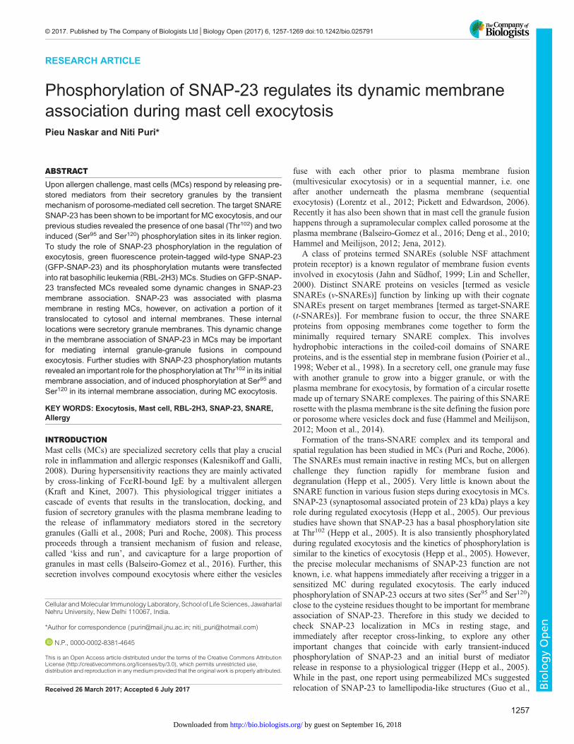
RESULTSActivation of RBL MCs by IgE cross-linking partially movesSNAP-23 from plasma membrane to intracellular locationsIn the present study, primed RBL-2H3 MCs were stimulated fordegranulation by cross-linking of FcεRI (Hepp et al., 2005). Theextent of MC degranulation was measured as β-hexosaminidaserelease at different time points, as shown in Fig. S1A. Thedegranulation reached a peak between 5-15 min. Using anti-phospho-SNAP-23-Ser120 ab (Fig. S1B) it was shown thatphosphorylation of SNAP-23 also reached peak at 5 min andpersisted until 20 min, indicating that phosphorylation of SNAP-23is an early and transient event during MC exocytosis (Fig. S1C).Therefore, to capture early changes, all further studies were carriedout 10 min after receptor cross-linking. Further, to check whetherIgE cross-linking affects the SNAP-23 synthesis/amount wequantified the total SNAP-23 pool at resting, IgE-sensitized, andallergen cross-linked stages of MC by western blotting (forendogenous SNAP-23) and flow cytometry (for EGFP-SNAP-23transfected RBL MCs) and found no significant difference in theamount of SNAP-23 in any of the stages (Fig. S1D,E). In order tostudy if these early events lead to any changes in membranelocalization of t-SNARE SNAP-23 during MC activation,localization of SNAP-23 was studied by confocal fluorescencemicroscopy. Endogenous SNAP-23 as well as transfected GFP-tagged SNAP-23 was analyzed. Visualization by fluorescenceconfocal microscopy of the endogenous SNAP-23 (by staining witha SNAP-23 specific antibody, data not shown) and the transfectedGFP SNAP-23 revealed a smooth pattern of plasma membrane-localized SNAP-23 in different Z-sections of IgE-sensitized MCs(Fig. 1A). But, 10 min after cross-linking the FcεRI, the cellsappeared flatter and the plasma membrane showed ruffles (Fig. 1B,DIC image). It can be seen that SNAP-23 now localized to theseplasma membrane ruffles and to some extent on some internalstructures (shown by white arrow heads in Fig. 1B). Theendogenous and transfected GFP-SNAP-23 were found to behavein a similar fashion both before and after MC exocytosis in terms oftheir membrane localization (data not shown). Further, to investigate
if the internal locations to which SNAP-23 relocated upon IgEcross-linking were internal organelle membranes or cytosoliclocations, we performed membrane/cytosol fractionation ofresting, IgE-sensitized, and IgE-cross-linked GFP-SNAP-23-transfected as well as untransfected RBL MCs. Quantitativeanalysis of western blots of the membrane cytosol fractionsshowed that most [87±2.7% for endogenous and 90±1.5% in caseof transfected GFP-SNAP-23 (mean±s.e.m.)] of the SNAP-23 wasassociated with membrane in resting and IgE-sensitized MCs(89±2.7% for endogenous and 86±1.5% in case of transfected GFP-SNAP-23), however, a small but significant decrease in membraneassociation (74±2.7% endogenous and 73±2% transfected) ofSNAP-23 was observed in MCs 10 min after FcεRI cross-linking(Fig. 1C,D). From this biochemical analysis it is now clear thatSNAP-23 relocates to internal cellular locations.
Many of the internal membranes to which SNAP-23 relocated10 min after receptor cross-linking in MCs were spherical,reminiscent of secretory granules, so we decided to investigate thenature of internal membranes by real-time live-cell microscopicstudy of GFP-SNAP-23-transfected RBL MCs. These transfectedMCs were sensitized with anti-DNP-IgE and lysosomes weremarked with Lysotracker Red dye to follow their fate duringexocytosis. Before observation in a live-cell imaging system one setof cells were mock stimulated (termed as IgE sensitized) and theother one was stimulated with DNP-BSA. The movie capture wasstarted 2 min after mock or allergen stimulation, and the cells werethen observed for 10 min thereafter. Snap shots were extracted froma representative 10 min movie (Movies 1 and 2) obtained from thelive-cell imaging system. A good staining of lysotracker dye wasobserved in both the cases. Like our confocal microscopic study, atIgE-sensitized stage (during mock stimulation) a smooth plasmamembrane staining of SNAP-23 (in green) and internal lysosomestaining (in red) was seen. During mock stimulation, the IgE-sensitized cells showed slight Brownian movement throughout the10 min video (Fig. 1E, three time points are shown). But, afterallergen addition various dynamic changes in plasma membraneand in lysosomal compartments were seen. At 0 min (which isactually 2 min after stimulation) membrane ruffles can be seen andSNAP-23 is associated with them. Some SNAP-23 startsassociating with round vesicles [Fig. 1F (0 min)]. SNAP-23-associated granules harboring lysotracker started appearing at0 min (2 min after allergen challenge) and they were tracked until5 min after allergen addition. These vesicles were found totranslocate towards plasma membrane (denoted with white arrowand arrow head). In allergen-stimulated cells a bunch of granuleswith SNAP-23-associated membrane gradually appeared at around4 min. These green granules were first seen to translocate inside thecell and then started to queue up towards the plasma membrane.During this process at around 7 min some green granules were seenfusing with each other. Ultimately (at 10 min) almost all granuleswith SNAP-23 ended up in a bigger granule most probably byhomotypic fusion. So, this real time imaging shows movement ofSNAP-23 to internal vesicle membranes, which may be lysosomalin nature, and fusion of some of these vesicles probably duringcompound exocytosis from MCs on allergen challenge.
SNAP-23 moves from plasma membrane to internallysosomal membranes during regulated exocytosis of MCsTo confirm the observation obtained from previous experiments weperformed immunofluorescence and confocal microscopy. We havestudied the association of SNAP-23 with different internalorganelles like Golgi apparatus, trans-Golgi network (TGN), and
1258
RESEARCH ARTICLE Biology Open (2017) 6, 1257-1269 doi:10.1242/bio.025791
BiologyOpen
by guest on September 16, 2018http://bio.biologists.org/Downloaded from
Fig. 1. Membrane localization of SNAP-23 in resting and IgE cross-linked RBLMCs. (A,B) Representative confocal images showing cross-sections of GFP-SNAP-23 (green) transfected RBL MCs along with DIC images. (A) In resting RBL MCs GFP-SNAP-23 wild type (WT) is associated with plasma membrane(n=28). (B) In receptor cross-linked RBL MCs GFP-SNAP-23WT is also seen in internal locations in cytosol (n=43). The white arrow heads are indicating GFP-SNAP-23WT in spherical-granule like structures in cytosol. Scale bar: 10 µm. (C) Immunoblot by SNAP-23-specific antibody, showing themembrane and cytosolassociation of endogenous SNAP-23 and transfected GFP-SNAP-23WT. (D) Quantitative analysis of the immunoblots in C showing a significant decrease inmembrane association of SNAP-23 (both endogenous and transfected SNAP-23 WT) in receptor cross-linked. Each data point is mean±s.e.m. of at least threeindependent experiments (*P≤0.05, student’s t-test, one-tailed distribution). (E,F) Representative still images from live cell imaging of GFP-SNAP-23WTtransfected RBL MCs with Lysotracker Red staining to track lysosomal compartments. SNAP-23 localization at plasma membrane is shown during mockstimulation at three representative time points with no colocalization with Lysotracker Red (E) (derived from live cell). (F) The receptor cross linked panel isshowing the snap shots from a representative video (at least 18 videos were captured), depicting SNAP-23 internal membrane localization. White arrows indicateSNAP-23 association with lysosome and arrow heads mark SNAP-23 associated lysotracker. Scale bar: 10 µm.
1259
RESEARCH ARTICLE Biology Open (2017) 6, 1257-1269 doi:10.1242/bio.025791
BiologyOpen
by guest on September 16, 2018http://bio.biologists.org/Downloaded from
also late endosome/lysosomes. RBL MCs were first stained withSNAP-23-specific antibody and then counter stained with TGN38[a type I integral membrane protein primarily localized to the TGN(Humphrey et al., 1993)]- and GM130 [Golgi matrix protein of130 kDa, peripherally associated with the cis-compartment(Nakamura et al., 1997)]-specific antibodies at IgE-sensitized andreceptor cross-linked states (Fig. 2A and B, respectively). Theconfocal microscopy images from Fig. 2A and B showed thatSNAP-23 is not associated with Golgi and TGN at IgE-sensitizedstates of MCs [Pearson coefficient 0.001±0.002 and 0.0357±0.0014(mean±s.e.m.), respectively, Fig. 2D] and also after receptor cross-linking it does not relocate to these internal organelle membranes[Pearson coefficient 0.15±0.042 and 0.054±0.002 (mean±s.e.m.),respectively, Fig. 2D]. In order to investigate if any SNAP-23localized to late endosome/lysosomal membranes in activated MCs,LAMP-3 lysosomal membrane marker in MCs was used. GFP-SNAP-23-transfected MCs were counterstained with anti-LAMP-3antibody. As shown in Fig. 2C, in IgE-sensitized GFP-SNAP-23expressing MCs SNAP-23 is mainly localized to plasma membraneand LAMP-3 staining is completely internal, and negligiblecolocalization is seen between the two (Pearson coefficient0.17±0.001, Fig. 2D). But after receptor cross-linking, transfectedGFP-SNAP-23 was found on plasma membrane ruffles and also oninternal membranes, a large number of which also showed stainingfor LAMP-3 (Fig. 2C). So, a very high level of colocalization wasobtained between GFP-SNAP-23 and LAMP-3 (Pearson coefficient0.7±0.03, Fig. 2D). This indicated that 10 min after receptor cross-linking SNAP-23 relocated to internal membranes in MCs, whichwere LAMP-3-positive and hence lysosomal in nature.
SNAP-23 relocates to secretory granule membranes duringregulated exocytosis in MCsSince lysosomes in MCs also have secretory functions, and aretherefore referred to as secretory lysosomes (Puri et al., 2003; Puriand Roche, 2008), we wanted to check whether SNAP-23 alsorelocates to the secretory granule. Rodent MCs are known to harborserotonin in granules that are lysosomal in nature, and there is aregulated release of serotonin in response to a physiological trigger(Puri and Roche, 2008). We decided to locate serotonin cargo inGFP-SNAP-23-transfected RBL cells by counterstaining serotoninwith serotonin-specific antibody in IgE-sensitized and allergen-activated RBL MCs. Syntaxin-3 (STX-3) in the IgE-sensitizedMCs, SNAP-23 showed plasma membrane association andserotonin showed a punctate staining pattern in intracellularorganelles (Fig. 3A, upper panel) with negligible or no co-localization (0.1±0.018, Fig. 3C). But, 10 min after FcεRI cross-linking, as SNAP-23 localization pattern changed, it showed partialco-localization with serotonin [Pearson coefficient 0.43±0.03(internal, i.e. excluding plasma membrane), Pearson coefficient0.54±0.018 (including plasma membrane, total)] (Fig. 3A, lowerpanel). The white arrows and inset image indicate serotonin punctaesurrounded by organelle membranes having GFP-SNAP-23staining. This indicated that SNAP-23 colocalized to internalsecretory granule membranes in MCs on activation by FcεRI cross-linking. Besides, from the literature it is known that t-SNARESyntaxin-3 (STX-3) resides in secretory granule membrane (Puriet al., 2003), so we also looked at STX-3 co-localization, if any,with SNAP-23 in GFP-SNAP-23-transfected RBL MCs bycounterstaining these cells with anti-STX-3 ab. In the case of IgE-sensitized MCs, STX-3 is seen on internal granule membranes anddoes not show any co-localization with GFP SNAP-23 (Fig. 3B,upper panel; Pearson coefficient 0.26±0.03, Fig. 3C) expressed on
plasma membrane. After receptor crosslinking, the relocatedSNAP-23 showed very high colocalization with STX-3 on almostcircular internal secretory granule membranes (Fig. 3B, lower panel;Pearson coefficient 0.77±0.02, Fig. 3C). Together these two resultsindicate that immediately after receptor crosslinking, SNAP-23relocates to STX-3 harboring internal organelles which may alsoenclose the secretory granule cargo like serotonin, and hence aresecretory granules.
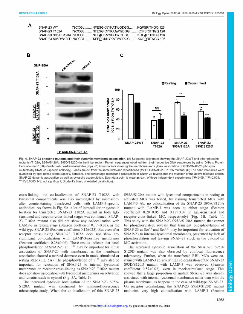
Phosphorylation of SNAP-23 is important for its membraneassociation in RBL MCsSNAP-23 lacks transmembrane domain and is thought to associatewith the plasma membrane through palmitoylation of its conservedcysteine residues present in the linker region (Vogel and Roche,1999; and V. Agarwal, P. N., N. P., unpublished data). Our previousstudies have identified one basal phosphorylation site, Threonine102
(T102), close to these cysteines in the linker region of SNAP-23. Toexplore if phosphorylation of T102 has any role in association ofSNAP-23 with membranes, T was mutated to A, so that it can nolonger be phosphorylated (Fig. 4A). GFP-SNAP-23 T102A-transfected RBL MCs were either mock-stimulated or receptor-cross-linked for 10 min and subjected to membrane cytosolfractionation. Immunoblotting of membrane cytosol fractions ofmock-stimulated cells revealed significantly lower association(55-60% decrease) of SNAP-23 T102A mutant with membrane incomparison to wild-type SNAP-23 (Fig. 4B,C). Similar results wereobtained for transfected cells activated by receptor cross-linking,with a major portion of mutant SNAP-23 residing in the cytosol(60% in cytosol) (Fig. 4B,C).
Further, the induced phosphorylation sites at S95 and S120 are alsovery close to the conserved cysteines in the linker region of SNAP-23.So, to investigate the role of SNAP-23 transient phosphorylationfollowed by dephosphorylation 20 min after receptor cross-linkingin membrane localization of SNAP-23, two kinds of mutants,GFP SNAP-23 S95A/S120A (phospho-negative, that cannot bephosphorylated) and GFP SNAP-23 S95D/S120D (phospho-mimetic, constitutively phosphorylated) (Hepp et al., 2005), wereused. The amino acid sequence comparison for these two mutantswith wild-type SNAP-23 is shown in Fig. 4A. In case of IgE-sensitized RBLMCs, SNAP-23 S95A/S120Amutant was shown, bymembrane cytosol fractionation, to be mainly associated withmembrane, though there was a small but significant decrease inmembrane association in comparison to wild-type SNAP-23(decrease from 90 to 73%) (Fig. 4B,C). In the receptor cross-linkedstage, the membrane association of SNAP-23 S95A/S120A mutantshowed a drastic decrease in comparison to wild-type SNAP-23(from 73 to 47%), and amajor portion of it was cytosolic (Fig. 4B,C).
The phospho-mimetic SNAP23 S95D/S120D mutant mimicsthe transient phosphorylated state which lasts 5 to 20 mins afterreceptor crosslinking. In mock-stimulated stage, membrane cytosolfractionation of SNAP-23 S95D/S120D-transfected RBL cellsrevealed much lower association of SNAP-23 mutant withmembrane in comparison to wild-type SNAP-23 (56% incomparison to 90%) (Fig. 4B,C). After activation, there is afurther decrease in membrane association, and increase in cytosoliclocalization, of SNAP-23 S95D/S120D mutant (43% membraneassociation) (Fig. 4B,C).
Role of SNAP-23 phosphorylation in its dynamic associationwith internal membranes during secretory response of MCsAs previous experiments with wild-type SNAP-23 hadshown relocation to internal lysosomal membranes on receptor
1260
RESEARCH ARTICLE Biology Open (2017) 6, 1257-1269 doi:10.1242/bio.025791
BiologyOpen
by guest on September 16, 2018http://bio.biologists.org/Downloaded from
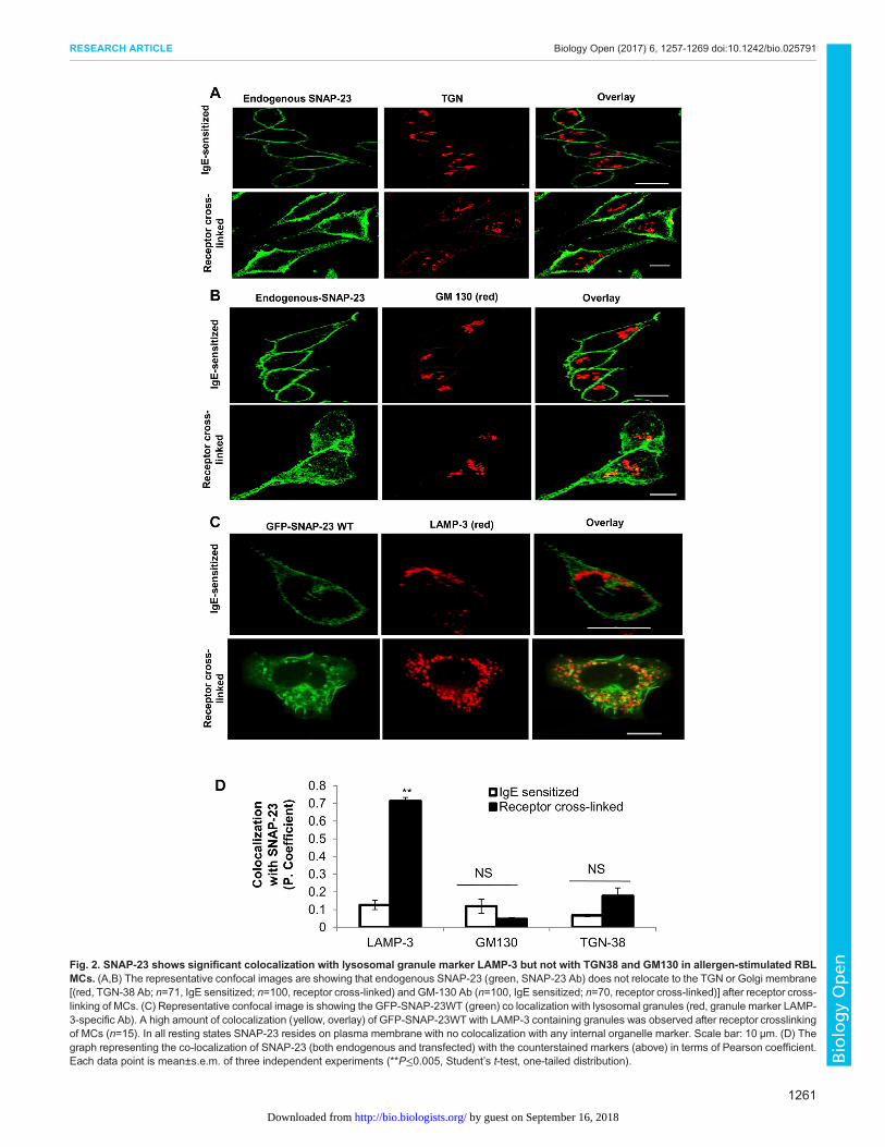
Fig. 2. SNAP-23 shows significant colocalization with lysosomal granule marker LAMP-3 but not with TGN38 and GM130 in allergen-stimulated RBLMCs. (A,B) The representative confocal images are showing that endogenous SNAP-23 (green, SNAP-23 Ab) does not relocate to the TGN or Golgi membrane[(red, TGN-38 Ab; n=71, IgE sensitized; n=100, receptor cross-linked) and GM-130 Ab (n=100, IgE sensitized; n=70, receptor cross-linked)] after receptor cross-linking of MCs. (C) Representative confocal image is showing the GFP-SNAP-23WT (green) co localization with lysosomal granules (red, granule marker LAMP-3-specific Ab). A high amount of colocalization (yellow, overlay) of GFP-SNAP-23WT with LAMP-3 containing granules was observed after receptor crosslinkingof MCs (n=15). In all resting states SNAP-23 resides on plasma membrane with no colocalization with any internal organelle marker. Scale bar: 10 μm. (D) Thegraph representing the co-localization of SNAP-23 (both endogenous and transfected) with the counterstained markers (above) in terms of Pearson coefficient.Each data point is mean±s.e.m. of three independent experiments (**P≤0.005, Student’s t-test, one-tailed distribution).
1261
RESEARCH ARTICLE Biology Open (2017) 6, 1257-1269 doi:10.1242/bio.025791
BiologyOpen
by guest on September 16, 2018http://bio.biologists.org/Downloaded from
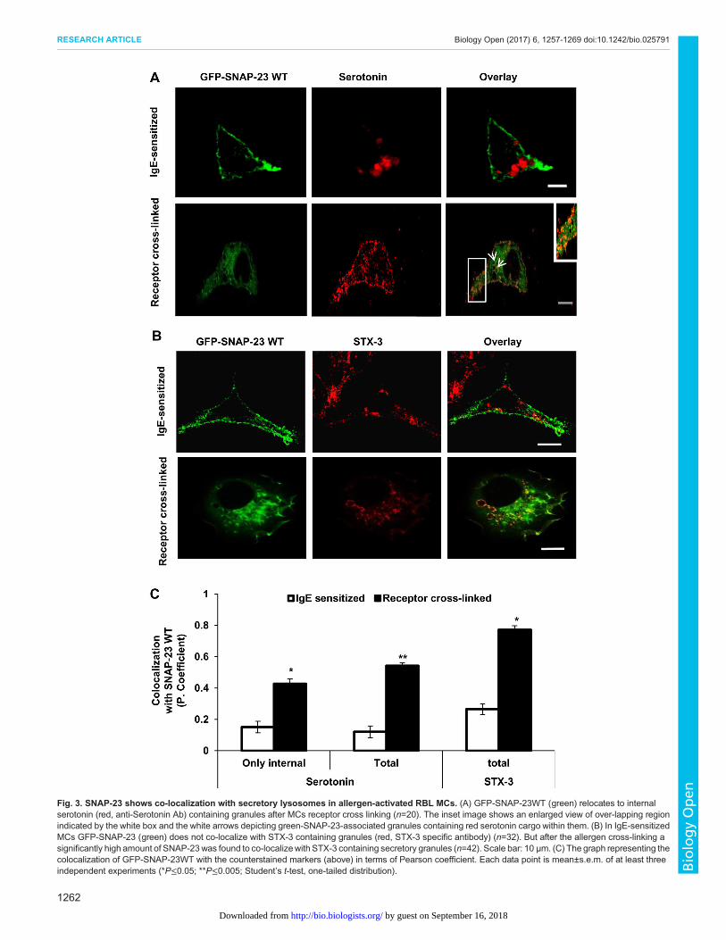
Fig. 3. SNAP-23 shows co-localization with secretory lysosomes in allergen-activated RBL MCs. (A) GFP-SNAP-23WT (green) relocates to internalserotonin (red, anti-Serotonin Ab) containing granules after MCs receptor cross linking (n=20). The inset image shows an enlarged view of over-lapping regionindicated by the white box and the white arrows depicting green-SNAP-23-associated granules containing red serotonin cargo within them. (B) In IgE-sensitizedMCs GFP-SNAP-23 (green) does not co-localize with STX-3 containing granules (red, STX-3 specific antibody) (n=32). But after the allergen cross-linking asignificantly high amount of SNAP-23 was found to co-localizewith STX-3 containing secretory granules (n=42). Scale bar: 10 μm. (C) The graph representing thecolocalization of GFP-SNAP-23WT with the counterstained markers (above) in terms of Pearson coefficient. Each data point is mean±s.e.m. of at least threeindependent experiments (*P≤0.05; **P≤0.005; Student’s t-test, one-tailed distribution).
1262
RESEARCH ARTICLE Biology Open (2017) 6, 1257-1269 doi:10.1242/bio.025791
BiologyOpen
by guest on September 16, 2018http://bio.biologists.org/Downloaded from
cross-linking, the co-localization of SNAP-23 T102A withlysosomal compartments was also investigated by microscopyafter counterstaining transfected cells with LAMP-3-specificantibodies. As shown in Fig. 5A, a lot of intracellular or cytosoliclocation for transfected SNAP-23 T102A mutant in both IgE-sensitized and receptor-cross-linked stages was confirmed. SNAP-23 T102A mutant also did not show any co-localization withLAMP-3 in resting stage (Pearson coefficient 0.17±0.03), as thewild-type SNAP-23 (Pearson coefficient 0.12±025). But even afterreceptor cross-linking SNAP-23 T102A does not show anysignificant co-localization with LAMP-3-positive membranes(Pearson coefficient 0.28±0.06). These results indicate that basalphosphorylation of SNAP-23 at T102 may be important for initialassociation of SNAP-23 with membranes as the membraneassociation showed a marked decrease even in mock-stimulated orresting stage (Fig. 5A). The phosphorylation of T102 may also beimportant for relocation of SNAP-23 to internal lysosomalmembranes on receptor cross-linking as SNAP-23 T102A mutantdoes not show association with lysosomal membranes on activationand remains stuck in cytosol (Fig. 5A, Table 1).The increased cytosolic localization of the SNAP-23 S95A/
S120A mutant was confirmed by immunofluorescencemicroscopic study. When the co-localization of this SNAP-23
S95A/S120A mutant with lysosomal compartments in resting oractivated MCs was tested, by staining transfected MCs withLAMP-3 Ab, no colocalization of the SNAP-23 S95A/S120Amutant with LAMP-3 was seen at either stage (Pearsoncoefficient 0.20±0.05 and 0.19±0.09 in IgE-sensitized andreceptor-cross-linked MC, respectively) (Fig. 5B, Table 1).This study with the SNAP-23 S95A/S120A mutant, that cannotbe phosphorylated, reveals that transient phosphorylation ofSNAP-23 at Ser95 and Ser120 may be important for relocation ofSNAP-23 to internal lysosomal membranes, prevented by lack ofphosphorylation and leaving SNAP-23 stuck in the cytosol onMC activation.
The increased cytosolic association of the SNAP-23 S95D/S120D mutant was also observed by confocal fluorescencemicroscopy. Further, when the transfected RBL MCs were co-stained with LAMP-3 ab, a very high colocalization of the SNAP-23S95D/S120D mutant with LAMP-3 was observed (Pearsoncoefficient 0.57±0.02), even in mock-stimulated stage. Thisshowed that a large proportion of mutant SNAP-23 was alreadyassociated with internal lysosomal membranes rather than with theplasma membrane, as happens in the case of wild-type SNAP-23.On receptor crosslinking, the SNAP-23 S95D/S120D mutantmaintains very high colocalization with LAMP-3 (Pearson
Fig. 4. SNAP-23 phospho mutants and their dynamic membrane association. (A) Sequence alignment showing the SNAP-23WT and other phosphomutants (T102A, S95A/S120A, S95D/S120D) in the linker region. Protein sequences obtained from their respective DNA sequences by using ‘DNA to Proteintranslation tool’ (http://insilico.ehu.es/translate/index.php). (B) Immunoblots showing the membrane and cytosol association of GFP-SNAP-23 phosphomutants (by SNAP-23-specific antibody). Lanes are cut from the same blots and repositioned (for GFP-SNAP-23 T102A mutant). (C) The band intensities werequantified by spot denso Alpha EaseFC software. The percentage membrane association of SNAP-23 reveals that the mutation of the above residues affectsSNAP-23 dynamic association as well as cytosolic accumulation. Each data point is mean±s.e.m. of three independent experiments (*P≤0.05; **P≤0.005;***P≤0.0005; NS, not significant; Student’s t-test, one-tailed distribution).
1263
RESEARCH ARTICLE Biology Open (2017) 6, 1257-1269 doi:10.1242/bio.025791
BiologyOpen
by guest on September 16, 2018http://bio.biologists.org/Downloaded from
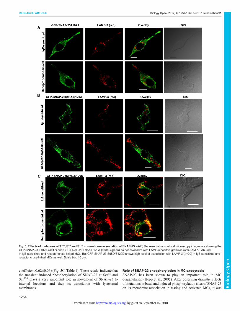
coefficient 0.62±0.06) (Fig. 5C, Table 1). These results indicate thatthe transient induced phosphorylation of SNAP-23 at Ser95 andSer120 plays a very important role in movement of SNAP-23 tointernal locations and then its association with lysosomalmembranes.
Role of SNAP-23 phosphorylation in MC exocytosisSNAP-23 has been shown to play an important role in MCdegranulation (Hepp et al., 2005). After observing dramatic effectsof mutations in basal and induced phosphorylation sites of SNAP-23on its membrane association in resting and activated MCs, it was
Fig. 5. Effects of mutations at T102, S95 and S120 in membrane association of SNAP-23. (A-C) Representative confocal microscopy images are showing theGFP-SNAP-23 T102A (n=17) and GFP-SNAP-23 S95A/S120A (n=34) (green) do not colocalize with LAMP-3 positive granules (anti-LAMP-3 Ab, red)in IgE-sensitized and receptor cross-linked MCs. But GFP-SNAP-23 S95D/S120D shows high level of association with LAMP-3 (n=20) in IgE-sensitized andreceptor cross-linked MCs as well. Scale bar: 10 μm.
1264
RESEARCH ARTICLE Biology Open (2017) 6, 1257-1269 doi:10.1242/bio.025791
BiologyOpen
by guest on September 16, 2018http://bio.biologists.org/Downloaded from
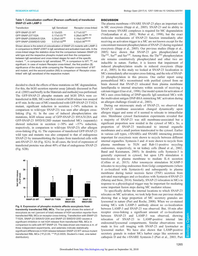
decided to check the effects of these mutations onMC degranulation.For this, the hGH secretion reporter assay [already discussed in Puriet al. (2003) and briefly in theMaterials andmethods] was performed.The GFP-SNAP-23 phospho mutants and hGH DNA were co-transfected in RBLMCs and their extent of hGH release was assayedat 45 min. In the case ofMCs transfectedwith GFP-SNAP-23 T102Amutant, significant reduction in secretion (∼54% reduction incomparison to wild-type SNAP-23) was seen after allergen cross-linking (Fig. 6). In the case of induced phosphorylation sitemutations, hGH release assay of GFP-SNAP-23 S95A/S120A andGFP-SNAP-23 S95D/S120D mutant transfected MCs (separately)showed reduction in secretion (∼30% and ∼57% reduction,respectively, in comparison to wild-type SNAP-23) after allergencross-linking (Fig. 6). The expression of transfected GFP-SNAP-23wild type and mutants was also compared to that of endogenousSNAP-23 by immunoblotting the lysates from transfected cells withanti-SNAP-23 Ab (Fig. S2A). In all cases, the level of expression oftransfected proteins was about 40% of that of endogenous SNAP-23(Fig. S2B).
DISCUSSIONThe plasma membrane t-SNARE SNAP-23 plays an important rolein MC exocytosis (Hepp et al., 2005). SNAP-23 and its ability toform ternary SNARE complexes is required for MC degranulation(Vaidyanathan et al., 2001; Weber et al., 1998), but the exactmolecular mechanisms of SNAP-23 function immediately afterreceiving an activation trigger in a MC are not known except for theconcomitant transient phosphorylation of SNAP-23 during regulatedexocytosis (Hepp et al., 2005). Our previous studies (Hepp et al.,2005) have shown that SNAP-23 gets phosphorylated atThr102, Ser95 and Ser120. Among these, the Thr102 phosphorylationsite remains constitutively phosphorylated and other two areinducible in nature. Further, it is known that impairment ofinduced phosphorylation results in reduced exocytosis (Heppet al., 2005). In this study we looked at SNAP-23 localization inMCs immediately after receptor cross-linking, and the role of SNAP-23 phosphorylation in this process. One earlier report usingpermeabilized MCs reconstituted with purified cytosol from ratbrain showed that SNAP-23 relocated from plasma membranelamellipodia to internal structures within seconds of receiving acalcium trigger (Guo et al., 1998). Ourmodel system for activation ofMCs uses cross-linking of DNP-specific IgE-sensitized MCs withthe multivalent antigen DNP-BSA mimicking MC activation duringan allergen challenge (Gould et al., 2003).
During our microscopic study of SNAP-23, we observed thatSNAP-23 membrane association changed dynamically uponallergen trigger and some of it seemed to relocate to intracellularsites. Membrane cytosol fractionation experiments revealed thata majority of SNAP-23 was still membrane-associated but asignificant proportion now resided in the cytosol of MCs. So, aproportion of SNAP-23 translocated to internal organellemembranes and a small portion translocated to the cytosol. Earlierin various cell types, t-SNAREs and SNARE interacting proteinsimportant for exocytosis were shown to recycle between differentinternal organelles. Syntaxin-3 and -4 were shown to recycle fromplasma membrane to TGN and Rab-11-positive recyclingendosomes, respectively, in rat kidney cells (Band et al., 2002;Band and Kuismanen, 2005). In dendritic cells Syntaxin-3 isgenerally expressed in cytosol, but upon LPS stimulation ittranslocates to plasma membrane to mediate IL-6 secretion(Collins et al., 2015). After ionomycin stimulation SCAMP-5relocates to recycling endosomes from Golgi compartments (whereit co-localized with Syntaxin-6) and subsequently to plasmamembrane during tumor necrosis factor (TNF) secretion fromactivated macrophages and co-localizes with Syntaxin-4/SNAP-23(Murray and Stow, 2014). Similarly, SNAP-23 relocation in MCs inresponse to a physiological trigger may be important for mediatingsome important fusion steps during MC mediator release.
To specifically define the internal locations to which SNAP-23relocates on MC activation, we took help from our previous studiesshowing that a large proportion of MC secretory granules arelysosomal in nature (Puri and Roche, 2008). When we co-stainedresting MCs with LAMP-3 antibody almost no co-localizationbetween LAMP-3 and SNAP-23 was observed. But, 10 min afterreceptor cross-linking a significant amount of co-localizationbetween SNAP-23 and LAMP-3 was observed, showingrelocation of SNAP-23 to LAMP-positive internal lateendosomal/lysosomal compartments. Similar observations weremade in live cell imaging with SNAP-23 and lysotracker aslysosomal marker. We have also shown that LAMP-positivesecretory granule in rodent MCs harbor cargo like serotonin orcathepsin D and the t-SNARE Syntaxin-3 (Puri et al., 2003; Puri
Fig. 6. Expression of phospho mutants affects exocytosis fromtransiently transfected RBL MCs. The bar graph shows the extent ofexocytosis as net (percent of total) release of hGH secretion reporter fromtransfected RBL MCs on receptor cross-linking. Transfection with SNAP-23T102A, SNAP-23 S95A/S120A and SNAP-23 S95D/S120D causes asignificant inhibition in net hGH release from transfected RBL MCs incomparison to cells with WT SNAP-23. The data shown are mean±s.e.m. ofthree independent experiments, and asterisks indicate statisticallysignificant differences in hGH release between SNAP-23WT versus mutanttransfected RBL MCs (*P≤0.05; ***P≤0.0005; Student’s t-test, one-taileddistribution).
Table 1. Colocalization coeffient (Pearson coefficient) of transfectedSNAP-23 with LAMP-3
Transfection IgE Sensitized Receptor cross-linked
GFP-SNAP-23 WT 0.12±025 0.71±0.02**GFP-SNAP-23T102A 0.17±0.03 NS 0.28±0.06$$$, NS
GFP-SNAP-23S95A/S120A 0.20±0.05NS 0.19±0.09$$$,NS
GFP-SNAP-23S95D/S120D 0.57±0.02$$$ 0.62±0.06NS,NS
Shown above is the extent of colocalization of SNAP-23 mutants with LAMP-3in comparison to SNAP-23WT in IgE sensitized and activatedmast cells. In thecross-linked stage the statistics show first the comparison between SNAP-23wild type and the respective phospho mutant and then the comparisonbetween IgE-sensitized and cross-linked stages for each of the specificmutant. **, in comparison to IgE sensitized; $$$, in comparison to WT; NS, notsignificant. In case of mutant-‘Receptor cross-linked’, the first position ($)significance of the study while comparing the ‘Receptor cross-linked’ of WTand mutant, and the second position (NS) is comparison of ‘Receptor cross-linked’ with IgE sensitized of the respective mutant.
1265
RESEARCH ARTICLE Biology Open (2017) 6, 1257-1269 doi:10.1242/bio.025791
BiologyOpen
by guest on September 16, 2018http://bio.biologists.org/Downloaded from
and Roche, 2008). So, we co-stained MC with either serotoninor Syntaxin-3-specific antibodies, respectively, and found noco-localization of plasma membrane-associated SNAP-23 withgranule-associated serotonin or Syntaxin-3 in resting MCs, but avery significant co-localization of SNAP-23 with serotonin andSyntaxin-3, 10 min after receptor cross-linking. This showsconclusively that SNAP-23 relocates to membrane of internalsecretory granules.The main questions are how and why does SNAP-23 end up on
internal membranes in MCs early after receptor cross-linking.Since some SNAP-23 still remained associated with plasmamembrane, only a portion of the total pool relocates to internalmembranes. There are two possible mechanisms by which SNAP-23 may relocate to internal membranes. One possibility is that itdissociates from plasma membrane, enters cytosol, and again bindsto internal lysosomal membranes. In fact, we do see a small butsignificant increase in SNAP-23 in the cytosolic fraction 10 minafter receptor cross-linking. This could be due to SNAP-23 beingon its way to relocate to internal membranes. There are reportsshowing that the extent of palmitoylation determines thedifferential membrane association of a protein (Greaves andChamberlain, 2011). It may be that the induced phosphorylationof SNAP-23 during exocytosis (Hepp et al., 2005) regulates thepalmitoylation of SNAP-23 so that it dissociates from plasmamembrane and associates with lysosomal membranes. The otherpossibility is that immediately after stimulation one or a fewgranules release their content via the ‘kiss and run’mechanism andquickly SNAP-23 relocates with this granule. It has been reportedearlier that MCs exhibit both ‘kiss and run’ and cavicapture types oftransient granule-plasma membrane fusion to maintain theirgranularity and to retain the capacity of undergoing repeatedexocytosis (Balseiro-Gomez et al., 2016; Cohen et al., 2012).Further, a recent study involving atomic force microscopy (AFM)and transmission electron microscope (TEM) detailed the captureof a typical porosome on activated RBL mast cells, wheremembrane-bound secretory vesicles dock and fuse (Deng et al.,2010; Jena, 2012). Studies on pancreatic acinar cells, whosesecretion is also dependent on the t-SNARE SNAP-23, haverevealed selective presence of SNAP-23 at the base of porosome,the site of secretory vesicle docking and fusion (Jena, 2012). Wealso could never find any LAMP translocating to plasma membraneafter activation, ruling out full fusion between lysosome/granuleand plasma membrane. By real time imaging, we have visualizedSNAP-23 relocation to internal granule membranes includinglysosomes that may be involved in homotypic fusion duringcompound exocytosis, as we have seen SNAP-23 in multiplegranule fusion (homotypic fusion) just beneath the plasmamembrane, upon activation. Granule-granule homotypic fusion isknown to require SNARE rosette formation (Hammel andMeilijson, 2012). Another least-likely possibility is that it is thenewly synthesized SNAP-23, which associates with the internallocations, and the older SNAP-23 are still associated with plasmamembrane. But, in our experiments, the amount of total SNAP-23was the same in resting, IgE-sensitized, and receptor crosslinkedstages by western blot analysis of endogenous SNAP-23, or flowcytometric analysis of transfected GFP-SNAP-23. So, it is unlikelythat in the short time span of 10 min after activation, newlysynthesized SNAP-23 associates with internal locations and theolder pool remains associated with the plasma membrane. In otherstudies involving secretory cells from adrenal medulla andpancreatic beta cells, t-SNARE SNAP-25 was shown to beimportant for sequential exocytosis and was supplied to primary
granules by lateral diffusion from plasma membrane on stimulation(Kishimoto et al., 2006; Takahashi et al., 2004).
We have shown that SNAP-23 gets differentiallyphosphorylated (Hepp et al., 2005) in resting and activatedMCs. In resting MCs, SNAP-23 is phosphorylated at Thr102, andthis basal phosphorylation is not affected by activation. Ser95
and Ser120 are transiently phosphorylated immediately afteractivation, and mirror the kinetics of MC secretion. Both theMC secretion and induced SNAP-23 phosphorylation peak at5-20 min, and show a decrease thereafter. Since all three majorphosphorylation sites are in the linker region of SNAP-23, inclose proximity to the conserved cysteine residues which mayplay an important role in anchoring SNAP-23 to membrane, wedecided to study the role of SNAP-23 phosphorylation in itsmembrane associations in resting or activated MCs, respectively.Transfection, and expression of basal phosphorylation mutantSNAP-23 T102A, which cannot be phosphorylated, revealed thata large portion of SNAP-23 T102A accumulated in cytosol inresting MCs and the situation remained the same even afteractivation of MCs. Also, SNAP-23 T102A transfected MCsshowed a 54% inhibition in exocytosis in comparison to controls.These results indicate that basal phosphorylation of SNAP-23 atThr102 is important for the initial membrane association ofSNAP-23. Due to the proximity of this site to conserved cysteinein the linker region of SNAP-23, it may function by affecting thepalmitoylation of these cysteine residues. Or, it may affect initialassociation with membrane for palmitoylation, either byfacilitating binding to some chaperone (Liu et al., 1996) or bychanges in overall hydrophobicity (Polyansky and Zagrovic,2012). Previous studies have indicated that the first association ofsimilar proteins with membrane may be by some mechanism otherthan palmitoylation (Dunphy and Linder, 1998). For example,members of the Src family of tyrosine kinases or G protein α i1subunit are cotranslationally myristoylated, and this helps in rapidmembrane association (van’t Hof and Resh, 1997) bringing them inclose proximity of cellular palmitoyltransferases localized tointracellular membranes (Berthiaume and Resh, 1995; Dunphy et al.,1996; Liu et al., 1996). SNAP-23 is not myristoylated, leavingunresolved the mechanism by which SNAP-23 initially associates withmembranes. SNAP-25, the key t-SNARE in neuronal cells thatmediates synaptic vesicle release, gets palmitoylated for its membraneassociation at steady-state level in neuroendocrine cells, but its initialplasma membrane association depends on interaction with Syntaxin 1while still in cytosol (Vogel et al., 2000). Hence some similarmechanism may be facilitated by Thr102 phosphorylation in SNAP-23in MCs for efficient initial association with plasma membrane.
Further, when the inducible phosphorylation sites Ser95 andSer120 of SNAP-23 were mutated to S95A/S120A for phospho-negative, and S95D/S120D for phospho-mimetic mutants,respectively, the dynamic regulated changes in SNAP-23subcellular localization on MC activation were completelycompromised. Membrane/cytosol fractionation and microscopystudies revealed that the phospho-negative SNAP-23 S95A/S120Amutant was able to dissociate from plasma membrane on activationas more of it ended up in cytosol in comparison to control, butunable to associate with internal LAMP-3-positive membranes.Hence, transient-induced phosphorylation of SNAP-23 at Ser95 andSer120 seems to be important for association of SNAP-23 withinternal lysosomal membranes. This conclusion was furthervalidated by subcellular localization studies on the transfectedphospho-mimetic SNAP-23 S95D/S120D mutant in resting andactivated MCs. In both stages, the SNAP23 S95D/S120D mutant
1266
RESEARCH ARTICLE Biology Open (2017) 6, 1257-1269 doi:10.1242/bio.025791
BiologyOpen
by guest on September 16, 2018http://bio.biologists.org/Downloaded from
shows a higher colocalization with LAMP-3 containing internalmembranes. Previously, in other studies, phosphorylation of claudin1 (French et al., 2009) and beta-catenin (Qian et al., 2014) has beenshown to regulate their subcellular localization and functions.Likewise, we have found that phosphorylation regulates membranerelocation of SNAP-23 during MC exocytosis. That means inducedphosphorylations are mediating the relocation and internalmembrane association of SNAP-23 to mediate the granule-granulefusion during MC mediator release. The present study reveals, forthe first time, that induced phosphorylation of SNAP-23 has a rolein dynamic changes in subcellular localizations of SNAP-23 inMCsundergoing degranulation. Mutations in phospho sites lead to apartial inhibition in exocytosis. So, maybe the inducedphosphorylation of SNAP-23 is involved in bringing SNAP-23 tothe right location, enabling it to participate in granule-granulefusion, which is an important step in compound exocytosis. Thephosphomimetic mutant of SNAP-23 shows good association withinternal LAMP-positive membranes, but its overall association withmembranes is significantly lower than that of SNAP-23 wild type.So, more than 50% of this mutant remains displaced to cytosol,and hence fails to reach the right locations. These results indicatethat the transient nature of phosphorylation is very important. Thedephosphorylation following the phosphorylation may be importantfor recycling of SNAREs by priming or recycling of SNAP-23back to plasma membrane (Hepp et al., 2005; Puri et al., 2003).As the SNAP23 S95D/S120D mutant is unable to show this
dephosphorylated stage it is either stuck on internal membranesor in the cytosol, and hence causes a significant inhibition inexocytosis.
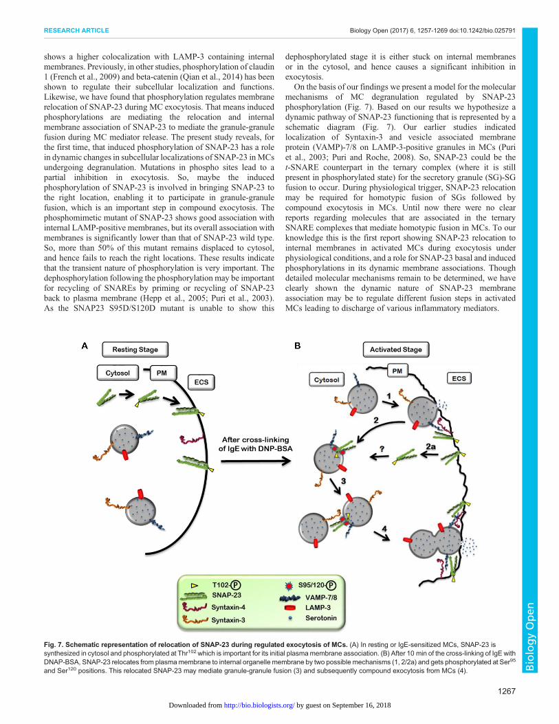
On the basis of our findings we present a model for the molecularmechanisms of MC degranulation regulated by SNAP-23phosphorylation (Fig. 7). Based on our results we hypothesize adynamic pathway of SNAP-23 functioning that is represented by aschematic diagram (Fig. 7). Our earlier studies indicatedlocalization of Syntaxin-3 and vesicle associated membraneprotein (VAMP)-7/8 on LAMP-3-positive granules in MCs (Puriet al., 2003; Puri and Roche, 2008). So, SNAP-23 could be thet-SNARE counterpart in the ternary complex (where it is stillpresent in phosphorylated state) for the secretory granule (SG)-SGfusion to occur. During physiological trigger, SNAP-23 relocationmay be required for homotypic fusion of SGs followed bycompound exocytosis in MCs. Until now there were no clearreports regarding molecules that are associated in the ternarySNARE complexes that mediate homotypic fusion in MCs. To ourknowledge this is the first report showing SNAP-23 relocation tointernal membranes in activated MCs during exocytosis underphysiological conditions, and a role for SNAP-23 basal and inducedphosphorylations in its dynamic membrane associations. Thoughdetailed molecular mechanisms remain to be determined, we haveclearly shown the dynamic nature of SNAP-23 membraneassociation may be to regulate different fusion steps in activatedMCs leading to discharge of various inflammatory mediators.
Fig. 7. Schematic representation of relocation of SNAP-23 during regulated exocytosis of MCs. (A) In resting or IgE-sensitized MCs, SNAP-23 issynthesized in cytosol and phosphorylated at Thr102 which is important for its initial plasmamembrane association. (B) After 10 min of the cross-linking of IgE withDNAP-BSA, SNAP-23 relocates from plasmamembrane to internal organelle membrane by two possible mechanisms (1, 2/2a) and gets phosphorylated at Ser95
and Ser120 positions. This relocated SNAP-23 may mediate granule-granule fusion (3) and subsequently compound exocytosis from MCs (4).
1267
RESEARCH ARTICLE Biology Open (2017) 6, 1257-1269 doi:10.1242/bio.025791
BiologyOpen
by guest on September 16, 2018http://bio.biologists.org/Downloaded from
MATERIALS AND METHODSCell cultureRat basophilic leukemia mast cells [RBL-2H3, a kind gift from Dr Paul ARoche (NIH, Bethesda, MD, USA)] were maintained in equal parts minimalessential medium (Sigma, MO, USA) and Iscove’s medium (Gibco, LifeTechnologies, Grand Island, NY, USA) containing 20% FBS (Gibco),25 mM HEPES (Sigma), and 120 μg ml−1 gentamicin (RBL completemedium) as described (Hepp et al., 2005). Cells were maintained as sub-confluent monolayers at 37°C in a humidified atmosphere containing 5%CO2 and passaged with trypsin.
PlasmidspCMV-FLAG-Rat SNAP-23 wild type, pCMV-FLAG-Rat SNAP-23T102A, pCMV-FLAG-Rat SNAP-23 95A/120A, and pCMV-FLAG-RatSNAP-23 95D/120Dwere a kind gift fromDr Paul A Roche (NIH, Bethesda,MD, USA). The cDNA encoding full length wild-type SNAP-23 and itsphosphomutants were subcloned from pCMV-FLAG-Rat SNAP-23 wildtype into EGFP-C2 plasmid (#6083-1) (Clontech, CA, USA) by using EcoRIand ApaI restriction sites to generate amino terminal GFP-tagged protein(called GFP-SNAP-23). The integrity of subcloned plasmids was confirmedby sequencing from GCC Biotech and SciGenom Labs Pvt Ltd., India.
AntibodiesPolyclonal rabbit anti-serum recognizing the SNAP-23 carboxyl terminuswas a gift from Dr Paul A Roche (NIH, Bethesda, MD, USA). Anti–DNPIgE (clone TIB 142) was obtained from the American Type CultureCollection (Manassas, VA, USA). Mouse anti-CD63/LAMP 3monoclonalantibody (mAb) AD1 (# 551458, 1:100), Mouse Anti-Rat trans-Golginetwork (TGN) 38 antibody (# 610899, 1:100) and Mouse Anti-Golgimatrix protein (GM) 130 antibody (# 610822, 1:100) (BD Biosciences,San Diego, CA, USA), and anti-GFP rabbit mAb from Clontech were usedin this study. Mouse anti serotonin antibody (# M0758, 1:50) was fromDako (Carpinteria, CA, USA). Alexa dye-conjugated secondaryantibodies were obtained from Molecular Probes (Eugene, OR, USA)(anti Rb Alexa 546: A11035; anti mouse Alexa 546: A11030; anti mouseAlexa 488: A11001 dilution 1:500). Lysotracker Red (# L7528) was fromMolecular Probes. Anti-Rabbit Protein A-HRP conjugated antibody (#7300-05, 1:7000, Southern Biotech, Birmingham, AL, USA) and anti-mouse IgG-HRP conjugated antibody ((# 1031-05, 1:7000, SouthernBiotech) were used. Antibody recognizing Syntaxin-3 (# ab133750,1:800) (rabbit monoclonal) was obtained from Abcam (UK).
Transfection of RBL cellsTransfection of RBL cells was performed as described earlier (Puri et al.,2003) with some modifications. Briefly, exponentially growing RBL cells(10×106/0.5 ml serum-free RBL media) were transfected by electroporation(320 mV, 950 μF) with 20 μg DNA. Immediately after electroporation, thecells were plated in RBL complete medium and analyzed 24 h later. Formicroscopy, transfected cells were plated on coverslips and then treated asper requirement.
Stimulation of RBL cell exocytosis in transfected RBL cellsRBL cells were transfected by electroporation with human growth hormone(2 µg) expression vector together with empty GFP vector or GFP-SNAP-23wild type or phospho mutant (20 µg). After 4-5 h culture, the cells weresensitized with IgE and after 16-18 h they were mock-stimulated orstimulated with DNP-BSA as described before (Hepp et al., 2005). Theamount of human growth hormone released into the medium or remainingcell associated was determined using a human growth hormone enzyme-linked immunosorbent assay (Roche Diagnostics Corp.) as describedpreviously (Puri et al., 2003). For quantitative experiments, statisticalanalyses were carried out by using a Student’s t-test. Results wereconsidered significant when a P value of less than 0.05 was obtained.
Confocal microscopyRBL cells with or without transfection were seeded on 10 mm diametercoverslips. For indirect immunofluorescence analysis, the cells were eitherfixed with 4% paraformaldehyde (PFA) in PBS for 30 min and excess
paraformaldehyde quenched with 50 mM NH4Cl in PBS or with coldmethanol at −20°C for 4 min. After washing, the fixed cells werepermeabilized with 1% IGEPAL (Sigma, MO, USA) in the presence of 3%normal goat serum (Sigma) and 0.05% saponin (SD Fine Chem. Limited,Boisar, India) in PBS. The cells were then incubated with 3% normal goatserum and 0.05% saponin in PBS for 1 h at RT to prevent nonspecific proteinbinding. Primary Abs diluted in the same buffer was added to the cells, andincubationwas conducted for 1 h at room temperature (RT). After washing, thecells were incubated for 30 min in the presence of secondary goat Absconjugated to Alexa Fluor 546 (red) (Molecular Probes). As a control, sampleswere stained with an irrelevant antibody and no staining was observed in therespective channel for all confocal fluorescence microscopy experiments.
Confocal images were collected with Olympus Fluoview FV1000microscope at 100× magnification (sometimes with 2× zoom) with anoptical slice thickness of 1.0 μm. Image Z-stacks were collected through thedepth of the cell using 0.4 μm step size. Colocalization analysis was done foreach plane of the individual image stacks using the colocalization analysisfeature of the Fluoview software Ver.1.7a (Olympus). Briefly, individualchannels were thresholded to include the structures of interest; regions ofinterest were then drawn to encompass the structures, resulting in scatterplots being generated and colocalization coefficients calculated. Thecolocalization coefficients represent colocalization in the green channelwith respect to the red channel. Single images were exported from theFluoview software Version 1.7a Software and organized into figures usingMicrosoft PowerPoint 2007.
Real time imaging of GFP SNAP-23 expressing cellsRBL cells were transfected with EGFP-SNAP-23 plasmid, plated in a3.5 cm culture dish and sensitized with anti-DNP IgE in RBL completemedium overnight at 37°C. Next day these were incubated withLysotracker Red [a dye which stains the lysosomes and secretorygranules as well in live cells (Marchini-Alves et al., 2012)] for 2 h inRBL complete medium. After washing with phenol red free RPMImedium they were either mock stimulated or stimulated with 100 ng ml−1
DNP-BSA and observed by a live cell imager Andor Spinning DiskConfocal microscope (Nikon Eclipse TiE, Software-Andor iQ 2.7) in 5%CO2 chamber at 37°C. The movies were captured 2 min after the additionof allergen and continued for 10 min. At least five movies were captured inthe above manner for each separate experiment. All the images wereanalyzed by NIS element AR ver4.
Membrane-cytosol fractionationMembrane-cytosol fractionation was done as described earlier (Hepp et al.,2005). Briefly, transfected RBL mast cells were harvested after 24 h andresuspended in hypotonic buffer (10 mM Tris, 1 mM KCl, 1 mM EGTA,0.5 mM MgCl2, pH 7.4) containing protease inhibitors (5 mMiodoacetamide, 50 mM PMSF, and 0.1 mM TLCK) and phosphataseinhibitors (5 mMEDTA, 5 mMEGTA, 50 mMNaF, 10 mMNa4P2O7, and1 mM Na3VO4). They were then disrupted by repeated passage of cellsthrough a 30.5 gauge syringe. Nuclei and unbroken cells were removed bycentrifugation at 1000×g and the post-nuclear supernatant was subjected tocentrifugation at 100,000×g for 1 h at 4°C to isolate membrane (pellet) andcytosol (supernatant). The membrane pellet and cytosolic supernatant werebrought to the same volume in hypotonic buffer and each was adjusted to afinal concentration of 1%Triton X-100. Equal portions of each fraction wereanalyzed by SDS-PAGE and immunoblotting.
SDS-polyacrylamide gel electrophoresis and immunoblottingRBL cell lysates and membrane-cytosol fractions were boiled in β-mercaptoethanol containing sample buffer and proteins were separated in12.5% SDS-polyacrylamide gel. Immunoblotting was performed withpolyclonal SNAP-23 C-terminus antibody or GFP antibody as previouslydescribed (Puri et al., 2003). As secondary antibodies anti-Rabbit Protein A-HRP and anti-mouse IgG-HRP were used. For immunoblotting separatedproteins were transferred to 0.2 μ PVDF membrane (Bio-Rad, USA) andvisualized by ECL using Immobilon Western Chemiluminescence HRPsubstrate (Millipore, MA, USA). Band intensity was determined by SpotDenso (AlphaEaseFC software, Alpha Innotech). For quantitative
1268
RESEARCH ARTICLE Biology Open (2017) 6, 1257-1269 doi:10.1242/bio.025791
BiologyOpen
by guest on September 16, 2018http://bio.biologists.org/Downloaded from
experiments, statistical analyses were carried out by using a Student’s t-testin one-tailed distribution. Results were considered significant when aP value of less than 0.05 was obtained.
AcknowledgementsWe thank Dr Paul A. Roche (National Institutes of Health, Bethesda, MD, USA),Prof. Shweta Saran (School of Life Sciences, JNU, New Delhi, India), andDr A. Selvapandiyan (JH-Institute of Molecular Medicine, Jamia Hamdard,New Delhi, India) for critical reading of the manuscript. We are grateful toDr Paul A. Roche (NIH, Bethesda, MD, USA) for the generous gift of cell lines andantibodies. We thank confocal fluorescent Microscopy Facility, AIRF (Advancedinstrument research facility), JNU for help with image analysis.
Competing interestsThe authors declare no competing or financial interests.
Author contributionsConceptualization: N.P.; Methodology: P.N., N.P.; Software: P.N., N.P.; Validation:P.N., N.P.; Formal analysis: P.N., N.P.; Investigation: P.N.; Resources: N.P.; Datacuration: P.N., N.P.;Writing - original draft: P.N., N.P.;Writing - review& editing: P.N.,N.P.; Visualization: P.N., N.P.; Supervision: N.P.; Project administration: N.P.;Funding acquisition: N.P.
FundingThis work was supported by research grants from Department of Science andTechnology, Ministry of Science and Technology (DST) Govt. of India (SR/SO/HS-0122/2009; and DST-PURSE), and University Grants Commission (UGC), India(UPE-II Project ID-54; and UGC-resource networking) to N.P. P.N. was supported bya grant from University Grants Commission (UGC), India (as a fellowship).
Supplementary informationSupplementary information available online athttp://bio.biologists.org/lookup/doi/10.1242/bio.025791.supplemental
ReferencesBalseiro-Gomez, S., Flores, J. A., Acosta, J., Ramirez-Ponce, M. P. and Ales, E.(2016). Transient fusion ensures granule replenishment to enable repeatedrelease after IgE-mediated mast cell degranulation. J. Cell Sci. 129, 3989-4000.
Band, A. M. and Kuismanen, E. (2005). Localization of plasma membranet-SNAREs syntaxin 2 and 3 in intracellular compartments. BMC Cell Biol. 6, 26.
Band, A. M., Ali, H., Vartiainen, M. K., Welti, S., Lappalainen, P., Olkkonen, V. M.and Kuismanen, E. (2002). Endogenous plasmamembrane t-SNARE syntaxin 4is present in rab11 positive endosomal membranes and associates with corticalactin cytoskeleton. FEBS Lett. 531, 513-519.
Berthiaume, L. and Resh, M. D. (1995). Biochemical characterization of a palmitoylacyltransferase activity that palmitoylates myristoylated proteins. J. Biol. Chem.270, 22399-22405.
Cohen, R., Corwith, K., Holowka, D. and Baird, B. (2012). Spatiotemporalresolution of mast cell granule exocytosis reveals correlation with Ca2+ waveinitiation. J. Cell Sci. 125, 2986-2994.
Collins, L. E., DeCourcey, J., Rochfort, K. D., Kristek, M. and Loscher, C. E.(2015). A role for syntaxin 3 in the secretion of IL-6 from dendritic cells followingactivation of toll-like receptors. Front Immunol 5, 691.
Deng, Z., Lulevich, V., Liu, F.-t. and Liu, G.-y. (2010). Applications of atomic forcemicroscopy in biophysical chemistry of cells. J. Phys. Chem. B 114, 5971-5982.
Dunphy, J. T. and Linder, M. E. (1998). Signalling functions of proteinpalmitoylation. Biochim. Biophys. Acta 1436, 245-261.
Dunphy, J. T., Greentree, W. K., Manahan, C. L. and Linder, M. E. (1996). G-protein palmitoyltransferase activity is enriched in plasma membranes. J. Biol.Chem. 271, 7154-7159.
French, A. D., Fiori, J. L., Camilli, T. C., Leotlela, P. D., O’Connell, M. P., Frank,B. P., Subaran, S., Indig, F. E., Taub, D. D. and Weeraratna, A. T. (2009). PKCand PKA phosphorylation affect the subcellular localization of claudin-1 inmelanoma cells. Int. J. Med. Sci. 6, 93-101.
Galli, S. J., Grimbaldeston, M. and Tsai, M. (2008). Immunomodulatory mast cells:negative, as well as positive, regulators of immunity. Nat. Rev. Immunol. 8,478-486.
Gould, H. J., Sutton, B. J., Beavil, A. J., Beavil, R. L., McCloskey, N., Coker,H. A., Fear, D. and Smurthwaite, L. (2003). The biology of IGE and the basis ofallergic disease. Annu. Rev. Immunol. 21, 579-628.
Greaves, J. and Chamberlain, L. H. (2011). Differential palmitoylation regulatesintracellular patterning of SNAP25. J. Cell Sci. 124, 1351-1360.
Guo, Z., Turner, C. and Castle, D. (1998). Relocation of the t-SNARE SNAP-23from lamellipodia-like cell surface projections regulates compound exocytosis inmast cells. Cell 94, 537-548.
Hammel, I. and Meilijson, I. (2012). Function suggests nano-structure:electrophysiology supports that granule membranes play dice. J. R. Soc.Interface 9, 2516-2526.
Hepp, R., Puri, N., Hohenstein, A. C., Crawford, G. L., Whiteheart, S. W. andRoche, P. A. (2005). Phosphorylation of SNAP-23 regulates exocytosis frommastcells. J. Biol. Chem. 280, 6610-6620.
Humphrey, J. S., Peters, P. J., Yuan, L. C. and Bonifacino, J. S. (1993).Localization of TGN38 to the trans-Golgi network: involvement of a cytoplasmictyrosine-containing sequence. J. Cell Biol. 120, 1123-1135.
Jahn, R. and Sudhof, T. C. (1999). Membrane fusion and exocytosis. Annu. Rev.Biochem. 68, 863-911.
Jena, B. P. (2012). Porosome: the secretory portal. Exp. Biol. Med. (Maywood) 237,748-757.
Kalesnikoff, J. and Galli, S. J. (2008). New developments in mast cell biology.Nat.Immunol. 9, 1215-1223.
Kishimoto, T., Kimura, R., Liu, T.-T., Nemoto, T., Takahashi, N. and Kasai, H.(2006). Vacuolar sequential exocytosis of large dense-core vesicles in adrenalmedulla. EMBO J. 25, 673-682.
Kraft, S. and Kinet, J.-P. (2007). New developments in FcepsilonRI regulation,function and inhibition. Nat. Rev. Immunol. 7, 365-378.
Lin, R. C. and Scheller, R. H. (2000). Mechanisms of synaptic vesicle exocytosis.Annu. Rev. Cell Dev. Biol. 16, 19-49.
Liu, L., Dudler, T. and Gelb, M. H. (1996). Purification of a proteinpalmitoyltransferase that acts on H-Ras protein and on a C-terminal N-Raspeptide. J. Biol. Chem. 271, 23269-23276.
Lorentz, A., Baumann, A., Vitte, J. and Blank, U. (2012). The SNARE machineryin mast cell secretion. Front Immunol. 3, 143.
Marchini-Alves, C. M., Nicoletti, L. M., Mazucato, V. M., de Souza, L. B., Hitomi, T.,Alves Cde, P., Jamur, M. C. and Oliver, C. (2012). Phospholipase D2: a pivotalplayer modulating RBL-2H3 mast cell structure. J. Histochem. Cytochem. 60,386-396.
Moon, T. C., Befus, A. D. and Kulka, M. (2014). Mast cell mediators: theirdifferential release and the secretory pathways involved. Front Immunol 5, 569.
Murray, R. Z. and Stow, J. L. (2014). Cytokine Secretion in Macrophages:SNAREs, Rabs, and Membrane Trafficking. Front Immunol 5, 538.
Nakamura, N., Lowe, M., Levine, T. P., Rabouille, C. and Warren, G. (1997). Thevesicle docking protein p115 binds GM130, a cis-Golgi matrix protein, in amitotically regulated manner. Cell 89, 445-455.
Pickett, J. A. and Edwardson, J. M. (2006). Compound exocytosis: mechanismsand functional significance. Traffic 7, 109-116.
Poirier, M. A., Xiao, W., Macosko, J. C., Chan, C., Shin, Y.-K. and Bennett, M. K.(1998). The synaptic SNARE complex is a parallel four-stranded helical bundle.Nat. Struct. Biol. 5, 765-769.
Polyansky, A. A. and Zagrovic, B. (2012). Protein electrostatic propertiespredefining the level of surface hydrophobicity change upon phosphorylation.J. Phys. Chem. Lett. 3, 973-976.
Puri, N. and Roche, P. A. (2006). Ternary SNARE complexes are enriched in lipidrafts during mast cell exocytosis. Traffic 7, 1482-1494.
Puri, N. and Roche, P. A. (2008). Mast cells possess distinct secretory granulesubsets whose exocytosis is regulated by different SNARE isoforms. Proc. Natl.Acad. Sci. USA 105, 2580-2585.
Puri, N., Kruhlak, M. J., Whiteheart, S. W. and Roche, P. A. (2003). Mast celldegranulation requires N-ethylmaleimide-sensitive factor-mediated SNAREdisassembly. J. Immunol. 171, 5345-5352.
Qian, H.-Y., Zhang, D.-G., Wang, H.-W., Pei, D.-S. and Zheng, J.-N. (2014).Tyrosine phosphorylation of beta-catenin affects its subcellular localization andtranscriptional activity of beta-catenin in Hela and Bcap-37 cells. Bioorg. Med.Chem. Lett. 24, 2565-2570.
Takahashi, N., Hatakeyama, H., Okado, H., Miwa, A., Kishimoto, T., Kojima, T.,Abe, T. and Kasai, H. (2004). Sequential exocytosis of insulin granules isassociated with redistribution of SNAP25. J. Cell Biol. 165, 255-262.
Vaidyanathan, V. V., Puri, N. and Roche, P. A. (2001). The last exon of SNAP-23regulates granule exocytosis from mast cells. J. Biol. Chem. 276, 25101-25106.
van’t Hof, W. and Resh, M. D. (1997). Rapid plasma membrane anchoring of newlysynthesized p59fyn: selective requirement for NH2-terminal myristoylation andpalmitoylation at cysteine-3. J. Cell Biol. 136, 1023-1035.
Vogel, K. and Roche, P. A. (1999). SNAP-23 and SNAP-25 are palmitoylated invivo. Biochem. Biophys. Res. Commun. 258, 407-410.
Vogel, K., Cabaniols, J.-P. and Roche, P. A. (2000). Targeting of SNAP-25 tomembranes is mediated by its association with the target SNARE syntaxin. J. Biol.Chem. 275, 2959-2965.
Weber, T., Zemelman, B. V., McNew, J. A., Westermann, B., Gmachl, M., Parlati,F., Sollner, T. H. and Rothman, J. E. (1998). SNAREpins: minimal machinery formembrane fusion. Cell 92, 759-772.
1269
RESEARCH ARTICLE Biology Open (2017) 6, 1257-1269 doi:10.1242/bio.025791
BiologyOpen
by guest on September 16, 2018http://bio.biologists.org/Downloaded from