Embed Size (px)
Citation preview

Helmholtz-Zentrum Dresden-Rossendorf (HZDR)
Protective effects of 2,3-diaryl-substituted indole-based cyclooxygenase-2 inhibitors on oxidative modification of human low
density lipoproteins in vitro
Pietzsch, J.; Laube, M.; Bechmann, N.; Pietzsch, F.-J.; Kniess, T.;
Originally published:
January 2016
Clinical Hemorheology and Microcirculation 61(2015)4, 615-632
DOI: https://doi.org/10.3233/CH-141923
Perma-Link to Publication Repository of HZDR:
https://www.hzdr.de/publications/Publ-23861
Release of the secondary publication based on the publisher's specified embargo time.

Clinical Hemorheology and Microcirculation 61 (2015) 615–632DOI 10.3233/CH-141923IOS Press
615
Protective effects of 2,3-diaryl-substitutedindole-based cyclooxygenase-2 inhibitorson oxidative modification of human lowdensity lipoproteins in vitro
Jens Pietzscha,b,∗, Markus Laubea,b, Nicole Bechmanna,b, Franz-Jacob Pietzscha,c
and Torsten Kniessa
aHelmholtz-Zentrum Dresden-Rossendorf, Institute of Radiopharmaceutical Cancer Research,Department Radiopharmaceutical and Chemical Biology, Dresden, GermanybTechnische Universitat Dresden, Department of Chemistry and Food Chemistry, Dresden, GermanycTechnische Universitat Dresden, Medical Faculty and University Hospital, Centrefor Translational Bone, Joint, and Soft Tissue Research, Dresden, Germany
Received: 2 December 2014Accepted: 15 December 2014
Abstract. It has been suggested that 2,3-diaryl-substituted indole-based cyclooxygenase-2 (COX-2) inhibitors (2,3-diaryl-indolecoxibs) do not only appear as potent anti-inflammatory agents but also show the ability to scavenge reactive oxygen species (ROS).This led to the hypothesis that 2,3-diaryl-indole coxibs also may act as potent inhibitors of oxidative modification of low-densitylipoprotein (LDL), which is considered a key factor in atherogenesis. The aim of this study was to explore i) the reactivity of aseries of new synthesized 2,3-diaryl-indoles with several well characterized LDL oxidation systems and ii) subsequent effectson an inflammatory/atherogenic microenvironment. The results demonstrate that under the present experimental conditions2,3-diaryl-indoles showed potent ROS scavenging activity and were able to markedly inhibit LDL oxidation. Subsequently,this led to a substantial decrease of modified LDL uptake by scavenger receptors in THP-1 macrophages in vitro and in ratsin vivo. Moreover, modified LDL-mediated monocyte/neutrophil adhesion to endothelial cells, macrophage NFκB activation,as well as macrophage and endothelial cell cytokine release was diminished in vitro. The reduction of modified LDL-inducedatherogenic effects by antioxidant 2,3-diaryl-indole coxibs may widen the therapeutic window of COX-2 targeted treatment.
Keywords: Antioxidants, atherogenesis, selective cyclooxygenase-2 (COX-2) inhibitors (coxibs), inflammation, lipid peroxida-tion, protein oxidation, radical scavenger, reactive oxygen species (ROS)
1. Introduction
Cyclooxygenase-2 (COX-2) is an inducible isoenzyme, whose overexpression is implicated in a num-ber of inflammatory or inflammation-associated disease processes. Equal to the constitutively expressedCOX-1 isoenzyme but more important in pathophysiological situations COX-2 catalyzes the conversionof arachidonic acid into prostaglandin H2. Starting from this precursor molecule subsequent enzymatic
∗Corresponding author: Jens Pietzsch. Tel.: +49 3512602622; Fax: +49 35126012622; E-mail: [email protected].
1386-0291/15/$35.00 © 2015 – IOS Press and the authors. All rights reserved

616 J. Pietzsch et al. / 2,3-Diaryl-indole COX-2 inhibitors and LDL oxidation
and/or non-enzymatic reactions lead to the formation of dozens of eicosanoids like prostaglandins, prosta-cyclin, thromboxanes, and isoprostanes as potent regulators of inflammatory response. This includesrecruitment of cells, activation of enzymes, generation of reactive oxygen species (ROS), vasodilation,endothelial fenestration, platelet aggregation, pain, and fever. Furthermore, an elevated COX-2 level is aprominent finding in neurodegenerative, cardiovascular, and neoplastic disorders and is correlated withcertain factors influencing disease progression and response to therapy [50]. Consequently, selectiveblocking of the COX-2 isoenzyme provides a number of therapeutic strategies [35]. This also is generallyaccepted for atherosclerosis. All the more, as one key atherogenic process, postsecretory modification inthe structure of low density lipoproteins (LDL) by ROS is strongly wedded to inflammatory processes.Besides other enzymes, which in an inflammatory microenvironment produce ROS, e.g., NADPH oxidaseand myeloperoxidase, COX-2 itself has been suggested to be an important source of ROS [21]. There isexperimental evidence that the anti-inflammatory effects of selective COX-2 blockade in part is causedby scavenging ROS. Most selective COX-2 inhibitors (coxibs) are characterized by a central monocyclicfive- or six-membered, or bicyclic heterocyclic core structure with two adjacent aromatic rings bearinga methylsulfonyl or aminosulfonyl group. Among them the indole motif is a classical pharmacophorepresent in the non-selective COX inhibitor indomethacin. More recently, N-substituted indole carboxyclicacid esters as well as various 2,3-, 3,6-, and 2,6-disubstituted indole derivatives were described [5, 20,22, 30, 32]. In line with this, a series of thirteen novel potential coxibs based on a 2,3-diaryl substitutedindole chemical lead with high affinity and selectivity for COX-2 has been developed by us as appropriatetemplates for radiotracer design. For our efforts the high potential of both varying the C-2/C-3 substitu-tion pattern in the indole scaffold, e.g., by introducing fluorine or methoxy groups, and using a highlyeffective synthesis route via McMurry cyclization was of particular interest [25, 33]. The indole motif,on the other hand, also is a prominent antioxidant pharmacophore [30]. Thus, not unexpectedly, these2,3-diaryl-substituted indoles exhibited physicochemical properties like high lipophilicity and distinctredox activity suggesting them also to be promising compounds positively affecting susceptibility ofLDL to oxidative modification [25, 33]. This prompted us to undertake a detailed in vitro study to explorei) the reactivity of a series of new synthesized 2,3-diaryl-indoles with several well characterized LDLoxidation systems and ii) the subsequent effects on an inflammatory/atherogenic microenvironment.
2. Materials and methods
2.1. Solvents and reagents
Melatonin, all other reagents and solvents were of the highest purity available from Sigma-Aldrich-Group (Taufkirchen, Germany), if not indicated otherwise.
2.2. Chemical synthesis of coxibs
For this investigation we selected five compounds (C1-C5) from a series of thirteen(physico)chemically and biochemically well characterized 2,3-diaryl-indole coxibs consider-ing promising data on their a) inhibitory activity against COX-1/COX-2, b) selectivity forCOX-2 and c) potential redox activity as published elsewhere [33]. In this regard, synthesis of2-[4-(aminosulfonyl)phenyl]-3-(4-methoxyphenyl)-1H-indole (C1), 2-[4-(aminosulfonyl)phenyl]-3-(4-fluorophenyl)-1H-indole (C2), 3-(4-fluorophenyl)-5-methoxy-2-[4-(methylsulfonyl)phenyl]-1H-indole
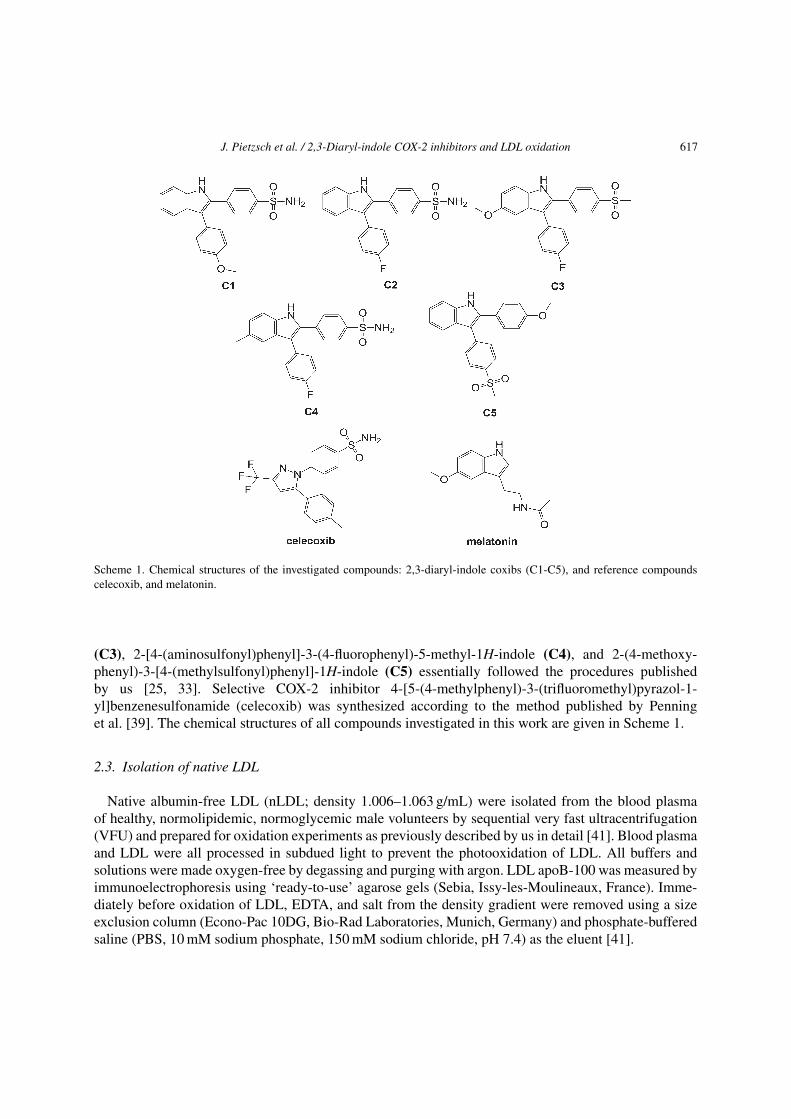
J. Pietzsch et al. / 2,3-Diaryl-indole COX-2 inhibitors and LDL oxidation 617
Scheme 1. Chemical structures of the investigated compounds: 2,3-diaryl-indole coxibs (C1-C5), and reference compoundscelecoxib, and melatonin.
(C3), 2-[4-(aminosulfonyl)phenyl]-3-(4-fluorophenyl)-5-methyl-1H-indole (C4), and 2-(4-methoxy-phenyl)-3-[4-(methylsulfonyl)phenyl]-1H-indole (C5) essentially followed the procedures publishedby us [25, 33]. Selective COX-2 inhibitor 4-[5-(4-methylphenyl)-3-(trifluoromethyl)pyrazol-1-yl]benzenesulfonamide (celecoxib) was synthesized according to the method published by Penninget al. [39]. The chemical structures of all compounds investigated in this work are given in Scheme 1.
2.3. Isolation of native LDL
Native albumin-free LDL (nLDL; density 1.006–1.063 g/mL) were isolated from the blood plasmaof healthy, normolipidemic, normoglycemic male volunteers by sequential very fast ultracentrifugation(VFU) and prepared for oxidation experiments as previously described by us in detail [41]. Blood plasmaand LDL were all processed in subdued light to prevent the photooxidation of LDL. All buffers andsolutions were made oxygen-free by degassing and purging with argon. LDL apoB-100 was measured byimmunoelectrophoresis using ‘ready-to-use’ agarose gels (Sebia, Issy-les-Moulineaux, France). Imme-diately before oxidation of LDL, EDTA, and salt from the density gradient were removed using a sizeexclusion column (Econo-Pac 10DG, Bio-Rad Laboratories, Munich, Germany) and phosphate-bufferedsaline (PBS, 10 mM sodium phosphate, 150 mM sodium chloride, pH 7.4) as the eluent [41].

618 J. Pietzsch et al. / 2,3-Diaryl-indole COX-2 inhibitors and LDL oxidation
2.4. LDL lipid and protein oxidation
In order to determine redox activity-structure relationships of 2,3-diaryl-substituted indole-basedcyclooxygenase-2 (COX-2) inhibitors (2,3-diaryl-indole coxibs) and, as controls, the non-indole-derivative celecoxib and the indole-derivative melatonin, three well-characterized LDL oxidation modelsgenerating OH•/O2
•− or hypochlorite (OCl−) were used. All compounds were tested at a final concentra-tion of 1 �M. For lipid peroxidation, to 200 �L-aliquots of nLDL (125 �g apoB-100/mL, equal to 0.25 �MLDL) in 96 well plates (UV-Star plates, UV transparent to 200 nm, Greiner Bio One, Frickenhausen,Germany) were added 25 �L of an aqueous solution of CuSO4 (16 �M) and 25 �L of stock solution ofthe compound (10 �M) to be tested for redox-activity. The same preparation without CuSO4 was used ascontrol. The oxidative process was monitored using a Synergy 4 thermostatic micro plate reader (BioTekInstruments, Bad Friedrichshall, Germany) by following the formation of conjugated dienes at 234 nmevery 5 min for 4 hours at 30◦C. This approach results in a curve exhibiting a lag phase, during which theabsorbance does not increase significantly, a propagation phase, during which the absorbance increasesrapidly, and a degradation phase, characterized by a slow fall in the absorbance [14, 33]. There is a posi-tive (negative) correlation between lag phase duration and the concentration of antioxidants (prooxidants)contained in the LDL sample [14]. For protein oxidation, 2 mL-aliquots of nLDL (125 �g apoB-100/mL,equal to 0.25 �M LDL) with or without 1 �M of the compound tested were subjected to two other wellcharacterized oxidation systems: A) hemin/H2O2 (10 �M/100 �M) and B) HOCl (100 �M) at 37◦C for40 hours in the dark [26, 40]. Then, on completion of the oxidation process, LDL were delipidated. Afterisolation, reduction, and enzymatic hydrolysis of apoB-100 the oxidative process was monitored in A) bymass spectrometric determination of 5-hydroxy-2-aminovaleric acid (HAVA) that is formed by reductionof �-glutamyl semialdehyde, which is a highly specific product of iron-(OH•/O2
•−)-mediated proteinoxidation and B) by mass spectrometric determination of formation of 3-chlorotyrosine (3Cl-TYR), ahighly specific marker of OCl−-mediated protein oxidation, as described elsewhere in detail [33, 40, 43].The results obtained by these LDL oxidation models are expressed as ratios between the duration of lagphase in the presence and in the absence of the compound (RDIENE), between the apoB-100 HAVA contentin the absence and in the presence of the compound (RHAVA), and between the apoB-100 3-chlorotyrosinecontent in the absence and in the presence of the compound (R3Cl-TYR). Thus, in these approaches Rvalues (RDIENE, RHAVA or R3Cl-TYR) higher than 1 indicate antioxidant activity, R values of about 1 meanthat the compound has no effect and R values lower than 1 suggests prooxidant activity. For reason ofcomparability RDIENE and RHAVA values were adopted from [33]. R3Cl-TYR values measured in this studycorrespond to four experiments, performed in triplicate.
2.5. OH• scavenging assay
In order to further characterize the redox activity of 2,3-diaryl-indole coxibs, in particular, to discrimi-nate possible OH• scavenging properties, a simplified deoxyribose degradation assay was used accordingto the procedure described by Lapenna et al. [31]. Results were expressed as percentage of OH• scav-enging activity %SAOH• of test sample compared to control without any antioxidant (100% deoxyriboseoxidation). In this study the use of EDTA in the reaction system is essential because inhibition of iron-dependent deoxyribose degradation in the absence of EDTA does not only depend on OH• scavenging,but also on its ability to form complexes with iron ions [17]. Each study corresponds to four experiments,performed in triplicate.

J. Pietzsch et al. / 2,3-Diaryl-indole COX-2 inhibitors and LDL oxidation 619
2.6. O2•− scavenging assay
In order to further characterize the redox activity of 2,3-diaryl-indole coxibs, in particular, to discrimi-nate possible O2
•− scavenging properties, a spectrophotometric assay monitoring O2•−-induced reduction
of nitroblue tetrazolium chloride (NBT) was used. In this assay the generation of O2•− was performed
using a nicotinamide adenine dinucleotide (NADH)/phenazine methosulfate (PMS) system according tothe procedure described by Costa et al. [9]. Results were expressed as percentage of O2
•− scavengingactivity (%SAO2
•-) of test sample compared to control without any antioxidant (100% diformazan for-mation). The selective effect of superoxide on the reduction of NBT to diformazan was confirmed usingsuperoxide dismutase. Each study corresponds to four experiments, performed in triplicate.
2.7. Radiolabeling of native and oxidized LDL
Native LDL and oxidatively modified LDL, which were obtained from both the hemin/H2O2
(oxLDL) and HOCl (OCl-LDL) LDL oxidation experiments, were radiolabeled with no-carrier addedN-succinimidyl-4-[18F]fluorobenzoate ([18F]SFB) as published in detail elsewhere [41]. In a typical LDLradiolabeling experiment, approximately 200 MBq of purified and sterile [18F]fluorobenzoylated LDLwere obtained, which had an effective specific activity in the range of 400–500 GBq/�mol for both[18F]FB-nLDL and [18F]FB-oxLDL/[18F]FB-OCl-LDL (each related to apoB-100; Mr 516.000, withoutcarbohydrate content) at the time of cell uptake studies. Furthermore, both [18F]FB-nLDL and [18F]FB-oxLDL/[18F]FB-OCl-LDL showed high in vitro stability at several time points (30 min, 2 h, and 4 h afterradiolabeling) using PBS (pH 6.5 to 7.5) as the solvent at 37◦C. At each time point after labeling morethan 96% of total activity (decay-corrected) could be recovered in the intact apoB-100 molecule. Of note,radiolabeling of LDL with no-carrier added [18F]SFB did not lead either to adverse oxidation of nativeLDL particles or to additional adverse modification of oxidatively modified LDL particles [42].
2.8. Lipoprotein uptake experiments in vitro
The influence of 2,3-diaryl-indole coxibs C1, C3 and C4 on the biological activity of LDL particlesin terms of specific cellular binding and uptake of both radiolabeled [18F]FB-nLDL and [18F]FB-oxLDL/[18F]FB-OCl-LDL obtained from hemin/H2O2 and HOCl LDL oxidation experiments wasassessed in human monocyte cell line THP-1 in vitro [41, 42]. For lipoprotein uptake experiments,cells were seeded in 24-well plates at a density of 1 × 10⁵ cells/mL. THP-1 cells were propagated inRPMI 1640, 10% FCS, containing penicillin (100 U/mL), streptomycin (100 �g/mL) at 37◦C in 5% CO2
[41]. For the uptake studies, cells were centrifuged and resuspended in fresh RPMI medium contain-ing 10% FCS in 24-well plates at a density of 1 × 10⁴ cells/mL. Transformation of THP-1 monocytes tomacrophages (THP-1Mφ) was performed by adding 64 nM phorbol myristate acetate for 72 h as described[41]. The cells were then washed extensively with serum-free RPMI medium and diluted to the appropri-ate density. In brief, for binding assays cells were incubated for 2 h at 4◦C with either [18F]FB-nLDL or[18F]FB-oxLDL/[18F]FB-OCl-LDL species (2.5–50 �g of protein/mL in 125 �L of media with 50 mMHepes, pH 7.4) in a total volume of 250 �L. At 4◦C, LDL bind to LDL receptors or scavenger receptors,but the lipoprotein-receptor complexes are not internalized. Non-specific binding was determined by theaddition of an excess (500 �g/mL) of unlabeled nLDL and oxLDL, respectively, and represented lessthan 15% of total lipoprotein binding in this cell type. The extent of specific binding was calculated by

620 J. Pietzsch et al. / 2,3-Diaryl-indole COX-2 inhibitors and LDL oxidation
subtracting the non-specific binding of [18F]FB-nLDL and [18F]FB-oxLDL/[18F]FB-OCl-LDL,respectively, from total binding. Assays for cell association of both [18F]FB-nLDL and [18F]FB-oxLDL/[18F]FB-OCl-LDL lasted for 2 h at 37◦C, as for the binding studies but without Hepes. Cellsincubated with LDL at 37◦C contain both membrane-bound and internalized lipoproteins. Lipoproteinuptake for 2 hours was calculated as the difference between total cell-associated lipoprotein (37◦C) andmembrane-bound lipoprotein (4◦C). At the end of all incubations the cells were washed twice with 1 mLof PBS containing 0.1% (w/v) bovine serum albumin, then twice with 1 mL of PBS. The cells weresolubilized in 1.5 mL of 1 M NaOH, assayed for protein content and counted for [18F]-activity in a CobraII gamma counter (Canberra-Packard, Meriden, CT, USA). Cell viability in this experimental settingwas assessed using the trypan blue dye exclusion test. Cellular protein was determined using the bicin-choninic acid protein assay (Pierce, Rockford, IL, USA) using bovine serum albumin as protein standard.Lipoprotein uptake is expressed as percent of injected dose per �g protein (%ID/�g protein) after 2 hours.
2.9. Lipoprotein uptake experiments in vivo
The influence of 2,3-diaryl-indole coxibs C1, C3 and C4 on the biological activity of LDL in terms ofblood clearance and specific organ uptake of radiolabeled [18F]FB-nLDL as well as [18F]FB-oxLDL and[18F]FB-OCl-LDL obtained from hemin/H2O2 and HOCl LDL oxidation experiments, respectively, wasassessed in rats in vivo [41, 42]. For investigation of in vivo catabolism of [18F]FB-nLDL and [18F]FB-oxLDL/[18F]FB-OCl-LDL, animal experiments were carried out with male Wistar rats (Kyoto-Wistarstrain; aged 6 weeks; 160–170 g; Harlan Laboratories, Venray, The Netherlands) according to the guide-lines of the German Regulations for Animal Welfare. The protocol was approved by the local EthicalCommittee for Animal Experiments and has been published elsewhere [42]. In brief, for biodistribu-tion studies, the animals were injected [18F]FB-nLDL, [18F]FB-oxLDL or [18F]FB-OCl-LDL (0.5 mL;0.8–1.2 MBq; radiochemical purity 96%; PBS, pH 7.2) into the tail vein under desfluran anaesthesia.Biodistribution was determined in groups of eight rats sacrificed 5 and 60 min post injection, respec-tively, by heart puncture under desfluran anaesthesia. Organs and tissues of interest were rapidly excised,weighed, and the [18F]-activity was determined (Cobra II gamma counter, Canberra-Packard, Meriden,CT, USA). The accumulated activity in organs and tissues was calculated as the percentage of the injecteddose per gram tissue (%ID/g tissue; corrected for decay).
2.10. Cell adhesion experiments in vitro
The potential of 2,3-diaryl-indole coxibs C1, C3 and C4 to influence the biological activity of oxLDL-promoted proinflammatory/proatherogenic processes in terms of adhesion of leukocytes to endothelialcells was assessed in human umbilical vein endothelial cells (HUVEC) in vitro. Adhesion experimentswere predominantly performed as co-incubation assays with HUVEC, THP-1 monocytes, and lipoproteinsfollowing a protocol published elsewhere with some modifications [28, 57]. In brief, HUVEC werepassaged onto 24-well plates, brought to confluence and treated simultaneously with native and oxidizedLDL species obtained from hemin/H2O2 and HOCl LDL oxidation experiments (50 �g/mL each finalconcentration) for 1 hour at 37◦C. Then HUVEC were washed once with 25 mM-Hepes-buffered M199(M199H) and, thereafter, incubated for further 60 min with THP-1 monocytes (106 cells per well) at 37◦C.Prior to adhesion THP-1 monocytes were labeled with the fluorescence dye BCECF/AM (Merck Group,Darmstadt, Germany). Therefore, 5 × 10⁶ cells/mL were incubated with 10 �g/mL BCECF/AM at 37◦C

J. Pietzsch et al. / 2,3-Diaryl-indole COX-2 inhibitors and LDL oxidation 621
for 30 min. Then, cells were centrifuged and resuspended in M199H medium at 2 × 10⁶ cells/mL. Foreach labeled cell preparation a calibration curve was prepared. The calibration curve was linear in therange from 12.500 to 200.000 cells per vial. At the end of the incubation period the supernatants werecarefully removed from the wells. HUVEC were washed once with M199H and the inverted test platewas centrifuged (50× g, 5 min). To lyse the cells 1 mL of 0.1 M Tris buffer plus 0.1% Triton X-100 wasadded to each well for 30 min. Fluorescence intensity (excitation 503 nm, emission 527 nm) of adheredcells was measured in triplicate, and the mean value was used for statistical analysis. Additionally, forthe most effective compound (C1) a subsequent adhesion experiment using human polymorphonuclearleukocytes (PMN) was performed. This approach allows for distinction of effects related to the use of ahuman primary inflammatory cell from those observed in an immortalized (leukemia-derived) cell line.Therefore, human PMN were isolated from heparinized blood of healthy volunteers by density gradientcentrifugation as described previously in detail [27, 29]. PMN then underwent the same procedure asreported above for THP-1 cells.
2.11. Cell inflammatory response in vitro
The potential of 2,3-diaryl-indole coxibs C1, C3 and C4 to influence the biological activity of oxLDL-mediated inflammatory/atherogenic processes in terms of specific cytokine release was assessed in THP-1Mφ (TNF�) and HUVEC (IL-8) [3]. Once differentiated, THP-1Mφ were incubated for 16 h (HUVECfor 4 h) with oxLDL obtained from hemin/H2O2 and HOCL LDL oxidation experiments in the absenceor presence of 1 �M of the compounds to be tested. At the end of each incubation, conditioned mediawere harvested and centrifuged, and TNF� and IL-8 levels were quantified on the supernatant fractionusing a commercial enzyme-linked immunosorbent assays (PromoKine, PromoCell GmbH, Heidelberg,Germany) according to the manufacturer’s instructions. The results (concentrations amounted to ng/mLor pg/mL) were expressed as fold over control and all were corrected for cell viability. Cell viability inthis experimental setting was determined by dimethylthiazoldiphenyltetrazolium bromide (MTT) assay[51]. Cell viability was calculated for cells upon every single/combined treatment (50 �g/mL nativeor oxidized LDL with/without 1 �M 2,3-diaryl-indole coxibs) and expressed as a percentage over thecontrol conditions. Under these conditions and within the respective time windows for each individualexperiment 2,3-diaryl-indole coxibs showed no sign of toxicity and cell viability of both THP-1 andHUVEC did not decrease below 90% (Data not shown in detail). Furthermore, nuclear factor κB (NF-κB)p56 subunit activity was measured in nuclear extracts from THP-1Mφ using an ELISA kit according to themanufacturer’s protocol (Pierce Biotechnology, Rockford, IL, USA). Chemoluminescence was measuredon Synergy 4 thermostatic micro plate reader (BioTek Instruments, Bad Friedrichshall, Germany) [16].
2.12. Statistical analysis
Results are presented as means ± SD from at least 3 independent experiments. Nonparametric statisticalanalyses were calculated by using the SPSS 20 software package (SPSS Inc., Chicago, IL, USA). Mann-Whitney test was used to compare 2 independent groups, Kruskal-Wallis test followed by Bonferronipost-hoc analysis was used to account for multiple testing. For all analyses a value of P < .05 wasconsidered as statistically significant.

622 J. Pietzsch et al. / 2,3-Diaryl-indole COX-2 inhibitors and LDL oxidation
3. Results and discussion
The aim of this work was to explore i) the reactivity of 2,3-diaryl-indole coxibs with several wellcharacterized LDL oxidation systems and ii) subsequent effects on a inflammatory/atherogenic microen-vironment. The five compounds investigated were selected from a series of novel 2,3-diaryl-indolecoxibs preferably bearing a fluorine or methoxy substituent, in order to gain a pool of versatile scaf-fold compounds promising for 18F or 11C radiotracer development for functional characterization ofCOX-2 activity/expression in vivo using positron emission tomography [33]. The indole heterocycle isa well-characterized antioxidant pharmacophore [30]. Thus, in our previous study on 2,3-diaryl indolecoxibs it consequently had to be considered that antioxidative properties might interfere with assays fordetermination of the COX inhibitory activity. Indeed, we observed disturbances of these assays, whichclearly could be attributed to redox-active properties of the novel compounds [33]. This hypothesis wassupported by another finding that one of the compounds (C1) was able to inhibit not only the formationof prostaglandin E2 but also the formation of F2�-isoprostanes after the exposure of endothelial cellsin both monolayer and organo-type aortic ring models to ionizing radiation [44, 56]. F2�-isoprostanesare prostaglandin-like products of nonenzymatic, free radical-catalyzed peroxidation of arachidonic acid,which are considered as potential biomarkers and have been correlated with conditions of oxidative stress,e.g., in inflammatory and atherogenic processes [38, 46]. Noteworthy, the combination of both COX-2inhibitory and antioxidative potential in one compound is hypothesized to be very attractive. Compoundscombining these properties potentially might overcome some adverse effects observed in long-term med-ication with coxibs, e.g., atherogenic properties in part associated with a prooxidative action [37, 58]. Onthe other hand, such compounds might act as double-edged swords in many pathophysiological situations,e.g., by inhibiting radiation-induced COX-2 expression and formation of ROS in parallel thus protectingnormal tissue from adverse effects of radiation therapy [44, 56]. Therefore, we decided to evaluate theantioxidant capacity of the 2,3-diaryl-indole coxibs in more detail by focusing on potential prevention ofLDL oxidation. There is experimental and clinical evidence supporting oxidative modification of LDLas an early and critical event in atherogenesis. Oxidation of LDL particles to atherogenic ones, on theone hand, generates high-uptake particles for macrophage and granulocyte scavenger receptor pathwaysleading to inappropriate accumulation of intracellular lipid deposits, and, on the other hand, generatesa large panoply of lipid mediators promoting endothelial dysfunction and an inflammatory endothe-lial/subendothelial microenvironment [6, 45, 52]. The finding from earlier investigations that 2,3-diarylindoles already show substantial antioxidative effects at pharmacologically relevant low levels promptedus to use consequently 1 �M of each test compound in all experimental settings [33].
3.1. Redox activity of 2,3-diaryl-indoles
Table 1 shows data obtained from lipid and protein oxidation assays as well as from radical scavengingassays summarized here as redox activity of 2,3-diaryl indoles C1 to C5 at a concentration of 1 �M in vitro.The well characterized antioxidative indole-derivative melatonin [2] and the potentially redox-neutral1,5-diaryl-pyrazole-derivative celecoxib [58] served as reference compounds.
Except C4, the 2,3-diaryl-indoles demonstrate potent antioxidative/ROS scavenging activity(C1 > C3 > C5 ∼ C2 » C4) towards OH•, O2
•−, and OCl−. The more detailed investigations hence con-firmed the earlier finding that various 2,3-diaryl indoles exhibit a substantial antioxidative behavior(RDIENE/RHAVA>1) at pharmacologically relevant low concentrations [33]. From these initial experimentsthree compounds were selected for further experiments: the most effective non-fluorine compound C1,

J. Pietzsch et al. / 2,3-Diaryl-indole COX-2 inhibitors and LDL oxidation 623
Table 1
Redox activity and lipophilicity of 2,3-diaryl-indole coxibs and reference compounds
Compound Redox activity Lipophilicity
RDIENE§ RHAVA
§ R3Cl-TYR %SAOH• %SAO2•- logP#
C1 2.85 1.98 1.56 ± 0.08 25.3 ± 4.4 8.4 ± 1.1 4.05 ± 0.70C2 1.32 1.36 1.22 ± 0.04 20.5 ± 3.2 5.5 ± 1.2 4.17 ± 0.73C3 1.78 1.71 1.38 ± 0.07 19.7 ± 3.7 7.9 ± 2.8 4.00 ± 0.72C4 0.98 0.99 0.96 ± 0.05 4.7 ± 1.2 0.9 ± 0.9 4.63 ± 0.73C5 1.54 1.29 1.31 ± 0.11 21.4 ± 4.6 4.6 ± 1.2 3.96 ± 0.58celecoxib 0.97 0.96 0.97 ± 0.05 1.9 ± 0.9 0.4 ± 1.1 4.21 ± 1.49melatonin 1.71 1.52 1.04 ± 0.08 17.6 ± 3.9 2.4 ± 2.2 0.96 ± 0.44
§RDIEN and RHAVA values were adopted from [33]. #log P values were calculated using ACD/ChemSketch software, version14.01 (Advanced Chemistry Development Inc., Toronto, ON, Canada; www.acdlabs.com).
the also very effective but fluorine compound C3, and, as control, the redox-neutral fluorine compoundC4. Of note, in our investigations melatonin exhibited already at a concentration of 1 �M a high redoxactivity with respect to the formation of dienes and HAVA that was comparable to the 2,3-diaryl-indolesC3 and C5. This is in contrast to the rather weak antioxidative activity of melatonin reported by others [49,59]. On the other hand, more consistently with the literature, in this work melatonin at 1 �M showed onlyvery low OCl− and O2
•− scavenging activities when compared to the most potent 2,3-diaryl-indoles. Thelack of aryl substituents and, logically, the higher hydrophilicity of melatonin might be one explanationfor this observation.
3.2. Influence of 2,3-diaryl-indoles on lipoprotein uptake in vitro
When differentiated to macrophages after stimulation with phorbol esters, monocytic THP-1 cells areconsidered scavenger receptor bearing human cells and are an accepted model for the examination of cel-lular interaction with oxidatively modified LDL. In agreement with former results, the present lipoproteinuptake experiments revealed a significantly higher specific binding and uptake in THP-1 macrophages ofboth radiolabeled [18F]FB-oxLDL and [18F]FB-OCl-LDL compared to [18F]FB-nLDL (Fig. 1) [19, 42].LDL that were oxidized in the presence of 1 �M 2,3-diaryl-indole coxibs still show scavenger receptor-mediated uptake in THP-1Mφ cells, which, however, was lower compared to untreated oxLDL (C1 > C3;P < .05) and OCl-LDL (C1 ∼ C3). The observed antioxidative effects appear to be more pronounced inoxLDL when compared to OCl-LDL, which is consistent with the parameters characterizing redox activ-ity of 2,3-diaryl-indoles towards OH•/O2
•−-mediated and OCl−-mediated oxidation as shown in Table 1.Of interest, compound C4 did not influence the scavenger receptor mediated uptake of oxLDL/OCl-LDL in THP-1Mφ cells. As an essential prerequisite for this investigation it has been demonstrated byus earlier that the [18F]fluorobenzoylation procedure used does not lead either to adverse oxidation ofnLDL particles or to additional adverse oxidative modification of oxLDL/OCl-LDL particles [41, 42,49]. Moreover, 2,3-diaryl-indoles did not significantly influence the cellular uptake of nLDL (Fig. 1).
3.3. Influence of 2,3-diaryl-indoles on lipoprotein uptake in vivo
Figure 2 summarizes the distribution of activity (decay-corrected) in male Wistar rats after a singleintravenous injection of [18F]FB-nLDL, [18F]FB-oxLDL or [18F]FB-OCl-LDL.

624 J. Pietzsch et al. / 2,3-Diaryl-indole COX-2 inhibitors and LDL oxidation
Fig. 1. Cellular uptake of [18F]FB-nLDL and [18F]FB-oxLDL/-OCl-LDL after 2 hours in phorbol ester-stimulated THP-1cells (THP-1Mφ). LDL particles were either untreated or treated with 1 �M of 2,3-diaryl-indole coxibs during the oxidationexperiment. #P < 0.05, vs. nLDL; ∗P < 0.05, vs. oxLDL/OCl-LDL.
Data were obtained at 5 and 60 min post injection. In agreement with former results, the presentbiodistribution experiments revealed both a significantly faster blood clearance and a significantly higherspecific association in liver, spleen, kidneys, and adrenals of both [18F]FB-oxLDL and [18F]FB-OCl-LDL compared to [18F]FB-nLDL. Oxidatively modified LDL that were oxidized in the presence of 1 �M2,3-diaryl-indole coxibs show a partly normalized blood clearance (P < .05) which is accompanied bysignificantly lowered uptake in liver and spleen (P < .05), and, in part, a normalized uptake in kidneys andadrenals when compared to untreated oxLDL (C1 > C3, P < .05) and OCl-LDL (C1 ∼ C3), respectively.In accordance with the in vitro uptake data this in vivo investigation indicates a substantial rerouting oftreated oxidatively modified LDL particles from the scavenger receptor pathways back to the LDL receptorpathway. In contrast, compound C4 did not significantly influence the metabolic behavior of oxLDL/OCl-LDL in the rat model. As an essential prerequisite for this investigation it has been demonstrated by usthat both [18F]FB-nLDL and [18F]FB-oxLDL/18F]FB-OCl-LDL exhibit high radiotracer stability in vivo[19, 41, 42].
3.4. Influence of 2,3-diaryl-indoles on oxidized lipoprotein-mediated cell adhesion in vitro
Incubation of HUVEC with 1 × 10⁶ THP-1 cells and PMN, respectively, and phosphate buffered salineshowed a basal adhesion of 65,000 ± 9,400 (THP-1) and 23,000 ± 5,100 (PMN) cells/well. This basaladhesion amounted to 6.5%/2.3% of added THP-1/PMN cells and corresponds well to data reportedby others and us [13, 28]. After co-incubation of HUVEC, THP-1 cells and LDL adhesion increasedsignificantly (P < 0.05) (Fig. 3). Similar results were obtained using PMN instead of THP-1 monocytes.Addition of nLDL in the presence of 1 �M 2,3-diaryl-indole coxibs did not show further effects. On theother hand, addition of both oxLDL and OCl-LDL instead of nLDL to the experimental system resulted in

J. Pietzsch et al. / 2,3-Diaryl-indole COX-2 inhibitors and LDL oxidation 625
Fig. 2. Biodistribution of [18F]FB-nLDL and [18F]FB-oxLDL/-OCl-LDL in male Kyoto-Wistar rats at 5 min (A) and 60 min (B)after intravenous injection.
a significantly higher adhesion. This increment could be markedly decreased by 2,3-diaryl-indole coxibs(C1 ∼ C3) for both cell types. In contrast, the use of compound C4 revealed no effect on cell adhesion.
3.5. Influence of 2,3-diaryl-indoles on oxidized lipoprotein-mediated inflammatory response in vitro
Macrophages are the major source of proinflammatory cytokines like TNF� within the atheroscleroticplaque [11]. These cytokines, in turn, amplify the inflammatory response in the cellular microenvironment,

626 J. Pietzsch et al. / 2,3-Diaryl-indole COX-2 inhibitors and LDL oxidation
Fig. 3. Effect of nLDL and oxLDL/OCl-LDL treated with/without 1 �M of 2,3-diaryl-indole coxibs on adherence of THP-1monocytes and PMN cells to HUVEC monolayers. §P < 0.05, vs. vehicle; #P < 0.05, vs. nLDL; ∗P < 0.05, vs. oxLDL/OCl-LDL.
e.g., by increasing macrophage adhesion to endothelial cells [61]. This process essentially contributesalso to oxidized lipoprotein-mediated cell adhesion [62]. In consequence, exposure of THP-1Mφ tooxidatively modified LDL resulted in an increase of TNF� secretion compared to nLDL. This incre-ment could be markedly decreased by 2,3-diaryl-indole coxibs (C1 ∼ C3) (Fig. 4). Vascular endothelialcells, on the other hand, usually are resistant to cholesterol accumulation by lipoprotein uptake. How-ever, these cells also have shown to express receptors that recognize oxidatively modified LDL andto have the biochemical pathways for sterol synthesis and receptor-mediated endocytosis of lipopro-teins [8, 10]. Furthermore, oxidatively modified LDL serve as important proinflammatory activators inHUVEC, e.g., by inducing and modulating expression/secretion of IL-8 [34, 54]. Consistently, exposureof HUVEC to oxidatively modified LDL resulted in a significant increase of IL-8 secretion comparedto nLDL. This increment also could be markedly decreased by 2,3-diaryl-indole coxibs (C1 ∼ C3)(Fig. 4).
The expression of inflammatory cytokines TNF� and IL-8 is regulated transcriptionally by NF-κB,which among other factors is activated by oxidatively modified lipoproteins and reactive oxygen species[36, 48]. In line with this, exposure of THP-1Mφ to oxidatively modified LDL resulted in a substantiallyhigher activity of NF-κB activity compared to THP-1Mφ incubated with nLDL. Oxidatively modifiedLDL that were oxidized in the presence of 1 �M 2,3-diaryl-indole coxibs resulted in a significant decre-ment in NF-κB activation when compared to untreated oxLDL/OCl-LDL (C1 > C3; P < .05) (Fig. 5).These experiments also showed that compound C4 at a concentration of 1 �M revealed no protectiveeffects.

J. Pietzsch et al. / 2,3-Diaryl-indole COX-2 inhibitors and LDL oxidation 627
Fig. 4. Effect of nLDL and oxLDL/OCl-LDL treated with/without 1 �M of 2,3-diaryl-indole coxibs on cytokine secretion inTHP-1Mφ and HUVEC. #P < 0.05, vs. nLDL; ∗P < 0.05, vs. oxLDL/OCl-LDL.
Fig. 5. Effect of nLDL and oxLDL/OCl-LDL treated with/without 1 �M of 2,3-diaryl-indole coxibs on NFκB activation inTHP-1Mφ. #P < 0.05, vs. nLDL; ∗P < 0.05, vs. oxLDL/OCl-LDL.

628 J. Pietzsch et al. / 2,3-Diaryl-indole COX-2 inhibitors and LDL oxidation
3.6. Redox activity of 2,3-diaryl-indoles: Structure-activity-relationships
Summarizing,theantioxidantpotencyof2,3-diaryl-indolecoxibswasinvestigatedusingacceptedmodelssimulating oxidative modification of LDL and, subsequently, experimental settings allowing for discrimi-nation of selected atherogenic effects typically mediated by oxidatively modified LDL in an inflammatoryenvironment. The data demonstrate that the examined 2,3-diaryl-indole coxibs exhibit scavenging activityfor ROS with variable effectiveness, depending on the kind of ROS and the compounds’ chemical struc-ture. In this work, compound C1 showed the overall most prominent antioxidative potential which wassignificantly higher than that of the known indole antioxidant melatonin used as positive control. The notfurther evaluated compounds C2 and C5, characterized by an unsubstituted 5-position in the indole hete-rocycle like C1, also showed an abundant antioxidative action that, however, was more comparable to thatof melatonin. Regarding the individual redox activity of 2,3-diaryl indoles the impact of the substituentin 5-position of the indole system is noteworthy. An alkyl (methyl) substituent in this position seems tohinder efficient ROS scavenging of compound C4. Additionally, the methyl substituent in this positionalso diminishes the COX-2 inhibitory activity as previously published by us [33]. By contrast, a methoxysubstituent in 5-position of the indole heterocycle seems to substantially support ROS scavenging as alsodemonstrated with compound C3 [15]. Moreover, a methoxy substituent also increases COX-2 inhibitoryactivity [33]. As expected, the 1,5-diaryl-pyrazole-derivative celecoxib did not exhibit any redox activityin the model systems used and was considered as negative control [33, 38]. A more detailed investigationto differentiate the ROS scavenging properties indicated i) a preferred inhibition of lipid oxidation com-pared to protein oxidation, ii) a preferred interaction with transition metal-catalyzed oxidation compared toHOCl-mediated oxidation, and iii) a preferred OH• scavenging compared to O2
•− scavenging. The over-all antioxidative action of the compounds tested is in line with the hypothesized mechanism discussed indetail for other indole antioxidants like 2-phenylindoles [53]. This mechanism essentially would allow thepresence of substituents like a methoxy group or a hydroxyl group at 5-position. Of interest, in most exper-iments performed here C1 showed a substantially higher antioxidative activity than did compound C3 thatnot alone can be explained by this mechanistic view [1, 23]. Of importance, this difference appeared to bemore pronounced in the OH•/O2
•−-mediated oxidation systems when compared to OCl−-mediated oxida-tion. In this regard, potential transition-metal binding properties of indole derivatives have to be taken intoconsideration [23, 24]. In compound C1 this possibly is supported by presence of both an aminosulfonyland a methoxy group in para-position in the 3-phenyl and 2-phenyl substituents, respectively [4]. Regardinglipoprotein oxidation the antioxidative action of the compounds tested in part also seems to be influencedby their lipophilicity. The 2,3-diaryl-indoles, as other coxibs, show low aqueous solubility and high logPs,allocating them, beside other factors, to the low-solubility, high-permeability BCS Class II compounds and,on the other hand, strongly suggesting a natural predisposition for increased plasma lipoprotein binding[60]. This is consistent with the data reported here. In this regard, a detailed investigation on both potentialtransition-metal binding properties and the distribution of 2,3-diaryl-indole coxibs in plasma lipoproteinsstill should be performed. Considering the overall setting in this investigation most effects observed clearlycan be attributed to the antioxidative action of 2,3-diaryl-indole coxibs on LDL oxidation. However, underthe experimental conditions employed radical scavenging should not consume the total amount of com-pounds. Thus, lipoprotein-bound 2,3-diaryl-indole coxibs are suggested to exert additional, direct actionson the cells used in vitro or on tissues in the biodistribution experiment in vivo. This might include, logi-cally, cyclooxygenase-2 inhibition but also direct antioxidative/regulative effects on intracellular pathwaysthat, in turn, should contribute to the observed effects. This would further explain the considerable effectsachieved at the concentration of 2,3-diaryl-indole coxibs used.

J. Pietzsch et al. / 2,3-Diaryl-indole COX-2 inhibitors and LDL oxidation 629
4. Conclusion
This work demonstrated ROS scavenging activity of novel 2,3-diaryl-indole coxibs with high potentialto decrease susceptibility of LDL to oxidation in vitro. Subsequently, this led to a substantial decreaseof LDL uptake by scavenger receptors in THP-1 macrophages in vitro and in a rodent model in vivo.Moreover, LDL-mediated macrophage adhesion to endothelial cells, macrophage NFκB activation, aswell as macrophage and endothelial cell cytokine release in vitro was substantially diminished. Regardingthe influence of most potent compounds C1 and C3 on these ‘downstream’ effects the tendency for eachof them is the same. Individual variation in effect strength, however, is assumed to depend on the actualinfluence of the tested compounds on the overall pattern of oxidized lipids and apolipoproteins withineach individual LDL particle. In line with the present data it can be hypothesized that lipophilicity andtransition-metal binding may contribute to variability. The LDL-associated antioxidant properties of 2,3-diaryl-indole coxibs essentially should positively contribute to other known antiatherogenic effects ofcyclooxygenase inhibition [7, 47]. This potentially may widen the therapeutic window of COX-2 targetedtreatment. In this regard, antioxidant coxibs potentially provide an opportunity to overcome in part someof the known adverse cardiovascular effects of long-term cyclooxygenase inhibition [18]. On the otherhand, antioxidant coxibs are promising adjuvant therapeutics for prevention of radiation-induced vasculardysfunction and atherogenesis [12, 44, 55, 56].
Acknowledgments
The authors are grateful to Mareike Barth, Catharina Heinig, Uta Lenkeit, and Regina Herrlich fortheir expert technical assistance in human LDL preparation, radiolabeling, and characterization. We arealso grateful to Sigrid Nitzsche from the former Lipoprotein Laboratory at the Department of InternalMedicine 3, Medical Faculty and University Hospital Carl Gustav Carus, Technische Universitat Dresden,for her expert technical assistance in LDL characterization, her advice and many stimulating discussions.We also thank Steffi Kopprasch, Ph.D., from the Pathobiochemistry Unit at the Department of InternalMedicine 3, Medical Faculty and University Hospital Carl Gustav Carus, Technische Universitat Dres-den, for her expert advice regarding primary leukocyte isolation and handling as well as many fruitfuldiscussions. This work was supported in part by the German Research Foundation (grant Pi 304/1-1). Theauthors also thank the Helmholtz Association for funding a part of this work through Helmholtz-PortfolioTopic “Technologie und Medizin – Multimodale Bildgebung zur Aufklarung des In-vivo-Verhaltens vonpolymeren Biomaterialien”. Franz-Jacob Pietzsch is graduate student member of the Integrated ResearchTraining Group “Matrixengineering” (within the Transregional Collaborative Research Centre 67 “Func-tional biomaterials for controlling healing processes in bone and skin – from material science to clinicalapplication” funded by German Research Foundation) at Medical Faculty and University Hospital CarlGustav Carus, Technische Universitat, Dresden.
References
[1] J. Antosiewicz, E. Damiani, W. Jassem, M. Wozniak, M. Orena and L. Greci, Influence of structure on the antioxidantactivity of indolinic nitroxide radicals, Free Radicals in Biology & Medicine 22 (1997), 249–255.
[2] D. Bonnefont-Rousselot, G. Cheve, A. Gozzo, A. Tailleux, V. Guilloz, S. Caisey, E. Teissier, J.C. Fruchart, J. Delattre, D.Jore, D. Lesieur, P. Duriez and M. Gardes-Albert, Melatonin related compounds inhibit lipid peroxidation during copperor free radical-induced LDL oxidation, Journal of Pineal Research 33 (2002), 109–117.

630 J. Pietzsch et al. / 2,3-Diaryl-indole COX-2 inhibitors and LDL oxidation
[3] K.Z. Boudjeltia, I. Legssyer, P. Van Antwerpen, R.L. Kisoka, S. Babar, N. Moguilevsky, P. Delree, J. Ducobu, C. Remacle,M. Vanhaeverbeek and D. Brohee, Triggering of inflammatory response by myeloperoxidase-oxidized LDL, Biochemistryand Cell Biology 84 (2006), 805–812.
[4] G.E. Cami, M.E. Chacon Villalba, P. Colinas, G.A. Echeverria, G. Estiu and D.B. Soria, Crystal structure, spectroscopyand theoretical studies of p-cyanobenzenesulfonamide and a Cu(II) complex, Journal of Molecular Structure 1024 (2012),110–116.
[5] J.A. Campbell, V. Bordunov, C.A. Broka, M.F. Browner, J.M. Kress, T. Mirzadegan, C. Ramesha, B.F. Sanpablo, R. Stabler,P. Takahara, A. Villasenor, K.A. Walker, J.H. Wang, M. Welch and P. Weller, Rational design of 6-methylsulfonylindolesas selective cyclooxygenase-2 inhibitors, Bioorganic Medicinal Chemistry Letters 14 (2004), 4741–4745.
[6] V. Capra, M. Back, S.S. Barbieri, M. Camera, E. Tremoli and G.E. Rovati, Eicosanoids and their drugs in cardiovasculardiseases: Focus on atherosclerosis and stroke, Medicinal Research Reviews 33 (2013), 364–438.
[7] F. Cipollone, G. Cicolini and M. Bucci, Cyclooxygenase and prostaglandin synthases in atherosclerosis: Recent insightsand future perspectives, Pharmacology & Therapeutics 118 (2008), 161–180.
[8] L. Cominacini, U. Garbin, A.F. Pasini, A. Davoli, M. Campagnola, A.M. Pastorino, G. Gaviraghi and V. Lo Cascio,Oxidized low-density lipoprotein increases the production of intracellular reactive oxygen species in endothelial cells:Inhibitory effect of lacidipine, Journal of Hypertension 16 (1998), 1913–1919.
[9] D. Costa, L. Moutinho, J.L.F.C. Lima and E. Fernandes, Antioxidant activity and inhibition of human neutrophil oxidativeburst mediated by arylpropionic acid non-steroidal anti-inflammatory drugs, Biological and Pharmaceutical Bulletin 29(2006), 1659–1670.
[10] S. Costa, F. Zimetti, M. Pedrelli, G. Cremonesi and F. Bernini, Manidipine reduces pro-inflammatory cytokines secretionin human endothelial cells and macrophages, Pharmacological Research 62 (2010), 265–270.
[11] C. Crisafulli, M. Galuppo and S. Cuzzocrea, Effects of genetic and pharmacological inhibition of TNF-alpha in theregulation of inflammation in macrophages, Pharmacological Research 60 (2009), 332–340.
[12] A. Eldor, Z. Fuks, Y. Matzner, L.D. Witte and I. Vlodavsky, Perturbation of endothelial functions by ionizing irradiation:Effects on prostaglandins, chemoattractants and mitogens, Seminars in Thrombosis and Hemostasis 15 (1989), 215–225.
[13] W. Erl, P.C. Weber and C. Weber, Monocytic cell adhesion to endothelial cells stimulated by oxidized low density lipoproteinis mediated by distinct endothelial ligands, Atherosclerosis 136 (1998), 297–303.
[14] H. Esterbauer, G. Striegl, H. Puhl and M. Rotheneder, Continuous monitoring of in vitro oxidation of human low densitylipoprotein, Free Radical Research Communications 6 (1989), 67–75.
[15] A. Gozzo, D. Lesieur, P. Duriez, J.C. Fruchart and E. Teissier, Structure-activity relationships in a series of melatoninanalogues with the low-density lipoprotein oxidation model, Free Radicals in Biology & Medicine 26 (1999), 1538–1543.
[16] C. Haase-Kohn, S. Wolf, J. Lenk and J. Pietzsch, Copper-mediated cross-linking of S100A4, but not of S100A2, results inproinflammatory effects in melanoma cells, Biochemical and Biophysical Research Communications 413 (2011), 494–498.
[17] B. Halliwell, J.M. Gutteridge and O.I. Aruoma, The deoxyribose method: A simple test-tube assay for determination ofrate constants for reactions of hydroxyl radicals, Analytical Biochemistry 165 (1987), 215–219.
[18] S. Harirforoosh, W. Asghar and F. Jamali, Adverse effects of nonsteroidal antiinflammatory drugs: An update of gastroin-testinal, cardiovascular and renal complications, Journal of Pharmacy & Pharmaceutical Sciences 16 (2013), 821–847.
[19] S. Hoppmann, B. Steiniger, K. Strobel, C. Haase and J. Pietzsch, In vivo catabolism of hypochlorite-modified low densitylipoproteins (LDL): Insights from small animal positron emission tomography studies, In: J. Pietzsch (ed.), ResearchSignpost, Trivandrum, India, 2006, pp. 197–219.
[20] W. Hu, Z. Guo, F. Chu, A. Bai, X. Yi, G. Cheng and J. Li, Synthesis and biological evaluation of substituted 2-sulfonyl-phenyl-3-phenyl-indoles: A new series of selective COX-2 inhibitors, Bioorganic Medicinal Chemistry 11 (2003),1153–1160.
[21] J. Y. Im, D. Kim, S. G. Paik, and P. L. Han, Cyclooxygenase-2-dependent neuronal death proceeds via superoxide aniongeneration, Free Radical Biology and Medicine 41 (2006), 960–972.
[22] B. Jawabrah Al-Hourani, S.K. Sharma, M. Suresh and F. Wuest, Cyclooxygenase-2 inhibitors: A literature and patentreview (2009 - 2010), Expert Opinion on Therapeutic Patents 21(9) (2011), 1339–1432.
[23] N.V. Kaminskaia, G.M. Ullmann, D.B. Fulton and N.M. Kostic, Spectroscopic, kinetic, and mechanistic study of a newmode of coordination of indole derivatives to platinum(II) and palladium(II) ions in complexes, Inorganic Chemistry 39(2000), 5004–5013.
[24] M. R. Karekal, V. Biradar and M. Bennikallu Hire Mathada, Synthesis, characterization, antimicrobial, DNA cleavage,and antioxidant studies of some metal complexes derived from schiff base containing indole and quinoline moieties,Bioinorganic Chemistry and Applications 2013 (2013), 315972.

J. Pietzsch et al. / 2,3-Diaryl-indole COX-2 inhibitors and LDL oxidation 631
[25] T. Kniess, M. Laube, R. Bergmann, F. Sehn, F. Graf, J. Steinbach, F. Wuest and J. Pietzsch, Radiosynthesis of a 18F-labeled2,3-diarylsubstituted indole via McMurry coupling for functional characterization of cyclooxygenase-2 (COX-2) in vitroand in vivo, Bioorganic Medicinal Chemistry 20 (2012), 3410–3421.
[26] S. Kopprasch, W. Leonhardt, J. Pietzsch and H. Kuhne, Hypochlorite-modified low-density lipoprotein stimulates humanpolymorphonuclear leukocytes for enhanced production of reactive oxygen metabolites, enzyme secretion, and adhesionto endothelial cells, Atherosclerosis 136 (1998), 315–324.
[27] S. Kopprasch, J. Pietzsch and J. Grassler, The protective effects of HDL and its constituents against neutrophil respiratoryburst activation by hypochlorite-oxidized LDL, Molecular and Cellular Biochemistry 258 (2004), 121–127.
[28] S. Kopprasch, J. Pietzsch, T. Westendorf, H-J Kruse and J. Grassler, The pivotal role of scavenger receptor CD36 andphagocyte-derived oxidants in oxidized low density lipoprotein-induced adhesion to endothelial cells, The InternationalJournal of Biochemistry & Cell Biology 36 (2004), 460–471.
[29] S. Kopprasch, K. Richter, W. Leonhardt, J. Pietzsch and J. Grassler, Urate attenuates oxidation of native low-densitylipoprotein by hypochlorite and the subsequent lipoprotein-induced respiratory burst activities of polymorphonuclearleukocytes, Molecular and Cellular Biochemistry 206 (2000), 51–56.
[30] I. Kruk, H.Y. Aboul-Enein, T. Michalska, K. Lichszteld, K. Kubasik-Kladna and S. Olgen, In vitro scavenging activity forreactive oxygen species by N-substituted indole-2-carboxylic acid esters, Luminescence 22 (2007), 379–386.
[31] D. Lapenna, G. Ciofani, D. Festi, M. Neri, S.D. Pierdomenico, M.A. Giamberardino and F. Cuccurullo, Antioxidantproperties of ursodeoxycholic acid, Biochemical Pharmacology 64(11) (2002), 1661–1667.
[32] M. Laube, T. Kniess and J. Pietzsch, Radiolabeled COX-2 inhibitors for non-invasive visualization of COX-2 expressionand activity–a critical update, Molecules 18 (2013), 6311–6355.
[33] M. Laube, C. Tondera, S.K. Sharma, N. Bechmann, F-J Pietzsch, A. Pigorsch, M. Kockerling, F. Wuest, J. Pietzsch andT. Kniess, 2,3-Diaryl-substituted indole based COX-2 inhibitors as leads for imaging tracer development, RSC Advances4 (2014), 38726–38742.
[34] D. Lussi, A. Potapovich, M. Riccardo, E. Dellambra, G. Pressi, V. Kostyuk, R. Dal Toso, C. De Luca, S. Pastore and L.Korkina, Anti-inflammatory effects of concentrated ethanol extracts of Edelweiss (Leontopodium alpinum Cass.) calluscultures towards human keratinocytes and endothelial cells, Mediators of Inflammation 2012 (2012), 498373.
[35] L.J. Marnett, The COXIB experience: A look in the rearview mirror, Annual Review of Pharmacology and Toxicology 49(2009), 265–290.
[36] C. Maziere and J.C. Maziere, Activation of transcription factors and gene expression by oxidized low-density lipoprotein,Free Radicals in Biology & Medicine 46 (2009), 127–137.
[37] D. Mukherjee, S.E. Nissen and E.J. Topol, Risk of cardiovascular events associated with selective COX-2 inhibitors,Journal oft the American Medical Association 286 (2001), 954–959.
[38] A. Oguogho, H. Kritz, O. Wagner and H. Sinzinger, 6-oxo-PGF1� and 8-epi-PGF2� in the arterial wall layers of variousspecies: A comparison between intact and atherosclerotic areas, Prostaglandins, Leukotrienes & Essential Fatty Acids 64(2001), 167–171.
[39] T.D. Penning, J.J. Talley, S.R. Bertenshaw, J.S. Carter, P.W. Collins, S. Docter, M.J. Graneto, L.F. Lee, J.W. Malecha,J.M. Miyashiro, R.S. Rogers, D.J. Rogier, S.S. Yu, G.D. Anderson, E.G. Burton, J.N. Cogburn, S.A. Gregory,C.M. Koboldt, W.E. Perkins, K. Seibert, A.W. Veenhuizen, Y.Y. Zhang and P.C. Isakson, Synthesis and biologicalevaluation of the 1,5-diarylpyrazole class of cyclooxygenase-2 inhibitors: Identification of 4-[5-(4-methylphenyl)-3-(trifluoromethyl)-1H-pyrazol-1-yl]benze nesulfonamide (SC-58635, celecoxib), Journal of Medicinal Chemistry 40 (1997),1347–1365.
[40] J. Pietzsch, Measurement of 5-hydroxy-2-aminovaleric acid as a specific marker of iron-mediated oxidation of prolineand arginine side-chain residues of low-density lipoprotein apolipoprotein B-100, Biochemical and Biophysical ResearchCommunications 270 (2000), 852–857.
[41] J. Pietzsch, R. Bergmann, K. Rode, C. Hultsch, B. Pawelke, F. Wuest and J. van den Hoff, Fluorine-18 radiolabeling oflow density lipoproteins (LDL): A potential approach for characterization and differentiation of metabolism of native andoxidized LDL in vivo, Nuclear Medicine and Biology 31 (2004), 1043–1050.
[42] J. Pietzsch, R. Bergmann, F. Wuest, B. Pawelke, C. Hultsch and J. van den Hoff, Catabolism of native and oxidized lowdensity lipoproteins: In vivo insights from small animal positron emission tomography studies, Amino Acids 29 (2005),389–404.
[43] J. Pietzsch, J., S. Kopprasch and R. Bergmann, Analysis of 3-chlorotyrosine as a specific marker of protein oxida-tion: The use of N(O,S)-ethoxycarbonyltrifluoroethyl ester derivatives and gas chromatography/mass spectrometry, RapidCommunications in Mass Spectrometry 17 (2003), 767–770.
632 J. Pietzsch et al. / 2,3-Diaryl-indole COX-2 inhibitors and LDL oxidation
[44] J. Pietzsch, M. Laube, F.J. Pietzsch, R. Bergmann and T. Kniess, Concomitant targeting of cyclooxygenase-2 and oxidantstress pathways for radioprotection of endothelial cells, In: B.S. Lewis, M.Y. Flugelman and D.A. Halon (eds.), MedimondS.r.l - Monduzzi Editore International, Bologna, Italy, 2011, pp. 107–110.
[45] D. Pratico, Prostanoid and isoprostanoid pathways in atherogenesis, Atherosclerosis 201 (2008), 8–16.[46] D. Pratico, J.A. Lawson, J. Rokach and G.A. FitzGerald, The isoprostanes in biology and medicine, Trends in Endocrinology
& Metabolism 12 (2001), 243–247.[47] E.D. Reis, M. Roque, H. Dansky, J.T. Fallon, J.J. Badimon, C. Cordon-Cardo, S.J. Shiff and E.A. Fisher, Sulindac inhibits
neointimal formation after arterial injury in wild-type and apolipoprotein E-deficient mice, Proceedings of the NationalAcademy of Sciences USA 97 (2000), 12764–12769.
[48] F. Robbesyn, R. Salvayre and A. Negre-Salvayre, Dual role of oxidized LDL on the NF-kappaB signaling pathway, FreeRadical Research 38 (2004), 541–551.
[49] H. Seeger, A.O. Mueck and T.H. Lippert, Effect of melatonin and metabolites on copper-mediated oxidation of low densitylipoprotein, British Journal of Clinical Pharmacology 44 (1997), 283–284.
[50] D.L. Simmons, R.M. Botting and T. Hla, Cyclooxygenase isozymes: The biology of prostaglandin synthesis and inhibition,Pharmacological Reviews 56 (2004), 387–437.
[51] Y. Steffen, T. Schewe and H. Sies, Epicatechin protects endothelial cells against oxidized LDL and maintains NO synthase,Biochemical and Biophysical Research Communications 331 (2005), 1277–1283.
[52] D. Steinberg, S. Parthasarathy, T.E. Carew, J.C. Khoo and J.L. Witztum, Beyond cholesterol. Modifications of low-densitylipoprotein that increase its atherogenicity, The New England Journal of Medicine 320 (1989), 915–924.
[53] S. Suzen, P. Bozkaya, T. Coban and D. Nebioglu, Investigation of the in vitro antioxidant behaviour of some 2-phenylindolederivatives: Discussion on possible antioxidant mechanisms and comparison with melatonin, Journal of Enzyme Inhibitionand Medicinal Chemistry 21 (2006), 4405–4411.
[54] R. Terkeltaub, C.L. Banka, J. Solan, D. Santoro, K. Brand and L.K. Curtiss, Oxidized LDL induces monocytic cellexpression of interleukin-8, a chemokine with T-lymphocyte chemotactic activity, Arteriosclerosis and Thrombosis 14(1994), 47–53.
[55] S. Ullm, M. Laube, N. Bechmann, T. Kniess and J. Pietzsch, Organotypical vascular model for characterization of radio-protective compounds: Studies on antioxidant 2,3-diaryl-substituted indole-based cyclooxygenase-2 inhibitors, ClinicalHemorheology and Microcirculation 58 (2014), 281–295.
[56] S. Ullm, F. Sehn, M. Laube, C. Tondera, N. Bechmann, B. Mosch, T. Kniess and J. Pietzsch, Targeting cyclooxygenase-2and oxidant stress pathways for attenuation of radiation-induced vascular dysfunction, In: A. Zarzuelo and R. Jimenez(eds.), Medimond S.r.l - Monduzzi Editore International, Bologna, Italy, 2013, pp. 87-90.
[57] A.A. Vaporciyan, M.L. Jones and P.A. Ward, Rapid analysis of leukocyte-endothelial adhesion, Journal of ImmunologicalMethods 159 (1993), 93–100.
[58] M.F. Walter, R.F. Jacob, C.A. Day, R. Dahlborg, Y. Weng and R.P. Mason, Sulfone COX-2 inhibitors increase suscepti-bility of human LDL and plasma to oxidative modification: Comparison to sulfonamide COX-2 inhibitors and NSAIDs,Atherosclerosis 177 (2004), 235–243.
[59] E. Walters-Laporte, C. Furman, S. Fouquet, F. Martin-Nizard, S. Lestavel, A. Gozzo, D. Lesieur, J.C. Fruchart, P. Duriezand E. Teissier, A high concentration of melatonin inhibits in vitro LDL peroxidation but not oxidized LDL toxicity towardcultured endothelial cells, Journal of Cardiovascular Pharmacology 32 (1998), 582–592.
[60] K.M. Wasan, D.R. Brocks, S.D. Lee, K. Sachs-Barrable and S.J. Thornton, Impact of lipoproteins on the biological activityand disposition of hydrophobic drugs: Implications for drug discovery, Nature Reviews Drug Discovery 7 (2008), 84–99.
[61] Y. You, J. Wang, Y. Tong, Q. Hao, Y. Li, H. Yang, L. Huang and F. Liao, Anti-inflammatory effect of acetylharpagidedemonstrated by its influence on leukocyte adhesion and transmigration in endothelial cells under controlled shear stress,Clinical Hemorheology and Microcirculation 56 (2014), 205–217.
[62] C. Zhang, The role of inflammatory cytokines in endothelial dysfunction, Basic Research in Cardiology 103 (2008),398–406.


![SYNTHESIS OF SOME SUBSTITUTED INDOLE ANALOGUES … · Indole or benzo[b]pyrrole 1 is a planar heteroaromatic molecule in which the benzene ring is fused at position 2 and 3 of the](https://img.pdfslide.net/doc/110x75/5eadaa0c3052c3210c3e6002/synthesis-of-some-substituted-indole-analogues-indole-or-benzobpyrrole-1-is-a.jpg)