Embed Size (px)
Citation preview
PhysiciansNneka O. Brooks, MDNicholas D. Chinskey, MDRishabh C. Date, MDLekha K. Desai, MDLeonard Feiner, MD, PhDHoward F. Fine, MD, MHScEric S. Friedman, MDLuis A. Gonzalez, MD, MPHJonathan P. Greenberg, MDPaul Hahn, MD, PhDVincent Y. Ho, MDBruce J. Keyser, MDDavid Y. Kim, MDJennifer M. Krawitz, MDMarisa K. Lau, MDSteven A. Madreperla, MD, PhDAkosua Nti, MDAlexander D. Port, MDJonathan L. Prenner, MDDaniel B. Roth, MDChristopher M. Seery, MDSumit P. Shah, MDHarris Sultan, MD, MSElizabeth Tegins, MDVinod B. Voleti, MDH. Matthew Wheatley, MD
North Jersey Belleville 973-450-5100
Elizabeth 908-409-4900
Morristown 973-630-7700
Ridgewood 201-445-6622
Teaneck 201-837-7300
Union City 201-867-2999
Vauxhall 908-349-8155
Wayne 973-633-9898
Central Jersey Bridgewater 908-218-4303
Eatontown 732-389-2333
Edison 732-906-1887
Lakewood 732-363-2396
Lawrenceville 609-896-3655
Monroe 609-655-8301
New Brunswick 732-220-1600
Toms River 732-797-3883
Locations
October // 2021 // njretina.com
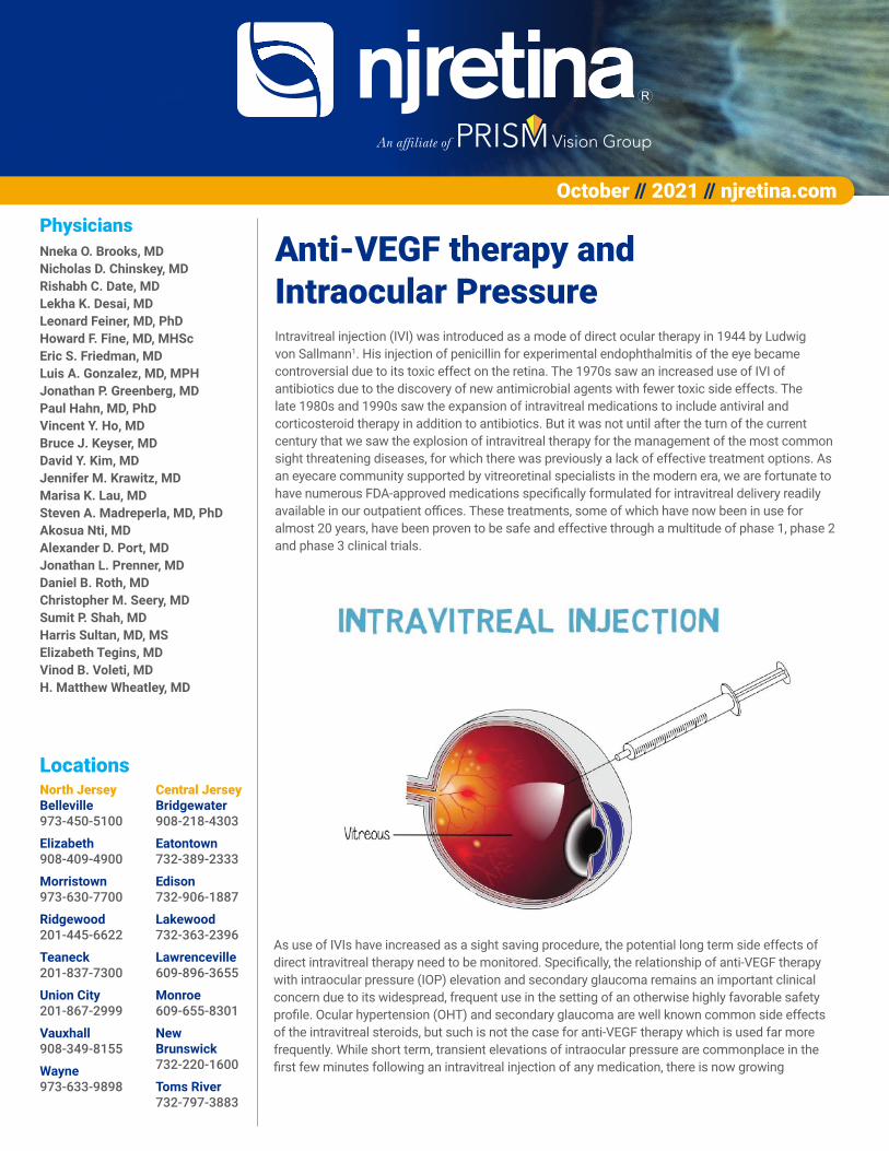
Anti-VEGF therapy and Intraocular PressureIntravitreal injection (IVI) was introduced as a mode of direct ocular therapy in 1944 by Ludwig von Sallmann1. His injection of penicillin for experimental endophthalmitis of the eye became controversial due to its toxic effect on the retina. The 1970s saw an increased use of IVI of antibiotics due to the discovery of new antimicrobial agents with fewer toxic side effects. The late 1980s and 1990s saw the expansion of intravitreal medications to include antiviral and corticosteroid therapy in addition to antibiotics. But it was not until after the turn of the current century that we saw the explosion of intravitreal therapy for the management of the most common sight threatening diseases, for which there was previously a lack of effective treatment options. As an eyecare community supported by vitreoretinal specialists in the modern era, we are fortunate to have numerous FDA-approved medications specifically formulated for intravitreal delivery readily available in our outpatient offices. These treatments, some of which have now been in use for almost 20 years, have been proven to be safe and effective through a multitude of phase 1, phase 2 and phase 3 clinical trials.
As use of IVIs have increased as a sight saving procedure, the potential long term side effects of direct intravitreal therapy need to be monitored. Specifically, the relationship of anti-VEGF therapy with intraocular pressure (IOP) elevation and secondary glaucoma remains an important clinical concern due to its widespread, frequent use in the setting of an otherwise highly favorable safety profile. Ocular hypertension (OHT) and secondary glaucoma are well known common side effects of the intravitreal steroids, but such is not the case for anti-VEGF therapy which is used far more frequently. While short term, transient elevations of intraocular pressure are commonplace in the first few minutes following an intravitreal injection of any medication, there is now growing
October // 2021 // njretina.com // 2
evidence that ongoing intravitreal therapy with anti-VEGF medication, which has restored and saved sight for otherwise blinding diseases such as neovascular macular degeneration, diabetic macular edema, and retinal vein occlusion, may in the long term lead to chronic elevation of the intraocular pressure in a subset of patients.
Immediate IOP changes following IVIs are directly related to the volume of medication injected. However, additional factors such as reflux, needle bore size and globe anatomy can also influence the immediate IOP elevation of the injected eye. Small-bore needles result in a higher immediate IOP elevation compared to large-bore needles after injections of anti-VEGF agents because of less reflux. Eyes with shorter axial lengths trend toward higher IOP elevations immediately after IVIs because of the relative increase in volume when injecting a smaller-sized eye. Scleral rigidity may also play a role in IOP spikes after IVIs. As scleral rigidity is positively correlated to increased age, older more rigid eyes may be more likely to have a spike in IOP.
For the reasons noted above, acute IOP elevations are common and somewhat expected. Kim et al showed that mean post-injection IOP is 44 mm Hg immediately after IVI2. These immediate IOP increases can occur with any medication and are typically transient. Pressure elevations were noted to quickly decline to less than 30 mm Hg in all patients within 15 minutes without intervention. Multiple studies have shown that IOP approaches normal levels in a majority of patients within 30-60 minutes of IVI.
The role of chronic anti-VEGF therapy in sustained elevation of IOP has been investigated through retrospective series, systematic reviews and meta-analyses. Real world data from The American Academy of Ophthalmology’s IRIS registry support the observation that a small subset of patients (2.6%) receiving intravitreal anti-VEGF therapy slowly develop chronic increases in their baseline IOP over time3. Mathalone et al reported a retrospective cohort of 201 eyes of patients receiving bevacizumab for neovascular AMD and showed sustained IOP elevations among 11% of the group4. The American Academy of Ophthalmology reviewed these two along with 12 other studies that reported on long term IOP elevation in patients who received anti-VEGF therapy in 20195. Eight of these fourteen studies reported that between 2.6% and 14.8% of patients had post-injection IOP elevations at 9 to 24 months of follow-up. The remaining six studies showed no change in IOP during a follow up period ranging from 1 to 36 months. Of note, the studies that reported IOP elevations had a higher mean and median number of patients and longer mean and median follow-up periods.
Potential mechanisms of chronic anti-VEGF IOP elevations include an inflammatory or a toxic drug effect on the trabecular meshwork and Schlemm’s canal. A mechanical blockade of the trabecular meshwork by protein aggregates or contaminant particles (such as silicone oil) have also been proposed. The nitric oxide pathway, known to relax smooth muscle and endothelial cells and thus increase anterior chamber aqueous outflow by decreasing trabeculocyte size and vasodilating Schlemm’s canal, has been
identified to be active in the anterior chamber through analyses of aqueous samples. Patients with open angle glaucoma have been shown to have lower levels of nitric oxide in their aqueous humor. Inhibition of nitric oxide synthesis by anti-VEGF drugs has been suggested as another means of IOP elevation6.
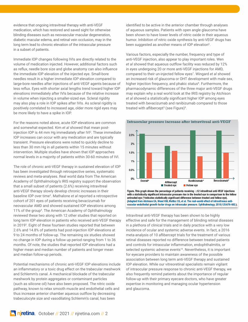
Various factors, especially the number, frequency and type of anti-VEGF injection, also appear to play important roles. Wen et al showed that aqueous outflow facility was reduced by 12% in eyes undergoing 20 or more anti-VEGF injections for AMD, compared to their un-injected fellow eyes7. Wingard et al showed an increased risk of glaucoma or OHT development with male sex, higher injection frequency, and phakic status8. Furthermore, the pharmacodynamic differences of the three major anti-VEGF drugs may explain why a real world look at the IRIS registry by Atchison et al showed a statistically significant higher IOP among eyes treated with bevacizumab and ranibizumab compared to those treated with aflibercept3 (see Figure)9.
Intravitreal anti-VEGF therapy has been shown to be highly effective and safe for the management of blinding retinal diseases in a plethora of clinical trials and in daily practice with a very low incidence of ocular and systemic adverse events. In fact, a 2016 meta-analysis of 10 aflibercept trials for the treatment of various retinal diseases reported no difference between treated patients and controls for intraocular inflammation, endophthalmitis, or selected systemic adverse events10. Nevertheless, it is important for eyecare providers to maintain awareness of the possible association between long term anti-VEGF therapy and sustained IOP elevation. While our vitreoretinal specialists remain vigilant of intraocular pressure response to chronic anti-VEGF therapy, we also frequently remind patients about the importance of regular follow-up with their primary eyecare doctors, who have greater expertise in monitoring and managing ocular hypertension and glaucoma.
October // 2021 // njretina.com // 3
References:1. Von Sallmann L, Meyer K , Di Grandi J. Experimental study of penicillin treatment of ectogenous infection of vitreous. Arch Ophthalmol. 1944;32:179
2. Kim JE, Mantravadi AV, Hur EY, Covert DJ. Short-term intraocular pressure changes immediately after intravitreal injections of anti-vascular endothelial growth factor agents. Am J Ophthalmol. 2008;146:930-934e.
3. Atchison EA, Wood KM, Mattoz CG, Barry CN, Lum F, MacCumber MW. The real-world effect of intravitreous anti-vascular endothelial growth factor drugs on intraocular pressure. Ophthalmology. 2018;125:676-682
4. Mathalone N, Arodi-Golan A, Sar S, Wolfson Y, Shalem M, Lavi I, Geyer O. Sustained elevation of intraocular pressure after intravitreal injections of bevacizumab in eyes with neovascular age-related macular degeneration. Graefes Arch Clin Exp Ophthalmol. 2012 Oct;250(10):1435-40.
5. Hoguet A, Chen PP, Junk AK, Mruthyunjaya P, Nouri-Mahdavi K, Radhakrishnan S, Takusagawa HL, Chen TC. The Effect of Anti-Vascular Endothelial Growth Factor Agents on Intraocular Pressure and Glaucoma: A Report by the American Academy of Ophthalmology. Ophthalmology. 2019 Apr;126(4):611-622.
6. Ricca AM, Morshedi RG, Wirostko BM. High intraocular pressure following anti-vascular endothelial growth factor therapy: proposed pathophysiology due to altered nitric oxide metabolism. J Ocul Pharmacol Ther. 2015;31:2-10.
7. Wen JC, Reina-Torres E, Sherwood JM, et al. Intravitreal anti-VEGF injections reduce aqueous outflow facility in patients with neovascular age-related macular degeneration. Invest Ophthalmol Vis Sci. 2017;58:1893–1898
8. Wingard JB, Delzell DA, Houlihan NV, Lin J, Gieser JP. Incidence of Glaucoma or Ocular Hypertension After Repeated Anti-Vascular Endothelial Growth Factor Injections for Macular Degeneration. Clin Ophthalmol. 2019 Dec 24;13:2563-2572.
9. Burget L, Maturi RK. IOP and anti-VEGF drugs: What we know so far. Retina Specialist. 2019 Nov 26
10. Kitchens JW, Do DV, Boyer DS, et al. Comprehensive review of ocular and systemic safety events with intravitreal aflibercept injection in randomized controlled trials. Ophthalmology. 2016;123: 1511–1520.
11. Wu H, Chen TC. The effects of intravitreal ophthalmic medications on intraocular pressure. Semin Ophthalmol. 2009 Mar-Apr;24(2):100-5.
12. Kähkönen M, Tuuminen R, Aaltonen V. Long-term effects of intravitreal bevacizumab and aflibercept on intraocular pressure in wet age-related macular degeneration. BMC Ophthalmol. 2021 Aug 28;21(1):312.
NJRetina Welcomes Our Newest Physician
What’s your philosophy of care?I believe that every patient should be seen as an individual and that their personal situation should be understood and empathized with. It is important to treat not only the patient’s eyes but the person as a whole. I feel that it is important to make sure a patient understands why I recommend what I do and to always give them the opportunity to clarify or ask questions. Lastly, I try to emphasize preventative care where appropriate.
What made you choose the Retina field as your specialty area?I love the field of retina. It provides the opportunity to make a real and lasting difference in peoples lives. From an interest perspective, I find the structure and physiology of the retina to be amazing and I enjoy the cutting-edge technology that the field employs and spearheads. Furthermore, I am meticulous and the complex and delicate surgeries inherent to the field suit my personality perfectly.
Why did you choose NJRetina?I was looking for a larger practice where the physicians form a community and where senior doctors act as mentors to the more junior ones - from what I have witnessed, NJRetina exemplifies these qualities. Furthermore, I wanted to be near New York City as my parents and both brothers live in and around the city.
What are some of your personal interests?I have two boys, aged four and one, and two Australian Sheppard puppies. I enjoy spending time with my family as well as being outside, going for walks and to parks. In winter, I like to ski.
Are you fluent in any language aside from English?I grew up in South Africa and can speak some Afrikaans, although I intend to learn to speak Spanish.
Jonathan P. Greenberg, MD

At the forefront of clinical researchNJRetina continuously conducts clinical trials at key locations. Our clinical research coordinators will be happy to discuss the inclusion/ exclusion criteria or any other aspect of these studies with you or your patients. If you have any questions, please feel free to contact:
Joe Martinez - Teaneck: 201-837-7300, [email protected] Elizabeth Silverio - Teaneck: [email protected] Joseph Portelli - Teaneck: [email protected] Dina Christodoro - Toms River: 732-797-3984 and Edison: 732-906-1887
October // 2021 // njretina.com // 4
Dry AMD Vauxhall GTSCOPE: A Study of Disease Progression in Genetically Defined Subjects With Geographic Atrophy Secondary to Age-Related Macular Degeneration
Teaneck and Toms River Gallego: A phase II, multicenter, randomized, single-masked, sham-controlled study to assess safety, tolerability, and efficacy of intravitreal injections of FHTR2163 in patients with geographic atrophy secondary to age-related macular degeneration (Gallego)
Diabetic Macular Edema (DME) Teaneck Gleam: A prospective, randomized, double-masked, active comparator-controlled, multi-center, two- arm, phase 3 study to evaluate the efficacy and safety of intravitreal KSI-301 compared with intravitreal aflibercept in participants with visual impairment secondary to treatment- naive diabetic macular edema.
Diabetic Retinopathy Teaneck Pavilion: A Phase III, Multicenter, Randomized Study of the Efficacy, Safety, and Pharmacokinetics of the Port Delivery System with Ranibizumab in Patients with Diabetic Retinopathy
TeaneckAltitude: A Phase 2, Randomized, Dose-escalation, Observation-controlled Study to Evaluate the Efficacy, Safety, and Tolerability of RGX-314 Gene Therapy Delivered via One or Two Suprachoroidal Space (SCS) Injections in Participants with Diabetic Retinopathy (DR) Without Center Involved-Diabetic Macular Edema (CI-DME) (ALTITUDE)
Retinal Vein Occlusion Balaton: A prospective, randomized, double-masked, active comparator-controlled, multi-center, two-arm, phase 3 study to evaluate the efficacy and safety of intravitreal KSI-301 compared with intravitreal aflibercept in participants with visual impairment due to treatment- naive macular edema secondary to retinal vein occlusion (RVO) – Teaneck
Beacon: A Phase III, Multicenter, Randomized, Double-Masked, Active Comparator-controlled Study To Evaluate The Efficacy And Safety Of Faricimab In Patients With Macular Edema Secondary To Branch Retinal Vein Occlusion – Toms River
Enrolling Studies:
To read past issues of our newsletter, visit njretina.com.
A 59-year old caucasian female presented with worsening photopsias and a visual field defect in the left eye.
Examination revealed 20/25 vision with a prominent nasal visual field defect. Anterior segment was normal.
Funduscopic examination revealed a large choroidal mass with intraretinal and subretinal hemorrhage. B-scan ultrasonography demonstrated a large mushroom-shaped choroidal mass consistent with a large choroidal melanoma. The patient was treated with plaque radiotherapy.
Melanoma Case Study