Embed Size (px)
Citation preview

Pi-Rads version 2
Masoom Haider, MD, FRCP(C)Professor of Radiology, University of TorontoClinician Scientist, Ontario Institute of Cancer ResearchSenior Scientist, Sunnybrook Research InstituteChief, Dept of Medical ImagingSunnybrook Health Sciences [email protected]
www.mybodymri.com

Addressing the Sampling Problem
• Variability is introduced by sampling error making it much hard to get a handle on tumor behavior in population studies
JAMA 2015 n = 1003• In men (mostly with a prior negative
systematic biopsy) MR/ultrasound fusion biopsy, c/w systematic biopsy had increased detection of Gleason >=4+3, (+30%) and decreased detection of low-risk prostate cancer (-17%)
• Prostatectomy cohort Sens 77% vs 53% Spec 68 vs 66% (Gleason 3+4 in >20% of prostate)October 22, 2016
Siddiqui MM, et al. Comparison of MR/ultrasound fusion-guided biopsy with ultrasound-guided biopsy for the diagnosis of prostate cancer. Jama. Jan 27 2015;313(4):390-7.

3 October 22, 2016
StandardizationPi-Rads v2.0 – 5 point scale
acr.org
4
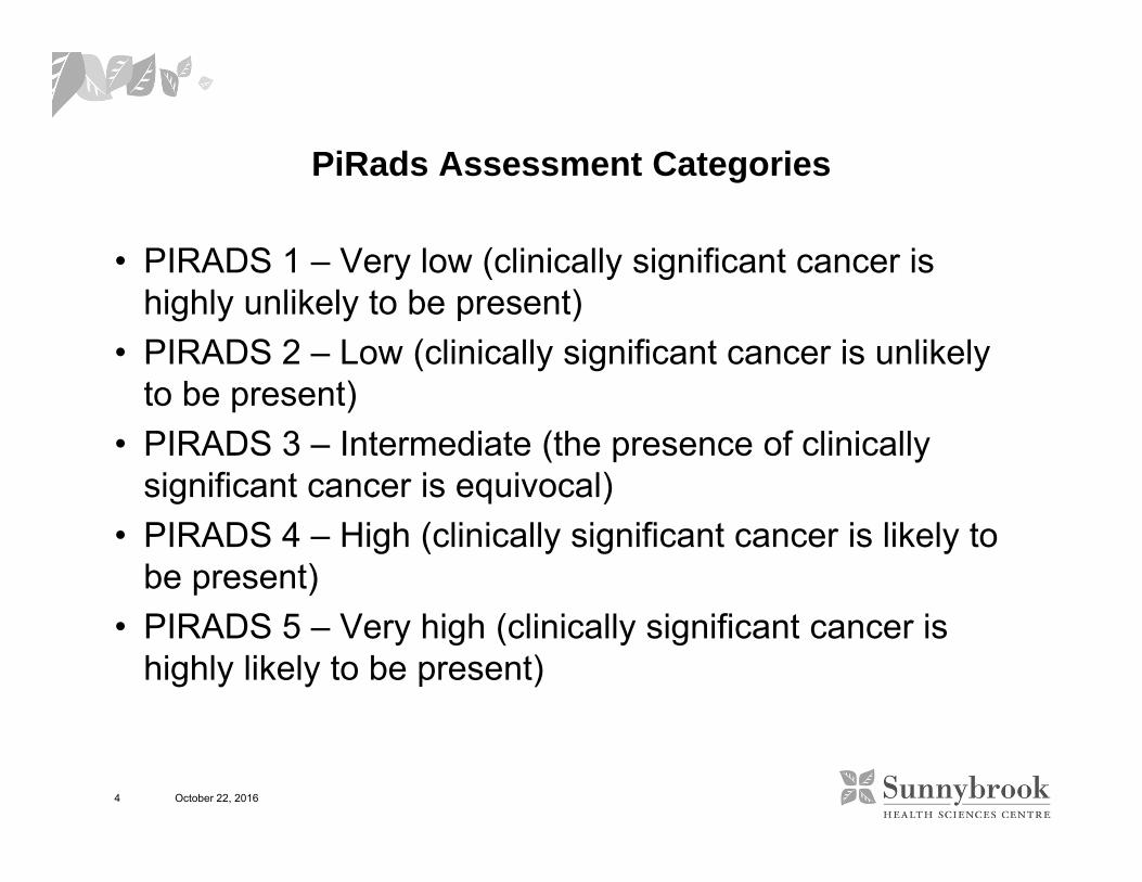
PiRads Assessment Categories
• PIRADS 1 – Very low (clinically significant cancer is highly unlikely to be present)
• PIRADS 2 – Low (clinically significant cancer is unlikely to be present)
• PIRADS 3 – Intermediate (the presence of clinically significant cancer is equivocal)
• PIRADS 4 – High (clinically significant cancer is likely to be present)
• PIRADS 5 – Very high (clinically significant cancer is highly likely to be present)
October 22, 2016

5
Pirads 1 or 2 means no CS cancer on BiopsyReduction in need for biopsyPirads 3 means low risk of CS cancerPirads 4/5 means much higher chance of CS cancer
October 22, 2016

6
Coil Combinations
1.5T 3T
No endorectal Thinner patients, current MRI, cardiac coil
Cardiac coil in thinner patients
Endorectal w/ Surface Thinner patients, cardiac coil Thinner patients, cardiac coil
Endorectal w/o surface Fatter patients Fatter patients partial phase FOV
October 22, 2016
3T 2NEX, ER only0.4x0.4x3mm, 18x9cm, 259s

7
Clinically Significant Cancer
Gleason score >= 7
And/or
Volume > 0.5cc
And/or
Extraprostatic extension (EPE)
October 22, 2016

8 October 22, 2016

9
Normal Anatomy(McNeal)
• 4 distinct zones– Peripheral Zone (PZ)– Central Zone (CZ)
• Lateral to Ejaculatory ducts towards base– Transition Zone (TZ)
• BPH nodules– Anterior Fibromuscular Stroma (AFMS)
• Non glandular tissues contiguous with the detrusor
October 22, 2016

10
Interpretation SchemeSimplifications in v 2
• Hierarchical scheme• In PZ DWI is dominant (PiRads = DWI score)• In TZ T2 is dominant (PiRads = T2 score)• The DCE interpretation has been reduced to positive or
negative (binary) and only has influence in PZ tumors that are equivocal on DWI (pushing Pi-Rads 3 to a 4 in some cases)– DCE assessment does not require curve analysis or typing
• Pirads 4 is the same as 5 but size is >15mm or ECE• B-values increased to >=1400 s/mm2
October 22, 2016

11

12
Overall Score for a Lesion
October 22, 2016
DWI T2 DCE Overall
1 Any Any 1
2 Any Any 2
3 Any- 3
+ 4
4 Any Any 4
5 Any Any 5
T2 DWI DCE Overall
1 Any Any 1
2 Any Any 2
3≤4 Any 3
5 Any 4
4 Any Any 4
5 Any Any 5
PZ TZ

13
DWI – PZ and TZ
1 No abnormality (i.e. normal) on ADC and high b‐value DWI
2 Indistinct hypointense on ADC
3 Focal mildly/moderately hypointense on ADC and isointense/mildly hyperintense on high b‐value DWI.
4Focal markedly hypointense on ADC and markedly hyperintense on high b‐value DWI; <1.5cm in greatest dimension
5 Same as 4 but ≥1.5cm in greatest dimension or definite extraprostatic extension/invasive behavior

14
Hambrock T et al. Radiology 2011
ADCCorrelates with Gleason Grade
Az =0.9 >G6

15
Technique – B-Values?
• Lower 0, 50, 100 s/mm2
• Higher 1400-2000s/mm2 best
• Minimum 2 b-valsbut can do more
• Can extrapolate from b values done less than 1000
October 22, 2016
0
0.5
1
1.5
2
2.5
3
0 500 1000 1500
Ln(S
I)
Flow driven
Slow Diffusion (ECF)
CalculatedDiffusion

16
DCE – PZ and TZ
-
• No early enhancementOR• diffuse enhancement not corresponding to a focal
finding on T2 and/or DWIOR• focal enhancement corresponding to a lesion
demonstrating features of BPH on T2WI
+focal, and earlier than or contemporaneously with enhancement of adjacent normal prostatic tissues andcorresponds to suspicious finding on T2W and/or DWI

17
Kinetic Analysis Models
C(t)
Time (min)
ve = 50%
Ktrans = 0.5 min-1Ktrans = 1.0 min-1Ktrans = 1.5 min-1Ktrans = 2.0 min-1Ktrans = 2.5 min-1Ktrans = 3.0 min-1Ktrans = 2.0 min-1
ve = 20%ve = 40%ve = 60%ve = 80%

18
DCE
• In plane dimension: ≤2mm X ≤2mm• Temporal resolution: ≤15sec (<7sec is preferred)• Total observation: >2min• Dose: 0.1mmol/kg standard GBCA or equivalent high relaxivity
GBCA• Injection rate: 2‐3cc/sec starting with continuous image data
acquisition (should• be the same for all exams)• 3D seq, FS typically off• If doing quantitation, modelling then follow manufacturers
suggestions– Multi flip angle T1 mapping
• Subtraction images if interpreting raw images• Watch for motion
October 22, 2016

19
T2 TZ
1 Homogeneous intermediate signal intensity(normal)
2 Circumscribed hypointense or heterogeneous encapsulated nodule(s) (BPH)
3Heterogeneous signal intensity with obscured margins. Includes others that do not qualify as 2, 4, or 5
4Lenticular or non‐circumscribed, homogeneous, moderately hypointense, and <1.5 cm in greatest dimension
5 Same as 4 but ≥1.5cm in greatest dimension or definite extraprostatic extension/invasive behavior
20
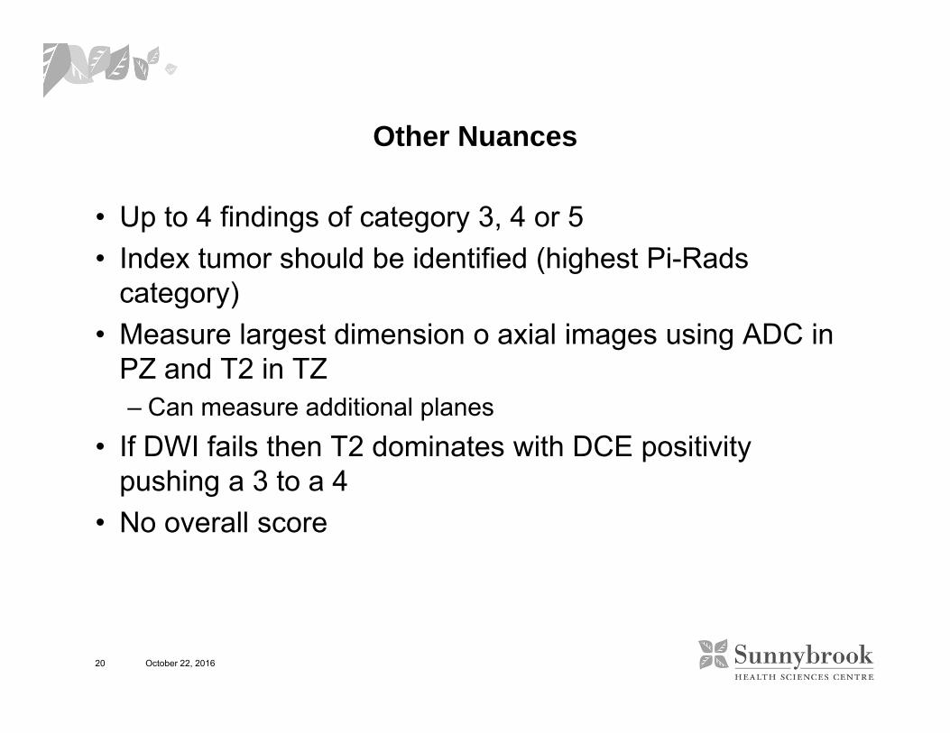
Other Nuances
• Up to 4 findings of category 3, 4 or 5• Index tumor should be identified (highest Pi-Rads
category)• Measure largest dimension o axial images using ADC in
PZ and T2 in TZ– Can measure additional planes
• If DWI fails then T2 dominates with DCE positivity pushing a 3 to a 4
• No overall score
October 22, 2016

21
Making Recommendations – Not in PiRads 1 or 2A work in progress
• PIRADS 1 or 2 – Biopsy* if high risk else followup MRI**• PIRADS 3- Biopsy* if intermediate or high risk otherwise
followup MRI**• PIRADS 4 & 5 – Biopsy*
*Biopsy– targets (if any) + systematic if no systematic in the last 2 years– targets only if systematic biopsy in the last two years
**Followup MRI– No consensus q1 or q2 years
• Risk profile– Being debated
October 22, 2016