Embed Size (px)
Citation preview
WELCOME
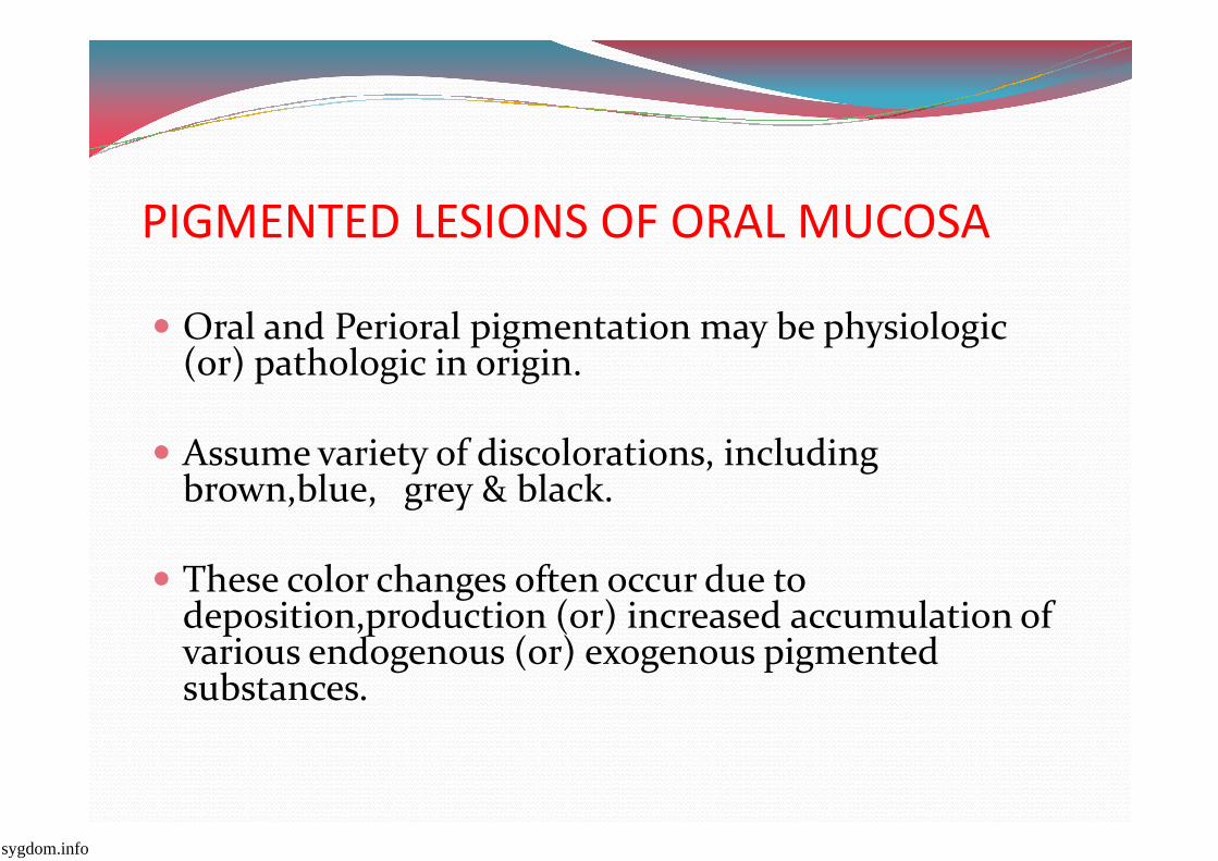
PIGMENTED LESIONS OF ORAL MUCOSA
� Oral and Perioral pigmentation may be physiologic (or) pathologic in origin.
Assume variety of discolorations, including � Assume variety of discolorations, including brown,blue, grey & black.
� These color changes often occur due to deposition,production (or) increased accumulation of various endogenous (or) exogenous pigmented substances.
sygdom.info
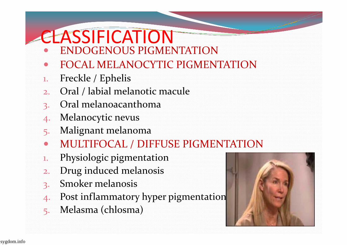
CLASSIFICATION� ENDOGENOUS PIGMENTATION
� FOCAL MELANOCYTIC PIGMENTATION.. Freckle / Ephelis
0. Oral / labial melanotic macule
1. Oral melanoacanthoma
2. Melanocytic nevus2. Melanocytic nevus
3. Malignant melanoma
� MULTIFOCAL / DIFFUSE PIGMENTATION.. Physiologic pigmentation
0. Drug induced melanosis
1. Smoker melanosis
2. Post inflammatory hyper pigmentation
3. Melasma (chlosma)
sygdom.info
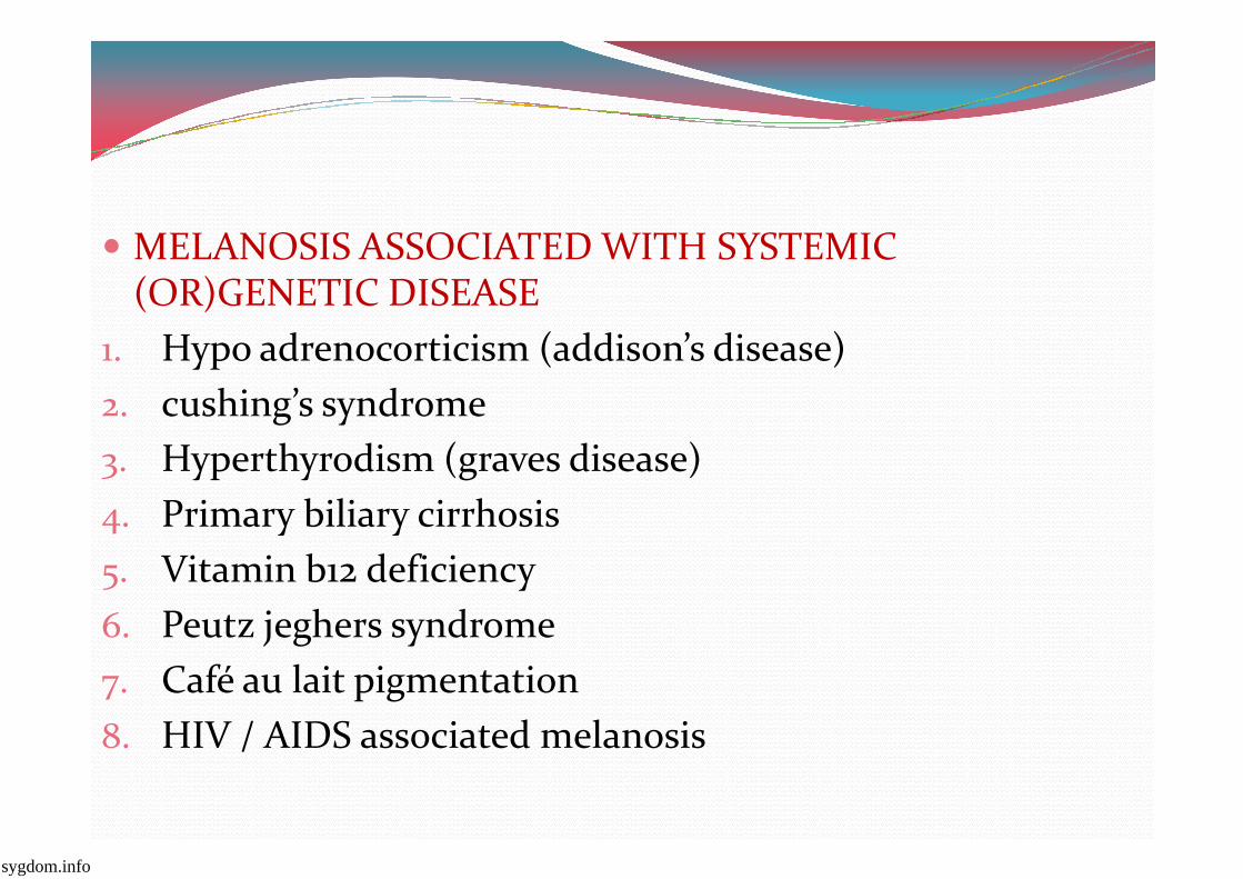
� MELANOSIS ASSOCIATED WITH SYSTEMIC (OR)GENETIC DISEASE
.. Hypo adrenocorticism (addison’s disease)
0. cushing’s syndrome
1. Hyperthyrodism (graves disease)1. Hyperthyrodism (graves disease)
2. Primary biliary cirrhosis
3. Vitamin b.0 deficiency
9. Peutz jeghers syndrome
<. Café au lait pigmentation
>. HIV / AIDS associated melanosis
sygdom.info
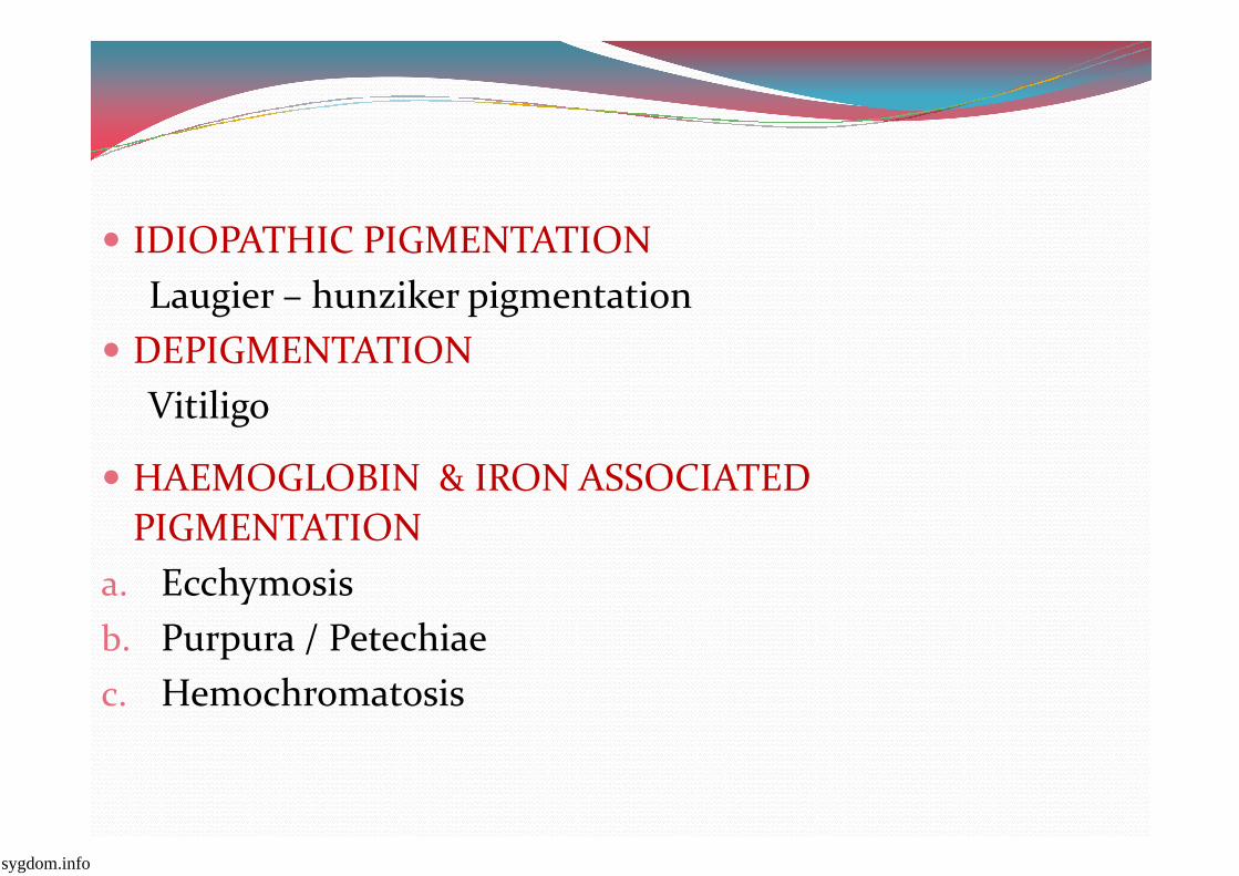
� IDIOPATHIC PIGMENTATION
Laugier – hunziker pigmentation
� DEPIGMENTATION
Vitiligo
� HAEMOGLOBIN & IRON ASSOCIATED
PIGMENTATION
a. Ecchymosis
b. Purpura / Petechiae
c. Hemochromatosis
sygdom.info
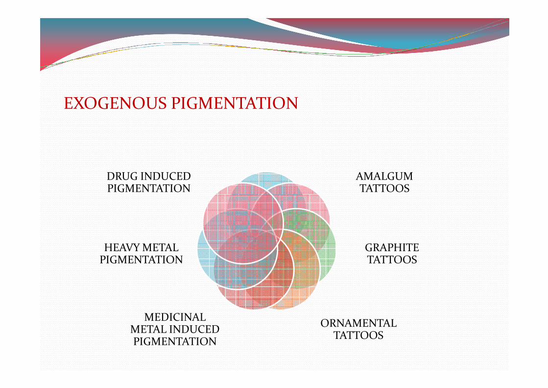
EXOGENOUS PIGMENTATION
AMALGUM TATTOOS
DRUG INDUCED PIGMENTATION TATTOOS
GRAPHITE TATTOOS
ORNAMENTAL TATTOOS
MEDICINAL METAL INDUCED PIGMENTATION
HEAVY METAL PIGMENTATION
PIGMENTATION
�
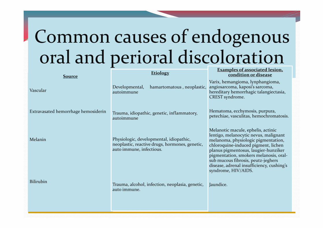
Common causes of endogenous oral and perioral discoloration
Source
Vascular
Extravasated hemorrhage hemosiderin
Etiology
Developmental, hamartomatous , neoplastic, autoimmune
Trauma, idiopathic, genetic, inflammatory,
Examples of associated lesion, condition or disease
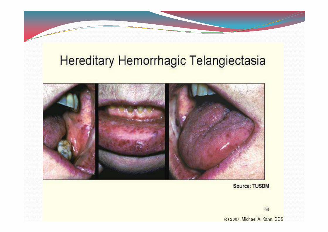
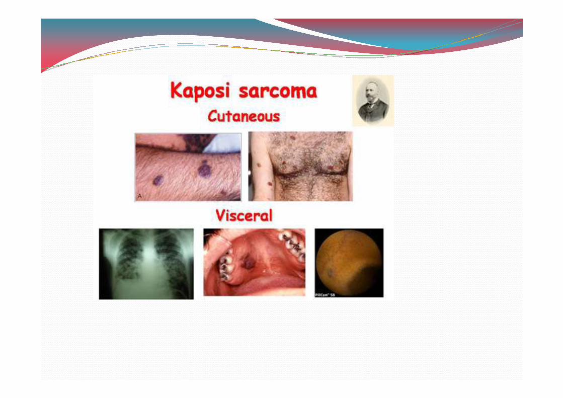
Varix, hemangioma, lynphangioma, angiosarcoma, kaposi’s sarcoma, hereditary hemorrhagic talangiectasia, CREST syndrome.
Hematoma, ecchymosis, purpura, petechiae, vasculitas, hemochromatosis.
Melanin
Bilirubin
Trauma, idiopathic, genetic, inflammatory, autoimmune
Physiologic, developmental, idiopathic, neoplastic, reactive drugs, hormones, genetic, auto immune, infectious.
Trauma, alcohol, infection, neoplasia, genetic, auto immune.
petechiae, vasculitas, hemochromatosis.
Melanotic macule, ephelis, actinic lentigo, melanocytic nevus, malignant melanoma, physiologic pigmentation, chloroquine-induced pigment, lichen planus pigmentosus, laugier-hunziker pigmentation, smokers melanosis, oral-sub mucous fibrosis, peutz-jeghers disease, adrenal insufficiency, cushing’s syndrome, HIV/AIDS.
Jaundice.
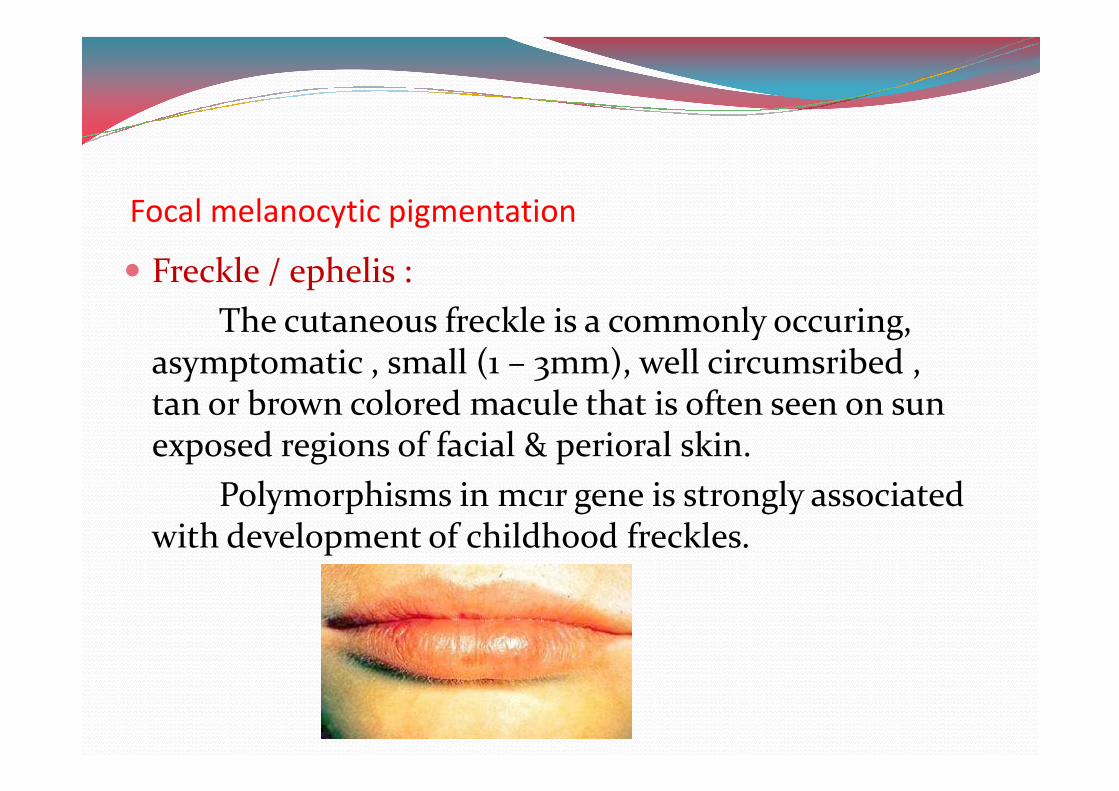
Focal melanocytic pigmentation
� Freckle / ephelis :
The cutaneous freckle is a commonly occuring, asymptomatic , small (. – 1mm), well circumsribed , tan or brown colored macule that is often seen on sun tan or brown colored macule that is often seen on sun exposed regions of facial & perioral skin.
Polymorphisms in mc.r gene is strongly associated with development of childhood freckles.
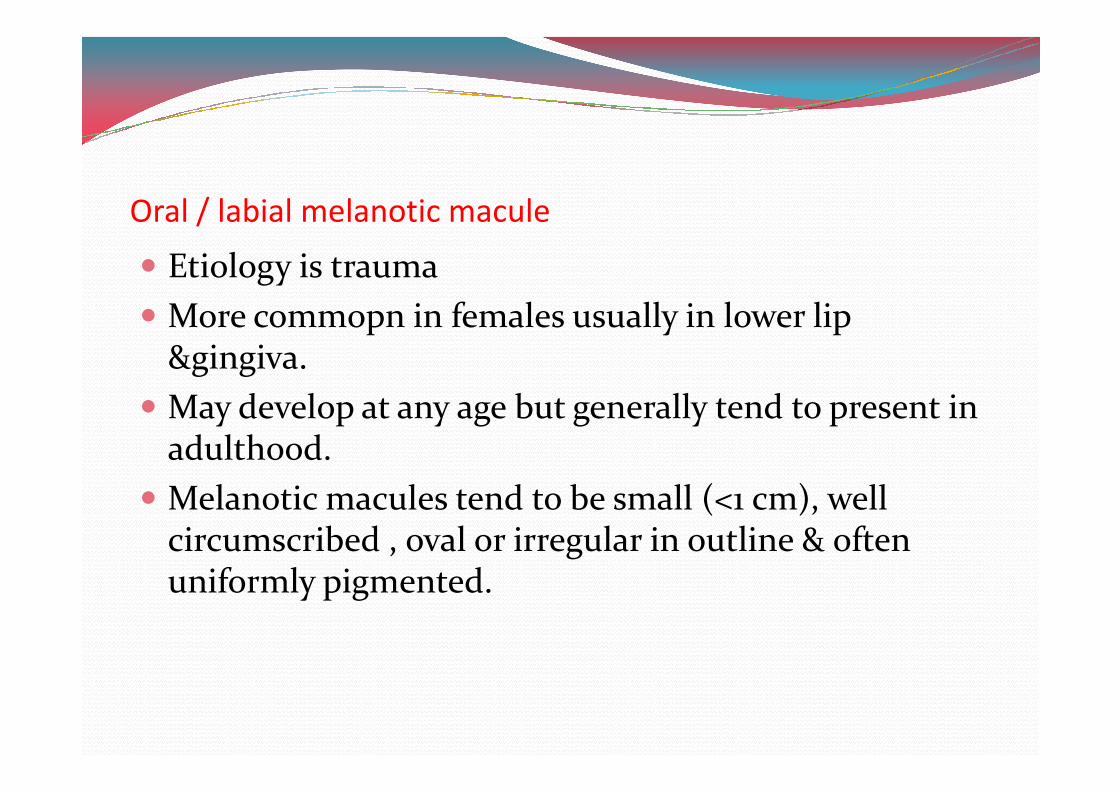
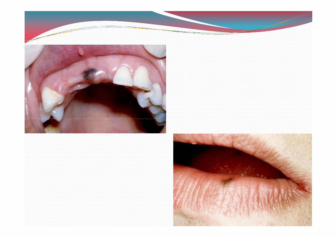
Oral / labial melanotic macule
� Etiology is trauma
� More commopn in females usually in lower lip &gingiva.
� May develop at any age but generally tend to present in � May develop at any age but generally tend to present in adulthood.
� Melanotic macules tend to be small (<. cm), well circumscribed , oval or irregular in outline & often uniformly pigmented.
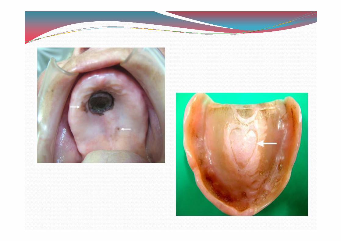
Oral melanoacanthoma
� Etiology is acute trauma or a history of chronic irritation
� Rapidly enlarging , ill defined , darkly pigmented � Rapidly enlarging , ill defined , darkly pigmented macular or plague like lesions.
� Buccal mucosa most common site of occurrence
� Dermatosis papulosa nigra is relatively common facial condition in older black females & represents multiple pigmented seborrheic keratoses
� Treatment is source of irritation should be removed
Melanocytic nevus
� Effect of sun exposure reconized in development of cutaneous nevi.
� Recent study shows GH% of dermal melanocytic nevi exhibit somatic activating mutations in BRAF exhibit somatic activating mutations in BRAF oncogene.
� Lesions are usually asymtomatic & often present as a small (<.cm) , solitary , brown or blue , well circumscribed nodule or macule.
� Oral nevi present in hard palate is the most common site , followed by buccal & labial mucosa & gingiva.
� Treatment of melanotic nevi is complete but conservative surgical excision is the treatment of choice for oral lesions.
� Rare recurrence
� Laser & intense pulse light therapies have been used successfully
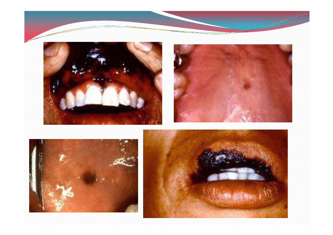
Malignant melanoma
� Etiology is episodes of acute sun exposure , especially at young age , immunosuppresion
� Exhibit mutation in the BRAF , HRAS & NRAS proto oncogenes.oncogenes.
� Palate represents most common site & next comes the maxillary gingiva.
� They are macular , plague like or mass forming , well circumscribed or irregular & exhibit focal or diffuse areas of brown , blue or black pigmentation.
� Treatment is ablative surgery with wide margins .
� Adjuvant radiotherapy is necessary.
� Recent development is antitumor vaccine adjuvant interferon alpha – 0B therapy is used to treat primary cutaneous melanomas >2mm in thickness.
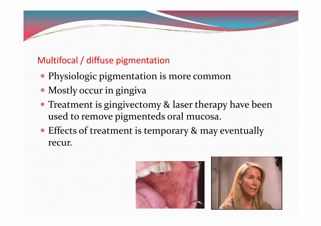
Multifocal / diffuse pigmentation
� Physiologic pigmentation is more common
� Mostly occur in gingiva
� Treatment is gingivectomy & laser therapy have been used to remove pigmenteds oral mucosa.used to remove pigmenteds oral mucosa.
� Effects of treatment is temporary & may eventually recur.
Drug induced melanosis
Chief drugs include
a) Minocycline – tetracycline derivative
b) Antimalarials include chloroquine , hydroxy chloroquine , quinacrine .chloroquine , quinacrine .
c) Phenothiazines such as chlorpromaqzine
d) Oral contraceptives
e) Cytotoxic medications such as cyclophosphamide & busulfan
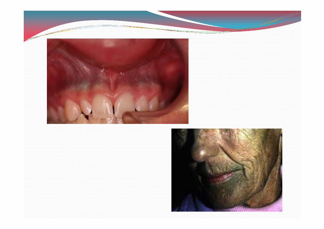
� Clinically the pigment can be diffuse yet localised to one mucosal surface , often hard palate
� Lesions are flat & without any evidence of nodularity or swelling.or swelling.
� Diagnosis & treatment is discolaration tends to fade within a few months after the drug is discontinued.
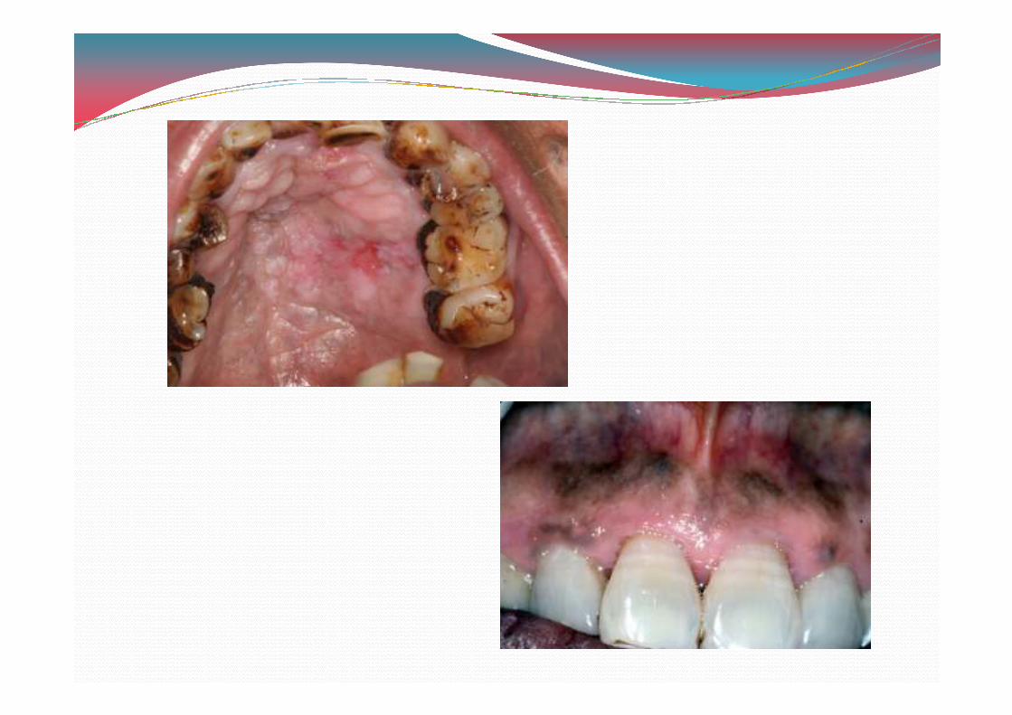
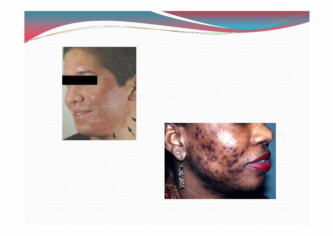
Smokers melanosis
� Diffuse melanosis of anterior facial maxillary & mandibular gingivae , buccal mucosa , lateral tongue , palate & floor of mouth occasionally seen among cigarette smokers.cigarette smokers.
� Pigmented areas are brown , flat & irregular.
� Melanin synthesis is stimuylated by tobbaco smoke products.
� Heat of smoke may stimulate pigmentation.
Post inflammatory pigmentation
� Focal or diffuse pigmentation in areas that were subjected to previous injury or inflammation.
� The mucosa overlying a non melanocytic malignancy may become pigmented.
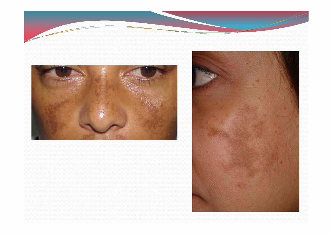
Melasma / chloasma
� Acquired symmetric melanosis that typically develops on sun exposed areas of skin & frequently on face.
� Forehead , cheeks , upperlips & chin are most � Forehead , cheeks , upperlips & chin are most commonly affected areas.
� Melasma has been used to describe any form of generalised facial hyperpigmentation including those related to post inflammatory changes & medication use.
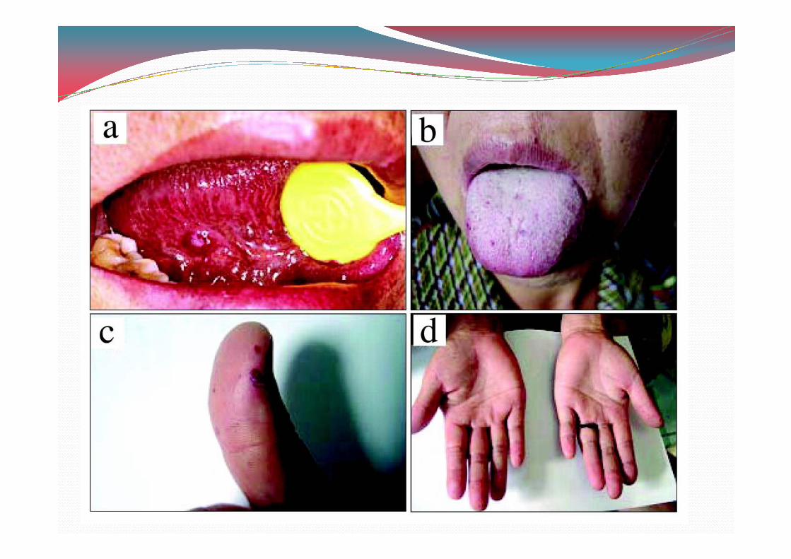
Melanosis associated with systemic or genetic disease
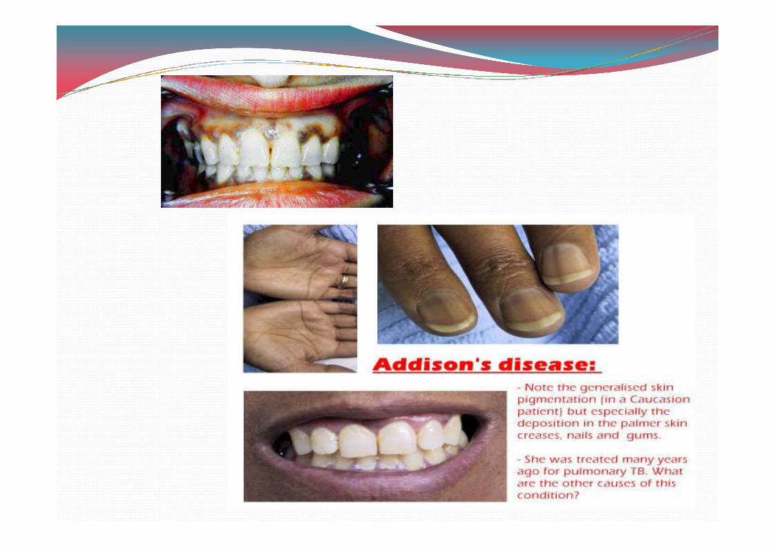
� Hypoadrenocortism:
� As steroid levels decrease , there is a compensatory activity by ACTH secretion , but if persists , the serum levels of alpha melanocyte stimulating hormone also levels of alpha melanocyte stimulating hormone also increase.
� Mucocutaneous hyperpigmentation
� Generalised bronzing of skin &diffuse but patchy melanosis of oral mucosa.
� Treatment is exogenous steroid replacement therapy.
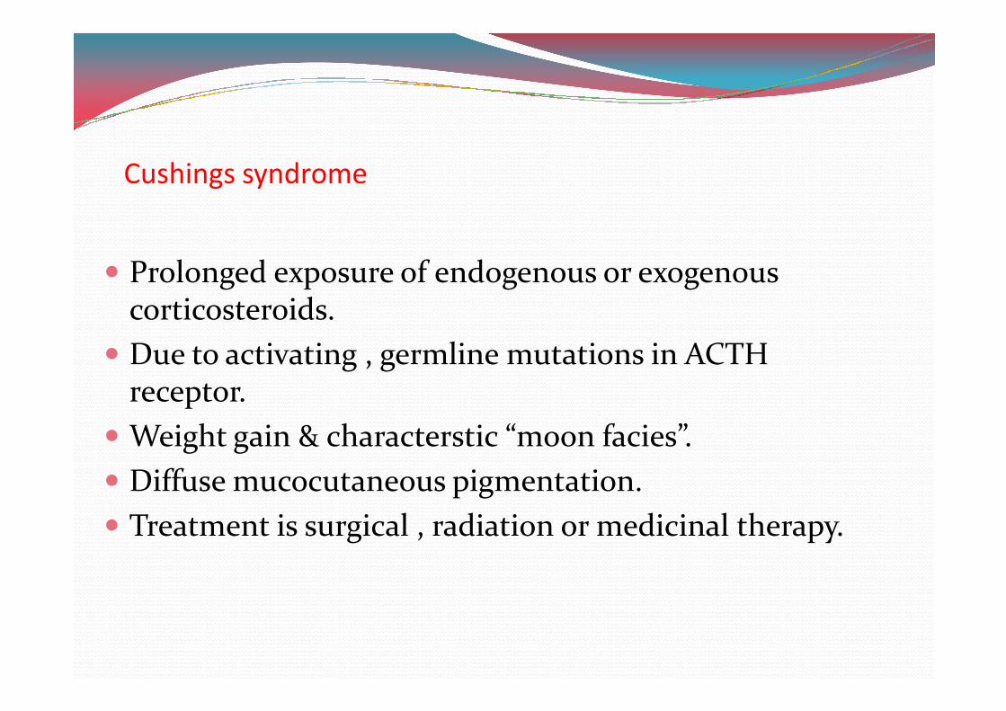
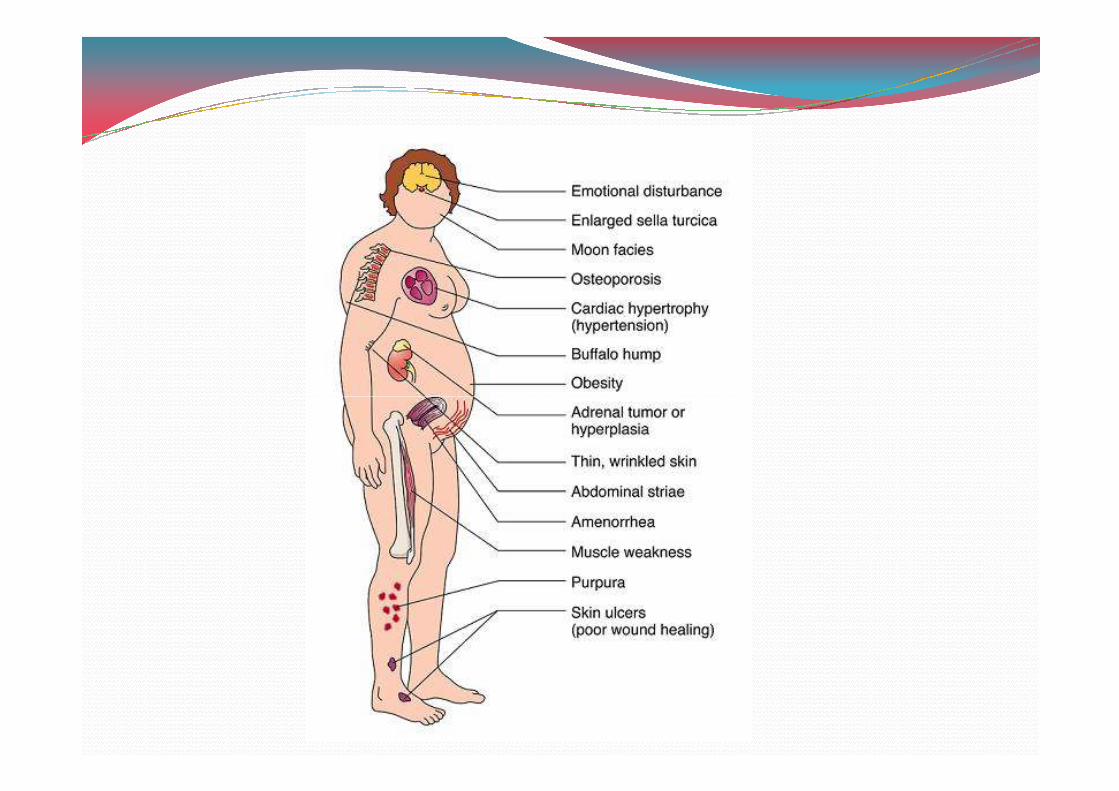
Cushings syndrome
� Prolonged exposure of endogenous or exogenous corticosteroids.
� Due to activating , germline mutations in ACTH � Due to activating , germline mutations in ACTH receptor.
� Weight gain & characterstic “moon facies”.
� Diffuse mucocutaneous pigmentation.
� Treatment is surgical , radiation or medicinal therapy.
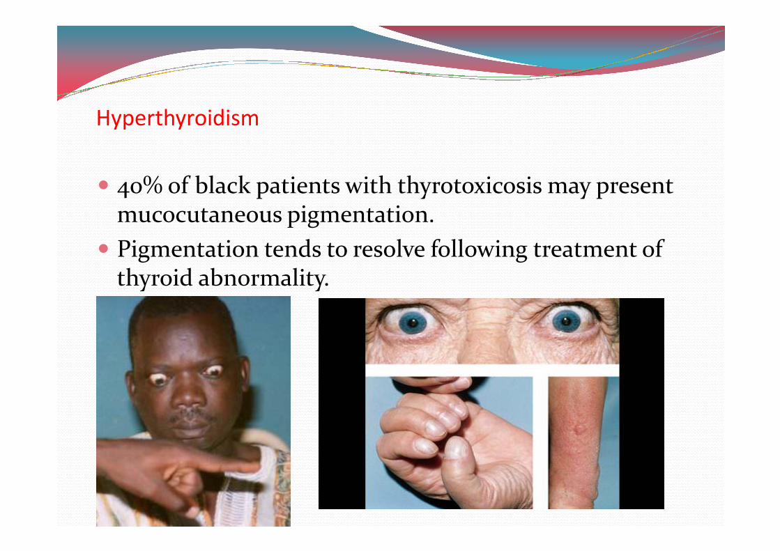
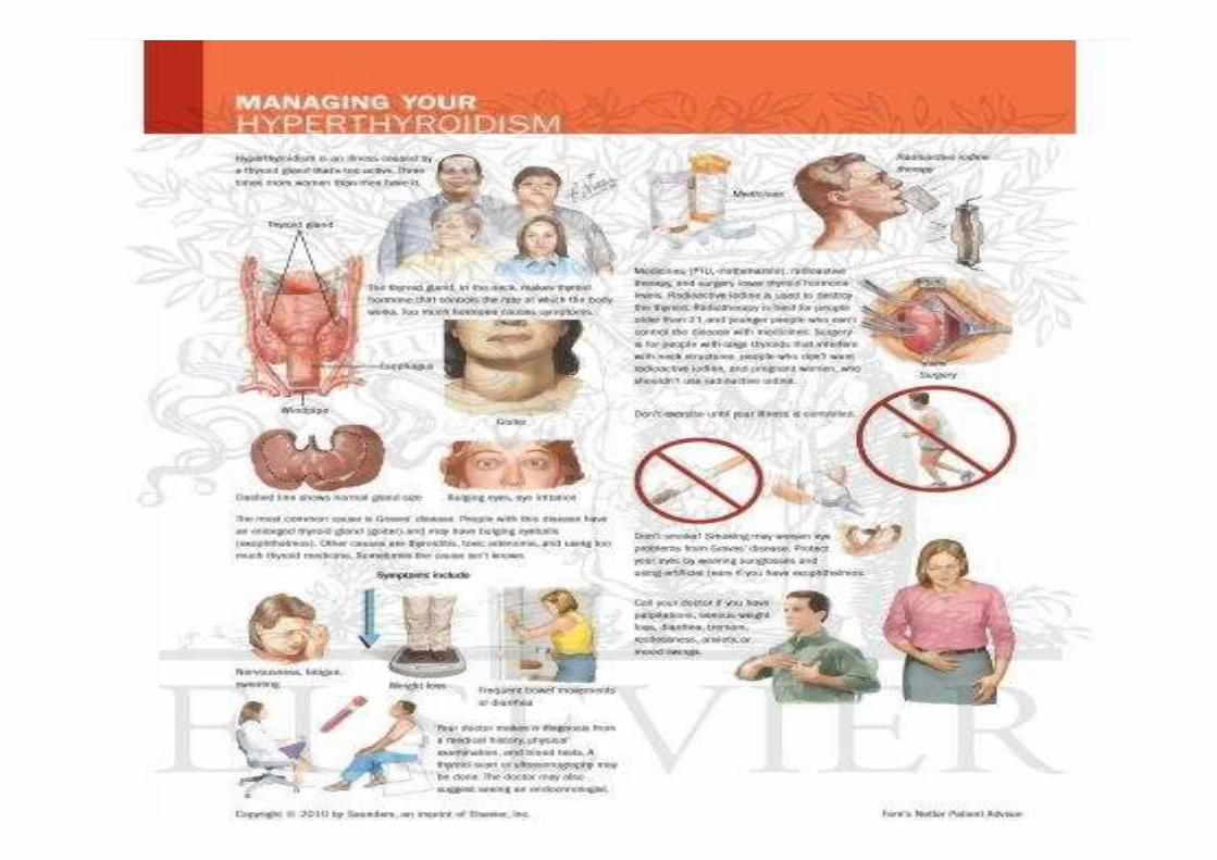
Hyperthyroidism
� 2H% of black patients with thyrotoxicosis may present mucocutaneous pigmentation.
� Pigmentation tends to resolve following treatment of thyroid abnormality.thyroid abnormality.
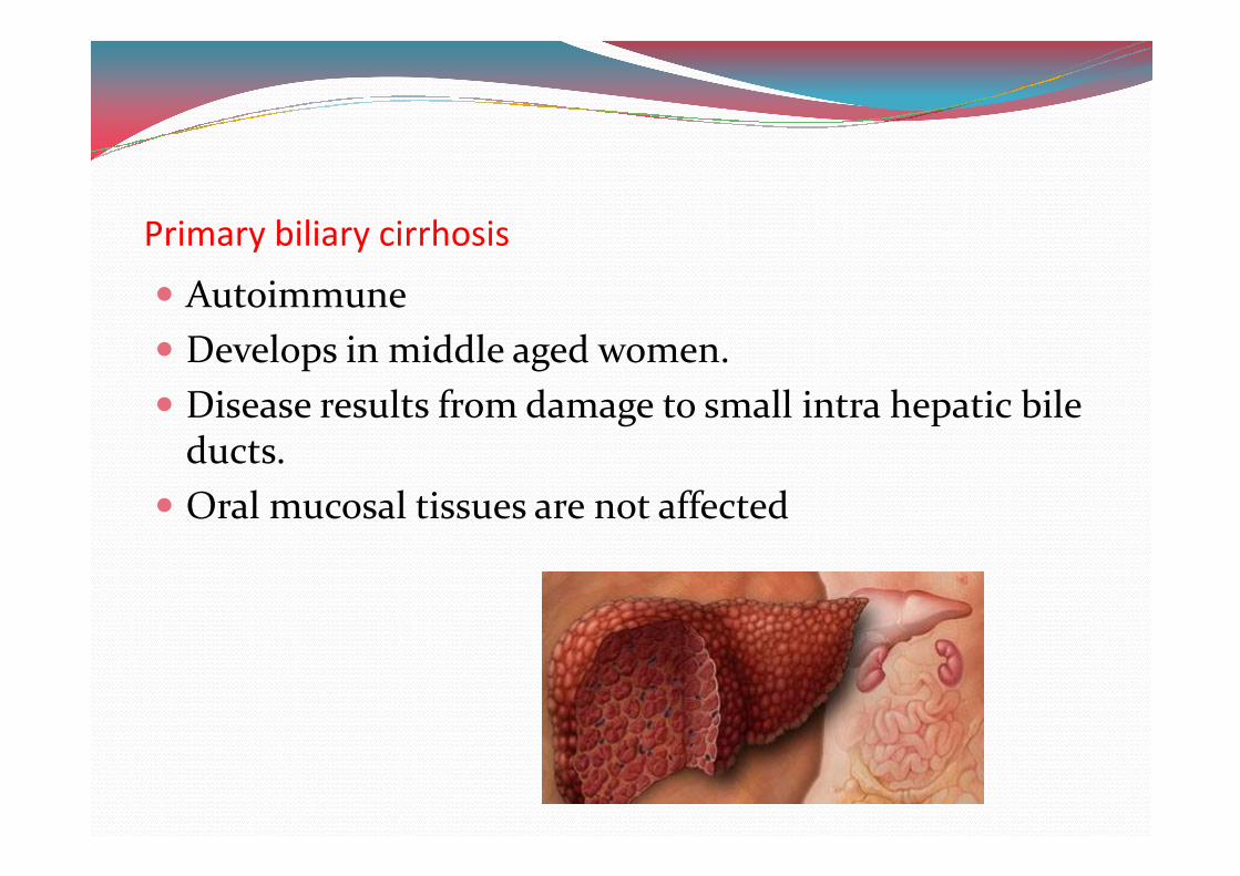
Primary biliary cirrhosis
� Autoimmune
� Develops in middle aged women.
� Disease results from damage to small intra hepatic bile ducts.ducts.
� Oral mucosal tissues are not affected
Vitamin B,- deficiency
� Generalised burning sensation & erythema & atrophy of the mucosal tissue.
� Pigmentation resolves followimg vitamin B.0 levels.
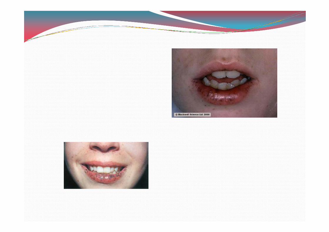
Peutz – jeghers syndrome
� Autosomal dominant disease associated with mutations in STK../LKB. tumor suppresor gene.
� Intestinal polyposis , cancer susceptibility & multiple , small , pigmented macules of lip , perioral skin , hand small , pigmented macules of lip , perioral skin , hand & feet.
� Resemble ephelides usually <H.3mm in diameter.
� Lesion may develop on anterior tongue , buccal & labial mucosa.
� Lip & perioral pigmentation is higly distinctive
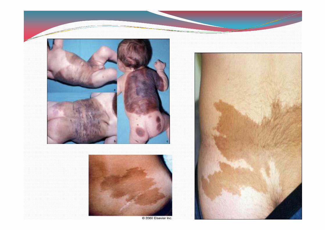
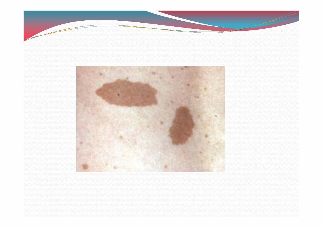
Café –au – lait pigmentation
� Identified in number of different genetic disorders include
a) Neuro fibromatous type .
b) Mccune – albright syndromeb) Mccune – albright syndrome
c) Noonans syndrome
� Present as tan or brown colored , irregularly shaped macules of variable size.
� Occur anywhere on skin , oral macular pigmentation have been reported.
HIV / AIDS
� Pigmentation may be related to intake of various medications , anti fungal & anti retoviral drugs.
� May also occur due to adreno cortical destruction by � May also occur due to adreno cortical destruction by virulent infectious organisms.
� Significant correlation between mucocutaneous pigment & CD2 counts / micro litre lessthan or equal to 0HH.
� Buccal mucosa is most affected site ,gingiva , palate &tongue involved.
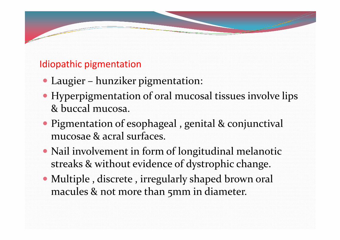
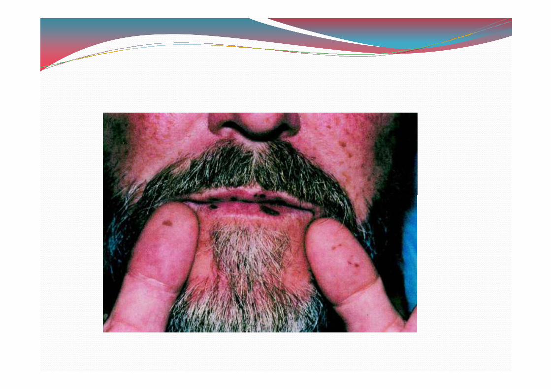
Idiopathic pigmentation
� Laugier – hunziker pigmentation:
� Hyperpigmentation of oral mucosal tissues involve lips & buccal mucosa.
� Pigmentation of esophageal , genital & conjunctival � Pigmentation of esophageal , genital & conjunctival mucosae & acral surfaces.
� Nail involvement in form of longitudinal melanotic streaks & without evidence of dystrophic change.
� Multiple , discrete , irregularly shaped brown oral macules & not more than 3mm in diameter.
Treatment of mucocutaneous melanosis
� Laser therapy has proven effective but recurrence occur in 0H% of treated patients.
� Various types of lasers.Various types of lasers.
a. Super pulsed CO0
b. Q – switched Nd – YAG
c. Switched alexandrite lasers
� Cryotherapy
� Phototherapy include intense pulsed light & fractional photothermolysis.
� First- line therapy involves application of tropical medicaments , that is bleaching cream.
� Simple agents such as azelaic acid or hydroquinone.
� Triple combination therapy
a. 2% hydroquinonea. 2% hydroquinone
b. H.H3% retinoic acid
c. H.H.% fluocinolone acetonide has proven effective in GH% of patients.
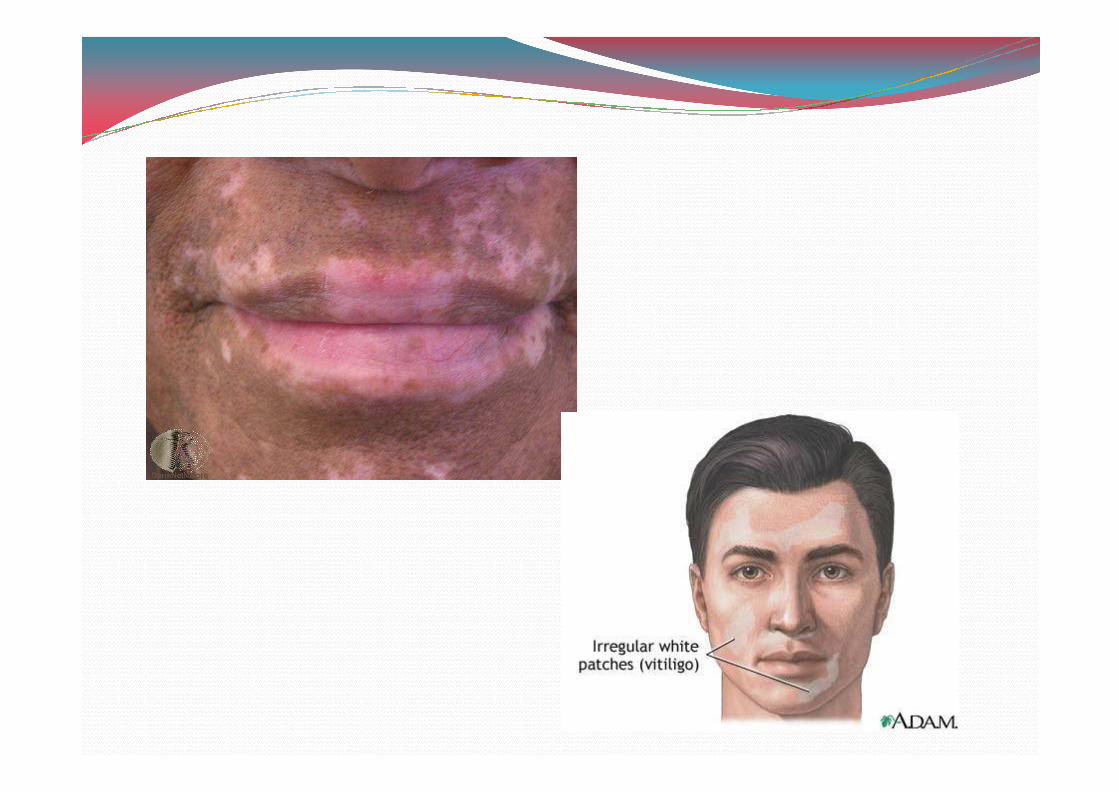
Depigmentation
Vitiligo :
� Relatively common , acquired autoimmune disease that is associated with hypomelanosis.
� Focal areas of depigmentation� Focal areas of depigmentation
� Vitiligenous lesion often present as well –circumscribed , round , oval ,or elongated pale or white colored macules that may coalese into larger areas of diffuse pigmentation.
� Arise in any patients undergoing immunotherapy.
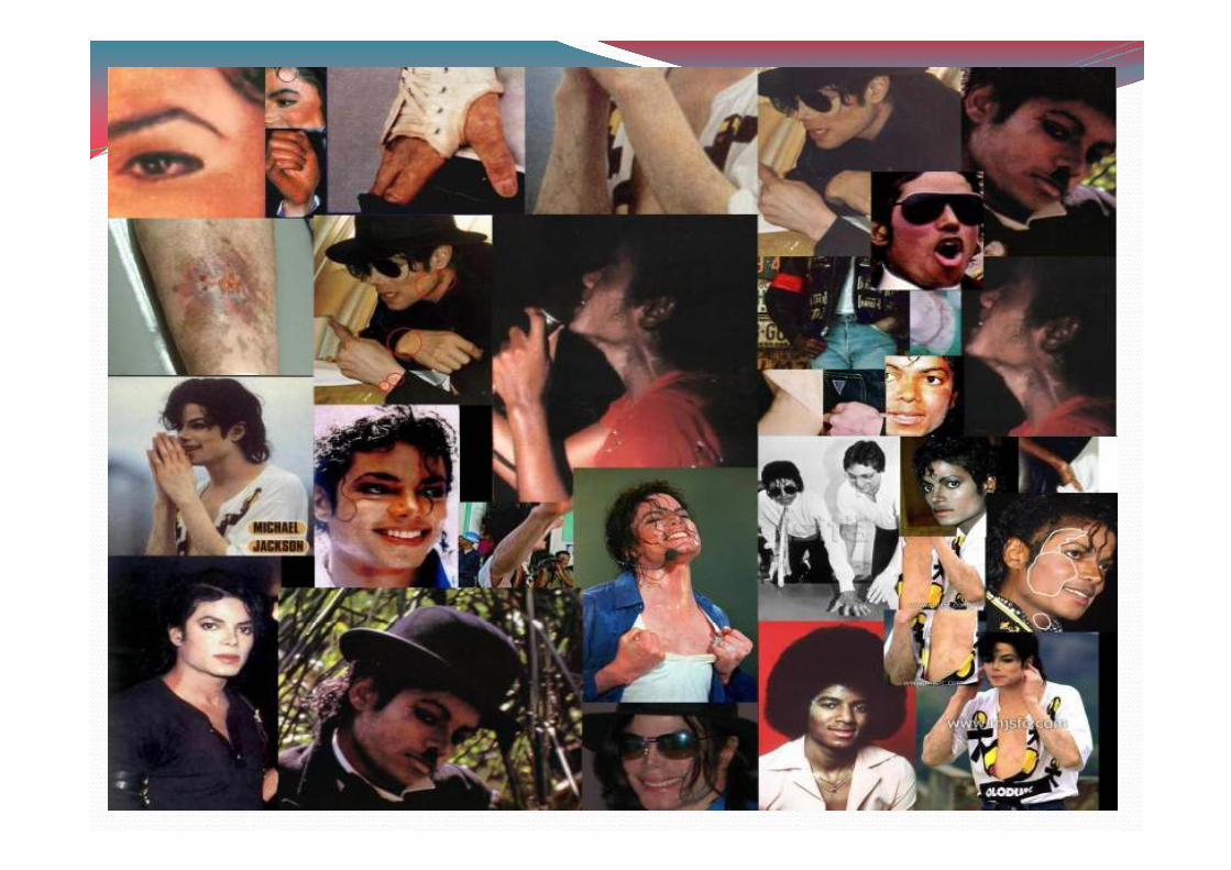
� Hypomelanosis of inner & outer surfaces of lips & perioral skin may be seen in up to 0H% of patients.
� Treatment with tropical corticosteroids , systemic photochemical therapies (psoralen & ultraviolet A exposure proven effective.exposure proven effective.
� Medicinal depigmentation that is cutaneoua bleaching to create unified skin color.
� Surgical intervention may be only optional (autologus epithelial grafts) used succesfully.
HEMOGLOBIN AND IRON ASSOCIATED PIGMENTATION
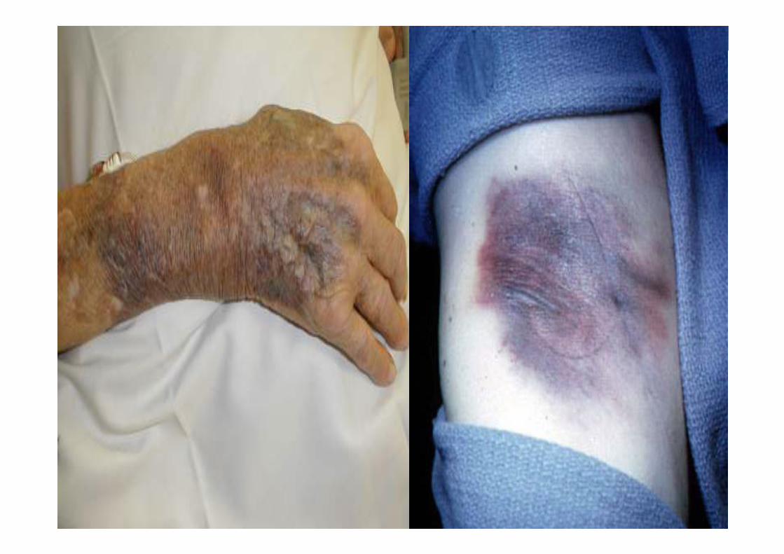
� Ecchymosis:
� Traumatic ecchymosis is common on the lips and face yet is uncommon in oral mucosa except in cases of blunt force trauma and oral intubation.force trauma and oral intubation.
� Immediately following traumatic event erythrocyte extra vasation into the sub mucosa will appear as bright red macule.
� The lesion will assume brown coloration within few days, after hemoglobin is degraded to hemosiderin.
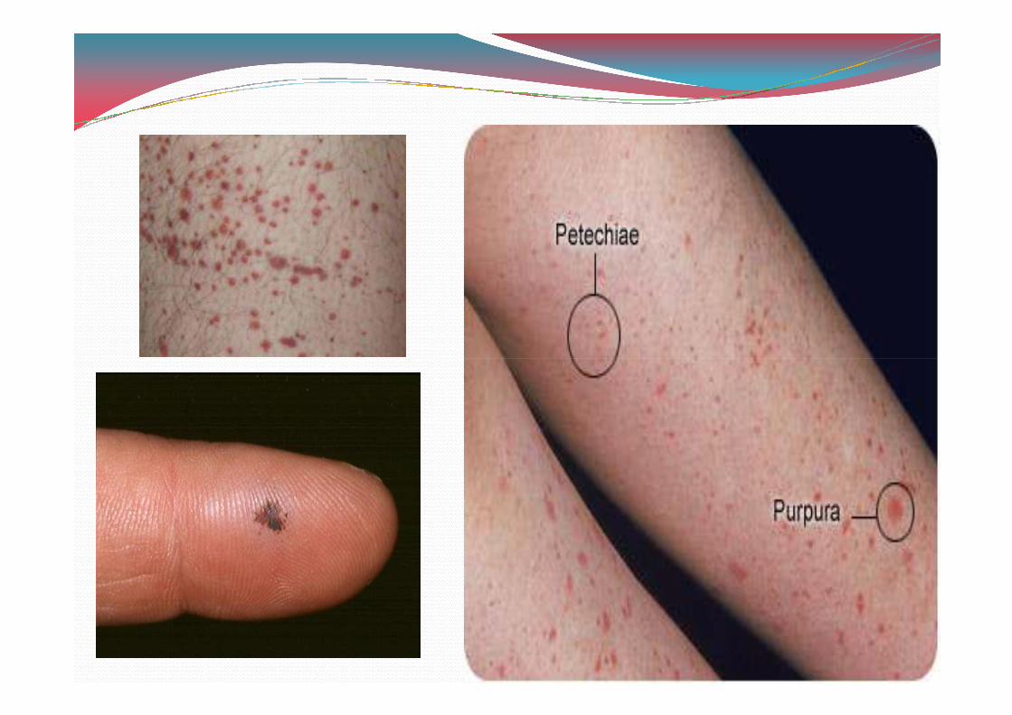
PURPURA/PETECHIAE
• Petechiae typically characterised as being pinpoint or slightly larger than pinpoint and purpura as multiple, small 0 to 2 mm collections of extravasated blood.
• Oral purpura may develop as a consequence of trauma or viral or systemic disease, identified in soft palate although any mucosal site may be affected.
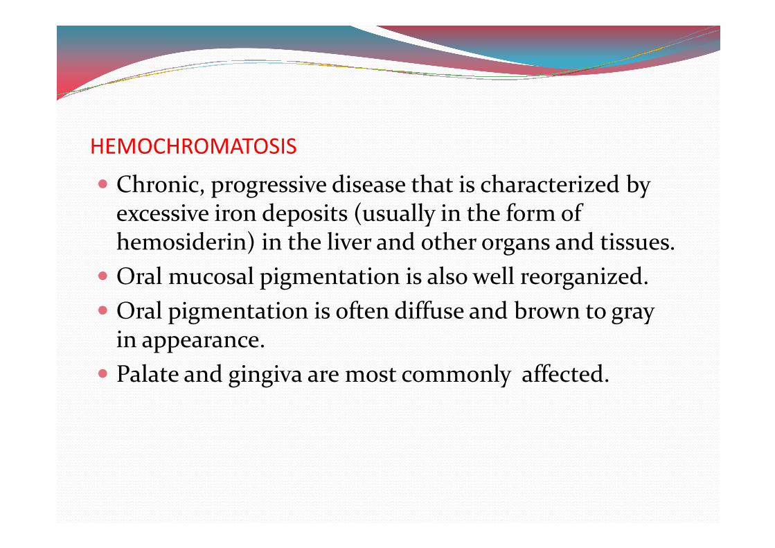
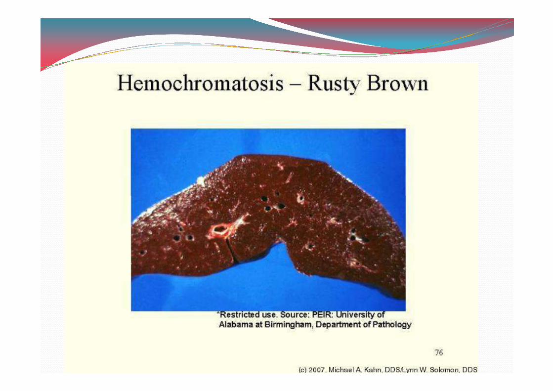
HEMOCHROMATOSIS
� Chronic, progressive disease that is characterized by excessive iron deposits (usually in the form of hemosiderin) in the liver and other organs and tissues.
� Oral mucosal pigmentation is also well reorganized.� Oral mucosal pigmentation is also well reorganized.
� Oral pigmentation is often diffuse and brown to gray in appearance.
� Palate and gingiva are most commonly affected.
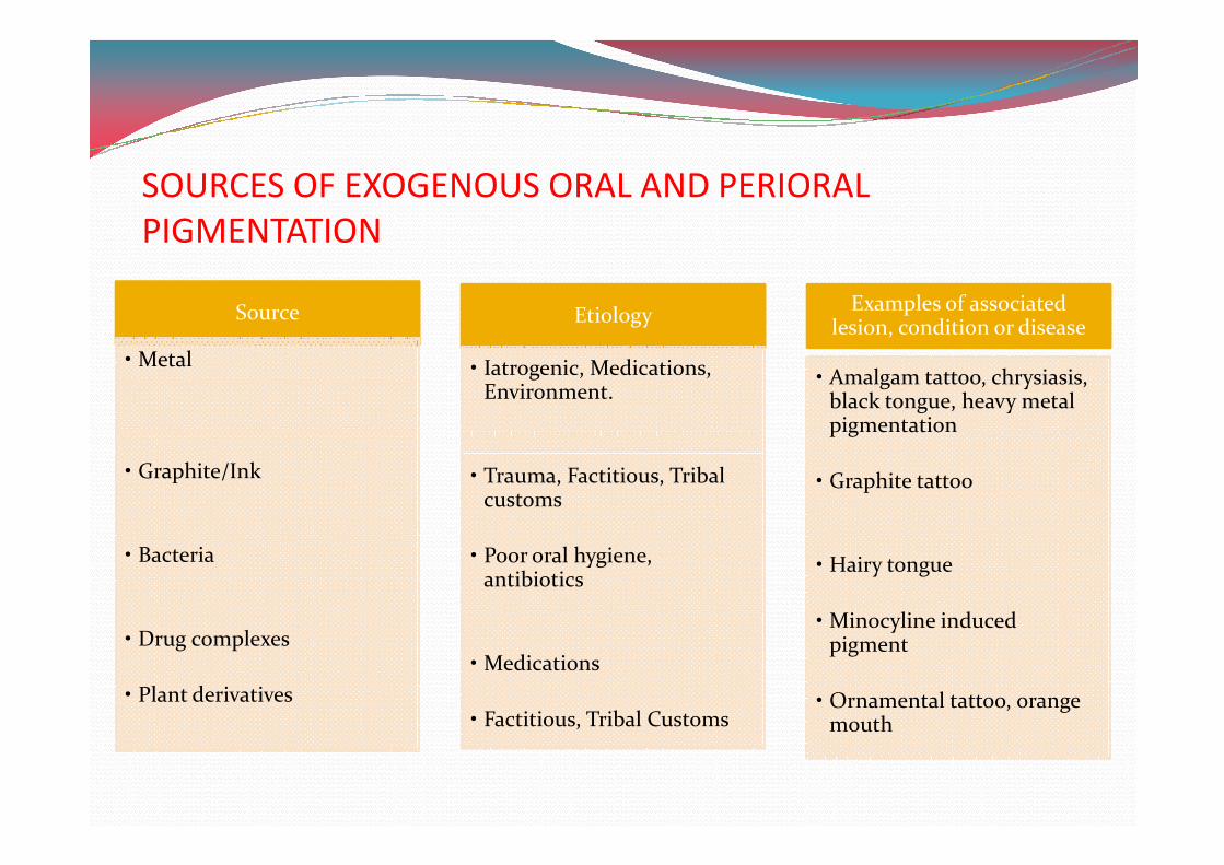
Source
• Metal
Etiology
• Iatrogenic, Medications, Environment.
Examples of associated lesion, condition or disease
• Amalgam tattoo, chrysiasis, black tongue, heavy metal pigmentation
SOURCES OF EXOGENOUS ORAL AND PERIORAL
PIGMENTATION
• Graphite/Ink
• Bacteria
• Drug complexes
• Plant derivatives
• Trauma, Factitious, Tribal customs
• Poor oral hygiene, antibiotics
• Medications
• Factitious, Tribal Customs
pigmentation
• Graphite tattoo
• Hairy tongue
• Minocyline induced pigment
• Ornamental tattoo, orange mouth
EXOGENOUS PIGMENTATION
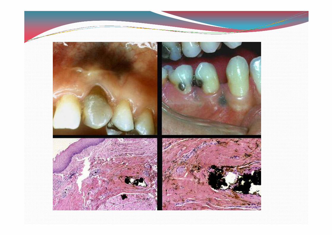
� AMALGUM TATTO:
� Etiology is deposition of amalgam material into sub mucosal tissue.
� Lesions small, asymptomatic, macular and bluish gray or � Lesions small, asymptomatic, macular and bluish gray or oven black in appearance.
� Gingiva, alveolar mucosa, buccal mucosa and floor of mouth are most common sites.
� Lesions found in vicinity of teeth with large amalgam restoration or crowned teeth and also in around healed extraction sites.
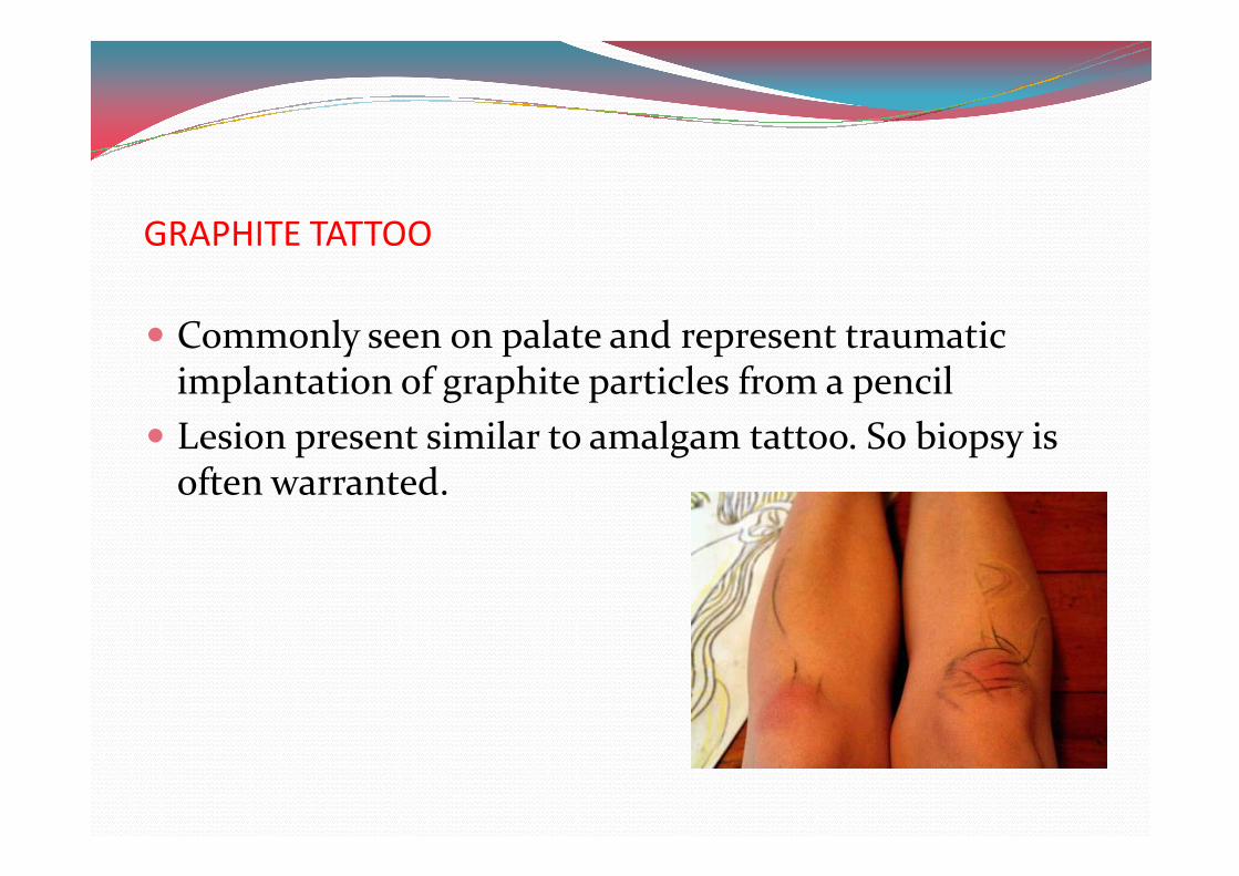
GRAPHITE TATTOO
� Commonly seen on palate and represent traumatic implantation of graphite particles from a pencil
� Lesion present similar to amalgam tattoo. So biopsy is � Lesion present similar to amalgam tattoo. So biopsy is often warranted.
ORNAMENTAL TATTOOS
� South African female tribal custom includes brushing the teeth and gums with a chewed root of the tree Euclea natalensis with the belief that promotes oral Euclea natalensis with the belief that promotes oral health.
� Plant root contain napthoquinones are pigmented and the mouths of root users are typically bright orange.
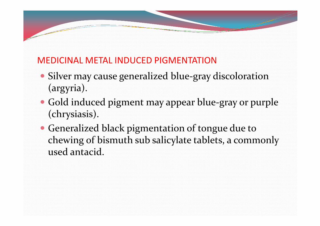
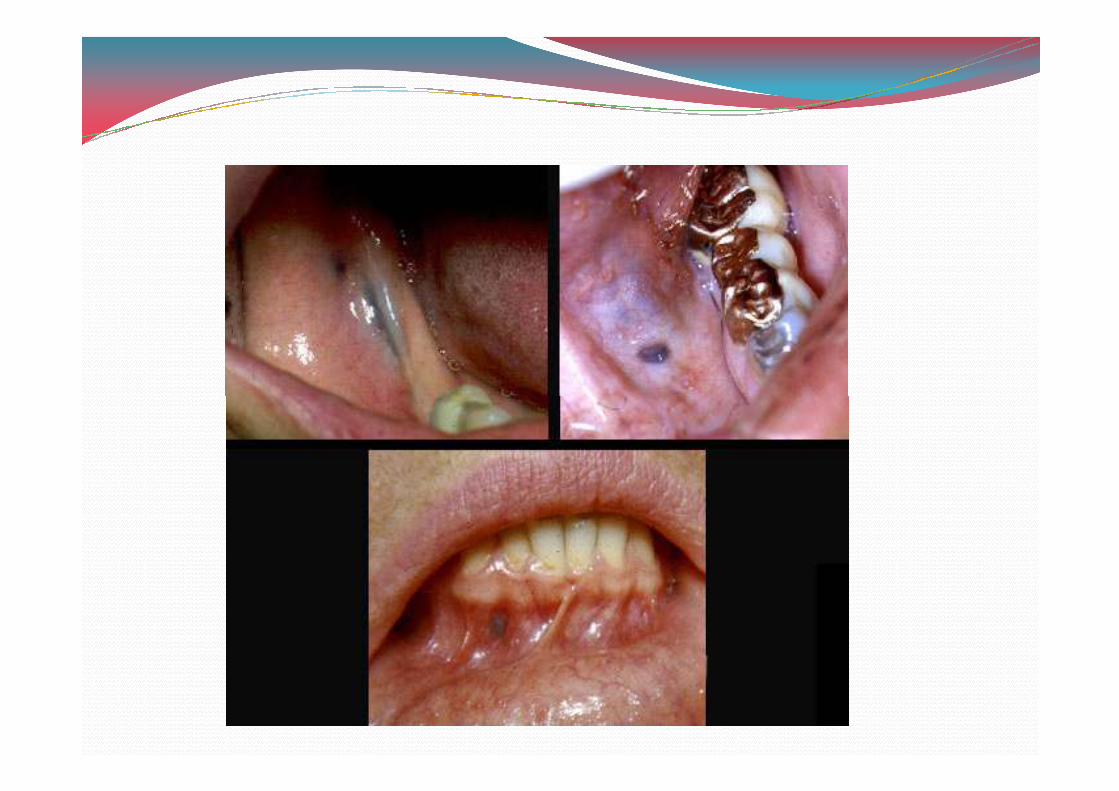
MEDICINAL METAL INDUCED PIGMENTATION
� Silver may cause generalized blue-gray discoloration (argyria).
� Gold induced pigment may appear blue-gray or purple (chrysiasis).(chrysiasis).
� Generalized black pigmentation of tongue due to chewing of bismuth sub salicylate tablets, a commonly used antacid.
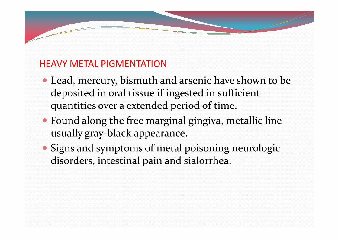
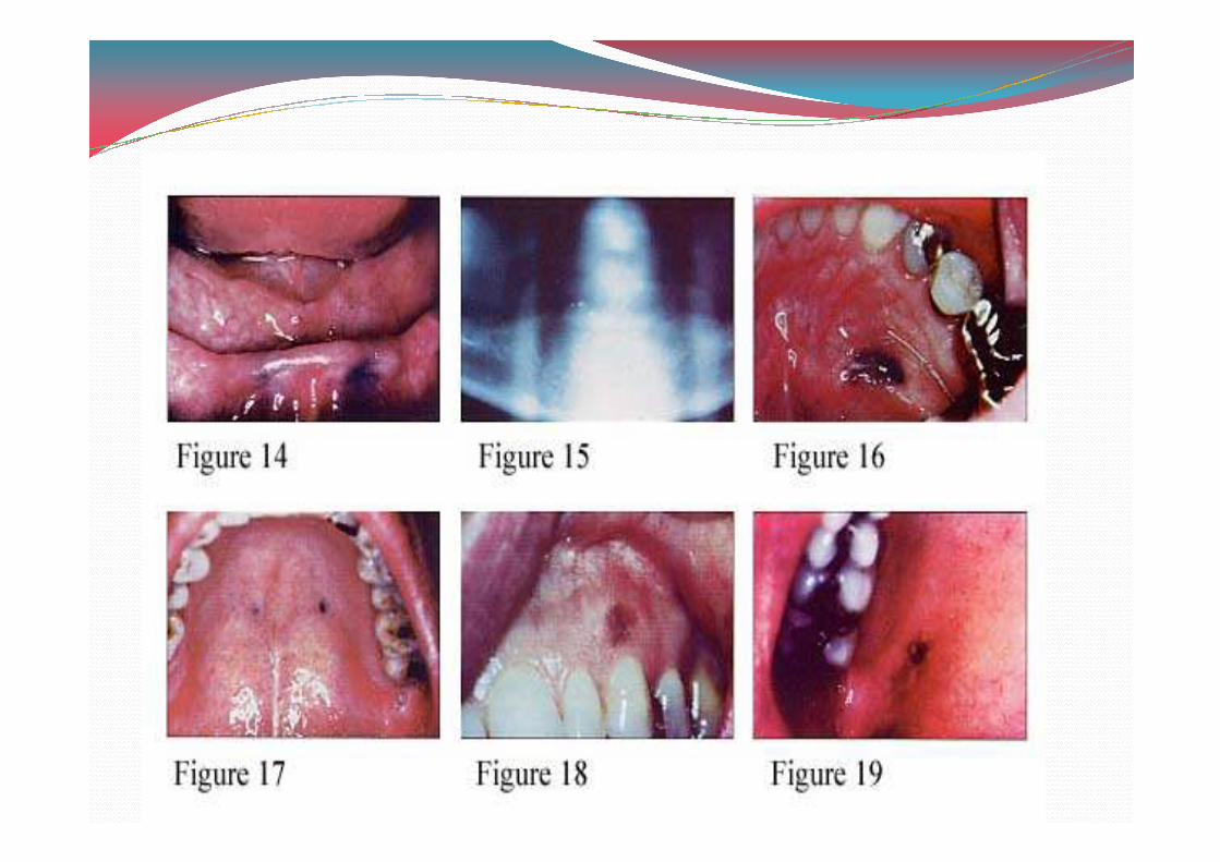
HEAVY METAL PIGMENTATION
� Lead, mercury, bismuth and arsenic have shown to be deposited in oral tissue if ingested in sufficient quantities over a extended period of time.
� Found along the free marginal gingiva, metallic line � Found along the free marginal gingiva, metallic line usually gray-black appearance.
� Signs and symptoms of metal poisoning neurologic disorders, intestinal pain and sialorrhea.
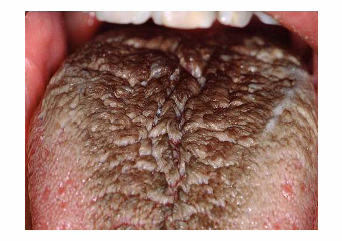
HAIRY TONGUE
� Change in oral flora associated with chromic antibiotic therapy.
� Colonization of chromogenic bacteria impart variety of colors white, green, brown or black.of colors white, green, brown or black.
� Various foods, drinks and also smoking of tobacco or crack cocaine has been shown in black hairy tongue.
� Treatment is using tongue scrapper and limit ingestion of coloring foods.

THANK YOU