Embed Size (px)
Citation preview
Pigments, Colours and Patterns – The contribution of eumelanin and
pheomelanin to molluscan shell ornamentation with a special focus on the
terrestrial snail Cepaea nemoralis
Dissertation
for award of the degree
"Doctor rerum naturalium" (Dr. rer. nat.)
of the Georg-August-Universität Göttingen
within the doctoral program Geosciences
of the Georg-August University School of Science (GAUSS)
submitted by
Susanne Affenzeller
from Linz a. d. Donau, Österreich
Göttingen 2019
Pigments, Colours and Patterns Dissertation Susanne Affenzeller 2019
Thesis Commitee:
Prof. Dr. Daniel J. Jackson, Dept. Geobiology, Georg-August-University Göttingen
Prof. Dr. Gregor Bucher, Dept. Developmental Biology GZMB, Georg-August-University
Göttingen
Dr. Klaus Wolkenstein, Dept. Geobiology, Georg-August-University Göttingen
Members of the Examination Board:
Reviewer: Prof. Dr. Daniel J. Jackson, Dept. Geobiology, Georg-August-
University Göttingen
Second Reviewer: Prof. Dr. Gregor Bucher, Dept. Developmental Biology GZMB,
Georg-August-University Göttingen
Further members of the Examination Board:
Dr. Klaus Wolkenstein, Dept. Geobiology, Georg-August-University Göttingen
Prof. Dr. Volker Thiel, Dept. Geobiology, Georg-August-University Göttingen
Prof. Dr. Christoph Bleidorn, Dept. Animal Evolution and Biodiversity, Georg-
August-University Göttingen
Dr. Sven Bradler, Dept. Animal Evolution and Biodiversity, Georg-August-
University Göttingen
Dr. Nico Posnien, Dept. Developmental Biology GZMB, Georg-August-University
Göttingen
Tag der mündlichen Prüfung: 7.10.2019
Pigments, Colours and Patterns Dissertation Susanne Affenzeller 2019
Versicherung
Hiermit versichere ich an Eides statt, dass die Dissertation mit dem Titel „Pigments,
Colours and Patterns – The contribution of eumelanin and pheomelanin to molluscan shell
ornamentation with a special focus on the terrestrial snail Cepaea nemoralis“ selbständig
und nur mit den angeführten Hilfsmitteln und Quellen angefertigt wurde.
Göttingen, den 20.08.2019

Pigments, Colours and Patterns Dissertation Susanne Affenzeller 2019
Pigments, Colours and Patterns Dissertation Susanne Affenzeller 2019
TABLE OF CONTENTS
CHAPTER 1: GENERAL INTRODUCTION ............................................................................... 9
CHAPTER 2: IDENTIFICATION AND VALIDATION OF REFERENCE GENES FOR QPCR IN THE
TERRESTRIAL GASTROPOD CEPAEA NEMORALIS. .............................................................. 22
CHAPTER 3: QUANTITATION OF EUMELANIN AND PHEOMELANIN MARKERS IN DIVERSE
BIOLOGICAL SAMPLES BY HPLC-UV-MS FOLLOWING SOLID-PHASE EXTRACTION .............. 38
CHAPTER 4: EUMELANIN AND PHEOMELANIN PIGMENTATION IN MOLLUSC SHELLS MAY
BE LESS COMMON THAN EXPECTED: INSIGHTS FROM MASS SPECTROMETRY .................. 60
CHAPTER 5: EUMELANIN IS NOT THE BANDED PIGMENT IN CEPAEA NEMORALIS ............. 80
CHAPTER 6: GENERAL DISCUSSION .................................................................................. 88
APPENDIX A: SUPPLEMENTARY MATERIAL FOR CHAPTER 2 .............................................. 97
APPENDIX B: SUPPLEMENTARY MATERIAL FOR CHAPTER 4 ............................................ 105
APPENDIX C: SUPPLEMENTARY MATERIAL FOR CHAPTER 5 ............................................ 107
APPENDIX D ................................................................................................................... 115
ACKNOWLEDGEMENTS .................................................................................................. 124
CURRICULUM VITAE ...................................................................................................... 126

Pigments, Colours and Patterns Dissertation Susanne Affenzeller 2019
6
Pigments, Colours and Patterns Dissertation Susanne Affenzeller 2019
7
ABSTRACT
In recent years interest into molluscan pigments increased. But a lot of techniques have to be adapted
to be usable with often difficult molluscan tissues and shell material. A comprehensive approach
needs to encompass both pigment chemistry and molecular biology. Here an improved method for
testing molluscan shells for the presence of characteristic melanin oxidation products is presented.
The established method of RT-qPCR relies heavily on sufficiently tested reference genes.
Comprehensive testing was carried out for both established house keeping genes and novel reference
genes in the terrestrial gastropod Cepaea nemoralis. Both of these techniques were used to test for
melanin in mollusc shell pigmentation. Evidence for eumelanin could be found in three conchiferan
classes: Nautilus pompilius (Cephalopoda), Mytilus edulis (Bivalvia), Clanculus pharaonius and
Steromphala adriatica (Gastropoda). Both eumelanin and pheomelanin were detected in the gastropod
C. nemoralis. In this species genes known to be involved in melanin synthesis in insects and mammals
were screened for their quantitative expression rates in shell producing mantle tissue. It was found
that Tyrosinase and Tyrosinase Related Protein are well expressed all over the mantle tissue, but show
no differential expression in band building mantle tissue. Together with evidence of both eumelanin
and pheomelanin oxidation products throughout the shell of C. nemoralis, this finding leads to the
conclusion that both types of melanin seem to be involved in shell colouration, but not band
patterning, of this gastropod shell. A surprisingly large number of other bivalve and gastropod species
tested for melanin show similar geometric patterns, that could not be verified as eumelanin. Future
research will hopefully shed light onto this very structurally stable molluscan shell pigmentation.

Pigments, Colours and Patterns Dissertation Susanne Affenzeller 2019
8
Pigments, Colours and Patterns Dissertation Susanne Affenzeller 2019
9
CHAPTER 1: GENERAL INTRODUCTION
Mollusca are the one of the most diverse and successful groups in the animal kingdom [1,2]. With an
estimated 85,000 extant and over 60,000 fossil species in nine recognized classes they represent the
second largest phylum of invertebrates[3,4]. Their rapid diversification during the Cambrian
explosion yielded a wide range of variations on the general mollusc Bauplan [5–7]. Through these
adaptations of their body plans and lifestyles they successfully conquered all regions of marine and
limnic habitats, with some gastropod linages even managing to adapt to the challenges of terrestrial
life [8]. On top of their many roles in recent ecosystems, the shell bearing Conchifera also play a
major role as markers in the fossil record [9,10].
Although less known mollusc classes like Solenogastres, Caudofoveates and Polyplacophora are
fascinating in their own right, it is the shell bearing molluscs that are best known. Especially the shells
of Gastropods and Bivalves have been collected and cherished for centuries by children as souvenirs
and scientific collectors alike. Perceived as especially precious and interesting are shells with
pigments and patterns. Some of them even play major cultural roles. Some cowrie species were used
as currency for trade in many different cultures in Africa, Asia and Oceania [11–13]. Conchs and
Tridacnas are not only used as symbols and building material in native cultures, but were also
integrated in European architecture and art from the renaissance period onwards [11,14–16]. The
colourful adornments of certain shells even dictate their value for use in jewellery, but also in
commercial food species [17–19]. It is these pigments and patterns and their variations on mollusc
shells that interest researches all around the globe [16]. Although the mathematical concepts of shell
pattering are well understood and can be computer simulated, little is known on the pigments they
use and the biological mechanisms employed [16,20–22]. What is the evolutionary purpose for these
patterns? Is it driven by camouflage to escape predation pressure or is it used as an intraspecific
communication tool? Why are the patterns of some species stable and useable as identification
Pigments, Colours and Patterns Dissertation Susanne Affenzeller 2019
10
markers, and other species show polymorphisms in both colouration and patterning? Research over
the last hundred years has given us some insights into possible answers to these questions. Molluscs
were shown to employ a number of chemically different pigments in their shells [16,23,24]. First
efforts were undertaken to understand how mollusc shells are built and in which way pigments could
be laid down into them [1,25–28]. And a number of studies tried to understand the evolution of
polymorphic shell patterns, prominently known from the terrestrial snail Cepaea nemoralis including
research on different selection pressures, underlying genetics and heritability patterns [29–36].
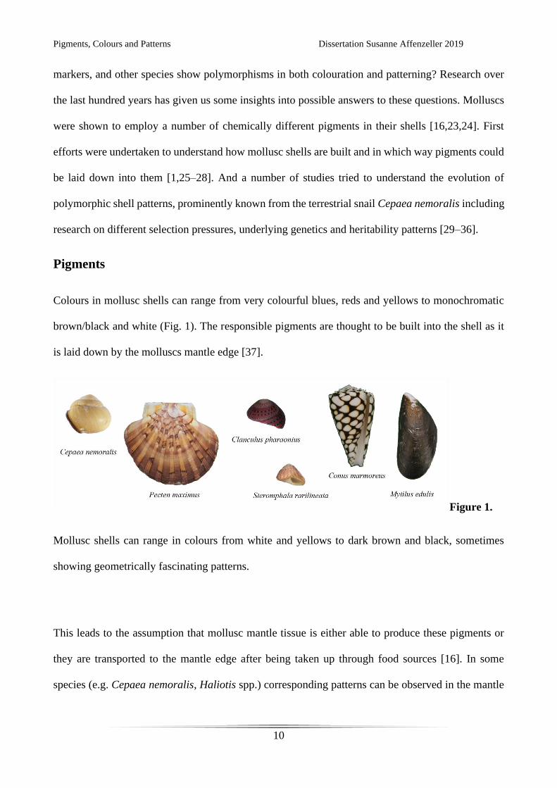
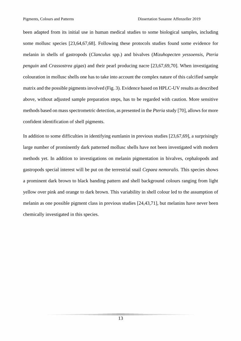
Pigments
Colours in mollusc shells can range from very colourful blues, reds and yellows to monochromatic
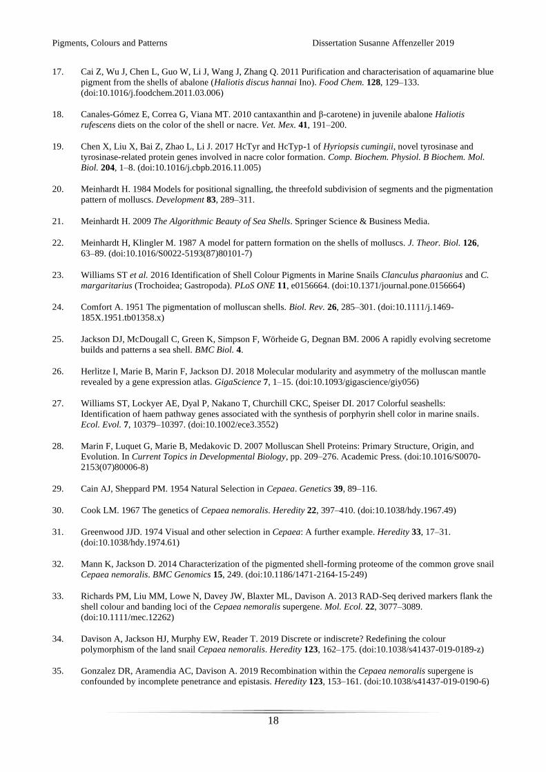
brown/black and white (Fig. 1). The responsible pigments are thought to be built into the shell as it
is laid down by the molluscs mantle edge [37].
Figure 1.
Mollusc shells can range in colours from white and yellows to dark brown and black, sometimes
showing geometrically fascinating patterns.
This leads to the assumption that mollusc mantle tissue is either able to produce these pigments or
they are transported to the mantle edge after being taken up through food sources [16]. In some
species (e.g. Cepaea nemoralis, Haliotis spp.) corresponding patterns can be observed in the mantle
Pigments, Colours and Patterns Dissertation Susanne Affenzeller 2019
11
tissue directly beneath shell colouration [25,37,38]. But although molluscs display a large amount of
different colours, little is known about the chemical composition of their pigments. In one of the
earliest comprehensive approaches Comfort [24,39–43] tested a range of conchiferan species for
different pigment classes and indicated the widespread presence of pyrroles and melanins in these
organisms. He found that tetrapyrroles, mainly as porphyrins and sometimes bile pigments, were
commonly found in marine gastropods and bivalves [24,39,40]. This pigment class is easily dissolved
in aqueous acids and shows a distinct reddish fluorescence [16]. Although Comforts’ work is an
invaluable basis to work from, one has to be aware that the methods available to him at the time were
only rudimentary compared to modern chemical methods. Many of his works are based on solubility
tests of these pigments and early chromatographic separations. While these investigations still yielded
light on basic chemical properties of many mollusc pigments, more in depth and precise methods
have to be applied to verify pigment classes. Efforts to that end were made in recent studies using
high performance liquid chromatography and UV detection (HPLC-UV) to further characterise
molluscan tetrapyrroles as uroporphyrins and protoporphyrins responsible for bright orange and red
colourations [23,44]. Within this colour range another abundant pigment class are carotenoids, which
are generally synthesised by plants and only secondarily taken up by animals for colouration [16,18].
Carotinoids are well known from plants, but also vertebrates and some Raman spectroscopy studies
on shell material indicated their possible presence in gastropod and bivalve shells [18,45–50].
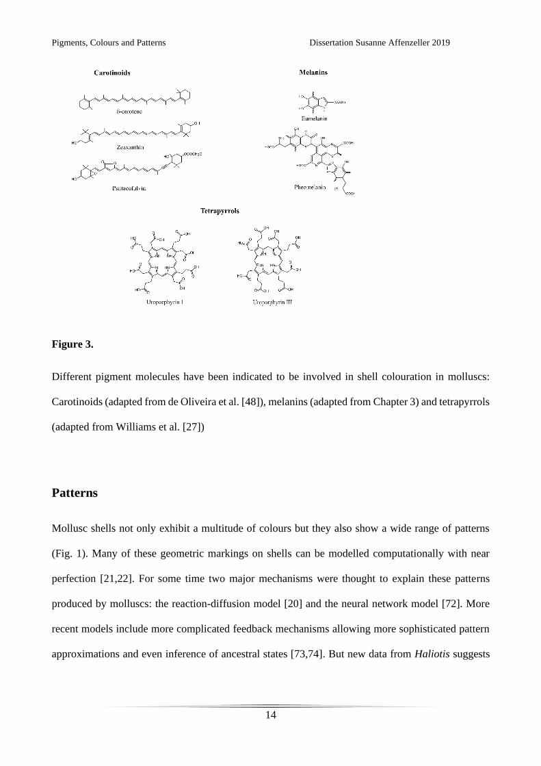
For dark geometric patterns, and generally darker colourations, melanins are often implicated as the
responsible pigments [16,24,43]. Although melanins are well known in vertebrates, the term was used
generically for most brown and black colourations in molluscs as well, often without chemically
analysing the pigments in question [24,42,51]. More accurately melanins are defined as
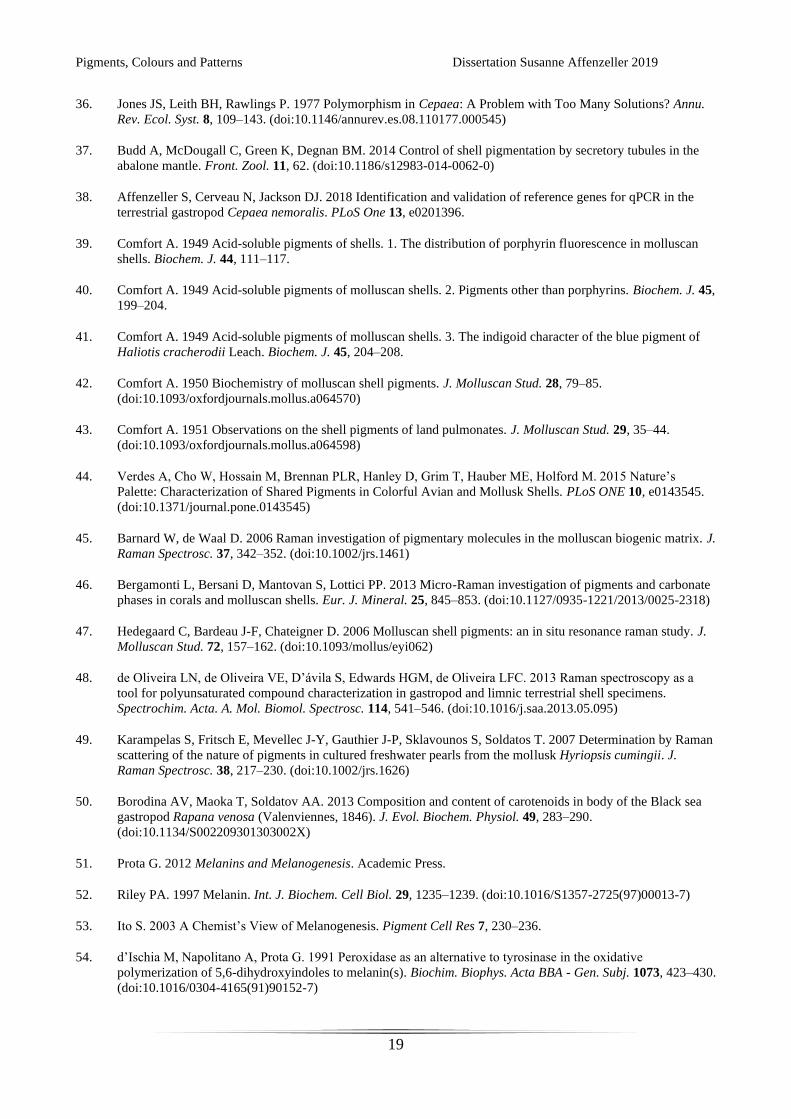
polymerisation products of DOPA (L-3,4-dihydroxyphenylalanine) subunits by enzymatic oxidation
(Fig. 2) [51–57].

Pigments, Colours and Patterns Dissertation Susanne Affenzeller 2019
12
Figure 2.
Eumelanin and pheomelanin are polymerisation products of DOPA subunits in the absence or
presence of cysteine. The process is catalysed be the enzyme Tyrosinase, and in some cases
Tyrosinase Related Proteins. (from d’Ischia et al. [57])
The resulting macromolecules, termed eumelanin, show a brown to black colour and are very thermo-
and chemo-stable [52,58–60]. The addition of cysteine during polymerisation results in pheomelanin
pigments, well known from human red hair [53,61–64]. This even more complex macromolecule can
produce a colour range of yellows, oranges and reds and is well known from mammals and birds
[62,64]. But it is chemically difficult to verify either of these melanins due to their specific
characteristics and complex nature [52,53,60]. An established method for vertebrate samples is
therefore to measure their characteristic oxidation products by HPLC-UV [65,66]. This method has
Pigments, Colours and Patterns Dissertation Susanne Affenzeller 2019
13
been adapted from its initial use in human medical studies to some biological samples, including
some mollusc species [23,64,67,68]. Following these protocols studies found some evidence for
melanin in shells of gastropods (Clanculus spp.) and bivalves (Mizuhopecten yessoensis, Pteria
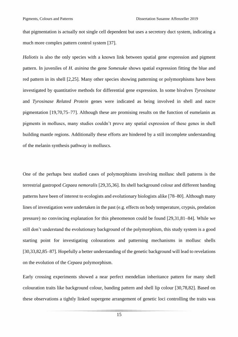
penguin and Crassostrea gigas) and their pearl producing nacre [23,67,69,70]. When investigating
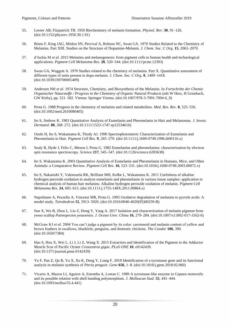
colouration in mollusc shells one has to take into account the complex nature of this calcified sample
matrix and the possible pigments involved (Fig. 3). Evidence based on HPLC-UV results as described
above, without adjusted sample preparation steps, has to be regarded with caution. More sensitive
methods based on mass spectrometric detection, as presented in the Pteria study [70], allows for more
confident identification of shell pigments.
In addition to some difficulties in identifying eumlanin in previous studies [23,67,69], a surprisingly
large number of prominently dark patterned mollusc shells have not been investigated with modern
methods yet. In addition to investigations on melanin pigmentation in bivalves, cephalopods and
gastropods special interest will be put on the terrestrial snail Cepaea nemoralis. This species shows
a prominent dark brown to black banding pattern and shell background colours ranging from light
yellow over pink and orange to dark brown. This variability in shell colour led to the assumption of
melanin as one possible pigment class in previous studies [24,43,71], but melanins have never been
chemically investigated in this species.
Pigments, Colours and Patterns Dissertation Susanne Affenzeller 2019
14
Figure 3.
Different pigment molecules have been indicated to be involved in shell colouration in molluscs:
Carotinoids (adapted from de Oliveira et al. [48]), melanins (adapted from Chapter 3) and tetrapyrrols
(adapted from Williams et al. [27])
Patterns
Mollusc shells not only exhibit a multitude of colours but they also show a wide range of patterns
(Fig. 1). Many of these geometric markings on shells can be modelled computationally with near
perfection [21,22]. For some time two major mechanisms were thought to explain these patterns
produced by molluscs: the reaction-diffusion model [20] and the neural network model [72]. More
recent models include more complicated feedback mechanisms allowing more sophisticated pattern
approximations and even inference of ancestral states [73,74]. But new data from Haliotis suggests
Pigments, Colours and Patterns Dissertation Susanne Affenzeller 2019
15
that pigmentation is actually not single cell dependent but uses a secretory duct system, indicating a
much more complex pattern control system [37].
Haliotis is also the only species with a known link between spatial gene expression and pigment
pattern. In juveniles of H. asinina the gene Somesuke shows spatial expression fitting the blue and
red pattern in its shell [2,25]. Many other species showing patterning or polymorphisms have been
investigated by quantitative methods for differential gene expression. In some bivalves Tyrosinase
and Tyrosinase Related Protein genes were indicated as being involved in shell and nacre
pigmentation [19,70,75–77]. Although these are promising results on the function of eumelanin as
pigments in molluscs, many studies couldn’t prove any spatial expression of these genes in shell
building mantle regions. Additionally these efforts are hindered by a still incomplete understanding
of the melanin synthesis pathway in molluscs.
One of the perhaps best studied cases of polymorphisms involving mollusc shell patterns is the
terrestrial gastropod Cepaea nemoralis [29,35,36]. Its shell background colour and different banding
patterns have been of interest to ecologists and evolutionary biologists alike [78–80]. Although many
lines of investigation were undertaken in the past (e.g. effects on body temperature, crypsis, predation
pressure) no convincing explanation for this phenomenon could be found [29,31,81–84]. While we
still don’t understand the evolutionary background of the polymorphism, this study system is a good
starting point for investigating colourations and patterning mechanisms in mollusc shells
[30,33,82,85–87]. Hopefully a better understanding of the genetic background will lead to revelations
on the evolution of the Cepaea polymorphism.
Early crossing experiments showed a near perfect mendelian inheritance pattern for many shell
colouration traits like background colour, banding pattern and shell lip colour [30,78,82]. Based on
these observations a tightly linked supergene arrangement of genetic loci controlling the traits was
Pigments, Colours and Patterns Dissertation Susanne Affenzeller 2019
16
assumed [33,36,87,88]. Efforts undertaken in recent years yielded a set of RAD-Seq markers flanking
this supergene, allowing further investigations into recombination events of the relevant loci [33,35].
This new data set reveals that recombination events within the supergene might not be as common as
previously assumed. Incomplete penetrance and epistasis are actually able to explain these
phenotypes [35]. Together with new investigations on transcriptomic and proteomic data the fine
mapping of the supergene of Cepaea nemoralis now seems to become possible. Still, data collected
over the last years with new and advanced sequencing techniques could not reveal conclusive
candidates for these patterning genes [32,89]. While Kerkvliet et al. [89] found metallothionein genes,
thought to inhibit melanin synthesis, as most promising candidates, Mann and Jackson [32] could not
detect any proteins associated directly with shell colouration. The genetic control for this complex
polymorphism is therefore still unsolved and further investigations into differential gene expression
in both bioinformatic and in situ experiments have to be undertaken it the future. The above
mentioned recent gain in molecular data and methods in C. nemoralis affords the opportunity of
combining molecular biology and shell chemistry to gain insights into the pigmentation and
patterning mechanisms involved [32,33,89].
In the following chapters I will describe insights gained from experiments on shell pigments and
patterning mechanisms in shell bearing molluscs, with a special focus on C. nemoralis. Chapter 2
contains the establishment of reference genes for reverse transcription quantitative polymerase chain
reaction in C. nemoralis. This method can be used to determine expression levels of certain genes.
To selectively and sensitively measure oxidation products of both eumelanin and pheomelanin in
complex biological sample matrices I developed and adapted a mass spectrometric method for use on
a wide variety of samples (chapter 3). Both of these methods were applied in chapter 4. These
chemical analyses were conducted on a range of mollusc species (bivalves, cephalopods and

Pigments, Colours and Patterns Dissertation Susanne Affenzeller 2019
17
gastropods) to determine the use of eumelanin and pheomelanin in mollusc shells (chapter 4). In
chapter 5 I used both methods to test eumelanin and pheomelanin composition in different shell
colour morphs and quantitative expression levels of known melanin pathway genes in C. nemoralis.
References
1. Kocot KM, Aguilera F, McDougall C, Jackson DJ, Degnan BM. 2016 Sea shell diversity and rapidly evolving
secretomes: insights into the evolution of biomineralization. Front. Zool. 13. (doi:10.1186/s12983-016-0155-z)
2. Jackson DJ, Wörheide G, Degnan BM. 2007 Dynamic expression of ancient and novel molluscan shell genes
during ecological transitions. BMC Evol. Biol. 7, 160. (doi:10.1186/1471-2148-7-160)
3. Taylor PD, Lewis DN. 2007 Fossil Invertebrates. Harvard University Press.
4. Rosenberg G. 2014 A New Critical Estimate of Named Species-Level Diversity of the Recent Mollusca*. Am.
Malacol. Bull. 32, 308–322. (doi:10.4003/006.032.0204)
5. Erwin DH, Laflamme M, Tweedt SM, Sperling EA, Pisani D, Peterson KJ. 2011 The Cambrian Conundrum:
Early Divergence and Later Ecological Success in the Early History of Animals. Science 334, 1091–1097.
(doi:10.1126/science.1206375)
6. Marshall CR. 2006 Explaining the Cambrian “Explosion” of Animals. Annu. Rev. Earth Planet. Sci. 34, 355–
384. (doi:10.1146/annurev.earth.33.031504.103001)
7. Smith MP, Harper DAT. 2013 Causes of the Cambrian Explosion. Science 341, 1355–1356.
(doi:10.1126/science.1239450)
8. Haszprunar G, Wanninger A. 2012 Molluscs. Curr. Biol. 22, R510–R514. (doi:10.1016/j.cub.2012.05.039)
9. Zuschin M, Harzhauser M, Mandic O. 2004 Taphonomy and Paleoecology of the lower Badenian (Middle
Miocene) Molluscan Assemblages at Grund (Lower Austria). Geol. Carpathica 55, 117–128.
10. Gallmetzer I, Haselmair A, Tomašových A, Stachowitsch M, Zuschin M. 2017 Responses of molluscan
communities to centuries of human impact in the northern Adriatic Sea. PLoS One 12, e0180820.
(doi:10.1371/journal.pone.0180820)
11. Ogundiran A. 2002 Of Small Things Remembered: Beads, Cowries, and Cultural Translations of the Atlantic
Experience in Yorubaland. Int. J. Afr. Hist. Stud. 35, 427–457. (doi:10.2307/3097620)
12. Gregory CA. 1996 Cowries and Conquest: Towards a Subalternate Quality Theory of Money. Comp. Stud. Soc.
Hist. 38, 195–217. (doi:10.1017/S0010417500020235)
13. Yang B. 2011 The Rise and Fall of Cowrie Shells: The Asian Story. J. World Hist. 22, 1–25.
14. Doran E. 1958 The Caicos Conch Trade. Geogr. Rev. 48, 388–401. (doi:10.2307/212258)
15. Pietak LM. 1998 Body Symbolism and Cultural Aesthetics: The Use of Shell Beads and Ornaments by
Delaware and Munsee Groups. North Am. Archaeol. 19, 135–161. (doi:10.2190/0BV6-Q0N1-37VU-PEQ7)
16. Williams ST. 2017 Molluscan shell colour: Molluscan shell colour. Biol. Rev. 92, 1039–1058.
(doi:10.1111/brv.12268)
Pigments, Colours and Patterns Dissertation Susanne Affenzeller 2019
18
17. Cai Z, Wu J, Chen L, Guo W, Li J, Wang J, Zhang Q. 2011 Purification and characterisation of aquamarine blue
pigment from the shells of abalone (Haliotis discus hannai Ino). Food Chem. 128, 129–133.
(doi:10.1016/j.foodchem.2011.03.006)
18. Canales-Gómez E, Correa G, Viana MT. 2010 cantaxanthin and β-carotene) in juvenile abalone Haliotis
rufescens diets on the color of the shell or nacre. Vet. Mex. 41, 191–200.
19. Chen X, Liu X, Bai Z, Zhao L, Li J. 2017 HcTyr and HcTyp-1 of Hyriopsis cumingii, novel tyrosinase and
tyrosinase-related protein genes involved in nacre color formation. Comp. Biochem. Physiol. B Biochem. Mol.
Biol. 204, 1–8. (doi:10.1016/j.cbpb.2016.11.005)
20. Meinhardt H. 1984 Models for positional signalling, the threefold subdivision of segments and the pigmentation
pattern of molluscs. Development 83, 289–311.
21. Meinhardt H. 2009 The Algorithmic Beauty of Sea Shells. Springer Science & Business Media.
22. Meinhardt H, Klingler M. 1987 A model for pattern formation on the shells of molluscs. J. Theor. Biol. 126,
63–89. (doi:10.1016/S0022-5193(87)80101-7)
23. Williams ST et al. 2016 Identification of Shell Colour Pigments in Marine Snails Clanculus pharaonius and C.
margaritarius (Trochoidea; Gastropoda). PLoS ONE 11, e0156664. (doi:10.1371/journal.pone.0156664)
24. Comfort A. 1951 The pigmentation of molluscan shells. Biol. Rev. 26, 285–301. (doi:10.1111/j.1469-
185X.1951.tb01358.x)
25. Jackson DJ, McDougall C, Green K, Simpson F, Wörheide G, Degnan BM. 2006 A rapidly evolving secretome
builds and patterns a sea shell. BMC Biol. 4.
26. Herlitze I, Marie B, Marin F, Jackson DJ. 2018 Molecular modularity and asymmetry of the molluscan mantle
revealed by a gene expression atlas. GigaScience 7, 1–15. (doi:10.1093/gigascience/giy056)
27. Williams ST, Lockyer AE, Dyal P, Nakano T, Churchill CKC, Speiser DI. 2017 Colorful seashells:
Identification of haem pathway genes associated with the synthesis of porphyrin shell color in marine snails.
Ecol. Evol. 7, 10379–10397. (doi:10.1002/ece3.3552)
28. Marin F, Luquet G, Marie B, Medakovic D. 2007 Molluscan Shell Proteins: Primary Structure, Origin, and
Evolution. In Current Topics in Developmental Biology, pp. 209–276. Academic Press. (doi:10.1016/S0070-
2153(07)80006-8)
29. Cain AJ, Sheppard PM. 1954 Natural Selection in Cepaea. Genetics 39, 89–116.
30. Cook LM. 1967 The genetics of Cepaea nemoralis. Heredity 22, 397–410. (doi:10.1038/hdy.1967.49)
31. Greenwood JJD. 1974 Visual and other selection in Cepaea: A further example. Heredity 33, 17–31.
(doi:10.1038/hdy.1974.61)
32. Mann K, Jackson D. 2014 Characterization of the pigmented shell-forming proteome of the common grove snail
Cepaea nemoralis. BMC Genomics 15, 249. (doi:10.1186/1471-2164-15-249)
33. Richards PM, Liu MM, Lowe N, Davey JW, Blaxter ML, Davison A. 2013 RAD-Seq derived markers flank the
shell colour and banding loci of the Cepaea nemoralis supergene. Mol. Ecol. 22, 3077–3089.
(doi:10.1111/mec.12262)
34. Davison A, Jackson HJ, Murphy EW, Reader T. 2019 Discrete or indiscrete? Redefining the colour
polymorphism of the land snail Cepaea nemoralis. Heredity 123, 162–175. (doi:10.1038/s41437-019-0189-z)
35. Gonzalez DR, Aramendia AC, Davison A. 2019 Recombination within the Cepaea nemoralis supergene is
confounded by incomplete penetrance and epistasis. Heredity 123, 153–161. (doi:10.1038/s41437-019-0190-6)
Pigments, Colours and Patterns Dissertation Susanne Affenzeller 2019
19
36. Jones JS, Leith BH, Rawlings P. 1977 Polymorphism in Cepaea: A Problem with Too Many Solutions? Annu.
Rev. Ecol. Syst. 8, 109–143. (doi:10.1146/annurev.es.08.110177.000545)
37. Budd A, McDougall C, Green K, Degnan BM. 2014 Control of shell pigmentation by secretory tubules in the
abalone mantle. Front. Zool. 11, 62. (doi:10.1186/s12983-014-0062-0)
38. Affenzeller S, Cerveau N, Jackson DJ. 2018 Identification and validation of reference genes for qPCR in the
terrestrial gastropod Cepaea nemoralis. PLoS One 13, e0201396.
39. Comfort A. 1949 Acid-soluble pigments of shells. 1. The distribution of porphyrin fluorescence in molluscan
shells. Biochem. J. 44, 111–117.
40. Comfort A. 1949 Acid-soluble pigments of molluscan shells. 2. Pigments other than porphyrins. Biochem. J. 45,
199–204.
41. Comfort A. 1949 Acid-soluble pigments of molluscan shells. 3. The indigoid character of the blue pigment of
Haliotis cracherodii Leach. Biochem. J. 45, 204–208.
42. Comfort A. 1950 Biochemistry of molluscan shell pigments. J. Molluscan Stud. 28, 79–85.
(doi:10.1093/oxfordjournals.mollus.a064570)
43. Comfort A. 1951 Observations on the shell pigments of land pulmonates. J. Molluscan Stud. 29, 35–44.
(doi:10.1093/oxfordjournals.mollus.a064598)
44. Verdes A, Cho W, Hossain M, Brennan PLR, Hanley D, Grim T, Hauber ME, Holford M. 2015 Nature’s
Palette: Characterization of Shared Pigments in Colorful Avian and Mollusk Shells. PLoS ONE 10, e0143545.
(doi:10.1371/journal.pone.0143545)
45. Barnard W, de Waal D. 2006 Raman investigation of pigmentary molecules in the molluscan biogenic matrix. J.
Raman Spectrosc. 37, 342–352. (doi:10.1002/jrs.1461)
46. Bergamonti L, Bersani D, Mantovan S, Lottici PP. 2013 Micro-Raman investigation of pigments and carbonate
phases in corals and molluscan shells. Eur. J. Mineral. 25, 845–853. (doi:10.1127/0935-1221/2013/0025-2318)
47. Hedegaard C, Bardeau J-F, Chateigner D. 2006 Molluscan shell pigments: an in situ resonance raman study. J.
Molluscan Stud. 72, 157–162. (doi:10.1093/mollus/eyi062)
48. de Oliveira LN, de Oliveira VE, D’ávila S, Edwards HGM, de Oliveira LFC. 2013 Raman spectroscopy as a
tool for polyunsaturated compound characterization in gastropod and limnic terrestrial shell specimens.
Spectrochim. Acta. A. Mol. Biomol. Spectrosc. 114, 541–546. (doi:10.1016/j.saa.2013.05.095)
49. Karampelas S, Fritsch E, Mevellec J-Y, Gauthier J-P, Sklavounos S, Soldatos T. 2007 Determination by Raman
scattering of the nature of pigments in cultured freshwater pearls from the mollusk Hyriopsis cumingii. J.
Raman Spectrosc. 38, 217–230. (doi:10.1002/jrs.1626)
50. Borodina AV, Maoka T, Soldatov AA. 2013 Composition and content of carotenoids in body of the Black sea
gastropod Rapana venosa (Valenviennes, 1846). J. Evol. Biochem. Physiol. 49, 283–290.
(doi:10.1134/S002209301303002X)
51. Prota G. 2012 Melanins and Melanogenesis. Academic Press.
52. Riley PA. 1997 Melanin. Int. J. Biochem. Cell Biol. 29, 1235–1239. (doi:10.1016/S1357-2725(97)00013-7)
53. Ito S. 2003 A Chemist’s View of Melanogenesis. Pigment Cell Res 7, 230–236.
54. d’Ischia M, Napolitano A, Prota G. 1991 Peroxidase as an alternative to tyrosinase in the oxidative
polymerization of 5,6-dihydroxyindoles to melanin(s). Biochim. Biophys. Acta BBA - Gen. Subj. 1073, 423–430.
(doi:10.1016/0304-4165(91)90152-7)
Pigments, Colours and Patterns Dissertation Susanne Affenzeller 2019
20
55. Lerner AB, Fitzpatrick TB. 1950 Biochemistry of melanin formation. Physiol. Rev. 30, 91–126.
(doi:10.1152/physrev.1950.30.1.91)
56. Binns F, King JAG, Mishra SN, Percival A, Robson NC, Swan GA. 1970 Studies Related to the Chemistry of
Melanins. Part XIII. Studies on the Structure of Dopamine-Melanin. J. Chem. Soc. C Org. 15, 2063–2070.
57. d’Ischia M et al. 2015 Melanins and melanogenesis: from pigment cells to human health and technological
applications. Pigment Cell Melanoma Res. 28, 520–544. (doi:10.1111/pcmr.12393)
58. Swan GA, Waggott A. 1970 Studies related to the chemistry of melanins. Part X. Quantitative assessment of
different types of units present in dopa-melanin. J. Chem. Soc. C Org. 0, 1409–1418.
(doi:10.1039/J39700001409)
59. Andersen NH et al. 1974 Structure, Chemistry, and Biosynthesis of the Melanins. In Fortschritte der Chemie
Organischer Naturstoffe / Progress in the Chemistry of Organic Natural Products (eds W Herz, H Grisebach,
GW Kirby), pp. 521–582. Vienna: Springer Vienna. (doi:10.1007/978-3-7091-7094-6_9)
60. Prota G. 1988 Progress in the chemistry of melanins and related metabolites. Med. Res. Rev. 8, 525–556.
(doi:10.1002/med.2610080405)
61. Ito S, Jimbow K. 1983 Quantitative Analysis of Eumelanin and Pheomelanin in Hair and Melanomas. J. Invest.
Dermatol. 80, 268–272. (doi:10.1111/1523-1747.ep12534616)
62. Ozeki H, Ito S, Wakamatsu K, Thody AJ. 1996 Spectrophotometric Characterization of Eumelanin and
Pheomelanin in Hair. Pigment Cell Res. 9, 265–270. (doi:10.1111/j.1600-0749.1996.tb00116.x)
63. Sealy R, Hyde J, Felix C, Menon I, Prota G. 1982 Eumelanins and pheomelanins: characterization by electron
spin resonance spectroscopy. Science 217, 545–547. (doi:10.1126/science.6283638)
64. Ito S, Wakamatsu K. 2003 Quantitative Analysis of Eumelanin and Pheomelanin in Humans, Mice, and Other
Animals: a Comparative Review. Pigment Cell Res. 16, 523–531. (doi:10.1034/j.1600-0749.2003.00072.x)
65. Ito S, Nakanishi Y, Valenzuela RK, Brilliant MH, Kolbe L, Wakamatsu K. 2011 Usefulness of alkaline
hydrogen peroxide oxidation to analyze eumelanin and pheomelanin in various tissue samples: application to
chemical analysis of human hair melanins: Alkaline hydrogen peroxide oxidation of melanin. Pigment Cell
Melanoma Res. 24, 605–613. (doi:10.1111/j.1755-148X.2011.00864.x)
66. Napolitano A, Pezzella A, Vincensi MR, Prota G. 1995 Oxidative degradation of melanins to pyrrole acids: A
model study. Tetrahedron 51, 5913–5920. (doi:10.1016/0040-4020(95)00259-B)
67. Sun X, Wu B, Zhou L, Liu Z, Dong Y, Yang A. 2017 Isolation and characterization of melanin pigment from
yesso scallop Patinopecten yessoensis. J. Ocean Univ. China 16, 279–284. (doi:10.1007/s11802-017-3162-6)
68. McGraw KJ et al. 2004 You can’t judge a pigment by its color: carotenoid and melanin content of yellow and
brown feathers in swallows, bluebirds, penguins, and domestic chickens. The Condor 106, 390.
(doi:10.1650/7384)
69. Hao S, Hou X, Wei L, Li J, Li Z, Wang X. 2015 Extraction and Identification of the Pigment in the Adductor
Muscle Scar of Pacific Oyster Crassostrea gigas. PLoS ONE 10, e0142439.
(doi:10.1371/journal.pone.0142439)
70. Yu F, Pan Z, Qu B, Yu X, Xu K, Deng Y, Liang F. 2018 Identification of a tyrosinase gene and its functional
analysis in melanin synthesis of Pteria penguin. Gene 656, 1–8. (doi:10.1016/j.gene.2018.02.060)
71. Vicario A, Mazon LI, Aguirre A, Estomba A, Lostao C. 1989 A tyrosinase-like enzyme in Cepaea nemoralis
and its possible relation with shell banding polymorphism. J. Molluscan Stud. 55, 441–444.
(doi:10.1093/mollus/55.4.441)
Pigments, Colours and Patterns Dissertation Susanne Affenzeller 2019
21
72. Ermentrout B, Campbell J, Oster G. 1986 A Model for Shell Patterns Based on Neural Activity. The Veliger 28,
369–388.
73. Boettiger A, Ermentrout B, Oster G. 2009 The neural origins of shell structure and pattern in aquatic mollusks.
Proc. Natl. Acad. Sci. 106, 6837–6842. (doi:10.1073/pnas.0810311106)
74. Gong Z, Matzke NJ, Ermentrout B, Song D, Vendetti JE, Slatkin M, Oster G. 2012 Evolution of patterns on
Conus shells. Proc. Natl. Acad. Sci. 109, E234–E241. (doi:10.1073/pnas.1119859109)
75. Zhang C, Xie L, Huang J, Chen L, Zhang R. 2006 A novel putative tyrosinase involved in periostracum
formation from the pearl oyster (Pinctada fucata). Biochem. Biophys. Res. Commun. 342, 632–639.
(doi:10.1016/j.bbrc.2006.01.182)
76. Nagai K, Yano M, Morimoto K, Miyamoto H. 2007 Tyrosinase localization in mollusc shells. Comp. Biochem.
Physiol. B Biochem. Mol. Biol. 146, 207–214. (doi:10.1016/j.cbpb.2006.10.105)
77. Zhang G et al. 2012 The oyster genome reveals stress adaptation and complexity of shell formation. Nature
490, 49–54. (doi:10.1038/nature11413)
78. Fisher RA, Diver C. 1934 Crossing-over in the Land Snail Cepaea nemoralis, L. Nature 133, 834–835.
(doi:10.1038/133834b0)
79. Cain AJ, Sheppard PM. 1950 Selection in the polymorphic land snail Cepaea nemoralis. Heredity 4, 275–294.
(doi:10.1038/hdy.1950.22)
80. Cain AJ, Sheppard PM. 1952 The effects of natural selection on body colour in the land snail Cepaea
nemoralis. Heredity 6, 217–231. (doi:10.1038/hdy.1952.22)
81. Surmacki A, Ożarowska-Nowicka A, Rosin ZM. 2013 Color polymorphism in a land snail Cepaea nemoralis
(Pulmonata: Helicidae) as viewed by potential avian predators. Naturwissenschaften 100, 533–540.
(doi:10.1007/s00114-013-1049-y)
82. Cain AJ, King JMB, Sheppard PM. 1960 New Data on the Genetics of Polymorphism in the Snail Cepaea
nemoralis L. Genetics 45, 393–411.
83. Lamotte M. 1959 Polymorphism of Natural Populations of Cepaea nemoralis. Cold Spring Harb. Symp. Quant.
Biol. 24, 65–86. (doi:10.1101/SQB.1959.024.01.009)
84. Jones JS. 1973 Ecological Genetics and Natural Selection in Molluscs: Climatic selection has an important
effect on some patterns of gene distribution in snail populations. Science 182, 546–552.
(doi:10.1126/science.182.4112.546)
85. Cuthill IC et al. 2017 The biology of color. Science 357, eaan0221. (doi:10.1126/science.aan0221)
86. Cook LM. 1998 A two-stage model for Cepaea polymorphism. Philos. Trans. R. Soc. B Biol. Sci. 353, 1577–
1593. (doi:10.1098/rstb.1998.0311)
87. Schwander T, Libbrecht R, Keller L. 2014 Supergenes and Complex Phenotypes. Curr. Biol. 24, R288–R294.
(doi:10.1016/j.cub.2014.01.056)
88. Murray J, Clarke B. 1976 Supergenes in polymorphic land snails. Heredity 37, 253.
89. Kerkvliet J, de Boer T, Schilthuizen M, Kraaijeveld K. 2017 Candidate genes for shell colour polymorphism in
Cepaea nemoralis. PeerJ 5, e3715. (doi:10.7717/peerj.3715)
Pigments, Colours and Patterns Dissertation Susanne Affenzeller 2019
22
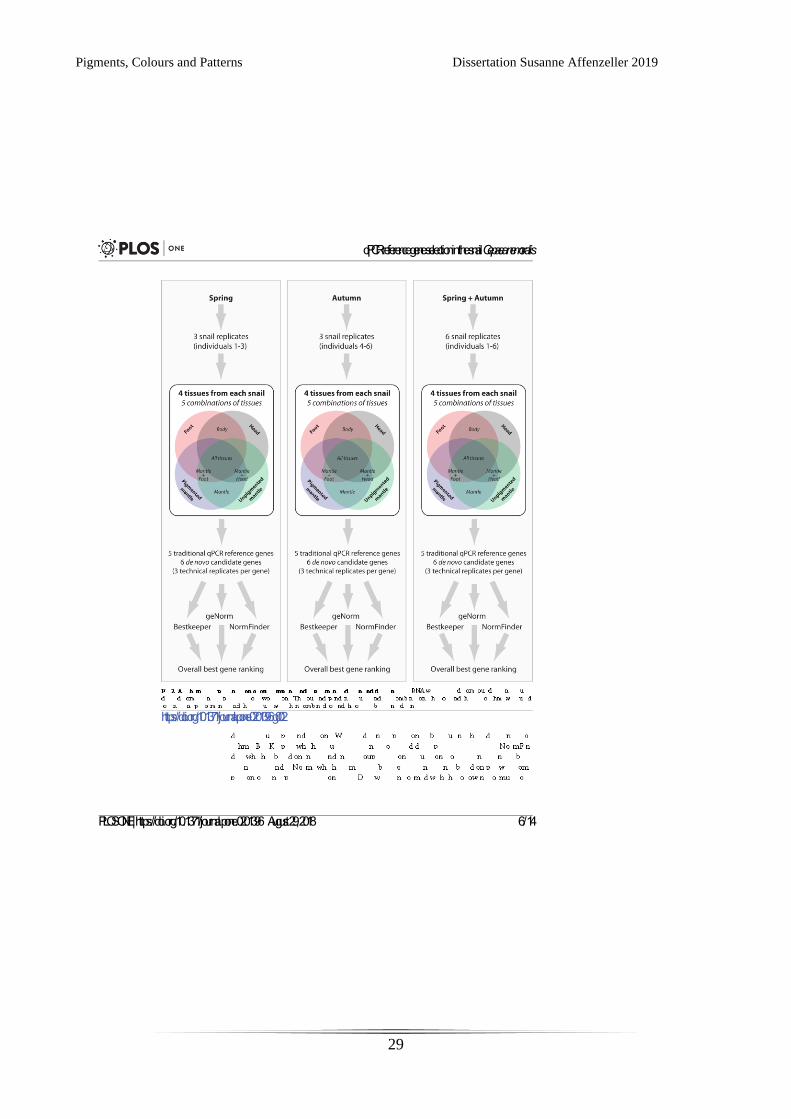
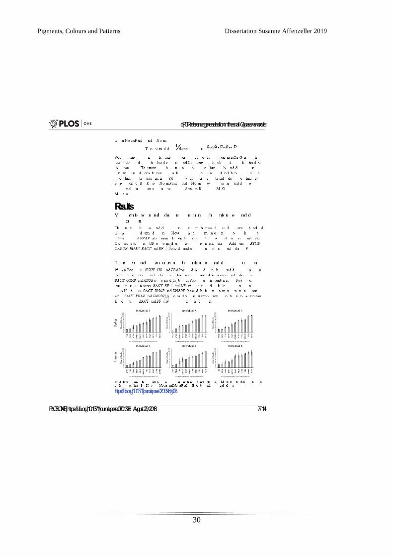
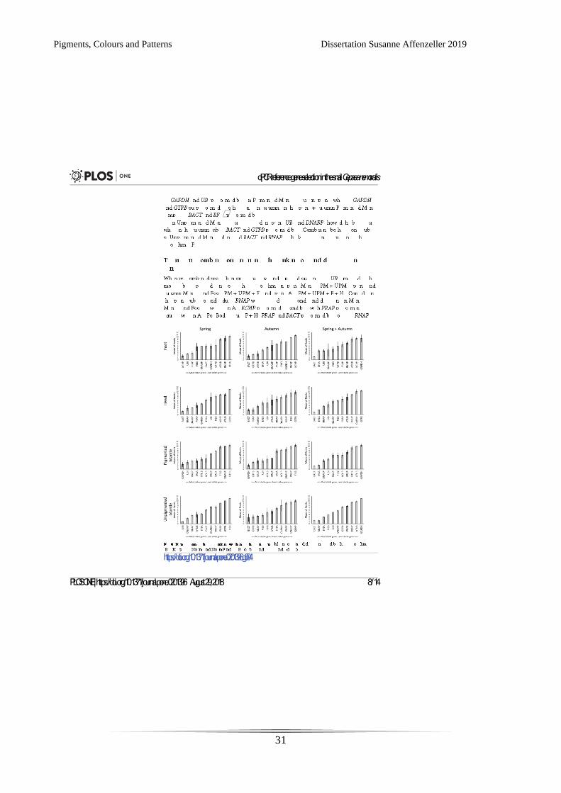
CHAPTER 2: IDENTIFICATION AND VALIDATION OF REFERENCE GENES FOR qPCR IN
THE TERRESTRIAL GASTROPOD CEPAEA NEMORALIS.
The following manuscript was published as follows
Affenzeller S, Cerveau N, Jackson DJ. Identification and validation of reference genes for qPCR in
the terrestrial gastropod Cepaea nemoralis. PloS one. 2018 Aug 29;13(8):e0201396.
and is reprinted here with permission form PLoS One.
Contributions by doctoral candidate: Formal analysis, Methodology, Writing – original draft, Writing
– review & editing

Pigments, Colours and Patterns Dissertation Susanne Affenzeller 2019
23
Pigments, Colours and Patterns Dissertation Susanne Affenzeller 2019
24
Pigments, Colours and Patterns Dissertation Susanne Affenzeller 2019
25
Pigments, Colours and Patterns Dissertation Susanne Affenzeller 2019
26
Pigments, Colours and Patterns Dissertation Susanne Affenzeller 2019
27

Pigments, Colours and Patterns Dissertation Susanne Affenzeller 2019
28
Pigments, Colours and Patterns Dissertation Susanne Affenzeller 2019
29
Pigments, Colours and Patterns Dissertation Susanne Affenzeller 2019
30
Pigments, Colours and Patterns Dissertation Susanne Affenzeller 2019
31
Pigments, Colours and Patterns Dissertation Susanne Affenzeller 2019
32
Pigments, Colours and Patterns Dissertation Susanne Affenzeller 2019
33

Pigments, Colours and Patterns Dissertation Susanne Affenzeller 2019
34
Pigments, Colours and Patterns Dissertation Susanne Affenzeller 2019
35
Pigments, Colours and Patterns Dissertation Susanne Affenzeller 2019
36
Pigments, Colours and Patterns Dissertation Susanne Affenzeller 2019
37
Pigments, Colours and Patterns Dissertation Susanne Affenzeller 2019
38
CHAPTER 3: QUANTITATION OF EUMELANIN AND PHEOMELANIN MARKERS IN
DIVERSE BIOLOGICAL SAMPLES BY HPLC-UV-MS FOLLOWING SOLID-PHASE
EXTRACTION
The following chapter was submitted to PloS One and is currently under revision.
Contributions by the doctoral candidate: Conceptualization, Formal analysis, Investigation,
Methodology, Validation, Writing – original draft, Writing – review & editing

Pigments, Colours and Patterns Dissertation Susanne Affenzeller 2019
39
Pigments, Colours and Patterns Dissertation Susanne Affenzeller 2019
40
Quantitation of eumelanin and pheomelanin markers in diverse biological
samples by HPLC-UV-MS following solid-phase extraction
Susanne Affenzeller1, Holm Frauendorf2, Tobias Licha3, Daniel J. Jackson1,* and Klaus
Wolkenstein1,*
1 Department of Geobiology, Georg-August University, 37077 Göttingen, Germany
2 Institute of Organic & Biomolecular Chemistry, Georg-August University, 37077 Göttingen,
Germany
3 Department of Applied Geology, Georg-August University, 37077 Göttingen, Germany
*Authors for correspondence:
Daniel J. Jackson
email: [email protected]
ORCID: 0000-0001-9045-381X
Klaus Wolkenstein
email: [email protected]
ORCID: 0000-0003-0944-6247
Pigments, Colours and Patterns Dissertation Susanne Affenzeller 2019
41
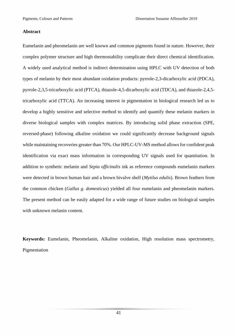
Abstract
Eumelanin and pheomelanin are well known and common pigments found in nature. However, their
complex polymer structure and high thermostability complicate their direct chemical identification.
A widely used analytical method is indirect determination using HPLC with UV detection of both
types of melanin by their most abundant oxidation products: pyrrole-2,3-dicarboxylic acid (PDCA),
pyrrole-2,3,5-tricarboxylic acid (PTCA), thiazole-4,5-dicarboxylic acid (TDCA), and thiazole-2,4,5-
tricarboxylic acid (TTCA). An increasing interest in pigmentation in biological research led us to
develop a highly sensitive and selective method to identify and quantify these melanin markers in
diverse biological samples with complex matrices. By introducing solid phase extraction (SPE,
reversed-phase) following alkaline oxidation we could significantly decrease background signals
while maintaining recoveries greater than 70%. Our HPLC-UV-MS method allows for confident peak
identification via exact mass information in corresponding UV signals used for quantitation. In
addition to synthetic melanin and Sepia officinalis ink as reference compounds eumelanin markers
were detected in brown human hair and a brown bivalve shell (Mytilus edulis). Brown feathers from
the common chicken (Gallus g. domesticus) yielded all four eumelanin and pheomelanin markers.
The present method can be easily adapted for a wide range of future studies on biological samples
with unknown melanin content.
Keywords: Eumelanin, Pheomelanin, Alkaline oxidation, High resolution mass spectrometry,
Pigmentation
Pigments, Colours and Patterns Dissertation Susanne Affenzeller 2019
42
1. Introduction
In the scientific literature the term ‘melanin’ has been used for any number of black, dark brown to
orange and yellow pigments that are non-soluble and very thermostable 1–7. A more accurate
definition of melanin would be that they are built through enzymatic oxidative polymerisation of
DOPA (L-3,4-dihydroxyphenylalanine) subunits 4,8–12. The stability and sizes of the resulting
macromolecules complicates their analysis by standard analytical methods 10,12,13. A well-established
method in human medical studies to identify the most common melanin types (eumelanin and
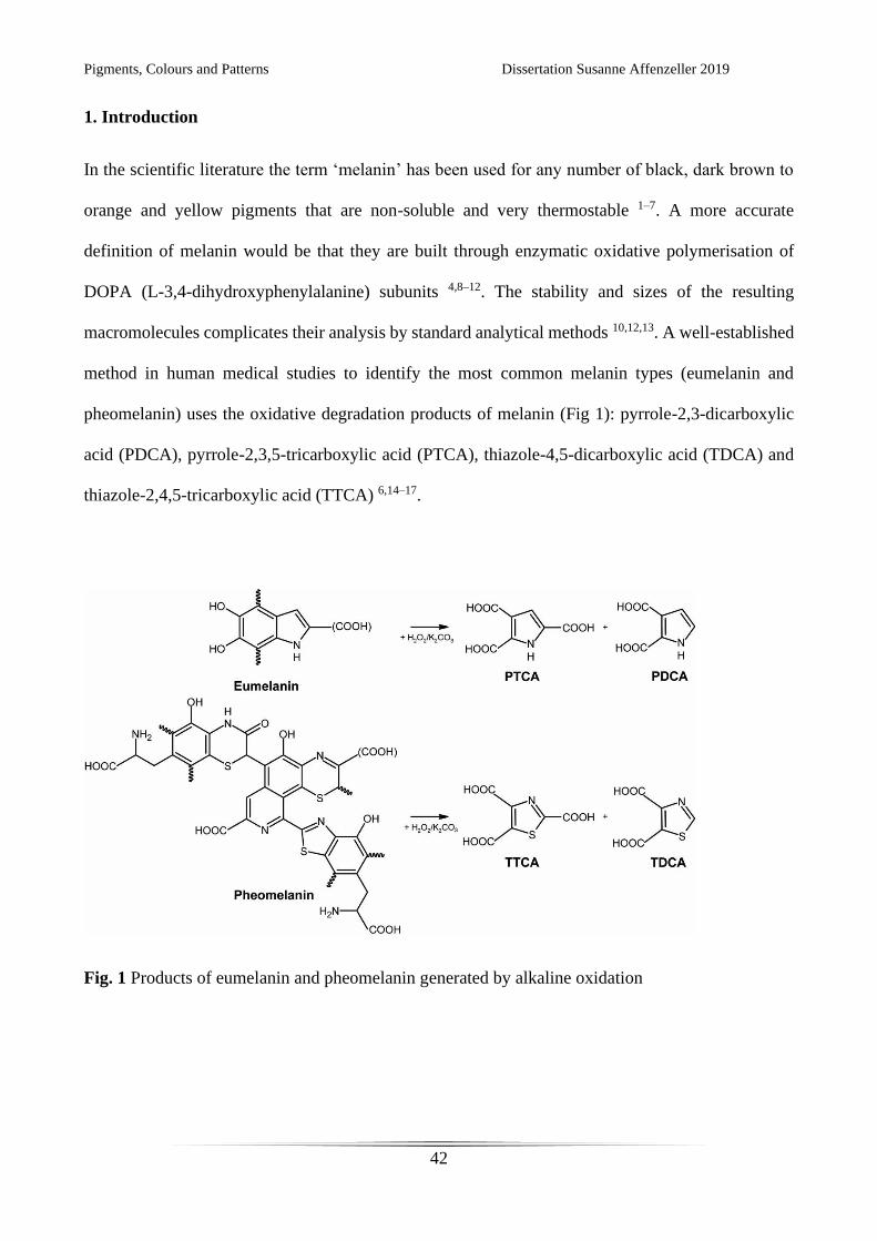
pheomelanin) uses the oxidative degradation products of melanin (Fig 1): pyrrole-2,3-dicarboxylic
acid (PDCA), pyrrole-2,3,5-tricarboxylic acid (PTCA), thiazole-4,5-dicarboxylic acid (TDCA) and
thiazole-2,4,5-tricarboxylic acid (TTCA) 6,14–17.
Fig. 1 Products of eumelanin and pheomelanin generated by alkaline oxidation

Pigments, Colours and Patterns Dissertation Susanne Affenzeller 2019
43
Further experiments based on the initially established method of potassium permanganate as
an oxidative agent 14–16 yielded an improved oxidation protocol using alkaline oxidation by hydrogen
peroxide 6,18–20. High-performance liquid chromatography (HPLC) with UV detection and a mobile
phase consisting of methanol–phosphate buffer (pH 2.1) has been used to separate and detect these
specific melanin oxidation products 6,20. This method was mainly developed to analyse human
melanin in medical applications like hair and skin samples, where the presence of melanin is
indisputable and interest lies mainly in quantitative analysis. But in recent years biologists have been
increasingly interested in pigments and patterns and their presence in a variety of biological settings
21–27. For these biological samples with unknown pigment content a more specific method is needed.
Furthermore, in biological samples like hair, feathers and shells, analyses of melanin is greatly
hindered by the presence of complex organic matrices resulting from the oxidation of proteins and
other compounds by H2O2 (compare chromatograms in Ito et al. 20, Williams et al. 25). Another effect
of these biological samples with naturally high background signals is that reliable peak identification
can be very difficult. The introduction of a simple sample preparation step that minimizes background
signals is therefore necessary. Yu et al. 28 used a liquid-liquid extraction method for this purpose, but
did not systematically test the effect of this step on established melanin oxidation markers. While
Rioux et al. 29 investigated the effects of solid phase extraction (SPE) using weak anion exchange
columns on melanin oxidation products, but focused solely on melanoma cells as a biological sample
and refrained from adapting the method for compatibility to mass spectrometric (MS) methods.
In order to overcome the limitation of low selectivity afforded by UV detection, recent
methods have replaced the phosphate buffer in the eluent with formic acid to allow MS detection. For
time of flight (TOF) -MS detection of eumelanin pigments, preparative separation of the analytes
prior to MS analysis was required 23, whereas more sensitive LC (liquid chromatography) MS/MS
methods for the analysis of the eumelanin markers PDCA and PTCA in bivalve tissue 28 and for
PTCA in hair samples 30 have been reported. However, to date, no method for the full
Pigments, Colours and Patterns Dissertation Susanne Affenzeller 2019
44
chromatographic separation and sensitive MS detection of eumelanin and pheomelanin markers has
been reported.
The aim of the present study is to establish a reliable analytical method for SPE sample clean-
up and subsequent detection and quantitation even of trace amounts of the eumelanin markers PTCA
and PDCA as well as the pheomelanin markers TTCA and TDCA from diverse biological samples.
2. Materials and methods
2.1 Chemicals and reagents
Water (HPLC gradient grade) was purchased from J.T. Baker (Deventer, The Netherlands).
Pestinorm® Supra Trace ethyl acetate was purchased from VWR Chemicals (Leuven, Belgium).
LiChrosolv® methanol (hypergrade for LC-MS), potassium carbonate (pro analysi grade) and
calcium carbonate (pro analysi grade) were obtained from Merck KGaA (Darmstadt, Germany).
Sodium sulphite (puriss p. a., ACS grade) and hydrogen peroxide solution (≥ 30%, trace analysis
grade) were obtained from Sigma-Aldrich (St. Louis, USA). Rotipuran® formic acid (≥98%, p.a.,
ACS grade), Rotipuran® hydrochloric acid (37%, p.a., ACS, ISO grade) and Pufferan® TRIS
hydrochloride (≥ 99%, p.a. grade) were purchased from Carl Roth GmbH + Co. KG (Karlsruhe,
Germany). Proteinase K was purchased from Qiagen GmbH (Hilden, Germany).
2.2 Standards and samples
Standards of the melanin oxidation products PDCA, PTCA, TDCA and TTCA (prepared according
to previously published protocols 31) were kindly provided by Prof. Shosuke Ito at stock solution
concentrations of 100 μg/mL. Synthetic melanin (prepared by oxidation of tyrosine with hydrogen
peroxide) and melanin from Sepia officinalis were obtained from Sigma-Aldrich (St. Louis, USA).
Pigments, Colours and Patterns Dissertation Susanne Affenzeller 2019
45
The human hair sample was donated by one of the authors (S.A.). Medium brown hair was
cut approximately 10 cm from the scalp and then cut into 5 mm pieces. A brown chicken feather was
obtained from a domestic chicken (Gallus gallus domesticus). Analyses were carried out on the distal
tip of the brown feather. A shell of the bivalve Mytilus edulis was commercially obtained from a food
market. Shell samples were taken from the distal growing edge which possessed brown longitudinal
stripes.
2.3 Sample preparation and melanin oxidation
All biological samples were cleaned with deionized water in an ultrasonic bath for 10 min and allowed
to dry. For synthetic melanin and Sepia ink melanin 0.2 mg, for feather and hair samples 5.5 mg and
for shell sample 1.5 g were used, respectively. Melanin oxidation was carried out as previously
published 20,25 with some modifications: Prepared shell pieces were dissolved in HCl (6 M,
approximately 7 mL) and centrifuged at 13,000 rpm for 15 min. The obtained supernatant was
discarded and the residue was washed twice with H2O.
Biological samples (hair, feather and shell) were treated with 10 μL proteinase K (10 mg/mL)
in 500 μL TRIS-HCl buffer (1 M, pH 8.0) for 30 min at 37 °C in a shaker. Treatment was stopped by
the addition of 300 μL HCl (6 M). Samples were centrifuged at 13,000 rpm for 15 min, the supernatant
discarded and the pellet was washed in water.
All oxidation reactions (synthetic melanin, Sepia ink melanin, feather, hair and shell) were
carried out for 20 h at 25 °C with vigorous shaking using 100 μL H2O, 375 μL K2CO3 (1 M) and 25
μL H2O2 (30%) as reactants. After this time any remaining H2O2 was inactivated by the addition of
50 μL Na2SO3 (10% (w/v) and 140 μL HCl (6 M). Samples were then centrifuged at 13,000 rpm for
30 min and the supernatant was transferred into a fresh tube. As a negative control 2.0 g of calcium
Pigments, Colours and Patterns Dissertation Susanne Affenzeller 2019
46
carbonate was treated like the shell sample. For comparison, a mixture of PTCA, PDCA, TTCA and
TDCA in H2O (2.5 μg/mL each) was run under the same conditions as the oxidised samples.
2.4 Sample treatment using SPE
Oxidised samples were treated by SPE on Strata™-X 33 μm Polymeric Reversed Phase cartridges
200 mg/6 mL (Phenomenex, Torrance, USA) under vacuum. Cartridges were conditioned with 5 mL
methanol followed by 5 mL H2O. Samples were loaded onto the SPE cartridges diluted in 5 ml formic
acid (0.3% (v/v)) and washed once with 5 mL formic acid (0.3% (v/v)). The cartridges were then
dried for 30 min and elution was carried out with 3 mL methanol followed by 3 mL ethyl acetate.
Solvents were removed under a constant nitrogen stream at 40 °C and samples were re-dissolved in
200 μL H2O.
2.5 Chromatographic separation with UV and MS detection
Measurements were carried out on a Thermo Fisher Scientific HPLC-MS system consisting of an
Accela HPLC with a Finnigan Surveyor PDA Detector and coupled to an LTQ Orbitrap XL mass
spectrometer equipped with an electrospray ionization (ESI) source. Chromatographic separation was
carried out on a Gemini C18 column (5 μm particle size, 250 × 2 mm i.d. (Phenomenex, Torrance,
USA)). Aliquots (10 μL) of the samples were injected into the HPLC system operating at a flow rate
of 0.2 mL/min. The mobile phase consisted of 0.3% formic acid (eluent A) and methanol (eluent B)
(80:20) was run at 45 °C for 20 min isocratically, followed by a wash step of A:B (5:95) for 10 min
and an equilibration phase to reach starting conditions for 10 min. UV data were recorded between
200 and 400 nm. Quantitation was conducted in the range of 250–290 nm. Mass spectra were acquired
in negative ion mode. The scan window was set to m/z = 120–220. Optimized MS conditions

Pigments, Colours and Patterns Dissertation Susanne Affenzeller 2019
47
included: gas flow rate of 50 (arbitrary units), a spray voltage of 5.0 kV and a heated capillary
temperature of 275 °C.
2.6 Calibration and validation
The linear range of the method was tested for each of the melanin oxidation products with a 9-point
calibration curve at concentrations ranging from 0.01 to 10 μg/mL by multiple injections. Limit of
detection (LOD) and limit of quantitation (LOQ) were determined with the signal-to-noise ratio
method for each of the standards based on HPLC measurements with UV detection. LOD was set at
3:1 and LOQ at 10:1 signal-to-noise respectively.
Recoveries after sample preparation by SPE were tested with a mixture of all four melanin
oxidation products in eluent A. Additionally, total method recovery was investigated in all three
natural matrices (feather, hair, shell) by a 3-point standard addition (2 times, 5 times and 10 times) of
all oxidation products. SPE recoveries without matrices were measured on an Agilent 1200 Series
HPLC system with diode array detector using the same chromatographic conditions as described
above.
Additional experiments on the oxidation protocol itself verified the linearity of PDCA and
PTCA from a synthetic eumelanin standard in the range of 0.05–0.4 mg. A test with an elongated
oxidation time (40 h) did not result in significantly higher amounts of oxidation products and even
yielded slightly less eumelanin markers in the case of shell samples.
3. Results and discussion
3.1 Method development
Pigments, Colours and Patterns Dissertation Susanne Affenzeller 2019
48
Our method for alkaline oxidation of melanin from a wide range of biological samples combines and
refines a variety of previously published protocols 20,21,23,25. We demonstrate that by including a
sample clean up step by SPE and adapting the chromatographic system to allow for dual detection
with UV and MS, two markers each for eumelanin (PDCA and PTCA) and pheomelanin (TDCA and
TTCA) can be analysed within one HPLC run. We could achieve baseline separation of all four
melanin oxidation markers for chromatographic conditions compatible with mass spectrometry. By
desalting and purifying samples via SPE we could significantly reduce the background of the diverse
samples we investigated. In addition, the evaporation step brings the advantage of concentrating the
analytes. A surprising observation during reversed-phase HPLC optimization was that the retention
time of the analytes was almost unaffected by the concentration of organic solvent. Instead, retention
times were strongly affected by the pH value of the eluent, an effect also observed in a recent study
29.
Pigments, Colours and Patterns Dissertation Susanne Affenzeller 2019
49
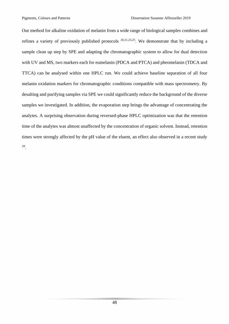
Fig. 2 Characterization of the melanin oxidation products PDCA, TDCA, PTCA and TTCA from
brown chicken feather by mass spectra (negative ion mode) and UV spectra
Pigments, Colours and Patterns Dissertation Susanne Affenzeller 2019
50
The high-resolution ESI mass spectra of PDCA, TDCA and PTCA show ion signals for the
deprotonated molecules [M–H]–. TDCA and PTCA show additional ions resulting from
fragmentation of CO2 from one of the carboxyl groups. In contrast, for TTCA only fragment ions
resulting from the loss of one and two carboxyl groups can be observed, yielding virtually the same
mass spectrum as TDCA (see Table 1 and Fig. 2). Therefore the identification and quantitation of
both TDCA and TTCA is only possible by the inclusion of a chromatographic separation. All four
melanin oxidation products can be characterized by their specific UV absorption spectra (Fig. 2),
when there are no interfering background peaks. Quantitation in MS requires appropriate internal
standards which are not commercially available for the analysed melanin markers. In contrast,
quantitation with UV detection can be done with melanin oxidation product standards via external
calibration or standard addition. However peak identification must be carried out very carefully due
to the naturally high backgrounds present in biological samples even after clean-up by SPE (compare
Fig. 3). By coupling UV with high-resolution MS detection, straight-forward quantitation based on
UV signals can be combined with reliable compound identification in complex biological samples.
In addition, mass spectrometric measurements yield an approximately 1.5 times higher sensitivity
than UV detection, an important factor when analysing biological samples with unknown melanin
content.
Previous method validations for the analyses of melanin oxidation products have not been
thoroughly performed 6,17,20,32. We present here calibration and validation data for the quantitation of
all four melanin oxidation product standards via UV detection (Table 2) following SPE. Linearity
could be shown for both eumelanin oxidation markers (PDCA and PTCA) in the range from 0.05–10
μg/mL. For pheomelanin oxidation markers (TDCA and TTCA) linearity ranged from 0.1–10 μg/mL.
Table 1 Accurate mass data of melanin oxidation products

Pigments, Colours and Patterns Dissertation Susanne Affenzeller 2019
51
Compound Molecular
formula
Calculated [M–H]–
m/z
Observed [M–H]–
m/z
Observed fragment
ions m/z
PDCA C6H5NO4 154.0140 154.0143 –
PTCA C7H5NO6 198.0039 198.0040, 154.0143
TDCA C5H3NO4S 171.9705 171.9706, 127.9810
TTCA C6H3NO6S 215.9603 – 171.9706, 127.9810
Fig. 3 Comparison of extracted ion
chromatograms (a) and corresponding UV
chromatogram (b) of oxidation products from a
brown chicken feather following SPE,
highlighting the need for peak confirmation in
biological samples

Pigments, Colours and Patterns Dissertation Susanne Affenzeller 2019
52
A recently published LC-MS/MS study on human hair determined LOD and LOQ for one of
the four standard melanin oxidation markers (PTCA) 30. Our method allows all four oxidation
products to be detected in very small amounts, for PTCA below or at a comparable level to LODs
previously published 21,29,30. Both eumelanin markers can be quantified at concentrations as low as
0.1 μg/mL. For pheomelanin markers the lowest quantifiable concentrations were 0.25 μg/mL for
TDCA and 0.33 μg/mL for TTCA.
Fig. 4 Extracted ion chromatograms of melanin oxidation
product standards and melanin reference compounds
(standard mixture of oxidation products, synthetic eumelanin,
Sepia ink melanin) as well as biological samples (human hair,
chicken feather, bivalve shell) following alkaline oxidation
Table 2 Limit of detection (LOD), limit of quantitation (LOQ) and linearity (R²) for HPLC with UV
detection
Pigments, Colours and Patterns Dissertation Susanne Affenzeller 2019
53
Compoun
d
LOD
(μg/mL)
LOQ
(μg/mL) Linearity (R²)
Range of Linearity
(μg/mL)
PDCA 0.03 0.08 0.995 0.05–10
PTCA 0.04 0.10 0.994 0.05–10
TDCA 0.08 0.25 0.994 0.1–10
TTCA 0.10 0.33 0.990 0.1–10
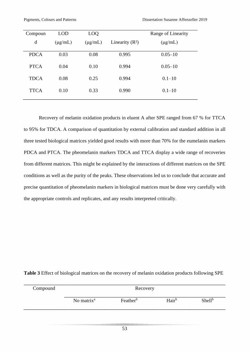
Recovery of melanin oxidation products in eluent A after SPE ranged from 67 % for TTCA
to 95% for TDCA. A comparison of quantitation by external calibration and standard addition in all
three tested biological matrices yielded good results with more than 70% for the eumelanin markers
PDCA and PTCA. The pheomelanin markers TDCA and TTCA display a wide range of recoveries
from different matrices. This might be explained by the interactions of different matrices on the SPE
conditions as well as the purity of the peaks. These observations led us to conclude that accurate and
precise quantitation of pheomelanin markers in biological matrices must be done very carefully with
the appropriate controls and replicates, and any results interpreted critically.
Table 3 Effect of biological matrices on the recovery of melanin oxidation products following SPE
Compound Recovery
No matrixa Featherb Hairb Shellb
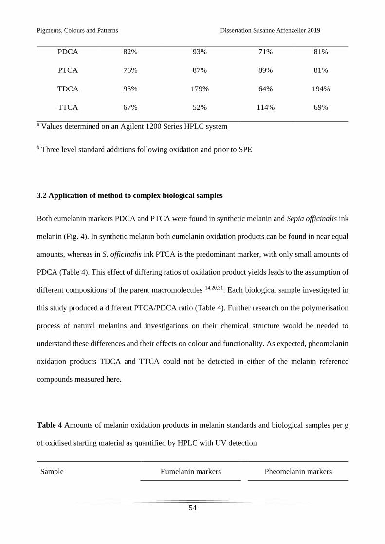
Pigments, Colours and Patterns Dissertation Susanne Affenzeller 2019
54
PDCA 82% 93% 71% 81%
PTCA 76% 87% 89% 81%
TDCA 95% 179% 64% 194%
TTCA 67% 52% 114% 69%
a Values determined on an Agilent 1200 Series HPLC system
b Three level standard additions following oxidation and prior to SPE
3.2 Application of method to complex biological samples
Both eumelanin markers PDCA and PTCA were found in synthetic melanin and Sepia officinalis ink
melanin (Fig. 4). In synthetic melanin both eumelanin oxidation products can be found in near equal
amounts, whereas in S. officinalis ink PTCA is the predominant marker, with only small amounts of
PDCA (Table 4). This effect of differing ratios of oxidation product yields leads to the assumption of
different compositions of the parent macromolecules 14,20,31. Each biological sample investigated in
this study produced a different PTCA/PDCA ratio (Table 4). Further research on the polymerisation
process of natural melanins and investigations on their chemical structure would be needed to
understand these differences and their effects on colour and functionality. As expected, pheomelanin
oxidation products TDCA and TTCA could not be detected in either of the melanin reference
compounds measured here.
Table 4 Amounts of melanin oxidation products in melanin standards and biological samples per g
of oxidised starting material as quantified by HPLC with UV detection
Sample Eumelanin markers
Pheomelanin markers
Pigments, Colours and Patterns Dissertation Susanne Affenzeller 2019
55
PDCA
(μg/g sample)
PTCA
(μg/g
sample)
TDCA
(μg/g sample)
TTCA
(μg/g sample)
Synthetic melanin 1206.23 963.80
< LOD < LOD
S. officinalis ink melanin 99.40 1808.25
< LOD < LOD
H. sapiens brown hair 3.75 60.60
< LOQ < LOD
G. g. domesticus brown feather 43.25 119.92
108.99 403.73
M. edulis brown shell 0.03 0.11
< LOD < LOD
In all three of the investigated biological matrices (feather, hair, shell) we were able to detect
eumelanin (Fig. 4 and Table 4). In the feather and hair samples we could also detect pheomelanin. It
has been shown already that feathers of North American barn swallows (Hirundo rustica
erythrogaster) contain both eumelanins and pheomelanins 21. The same study investigated yellow
chicken plumage from nestlings and found trace amounts of eumelanin. In applying our method to
adult brown chicken feathers we detected an abundance of all four oxidation products, confirming
the results of previous electron spin resonance investigations 33.
A difficult biological matrix from which to extract organic macromolecules from are the
calcified shells of molluscs. The pigment bearing layer of Mytilus edulis is very thin, providing only
small amounts of pigment from relatively large amounts of shell material. Nonetheless, we were able
to detect eumelanin in this bivalve, providing further evidence that our method is sensitive enough to
detect these pigments in a range of biological matrices. Pigmentation and the use of melanin to pattern
shells and nacreous materials by a variety of molluscs has seen an increase in research in recent years
28,34,35. Our method facilitates working with these complex samples and will hopefully lead to further
investigations in melanic mollusc shell pigmentation.
An especially challenging sample type are fossilized tissues and matrices containing melanin.
Although melanin seems to fossilize very well 23,36,37 only few researchers have access to enough

Pigments, Colours and Patterns Dissertation Susanne Affenzeller 2019
56
material to analyse these samples with chromatographic methods with UV detection. Mass
spectrometric measurements were previously performed for fossilized Sepia ink 23 which found
evidence for eumelanin. The protocols presented here are a good starting point for further adjustments
of sample preparation with SPE, and the development of even more sensitive MS methods for fossil
samples suspected to contain melanin. Preliminary measurements using MS/MS detection have
shown that the sensitivity of our method can be further improved by several orders of magnitude.
4. Conclusion
The method we present here allows researchers to detect eumelanin and pheomelanin in a variety of
complex biological samples. The cleaning and concentrating effect afforded by SPE and the addition
of mass spectrometry allows for the selective identification of even small amounts of known melanin
oxidation products. High resolution mass spectrometry allows confident peak identification even in
complex biological samples with interfering background and overlapping peaks in chromatograms
provided by UV detection.
In contrast to the difficult analysis of their parent macromolecules, oxidation products for
eumelanin and pheomelanin can be quantitated with the present method. However, due to the different
compositions of natural melanins (e.g. PDCA/PTCA ratio differences in reference compounds and
biological samples) and matrix effects of biological samples on SPE (see Table 3), we recommend
the use of both oxidation markers for each type of melanin as indicators for the abundance of
eumelanin and pheomelanin pigments in the original sample.
The highly sensitive method that we report here improves our ability to simultaneously detect
eumelanin and pheomelanin in a variety of complex biological samples.
Pigments, Colours and Patterns Dissertation Susanne Affenzeller 2019
57
Conflict of interests
There are no conflicts of interest to declare.
Acknowledgements
The authors are very grateful to Professors Shosuke Ito and Kazumasa Wakamatsu (Department of
Chemistry, Fujita Health University School of Health Sciences) for providing standard solutions for
melanin oxidation products. We thank Gabriele Müller for the donation of the brown chicken feather.
This research was funded by DFG grant WO 1491/4-2 to KW, and JA 2108/2-1 and JA 2108/6-1 to
DJJ.
References
1. Comfort A. The pigmentation of molluscan shells. Biol Rev. 1951;26(3):285–301.
2. Swan GA, Waggott A. Studies related to the chemistry of melanins. Part X. Quantitative assessment of
different types of units present in dopa-melanin. J Chem Soc C: Org. 1970;(10):1409–18.
3. Swan G. Structure, chemistry, and biosynthesis of the melanins. Fortschr Chem Org Naturst. Springer, Vienna;
1974. pp. 521–582.
4. Riley PA. Melanin. Int J Biochem Cell Biol. 1997;29(11):1235–9.
5. Prota G. Progress in the chemistry of melanins and related metabolites. Med Res Rev . 1988;8(4):525–56.
6. Wakamatsu K, Ito S. Advanced chemical methods in melanin determination. Pigment Cell Res.
2002;15(3):174–83.
7. Ito S, Wakamatsu K. Quantitative analysis of eumelanin and pheomelanin in humans, mice, and other animals:
a comparative review. Pigment Cell Res. 2003;16(5):523–31.
8. Lerner AB, Fitzpatrick TB. Biochemistry of melanin formation. Physiol Rev. 1950;30(1):91–126.
9. Binns F, King JA, Mishra SN, Percival A, Robson NC, Swan GA, Waggott A. Studies related to the chemistry
of melanins. Part XIII. Studies on the structure of dopamine-melanin. J Chem Soc C: Org. 1970(15):2063–70.
10. Ito S. Reexamination of the structure of eumelanin. Biochim Biophys Acta Gen Subj. 1986;883(1):155–61.
11. d'Ischia M, Napolitano A, Prota G. Peroxidase as an alternative to tyrosinase in the oxidative polymerization of
5,6-dihydroxyindoles to melanin (s). Biochim Biophys Acta Gen Subj. 1991;1073(2):423–30.
12. Prota G. Melanins and Melanogenesis. New York: Academic Press; 1992. pp. 1–290.
Pigments, Colours and Patterns Dissertation Susanne Affenzeller 2019
58
13. Prota G. The chemistry of melanins and melanogenesis. Fortschr Chem Org Naturst. Springer, Vienna; 1995.
pp. 93–148.
14. Piattelli M, Nicolaus RA. The structure of melanins and melanogenesis—I: The structure of melanin in Sepia.
Tetrahedron. 1961;15(1–4):66–75.
15. Nicolaus RA, Piattelli M, Fattorusso E. The structure of melanins and melanogenesis—IV: On some natural
melanins. Tetrahedron. 1964;20(5):1163–72.
16. Ito S, Fujita K. Microanalysis of eumelanin and pheomelanin in hair and melanomas by chemical degradation
and liquid chromatography. Anal Biochem. 1985;144(2):527–36.
17. Napolitano A, Perzzella A, Vincensi MR, Prota G. Oxidative degradation of eumelanins to pyrrole acids: a
model study. Tetrahedron 1995;51:5913–5920.
18. Ito S, Wakamatsu K. Chemical degradation of melanins: application to identification of dopamine melanin.
Pigment Cell Res. 1998;11(2):120–6.
19. Wakamatsu K, Fujikawa K, Zucca FA, Zecca L, Ito S. The structure of neuromelanin as studied by chemical
degradative methods. J Neurochem. 2003;86(4):1015–23.
20. Ito S, Nakanishi Y, Valenzuela RK, Brilliant MH, Kolbe L, Wakamatsu K. Usefulness of alkaline hydrogen
peroxide oxidation to analyze eumelanin and pheomelanin in various tissue samples: application to chemical
analysis of human hair melanins. Pigment Cell Melanoma Res. 2011;24(4): 605–613.
21. McGraw KJ, Wakamatsu K, Ito S, Nolan PM, Jouventin P, Dobson FS, Austic RE, Safran RJ, Siefferman LM,
Hill GE, Parker RS. You can't judge a pigment by its color: carotenoid and melanin content of yellow and
brown feathers in swallows, bluebirds, penguins, and domestic chickens. The Condor. 2004;106(2):390–5.
22. Chen SR, Jiang B, Zheng JX, Xu GY, Li JY, Yang N. Isolation and characterization of natural melanin derived
from silky fowl (Gallus gallus domesticus Brisson). Food Chem. 2008;111(3):745–9.
23. Glass K, Ito S, Wilby PR, Sota T, Nakamura A, Bowers CR, Vinther J, Dutta S, Summons R, Briggs DEG,
Wakamatsu K, Simon JD. Direct chemical evidence for eumelanin pigment from the Jurassic period. Proc Natl
Acad Sci U S A. 2012;109(26):10218–10223.
24. Kronforst MR, Barsh GS, Kopp A, Mallet J, Monteiro A, Mullen SP, Protas M, Rosenblum EB, Schneider CJ,
Hoekstra HE. Unraveling the thread of nature’s tapestry: the genetics of diversity and convergence in animal
pigmentation. Pigment Cell Melanoma Res. 2012;25(4):411–33.
25. Williams ST, Ito S, Wakamatsu K, Goral T, Edwards NP, Wogelius RA, Henkel T, de Oliveira LF, Maia LF,
Strekopytov S, Jeffries T. Identification of shell colour pigments in marine snails Clanculus pharaonius and C.
margaritarius (Trochoidea; Gastropoda). PloS One. 2016;11(7):e0156664.
26. Cuthill IC, Allen WL, Arbuckle K, Caspers B, Chaplin G, Hauber ME, Hill GE, Jablonski NG, Jiggins CD,
Kelber A, Mappes J. The biology of color. Science. 2017;357(6350):eaan0221.
27. Williams ST. Molluscan shell colour. Biol Rev. 2017;92(2):1039–58.
28. Yu F, Pan Z, Qu B, Yu X, Xu K., Deng Y, Liang, F. Identification of a tyrosinase gene and its functional
analysis in melanin synthesis of Pteria penguin. Gene. 2018;656:1–8.
29. Rioux B, Rouanet J, Akil H, Besse S, Debiton E, Bouchon B, Degoul F, Quintana M. Determination of
eumelanin and pheomelanin in melanomas using solid-phase extraction and high performance liquid
chromatography–diode array detection (HPLC-DAD) analysis. Journal of Chromatography B. 2019; 1113:60–
68
30. Petzel-Witt S, Meier SI, Schubert Zsilavecz M, Toennes SW. PTCA (1H pyrrole 2,3,5 tricarboxylic acid) as a
marker for oxidative hair treatment. Drug Test Anal. 2018;10(4):768–73.
Pigments, Colours and Patterns Dissertation Susanne Affenzeller 2019
59
31. d’Ischia M, Wakamatsu K, Napolitano A, Briganti S, Garcia-Borron J-C, Kovacs D, u.a. Melanins and
melanogenesis: methods, standards, protocols. Pigment Cell & Melanoma Research. September
2013;26(5):616–33.
32. Magarelli M, Passamonti P, Renieri C. Purification, characterization and analysis of sepia melanin from
commercial sepia ink (Sepia officinalis)––Purificación, caracterización y análisis de la melanina de sepia a
partir de la tinta de sepia (Sepia officinalis). CES Medicina Veterinaria y Zootecnia. 2010;5(2):18–28.
33. Sealy RC, Hyde JS, Felix CC, Menon IA, Prota G. Eumelanins and pheomelanins: characterization by electron
spin resonance spectroscopy. Science. 1982;217(4559):545–7.
34. Hao S, Hou X, Wei L, Li J, Li Z, Wang X. Extraction and identification of the pigment in the adductor muscle
scar of pacific oyster Crassostrea gigas. PloS One. 2015;10(11):e0142439.
35. Sun X, Wu B, Zhou L, Liu Z, Dong Y, Yang A. Isolation and characterization of melanin pigment from yesso
scallop Patinopecten yessoensis. J Ocean Univ China. 2017;16(2):279–84.
36. Colleary C, Dolocan A, Gardner J, Singh S, Wuttke M, Rabenstein R, Habersetzer J, Schaal S, Feseha M,
Clemens M, Jacobs BF. Chemical, experimental, and morphological evidence for diagenetically altered
melanin in exceptionally preserved fossils. Proc Natl Acad Sci U S A. 2015;112(41):12592–7.
37. Lindgren J, Sjövall P, Thiel V, Zheng W, Ito S, Wakamatsu K, Hauff R, Kear BP, Engdahl A, Alwmark C,
Eriksson ME, Jarenmark M, Sachs S, Ahlberg PE, Marone F, Kuriyama T, Gustafsson O, Malmberg P,
Thomen A, Rodríguez-Meizoso I, Uvdal P, Ojika M, Schweitzer MH. Soft-tissue evidence for homeothermy
and crypsis in a Jurassic ichthyosaur. Nature. 2018;564(7736):359–365.
Pigments, Colours and Patterns Dissertation Susanne Affenzeller 2019
60
CHAPTER 4: EUMELANIN AND PHEOMELANIN PIGMENTATION IN MOLLUSC SHELLS
MAY BE LESS COMMON THAN EXPECTED: INSIGHTS FROM MASS SPECTROMETRY
The following chapter is prepared for publication in Proceedings of the Royal Society B and is
currently being revised by my co-authors.
Contributions by the doctoral candidate: Conceptualization, Formal analysis, Investigation,
Methodology, Validation, Writing – original draft, Writing – review & editing

Pigments, Colours and Patterns Dissertation Susanne Affenzeller 2019
61
Pigments, Colours and Patterns Dissertation Susanne Affenzeller 2019
62
Eumelanin and pheomelanin pigmentation in mollusc shells may be less common
than expected: insights from mass spectrometry
Susanne Affenzeller1, Klaus Wolkenstein1,*, Holm Frauendorf2 and Daniel J. Jackson1,*
1 Department of Geobiology, Georg-August University of Göttingen, Goldschmidtstrasse 3, 37077
Göttingen, Germany
2 Institute of Organic & Biomolecular Chemistry, Georg-August University, 37077 Göttingen,
Germany
*Author for correspondence:
Klaus Wolkenstein
email: [email protected]
Daniel J. Jackson
email: [email protected]
Pigments, Colours and Patterns Dissertation Susanne Affenzeller 2019
63
Abstract
Both simple and complex geometric patterns adorn the shells of many disparate molluscan species,
and are expressed in many colours from the visible spectrum. Although early chemical studies
implicated melanin as a commonly employed pigment, surprisingly little evidence generated with
more recent and sensitive techniques exists to support these observations. Here we present the first
mass spectrometric investigations for the presence of eumelanin and pheomelanin in 13 different
species from three conchiferan classes: Bivalvia, Cephalopoda and Gastropoda. In the bivalve Mytilus
edulis we demonstrate that eumelanin mainly occurs in the outermost and highly pigmented layer of
the shell, the periostractum. Eumelanin was also identified in the shells of the cephalopod Nautilus
pompilius and the marine gastropods Clanculus pharaonius and Steromphala adriatica. In the
terrestrial gastropod Cepaea nemoralis the presence of pheomelanin in a mollusc shell is verified
using mass spectrometry for the first time. Surprisingly, in a large number of brown/black coloured
shells we did not find any evidence for either type of melanin, suggesting that these species employ
as yet unidentified pigments to pattern their shells.
Keywords: Eumelanin, Pheomelanin, molluscs, shell pigmentation, liquid chromatography mass
spectroscopy
Pigments, Colours and Patterns Dissertation Susanne Affenzeller 2019
64
1. Introduction
Shell bearing molluscs (Conchifera Gegenbauer, 1878) constitute one of the most abundant and
diverse groups of extant and extinct life [1–4]. The colouration and patterning of the molluscan shell
and associated biominerals (i.e. pearls) have fascinated human cultures since prehistoric times [5–
10]. The pigmentation of these structures hold not only aesthetic beauty, but can also dictate their
commercial value [11–13]. It is therefore surprising that these pigments (which range from blue, red
and yellow to monochromatic brown/black and white) are not well characterised [14]. Early chemical
studies based on chromatographic properties and UV–visible spectra of pigments carried out by
Comfort [7,15–19] identified the presence of different classes of organic pigments, including
tetrapyrroles and melanins. More recent studies have shown that tetrapyrroles (porphyrins and
biliverdins) and carotenoids are present in colourful mollusc shells [14,20–25]), with melanins being
associated with dark purple, brown and black shell patterns most often [7,19,20,20,26,27]. For
example black–brown eumelanin has been linked to the dark colouration of pearls [11,28–31].
Despite the common association of melanin with dark colours in mollusc shells, very few studies
have used extensive analytical methods to support its presence. Evidence of eumelanin was reported
recently in the shells of Clanculus (Gastropoda) which bear black dots (via high performance liquid
chromatography with UV detection (HPLC-UV)), and in the bivalves Mizuhopecten yessoensis
(HPLC-–UV), Pteria penguin (HPLC with mass spectrometric detection (HPLC–MS) and
Crassostrea gigas (infrared absorbtion spectra)[20,29,30,32]. However, analysing mollusc shells for
melanins is challenging due to the presence of complex organic matrices, leading to a high
background of signals (see chromatograms in the above publications). Moreover, melanins are
complex macromolecules that are generally very difficult to analyse [33,34]. Finally, to complicate
matters further, the term ‘melanin’ has been used in the literature as an umbrella term in reference to
black/brown and reddish to yellow pigments that are non–soluble and very stable. Here we define
melanin as the product of enzymatic oxidative polymerisation of DOPA (L-3,4-
Pigments, Colours and Patterns Dissertation Susanne Affenzeller 2019
65
dihydroxyphenylalanine) subunits. While different methods to characterize melanins in biological
samples have been reported, for example Raman spectroscopy [22,28,35], electron resonance
spectroscopy [36,37] and pyrolysis–gas chromatography–mass spectroscopy [37,38], these all have
their short–comings. Currently, only one identification method is well established and accepted in
melanin research [39,40], namely analysis of characteristic oxidation products following alkaline
oxidation of the melanin polymers [40,41]. After alkaline oxidation the products PDCA (pyrrole-2,3-
dicarboxylic acid) and PTCA (pyrrole-2,3,5-tricarboxylic acid) for eumelanin and TDCA (thiazole-
4,5-dicarboxylic acid) and TTCA (thiazole-2,4,5-tricarboxylic acid) for pheomelanin can be analysed
with HPLC–UV [37,40,42]. However, distinguishing these specific melanin markers from
background signals resulting from the oxidation of proteins and other compounds without mass
information is challenging. We have recently demonstrated that a sample preparation and clean–up
step after alkaline oxidation, followed by HPLC–MS permits the unequivocal detection of even trace
amount of melanins in mollusc shells (Affenzeller et al. in revision).
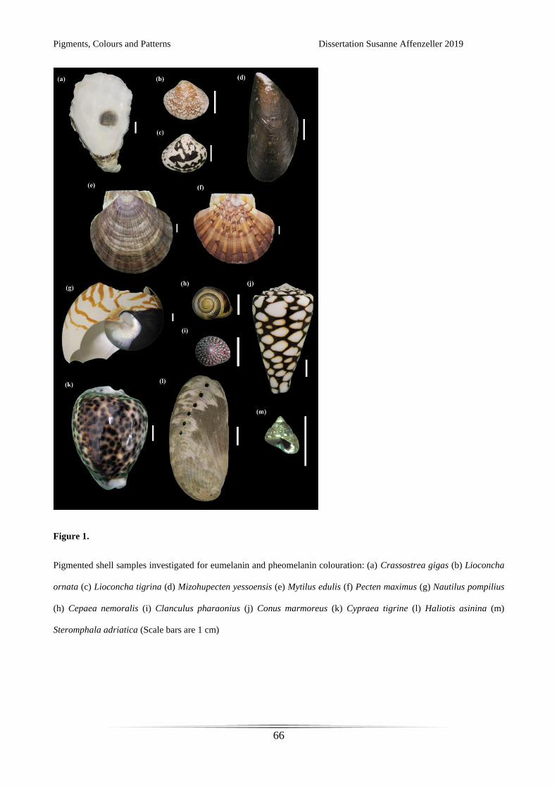
Pigments, Colours and Patterns Dissertation Susanne Affenzeller 2019
66
Figure 1.
Pigmented shell samples investigated for eumelanin and pheomelanin colouration: (a) Crassostrea gigas (b) Lioconcha
ornata (c) Lioconcha tigrina (d) Mizohupecten yessoensis (e) Mytilus edulis (f) Pecten maximus (g) Nautilus pompilius
(h) Cepaea nemoralis (i) Clanculus pharaonius (j) Conus marmoreus (k) Cypraea tigrine (l) Haliotis asinina (m)
Steromphala adriatica (Scale bars are 1 cm)
Pigments, Colours and Patterns Dissertation Susanne Affenzeller 2019
67
Here we look for the presence of eumelanin and pheomelanin pigmentation in 13 different species of
shell bearing molluscs (Table 1) using alkaline oxidation followed by HPLC–MS. All of these species
display prominent patterns on their shells with colours ranging from yellow, light brown, orange and
red to dark brown and black (Fig. 1). A number of these species are of significant commercial or
cultural value (Mytilus edulis, Pecten maximus, Cypraea tigrina, Haliotis asinina), while others
(Crassostrea gigas, Mizohupecten yessoensis, Cepaea nemoralis, Clanculus pharaonius, were
chosen due to previous reports of melanic pigmentation [7,20,27,29,32].
2. Material and methods
(a) Samples and standards
Shells from 13 different mollusc species were obtained either commercially or by donation from the
Natural History Museum Vienna or private collectors for analysis (Figure 1 for images of used
samples and Table 1 for previous literature and sample sources). For species previously reported to
contain eumelanin in their shells (C. gigas, M, yessoensis, C. pharaonius (19,27,30)) three replicates
were analysed. For C. nemoralis a morph with yellow background and multiple brown bands was
analysed. For M. edulis the periostracum was removed by scrubbing the shell with sand for one shell
valve, while the other valve remained intact. As S. adriatica are very small seven shells were
combined into one sample. Samples contained approximately 1.5 g of shell material each. For L.
ornata 0.6 g and for C. pharaonius approximatly 1 g of shell material per sample was available due
to shell sizes.
For comparison, standards of the melanin oxidation products PDCA, PTCA, TDCA and TTCA kindly
provided by Prof. Ito were used.
Pigments, Colours and Patterns Dissertation Susanne Affenzeller 2019
68
(b) Sample preparation, melanin oxidation and HPLC–MS analysis
Samples were processed as previously described (Affenzeller et al. in revision). In brief, shells were
cleaned in deionized water, dried and weighed, and then dissolved in 6 M HCl. Residues were washed
with water and were treated with proteinase K in 1 M Tris-HCl buffer at 37 °C for 2 h. Pigmented
residues were treated with alkaline oxidation via H2O2 [40]: Oxidation reactions for each sample were
carried out for 20 h at 25 °C under vigorous shaking using 100 μL H2O, 375 μL 1 M K2CO3 and 25
μL 30% H2O2 as reactants. Remaining H2O2 was decomposed by the addition of 50 μL 10% Na2SO3
and the mixture was acidified with 140 μL 6 M HCl. The solutions were centrifuged and supernatants
were transferred to fresh tubes.
Samples were treated by solid-phase extraction (Phenomenex Strata-X Polymeric Reversed Phase
columns, 33 μm). Columns were conditioned with methanol (MeOH) followed by H2O. Shell extracts
were loaded onto the columns and washed with 0.3% formic acid. Columns were dried and elution
was carried out with MeOH followed ethyl acetate. Solvents were removed under constant nitrogen
stream at 40 °C and samples were dissolved in 200 μL H2O.
Measurements were carried out on a Thermo Fisher Scientific HPLC–MS system consisting of an
Accela HPLC with a Finnigan Surveyor PDA Detector coupled to an LTQ Orbitrap XL mass
spectrometer equipped with an electrospray ionisation (ESI) source. Separation was performed on a
Phenomenex Gemini C18 column (250 × 2 mm, 5 μm). The mobile phase was 0.3% formic acid in
H2O:MeOH (80:20). Analyses were performed at 45 °C at a flow rate of 0.2 ml/min. Mass spectra
were acquired in negative ion mode over an m/z range of 120–220.
Identification of melanin oxidation products were based on exact mass data. Quantitation was carried
out by HPLC–UV using external calibration with melanin oxidation product standards.
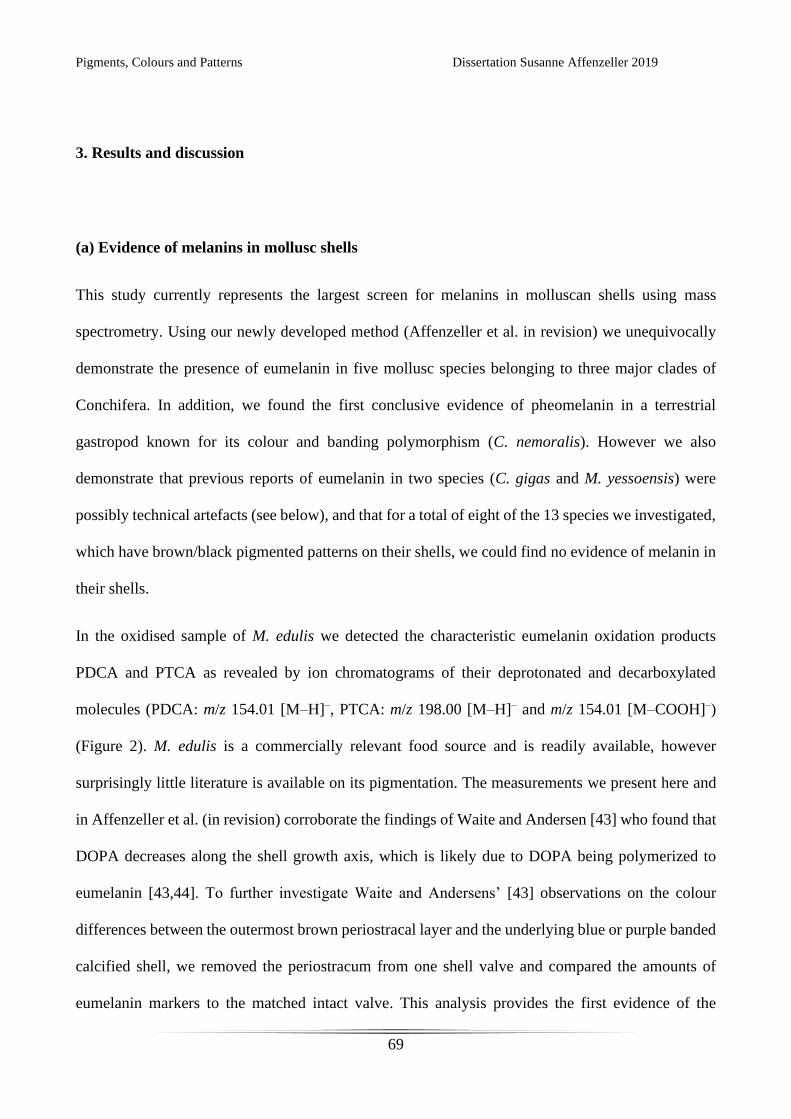
Pigments, Colours and Patterns Dissertation Susanne Affenzeller 2019
69
3. Results and discussion
(a) Evidence of melanins in mollusc shells
This study currently represents the largest screen for melanins in molluscan shells using mass
spectrometry. Using our newly developed method (Affenzeller et al. in revision) we unequivocally
demonstrate the presence of eumelanin in five mollusc species belonging to three major clades of
Conchifera. In addition, we found the first conclusive evidence of pheomelanin in a terrestrial
gastropod known for its colour and banding polymorphism (C. nemoralis). However we also
demonstrate that previous reports of eumelanin in two species (C. gigas and M. yessoensis) were
possibly technical artefacts (see below), and that for a total of eight of the 13 species we investigated,
which have brown/black pigmented patterns on their shells, we could find no evidence of melanin in
their shells.
In the oxidised sample of M. edulis we detected the characteristic eumelanin oxidation products
PDCA and PTCA as revealed by ion chromatograms of their deprotonated and decarboxylated
molecules (PDCA: m/z 154.01 [M–H]–, PTCA: m/z 198.00 [M–H]– and m/z 154.01 [M–COOH]–)
(Figure 2). M. edulis is a commercially relevant food source and is readily available, however
surprisingly little literature is available on its pigmentation. The measurements we present here and
in Affenzeller et al. (in revision) corroborate the findings of Waite and Andersen [43] who found that
DOPA decreases along the shell growth axis, which is likely due to DOPA being polymerized to
eumelanin [43,44]. To further investigate Waite and Andersens’ [43] observations on the colour
differences between the outermost brown periostracal layer and the underlying blue or purple banded
calcified shell, we removed the periostracum from one shell valve and compared the amounts of
eumelanin markers to the matched intact valve. This analysis provides the first evidence of the
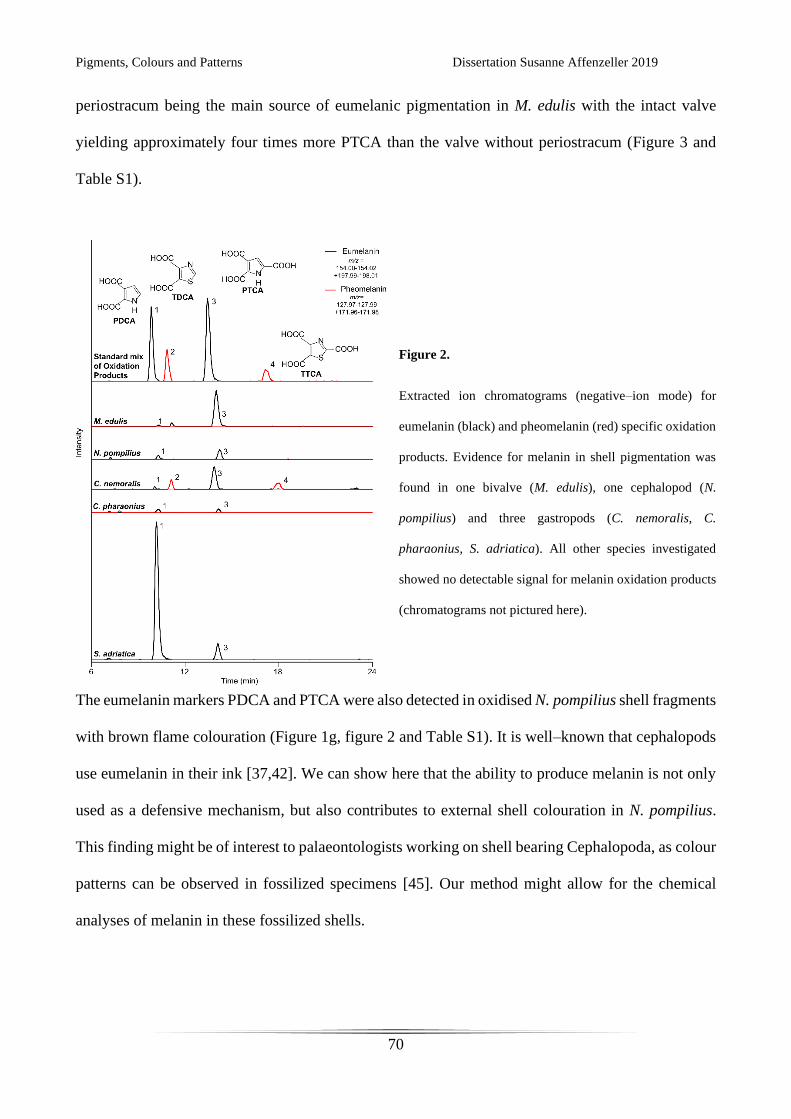
Pigments, Colours and Patterns Dissertation Susanne Affenzeller 2019
70
periostracum being the main source of eumelanic pigmentation in M. edulis with the intact valve
yielding approximately four times more PTCA than the valve without periostracum (Figure 3 and
Table S1).
Figure 2.
Extracted ion chromatograms (negative–ion mode) for
eumelanin (black) and pheomelanin (red) specific oxidation
products. Evidence for melanin in shell pigmentation was
found in one bivalve (M. edulis), one cephalopod (N.
pompilius) and three gastropods (C. nemoralis, C.
pharaonius, S. adriatica). All other species investigated
showed no detectable signal for melanin oxidation products
(chromatograms not pictured here).
The eumelanin markers PDCA and PTCA were also detected in oxidised N. pompilius shell fragments
with brown flame colouration (Figure 1g, figure 2 and Table S1). It is well–known that cephalopods
use eumelanin in their ink [37,42]. We can show here that the ability to produce melanin is not only
used as a defensive mechanism, but also contributes to external shell colouration in N. pompilius.
This finding might be of interest to palaeontologists working on shell bearing Cephalopoda, as colour
patterns can be observed in fossilized specimens [45]. Our method might allow for the chemical
analyses of melanin in these fossilized shells.
Pigments, Colours and Patterns Dissertation Susanne Affenzeller 2019
71
Figure 3.
To test the predominance of eumelanin in different shell layers of M. edulis the outermost brown periostracum was
removed from one shell valve while the other valve was measured with intact periostracum. Eumelanin oxidation products
PDCA and PTCA were quantitated by HPLC with UV detection with external calibration and measurements were
normalized to initial sample weight.
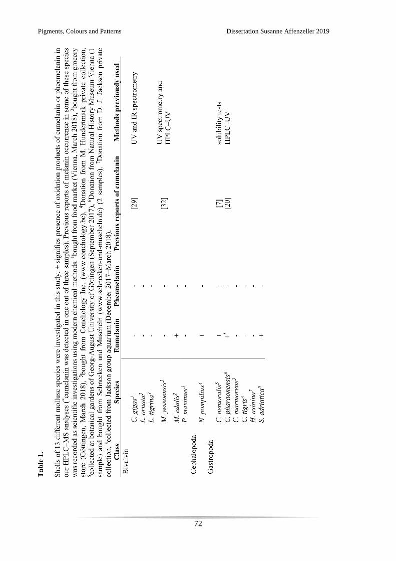
Pigments, Colours and Patterns Dissertation Susanne Affenzeller 2019
72
Pigments, Colours and Patterns Dissertation Susanne Affenzeller 2019
73
Within the gastropods we found melanin markers in the oxidised samples of three species: C.
nemoralis, C. pharaonius and S. adriatica (table 1, Table S1). For the terrestrial gastropod C.
nemoralis we found mass spectrometric evidence for all four melanin oxidation products (Figure 2
and Table S1), the characteristic pheomelanin markers TDCA and TTCA revealed by the ion
chromatograms of the deprotonated molecule and ions resulting from the loss of one and two carboxyl
groups (TDCA: m/z 171.97 [M–H]– and m/z 127.98 [M–COOH]–, TTCA: m/z 171.97 [M–COOH]–
and m/z 127.98 [M–C2HO4]–). This is the first mass spectrometric evidence of simultaneous
eumelanin and pheomelanin use in a molluscan shell. Further investigation on the spatial distribution
of these melanic pigments within the shell are needed to clarify their contribution to band and
background colouration.
We could identify both eumelanin markers in one out of three replicates of the colourful marine
gastropod C. pharaonius (strawberry topshell) (Figure 1i and Figure 2). Using HPLC with UV
detection but without additional mass information, the eumelanin marker PTCA was recently
identified in another study of C. pharaonius [20]. In our study of this species we detected a peak with
nearly the same retention time as PTCA. The use of exact mass data allowed us to verify that for two
shells this is in fact not the eumelanin marker, but another substance with a very similar
chromatographic behaviour. This sporadic finding of eumelanin with no obvious linkage to the shell
phenotype (see Figure S1) complicates the interpretation of this data.
In the marine gastropod S. adriatica (Figure 1m) we found an abundance of eumelanin markers
(Figure 2 and Table S1). This species is known to live in shallow waters in the Mediterranean sea
grazing on microfilm algae [46]. Melanin incorporation into the shell might therefore play a role in
Pigments, Colours and Patterns Dissertation Susanne Affenzeller 2019
74
sun protection or habitat blending, but further research is needed functionally characterise this
melanic pigment in S. adriatica.
(b) The surprising absence of melanins in diverse pigmented mollusc shells
Surprisingly, no traces of melanin oxidation products were detected for many prominently patterned
and brown coloured mollusc shells (Figure 1, Table 1). This is especially surprising as brown and
black colour patterns on bivalve and gastropod shells have generally been believed to be of melanic
origin since the early studies of Comfort [7,14,16,18]. Moreover, for some of the species the absence
of melanins is in contrast to previous studies. In a recent study, analysis of melanin oxidation products
by HPLC with UV detection suggested that the brown valve of the bicoloured bivalve M. yessoensis
contain eumelanin and pheomelanin (as Patinopecten yessoensis in Sun et al. [32]). However in that
study peak identification relied solely on retention times and no identification with mass data was
used to verify those results. This practice can easily lead to the misidentification of melanin oxidation
products (see Affenzeller et. al. in review). Pigmentation present in the dark adductor scar of C. gigas
was assumed to be (eu)melanin. However, this result is based solely on measurements obtained by
UV spectrophotometry and IR spectrometry [29]. During sample preparation we observed acid
solubility and fluorescence of pigments from the shell of C. gigas, possibly indicating porphyrin–like
pigments known to be produced by the bivalves Pinctada spp. and Pteria penguin [47,48].
The method we have used to detect melanin oxidation products was developed and adapted for
challenging biological sample matrices such as molluscan shells and is highly sensitive (limit of
detection ranging from 0.03 μg/mL to 0.10 μg/mL). Further efforts to detect melanins in the intensely
brown coloured Conus shell (grinding of shell before dissolution, longer oxidation time among
others) were unsuccessful. We are therefore confident that in the indicated specimens melanin is
Pigments, Colours and Patterns Dissertation Susanne Affenzeller 2019
75
genuinely absent (or exists in trace amounts inadequate to appreciably pigment the shell). This leads
us to question what the prominent brown to black pigments are in shells where no melanin is detected.
For some cases (e.g. L. tigrina) we observed the pigmented pattern on the shell preserves its geometric
configuration even after the calcium carbonate is dissolved in high molarity acid. This suggests a
complex and very stable pigment, possibly a macromolecule tightly bound to shell proteins.
Unfortunately we were not able to identify other oxidation products in our samples that would
indicate the chemical composition of this pigment. Further investigations are necessary to unravel its
nature.
4. Conclusions
We have found mass spectrometric evidence for melanins in three conchiferan classes: Cephalopoda,
Gastropoda and Bivalvia. This is the first time melanin has been detected in cephalopod shells (N.
pompilius). In the marine bivalve M. edulis eumelanin is predominantly located in the periostracum
layer relative to the calcified shell. For the first time both eumelanin and pheomelanin were detected
in a mollusc shell (the terrestrial gastropod C. nemoralis), however further study is needed to spatially
localise the distribution of these pigments in this shell. Eumelanin markers could only be detected in
one out of three marine C. pharaonius individuals. In another marine gastropod (S. adriatica)
eumelanin was abundant. We could not detect melanin in a surprisingly large number of prominently
patterned gastropod and bivalve shells. Further investigations are needed to identify the underlying
pigmentation mechanism responsible for these complex geometric colourations.
Acknowledgements
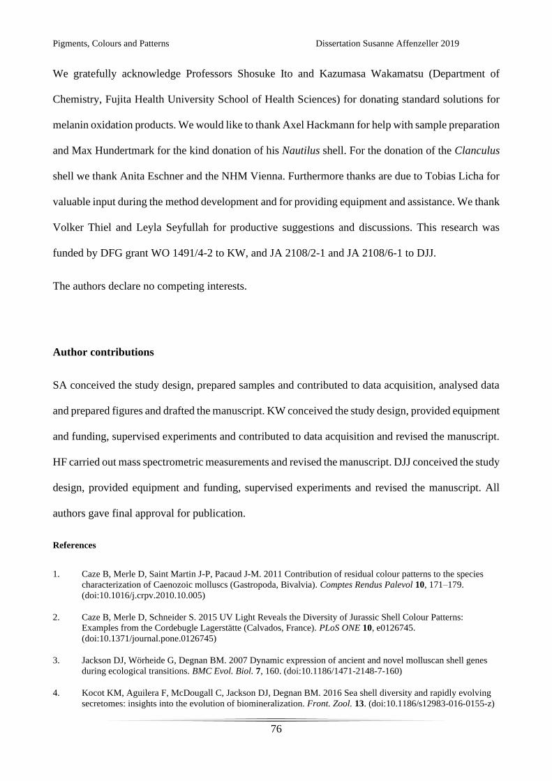
Pigments, Colours and Patterns Dissertation Susanne Affenzeller 2019
76
We gratefully acknowledge Professors Shosuke Ito and Kazumasa Wakamatsu (Department of
Chemistry, Fujita Health University School of Health Sciences) for donating standard solutions for
melanin oxidation products. We would like to thank Axel Hackmann for help with sample preparation
and Max Hundertmark for the kind donation of his Nautilus shell. For the donation of the Clanculus
shell we thank Anita Eschner and the NHM Vienna. Furthermore thanks are due to Tobias Licha for
valuable input during the method development and for providing equipment and assistance. We thank
Volker Thiel and Leyla Seyfullah for productive suggestions and discussions. This research was
funded by DFG grant WO 1491/4-2 to KW, and JA 2108/2-1 and JA 2108/6-1 to DJJ.
The authors declare no competing interests.
Author contributions
SA conceived the study design, prepared samples and contributed to data acquisition, analysed data
and prepared figures and drafted the manuscript. KW conceived the study design, provided equipment
and funding, supervised experiments and contributed to data acquisition and revised the manuscript.
HF carried out mass spectrometric measurements and revised the manuscript. DJJ conceived the study
design, provided equipment and funding, supervised experiments and revised the manuscript. All
authors gave final approval for publication.
References
1. Caze B, Merle D, Saint Martin J-P, Pacaud J-M. 2011 Contribution of residual colour patterns to the species
characterization of Caenozoic molluscs (Gastropoda, Bivalvia). Comptes Rendus Palevol 10, 171–179.
(doi:10.1016/j.crpv.2010.10.005)
2. Caze B, Merle D, Schneider S. 2015 UV Light Reveals the Diversity of Jurassic Shell Colour Patterns:
Examples from the Cordebugle Lagerstätte (Calvados, France). PLoS ONE 10, e0126745.
(doi:10.1371/journal.pone.0126745)
3. Jackson DJ, Wörheide G, Degnan BM. 2007 Dynamic expression of ancient and novel molluscan shell genes
during ecological transitions. BMC Evol. Biol. 7, 160. (doi:10.1186/1471-2148-7-160)
4. Kocot KM, Aguilera F, McDougall C, Jackson DJ, Degnan BM. 2016 Sea shell diversity and rapidly evolving
secretomes: insights into the evolution of biomineralization. Front. Zool. 13. (doi:10.1186/s12983-016-0155-z)
Pigments, Colours and Patterns Dissertation Susanne Affenzeller 2019
77
5. Sakalauskaite J et al. 2019 ‘Palaeoshellomics’ reveals the use of freshwater mother-of-pearl in prehistory. eLife
8, e45644. (doi:10.7554/eLife.45644)
6. Adamkewicz L, Castagna M. 1988 Genetics of Shell Color and Pattern in the Bay Scallop Argopecten
irradians. J. Hered. 79, 14–17. (doi:10.1093/oxfordjournals.jhered.a110436)
7. Comfort A. 1951 The pigmentation of molluscan shells. Biol. Rev. 26, 285–301. (doi:10.1111/j.1469-
185X.1951.tb01358.x)
8. Ogundiran A. 2002 Of Small Things Remembered: Beads, Cowries, and Cultural Translations of the Atlantic
Experience in Yorubaland. Int. J. Afr. Hist. Stud. 35, 427–457. (doi:10.2307/3097620)
9. Doran E. 1958 The Caicos Conch Trade. Geogr. Rev. 48, 388–401. (doi:10.2307/212258)
10. Pietak LM. 1998 Body Symbolism and Cultural Aesthetics: The Use of Shell Beads and Ornaments by
Delaware and Munsee Groups. North Am. Archaeol. 19, 135–161. (doi:10.2190/0BV6-Q0N1-37VU-PEQ7)
11. Bai Z, Zheng H, Lin J, Wang G, Li J. 2013 Comparative Analysis of the Transcriptome in Tissues Secreting
Purple and White Nacre in the Pearl Mussel Hyriopsis cumingii. PLoS ONE 8, e53617.
(doi:10.1371/journal.pone.0053617)
12. Cai Z, Wu J, Chen L, Guo W, Li J, Wang J, Zhang Q. 2011 Purification and characterisation of aquamarine blue
pigment from the shells of abalone (Haliotis discus hannai Ino). Food Chem. 128, 129–133.
(doi:10.1016/j.foodchem.2011.03.006)
13. Canales-Gómez E, Correa G, Viana MT. 2010 cantaxanthin and β-carotene) in juvenile abalone Haliotis
rufescens diets on the color of the shell or nacre. Vet. Mex. 41, 191–200.
14. Williams ST. 2017 Molluscan shell colour. Biol. Rev. 92, 1039–1058. (doi:10.1111/brv.12268)
15. Comfort A. 1949 Acid-soluble pigments of shells. 1. The distribution of porphyrin fluorescence in molluscan
shells. Biochem. J. 44, 111–117.
16. Comfort A. 1949 Acid-soluble pigments of molluscan shells. 2. Pigments other than porphyrins. Biochem. J. 45,
199–204.
17. Comfort A. 1949 Acid-soluble pigments of molluscan shells. 3. The indigoid character of the blue pigment of
Haliotis cracherodii Leach. Biochem. J. 45, 204–208.
18. Comfort A. 1950 Biochemistry of molluscan shell pigments. J. Molluscan Stud. 28, 79–85.
(doi:10.1093/oxfordjournals.mollus.a064570)
19. Comfort A. 1951 Observations on the shell pigments of land pulmonates. J. Molluscan Stud. 29, 35–44.
(doi:10.1093/oxfordjournals.mollus.a064598)
20. Williams ST et al. 2016 Identification of Shell Colour Pigments in Marine Snails Clanculus pharaonius and C.
margaritarius (Trochoidea; Gastropoda). PLoS ONE 11, e0156664. (doi:10.1371/journal.pone.0156664)
21. Barnard W, de Waal D. 2006 Raman investigation of pigmentary molecules in the molluscan biogenic matrix. J.
Raman Spectrosc. 37, 342–352. (doi:10.1002/jrs.1461)
22. Bergamonti L, Bersani D, Mantovan S, Lottici PP. 2013 Micro-Raman investigation of pigments and carbonate
phases in corals and molluscan shells. Eur. J. Mineral. 25, 845–853. (doi:10.1127/0935-1221/2013/0025-2318)
23. Hedegaard C, Bardeau J-F, Chateigner D. 2006 Molluscan shell pigments: an in situ resonance raman study. J.
Molluscan Stud. 72, 157–162. (doi:10.1093/mollus/eyi062)

Pigments, Colours and Patterns Dissertation Susanne Affenzeller 2019
78
24. de Oliveira LN, de Oliveira VE, D’ávila S, Edwards HGM, de Oliveira LFC. 2013 Raman spectroscopy as a
tool for polyunsaturated compound characterization in gastropod and limnic terrestrial shell specimens.
Spectrochim. Acta. A. Mol. Biomol. Spectrosc. 114, 541–546. (doi:10.1016/j.saa.2013.05.095)
25. Verdes A, Cho W, Hossain M, Brennan PLR, Hanley D, Grim T, Hauber ME, Holford M. 2015 Nature’s
Palette: Characterization of Shared Pigments in Colorful Avian and Mollusk Shells. PLoS ONE 10, e0143545.
(doi:10.1371/journal.pone.0143545)
26. Nagai K, Yano M, Morimoto K, Miyamoto H. 2007 Tyrosinase localization in mollusc shells. Comp. Biochem.
Physiol. B Biochem. Mol. Biol. 146, 207–214. (doi:10.1016/j.cbpb.2006.10.105)
27. Mao J, Zhang W, Wang X, Song J, Yin D, Tian Y, Hao Z, Han B, Chang Y. 2019 Histological and Expression
Differences Among Different Mantle Regions of the Yesso Scallop (Patinopecten yessoensis) Provide Insights
into the Molecular Mechanisms of Biomineralization and Pigmentation. Mar. Biotechnol. (doi:10.1007/s10126-
019-09913-x)
28. Karampelas S, Fritsch E, Mevellec J-Y, Gauthier J-P, Sklavounos S, Soldatos T. 2007 Determination by Raman
scattering of the nature of pigments in cultured freshwater pearls from the mollusk Hyriopsis cumingii. J.
Raman Spectrosc. 38, 217–230. (doi:10.1002/jrs.1626)
29. Hao S, Hou X, Wei L, Li J, Li Z, Wang X. 2015 Extraction and Identification of the Pigment in the Adductor
Muscle Scar of Pacific Oyster Crassostrea gigas. PLoS ONE 10, e0142439.
(doi:10.1371/journal.pone.0142439)
30. Yu F, Pan Z, Qu B, Yu X, Xu K, Deng Y, Liang F. 2018 Identification of a tyrosinase gene and its functional
analysis in melanin synthesis of Pteria penguin. Gene 656, 1–8. (doi:10.1016/j.gene.2018.02.060)
31. Chen X, Liu X, Bai Z, Zhao L, Li J. 2017 HcTyr and HcTyp-1 of Hyriopsis cumingii, novel tyrosinase and
tyrosinase-related protein genes involved in nacre color formation. Comp. Biochem. Physiol. B Biochem. Mol.
Biol. 204, 1–8. (doi:10.1016/j.cbpb.2016.11.005)
32. Sun X, Wu B, Zhou L, Liu Z, Dong Y, Yang A. 2017 Isolation and characterization of melanin pigment from
yesso scallop Patinopecten yessoensis. J. Ocean Univ. China 16, 279–284. (doi:10.1007/s11802-017-3162-6)
33. Ito S. 1986 Reexamination of the structure of eumelanin. Biochim. Biophys. Acta 883, 155–161.
34. Prota G. 1995 The Chemistry of Melanins and Melanogenesis. In Fortschritte der Chemie organischer
Naturstoffe / Progress in the Chemistry of Organic Natural Products (eds W Herz, GW Kirby, RE Moore, W
Steglich, Ch Tamm), pp. 93–148. Vienna: Springer Vienna. (doi:10.1007/978-3-7091-9337-2_2)
35. Huang Z, Lui H, Chen XK, Alajlan A, McLean DI, Zeng H. 2004 Raman spectroscopy of in vivo cutaneous
melanin. J. Biomed. Opt. 9, 1198. (doi:10.1117/1.1805553)
36. Sealy R, Hyde J, Felix C, Menon I, Prota G. 1982 Eumelanins and pheomelanins: characterization by electron
spin resonance spectroscopy. Science 217, 545–547. (doi:10.1126/science.6283638)
37. Glass K et al. 2012 Direct chemical evidence for eumelanin pigment from the Jurassic period. Proc. Natl. Acad.
Sci. 109, 10218–10223. (doi:10.1073/pnas.1118448109)
38. Stępień K, Dzierżęga-Lęcznar A, Kurkiewicz S, Tam I. 2009 Melanin from epidermal human melanocytes:
Study by pyrolytic GC/MS. J. Am. Soc. Mass Spectrom. 20, 464–468. (doi:10.1016/j.jasms.2008.11.003)
39. Napolitano A, Pezzella A, Vincensi MR, Prota G. 1995 Oxidative degradation of melanins to pyrrole acids: A
model study. Tetrahedron 51, 5913–5920. (doi:10.1016/0040-4020(95)00259-B)
40. Ito S, Nakanishi Y, Valenzuela RK, Brilliant MH, Kolbe L, Wakamatsu K. 2011 Usefulness of alkaline
hydrogen peroxide oxidation to analyze eumelanin and pheomelanin in various tissue samples: application to
chemical analysis of human hair melanins: Alkaline hydrogen peroxide oxidation of melanin. Pigment Cell
Melanoma Res. 24, 605–613. (doi:10.1111/j.1755-148X.2011.00864.x)
Pigments, Colours and Patterns Dissertation Susanne Affenzeller 2019
79
41. Wakamatsu K, Fujikawa K, Zucca FA, Zecca L, Ito S. 2003 The structure of neuromelanin as studied by
chemical degradative methods: Chemical structure of neuromelanin. J. Neurochem. 86, 1015–1023.
(doi:10.1046/j.1471-4159.2003.01917.x)
42. Magarelli M, Passamonti P, Renieri C. 2010 Purification, characterization and analysis of sepia melanin from
commercial sepia ink (Sepia officinalis). Rev. CES Med. Vet. Zootec. 5, 18–28.
43. Waite JH, Andersen SO. 1980 3,4-dihydroxyphenylalanine (dopa) and sclerotization of periostracum in Mytilus
edulis L. Biol. Bull. 158, 164–173. (doi:10.2307/1540767)
44. Ito S, Wakamatsu K. 1989 Melanin Chemistry and Melanin Precursors in Melanoma. J. Invest. Dermatol. 92,
S261–S265. (doi:10.1038/jid.1989.78)
45. Mapes RH, Sneck DA. 1987 The oldest ammonoid ‘colour’ patterns: description, comparison with Nautilus,
and implications. Paleontology 30, 299–309.
46. Affenzeller S, Haar N, Steiner G. 2017 Revision of the genus complex Gibbula: an integrative approach to
delineating the Eastern Mediterranean genera Gibbula Risso, 1826, Steromphala Gray, 1847, and Phorcus
Risso, 1826 using DNA-barcoding and geometric morphometrics (Vetigastropoda, Trochoidea). Org. Divers.
Evol. 17, 789–812. (doi:10.1007/s13127-017-0343-5)
47. Iwahashi Y, Akamatsu S. 1994 Porphyrin Pigment in Black-Lip Pearls and Its Application to Pearl
Identification. Fish. Sci. 60, 69–71. (doi:10.2331/fishsci.60.69)
48. Miyoshi T, Matsuda Y, Komatsu H. 1987 Fluorescence from Pearls and Shells of Black Lip Oyster, Pinctada
margaritifera , and Its Contribution to the Distinction of Mother Oysters Used in Pearl Culture. Jpn. J. Appl.
Phys. 26, 1069–1072. (doi:10.1143/JJAP.26.1069)
Pigments, Colours and Patterns Dissertation Susanne Affenzeller 2019
80
CHAPTER 5: EUMELANIN IS NOT THE BANDED PIGMENT IN CEPAEA NEMORALIS
The following chapter is prepared for publication as Correspondence in Current Biology and is
currently being revised by my co-authors.
Contributions by the doctoral candidate: Conceptualization, Formal analysis, Investigation,
Methodology, Validation, Writing – review & editing

Pigments, Colours and Patterns Dissertation Susanne Affenzeller 2019
81
Pigments, Colours and Patterns Dissertation Susanne Affenzeller 2019
82
Eumelanin is not the banded pigment in Cepaea nemoralis
Susanne Affenzeller1, Klaus Wolkenstein1 and Daniel J. Jackson1,*
1 Department of Geobiology, Georg-August University of Göttingen, Goldschmidtstrasse 3, 37077
Göttingen, Germany
*Author for correspondence:
Daniel J. Jackson
email: [email protected]
Keywords: Eumelanin, Pheomelanin, mollusc, shell pigmentation, mass spectroscopy, qPCR
Pigments, Colours and Patterns Dissertation Susanne Affenzeller 2019
83
Main text
With highly variable and colourful patterns of shell pigmentation (Fig. 1A) the terrestrial snail
Cepaea nemoralis has long been a textbook example of phenotypic variability and a model system
for evolution, ecology and population genetics (Cain and Sheppard, 1954). Early studies established
that the shell background colour, the number of dark pigmented bands on the shell and several other
shell-pigment characteristics are inherited in a Mendelian fashion (Cain et al., 1960). It has also been
long assumed that melanin (specifically eumelanin) is the pigment that underlies the dark pigmented
bands (Comfort, 1951; Helmcke, 1935). We tested this assumption using a combination of sensitive
chemical and molecular techniques and find no evidence that eumelanin is differentially distributed
within the shells of C. nemoralis. This implies that an unknown pigment adorns the shells of C.
nemoralis, and has implications for the continuing search for the supergene that generates the various
pigmentation morphotypes.
Recently we developed a highly sensitive LC-MS based method for the detection of eu- and
pheomelanin from a variety of biological matrices including molluscan shells (Affenzeller et al., in
revision). Our initial experiments on whole shells of the common yellow banded morph of C.
nemoralis detected all four common melanin oxidation products: PDCA (pyrrole-2,3-dicarboxylic
acid) and PTCA (pyrrole-2,3,5-tricarboxylic acid) for eumelanin, and TDCA (thiazole-4,5-
dicarboxylic acid) and TTCA (thiazole-2,4,5-tricarboxylic acid) for pheomelanin, albeit for
eumelanin at levels significantly lower than the darkly pigmented shells of Mytillus edulis
(Affenzeller et al., in prep). To further investigate the spatial distribution of eu- and pheomelanin in
C. nemoralis shells we have conducted quantitative measurements of colour-sorted shell fragments
(pink banded, pink background, yellow banded and yellow background; Figure 1A). All four shell
colour fractions contained the markers PDCA and PTCA, indicating a broad distribution of
eumelanin, however we could not detect significant differences in the amounts of these eumelanin
Pigments, Colours and Patterns Dissertation Susanne Affenzeller 2019
84
markers between banded and background shell fragments for either yellow or pink shells (Figure 1B).
We also detected the pheomelanin markers TDCA and TTCA in both yellow banded and yellow
background shell fragments in high abundance, but with no significant difference between the two
(Figure 1B). In pink morphs we detected far less TDCA in both fractions while TTCA was below the
limit of quantitation but could still be detected (Supplementary Table 1).
To further investigate the role of melanin in the banding pattern of C. nemoralis shells we studied the
expression of four known melanin synthesis pathway genes using recently established RT-qPCR
reference genes (Affenzeller et al., 2018). We selected the genes Yellow, Tyrosinase Related Protein,
Tyrosinase and Laccase, all of which are known to be involved in melanin synthesis in a variety of
species including molluscs. Tyrosinases and Tyrosinase Related Proteins have been the focus of
recent research due to their proposed involvement in nacre colour formation in bivalves, thus
influencing the value of pearls. Despite considerable variation Yellow was significantly differentially
expressed in pigmented vs. non-pigmented regions of the mantle (p<0.05 but not <0.01; Fig. 1C).
However Yellow was significantly more abundant (approximately three fold) in foot tissue relative to
mantle tissue overall, suggesting that this gene either has pleiotropic roles in these tissues or is not
involved in melanogenesis in C. nemoralis. Supporting this latter scenario the Yellow homolog we
identified here has relatively low sequence similarity (35% identity) with insect isoforms known to
be involved in pigmentation. Tyrosinase Related Protein was significantly upregulated in mantle vs.
foot tissues suggesting it plays either a role in biomineralization as is known for molluscs, or
pigmentation. However comparisons between pigmented and non-pigmented mantle tissue revealed
no significant differences in Tyrosinase Related Protein expression (Fig. 1C). Finally Tyrosinase and
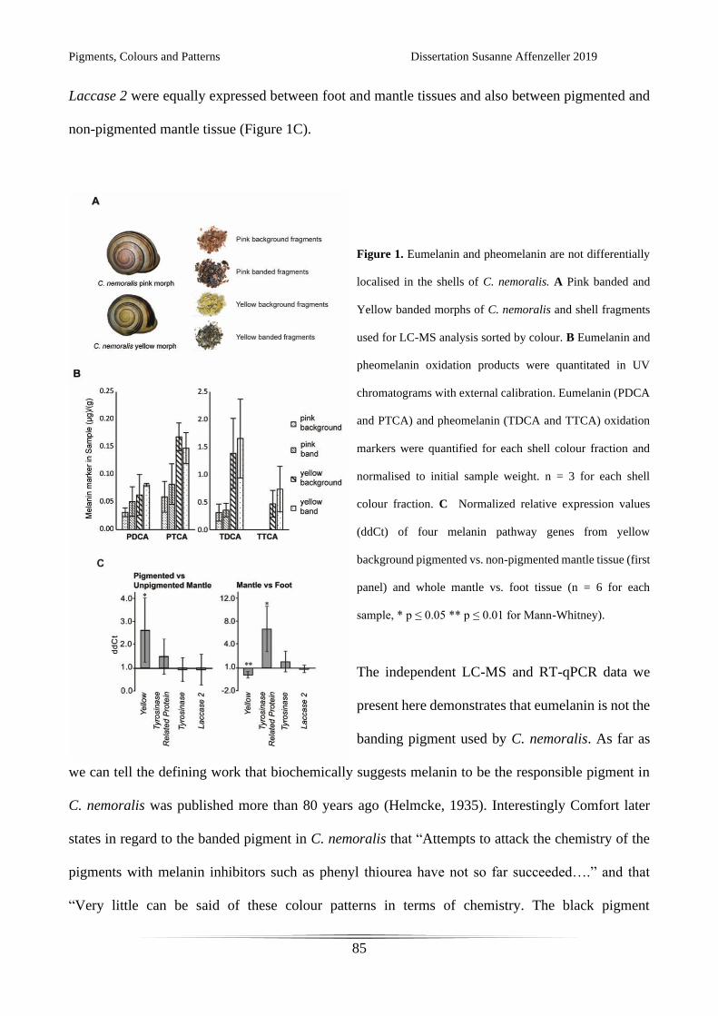
Pigments, Colours and Patterns Dissertation Susanne Affenzeller 2019
85
Laccase 2 were equally expressed between foot and mantle tissues and also between pigmented and
non-pigmented mantle tissue (Figure 1C).
Figure 1. Eumelanin and pheomelanin are not differentially
localised in the shells of C. nemoralis. A Pink banded and
Yellow banded morphs of C. nemoralis and shell fragments
used for LC-MS analysis sorted by colour. B Eumelanin and
pheomelanin oxidation products were quantitated in UV
chromatograms with external calibration. Eumelanin (PDCA
and PTCA) and pheomelanin (TDCA and TTCA) oxidation
markers were quantified for each shell colour fraction and
normalised to initial sample weight. n = 3 for each shell
colour fraction. C Normalized relative expression values
(ddCt) of four melanin pathway genes from yellow
background pigmented vs. non-pigmented mantle tissue (first
panel) and whole mantle vs. foot tissue (n = 6 for each
sample, * p ≤ 0.05 ** p ≤ 0.01 for Mann-Whitney).
The independent LC-MS and RT-qPCR data we
present here demonstrates that eumelanin is not the
banding pigment used by C. nemoralis. As far as
we can tell the defining work that biochemically suggests melanin to be the responsible pigment in
C. nemoralis was published more than 80 years ago (Helmcke, 1935). Interestingly Comfort later
states in regard to the banded pigment in C. nemoralis that “Attempts to attack the chemistry of the
pigments with melanin inhibitors such as phenyl thiourea have not so far succeeded….” and that
“Very little can be said of these colour patterns in terms of chemistry. The black pigment
Pigments, Colours and Patterns Dissertation Susanne Affenzeller 2019
86
(‘melanocochlin’) is alkali-soluble (Helmcke, 1935).” (Comfort, 1951). Nonetheless subsequent
works refer to pigmented cells in the mantle as melanocytes (Emberton, 1963) and have worked from
the assumption that melanin is the banded pigment (Vicario et al., 1989). We previously demonstrated
that the banded pigment is apparently not covalently linked to a protein (Mann and Jackson, 2014),
however the banded and background pigments are yet to be identified. Taken together, our results
indicate that a novel molecular mechanism which deposits a dark brown pigment in a banded fashion
is operating in the mantle tissue of C. nemoralis. A knowledge of what this dark pigment is (together
with the background pigments and their respective synthesis pathways) would likely assist efforts to
identify the supergene that is thought to regulate the famous Cepaea colour polymorphism. Such a
complete understanding of this process at the genetic and chemical scales would in turn provide deep
insight into the long studied population genetics and evolutionary biology of this system.
Acknowledgements
We are grateful to Professors Shosuke Ito and Kazumasa Wakamatsu (Department of Chemistry,
Fujita Health University School of Health Sciences) for donating standard solutions for melanin
oxidation products. Furthermore thanks are due to Tobias Licha for valuable input on understanding
the chemistry and for providing equipment and assistance. We thank Leyla Seyfullah and Volker
Thiel for productive suggestions and discussions. This research was funded by DFG grant WO
1491/4-2 to KW, and JA 2108/2-1 and JA 2108/6-1 to DJJ.
References
Affenzeller, S., Frauendorf, H., Licha, T., Jackson, D.J., and Wolkenstein, K. (in revision). Quantitation of eumelanin
and pheomelanin markers in diverse biological samples by HPLC-UV-MS following solid-phase extraction.
PLoS One.
Pigments, Colours and Patterns Dissertation Susanne Affenzeller 2019
87
Affenzeller, S., Wolkenstein, K., Frauendorf, H., and Jackson, D.J. (in prep). Eumelanin and pheomelanin pigmentation
in mollusc shells may be less common than expected: insights from mass spectrometry.
Affenzeller, S., Cerveau, N., and Jackson, D.J. (2018). Identification and validation of reference genes for qPCR in the
terrestrial gastropod Cepaea nemoralis. PLoS One 13, e0201396.
Cain, A.J., and Sheppard, P.M. (1954). Natural Selection in Cepaea. Genetics 39, 89–116.
Cain, A.J., King, J., and Sheppard, P. (1960). New data on the genetics of polymorphism in the snail Cepaea nemoralis
L. Genetics 45, 393.
Comfort, A. (1951). Observations on the shell pigments of land pulmonates. J. Molluscan Stud. 29, 35–44.
Emberton, L.R.B. (1963). Relationships Between Pigmentation of Shell and of Mantle in the Snails Cepaea Nemoralis
(l.) and Cepaea Hortensis (mull.). Proc. Zool. Soc. Lond. 140, 273–293.
Helmcke, J.-G. (1935). Untersuchungen über die Schalen- und Körperfärbung der Landschnecken. Fischer.
Mann, K., and Jackson, D. (2014). Characterization of the pigmented shell-forming proteome of the common grove
snail Cepaea nemoralis. BMC Genomics 15, 249.
Vicario, A., Mazon, L.I., Aguirre, A., Estomba, A., and Lostao, C. (1989). A tyrosinase-like enzyme in Cepaea
nemoralis and its possible relation with shell banding polymorphism. J. Molluscan Stud. 55, 441–444.
Pigments, Colours and Patterns Dissertation Susanne Affenzeller 2019
88
CHAPTER 6: GENERAL DISCUSSION
Mollusc shell pigmentation and the underlying genetic mechanisms responsible for these patterns are
a fascinating topic. But for many investigations basic protocols for dealing with molluscs are missing.
One very useful tool to quantitatively measure gene expression levels is reverse transcription
quantitative polymerase chain reaction (qPCR). To be able to accurately use this technique I tested
potential reference genes for a terrestrial gastropod Cepaea nemoralis (chapter 2, [1]). In addition to
finding suitable reference genes, this study highlighted how difficult and complicated gene expression
studies can be. I found that expression levels of so-called housekeeping genes vary greatly, not only
depending on tissue, but also on season. As even constantly used genes in the organism show such
high variability, one has to be very careful with interpreting any results gained from qPCR on
seasonally employed genes. Especially genes involved in shell building and pigmentation in a
temperate climate snail like C. nemoralis will very likely undergo seasonal fluctuations, as shell
material and the pigments responsible for the well known C. nemoralis polymorphism are mainly
laid down in spring and summer[2–4].
Eumelanin has long since been thought to be the banding pigment in C. nemoralis [5–9]. I aimed to
test this assumption. To this end a specialized protocol to sensitively detect melanin oxidation
markers in complex biological samples was developed (chapter 3). Mollusc shell samples have to be
prepared carefully for any chemical analyses on high performance liquid chromatography (HPLC) to
guarantee quality and reproducibility of these experiments. One way to obtain desalted and cleaned
mollusc shell samples suitable for HPLC is solid phase extraction. Additionally this method allowed
me to concentrate the samples through evaporation, an important advantage for species with small
shells or little pigmentation. By adjusting existing protocols [10,11] to allow for mass spectrometric
(MS) measurements, identification of trace amounts of melanin oxidation products via exact masses
became possible. This was necessary as on top of small pigment amounts in some shells, even cleaned

Pigments, Colours and Patterns Dissertation Susanne Affenzeller 2019
89
mollusc samples still contain a complex matrix. With these adaptations the presented method allows
for confident identification and quantitation of eumelanin and pheomelanin oxidation products in
diverse biological samples including feathers, hair and mollusc shells.
The new chemical method was applied to 13 different mollusc species with prominent shell
pigmentation (chapter 4). Some of them have been previously indicated as containing eumelanin
[10,12–14], while others were chosen due to their brown or black colouration. In C. nemoralis I was
able to detect the first mass spectrometric evidence od pheomelanin use in mollusc shell
pigmentation. We found evidence for eumelanin in three prominent mollusc classes (Mytilus edulis,
Bivalvia; Nautilus pompilius, Cephalopoda; Cepaea nemoralis, Clanculus pharaonius, Steromphala
adriatica, all Gastropoda). Still, a surprisingly large number of species showed no trace of eumelanin
in their dark patterned shells. This contradicts some of the previous findings for Mizuhopecten
yessoensis and Crassostrea gigas, [12,13]. In C. pharaonius the data is inconclusive as eumelanin
could only be detected in one out of three shells.
To further investigate the use of eumelanin and pheomelanin in C. nemoralis I used a combination of
the developed methods to gain more insight (chapter 5). I analysed colour sorted shell fragments for
yellow and pink background coloured snails and their corresponding bands to test the contribution of
pheomelanin and eumelanin to background and band pigmentation. Although both pigments are
present in C. nemoralis shells, I wasn’t able to find any conclusive evidence for eumelanin as the
banding pigment. Pheomelanin might contribute to the background colour, but comparisons of yellow
and pink background indicate that this pigment is not solely responsible for these colours. I further
tested expression levels of known melanin pathway genes in shell producing mantle tissue. As
expected from studies on other mollusc species (namely Mizuhopecten yessoensis, Pteria penguin,
Hyriopsis cumingii and Crassostrea gigas [12,13,15–17]) these genes are active in the mantle, but
they don’t show differential expression between band building and background building tissue. This
Pigments, Colours and Patterns Dissertation Susanne Affenzeller 2019
90
adds to the conclusion that while C. nemoralis does lay down melanin pigments into its shell,
eumelanin and pheomelanin are not the primary pigments used for the well known polymorphism.
One of the most surprising conclusions to be drawn from the here presented studies is that although
shell bearing molluscs have the ability to produce eumelanin, and probably also pheomelanin, many
species don’t use that pigment for patterning their shell. This is contradicting many early works
indicating eumelanin as a broadly used pigment in molluscs [7,18,19]. For those species shown here
to have eumelanin in their shells, only C. nemoralis was available in enough sample quantities to
investigate the spatial distribution of pigment within the shell. This experiment showed that
eumelanin is not the pigment building the band, as previously assumed [5,6,9]. It would of cause be
interesting to see wither eumelanin is involved in the patterns of N. pompilius, C. pharaonius and S.
adriatica. In Clanculus first efforts were undertaken to this end by Williams et al. [10], who tested
for eumelanin after removing the uppermost pigment bearing shell layer. They found no traces of
eumelanin were left, proving that the pigment is part of the visible shell colouration. In light of our
inconclusive results in addition to previous work described above, further experiments on eumelanin
shell pigmentation in this species would be very interesting, but might be difficult to archive. On top
of technical difficulties in separating black coloured shell fragments, the availability of enough shells
might pose a problem. Clanculus pharaonius is one of the most beautiful mollusc shells, and though
not severely protected at the moment, should be collected with care.
If these patterns are not built by melanin, the question remains: What pigment is it? At the current
moment, this is a difficult question to answer. Two other major pigment classes have been shown in
mollusc shells up to now: Carotinoids and tetrapyrrols [10,14,20–23]. Tetrapyrrols, as porphyrins,
can appear in a multitude of colours, but they all share a strong fluorescence under UV light [7,10].
All species testing negative for eumelanin in chapter 4, except Crassostrea gigas, show no such
Pigments, Colours and Patterns Dissertation Susanne Affenzeller 2019
91
fluorescence (data not shown here). I would therefore assume that porphyrins can be disregarded as
likely candidate pigments in these species. Carotinoids on the other hand are a possible source for
light brown patterns observed. Employing Raman spectroscopy might shed some light on the
likelihood of carotinoid pigments being responsible for the patterns in these shells.
Still, the mystery of the dark brown to black pigment (e.g. in Conus marmoreus, but also in C.
nemoralis) remains unsolved. For C. nemoralis Mann and Jackson [24] observed that there is no
obvious linkage of pigment to a shell protein as seen in juveniles of Haliotis asinina [25,26]. The
pigment itself is thermo- and chemo stable and according to Comfort [6] also resists enzymatic
attempts to attack it. From a chemistry point of view gaining the pigment in clean state might be the
next step in understanding its nature and allow further tests. A possible hinderance is the amount of
pigment need, and therefore the amount of shell material required to extract the pigment. Furthermore
extraction of the pure pigment is difficult to undertake with no clear idea about the chemical nature
of the molecule. A solution to circumvent these challenges might be to understand the genetic
background for banding in C. nemoralis. If we were able to understand the genes involved in the
polymorphism, especially the locus for band pigmentation, we might discover what kind of pigment
we are looking for. Recent efforts on understanding recombination, epistasis and incomplete
penetrance in the C. nemoralis supergene, together with more transcriptomic data being available,
are a solid groundwork for future studies [27,28]. Especially long read sequencing technology might
prove useful in finally unravelling the C. nemoralis supergene [27].
Research on patterning mechanisms in the 1980s allowed computational reconstructions of most shell
patterns found in nature by reaction-diffusion and neural network models [29–31]. But although this
knowledge is readily available, further investigations were hindered by lacking methods for molluscs
in both molecular biology and analytical chemistry. Now, new protocols and sequencing techniques

Pigments, Colours and Patterns Dissertation Susanne Affenzeller 2019
92
are available and we can start understanding these mechanisms. Further investigations based on
transcriptomic and genomic data, together with in situ expression experiments and investigations on
pigment chemistry, will hopefully help further our knowledge on these beautiful works of art built by
nature.
References
1. Affenzeller S, Cerveau N, Jackson DJ. 2018 Identification and validation of reference genes for qPCR in the
terrestrial gastropod Cepaea nemoralis. PLoS One 13, e0201396.
2. Cain AJ, Sheppard PM. 1950 Selection in the polymorphic land snail Cepaea nemoralis. Heredity 4, 275–294.
(doi:10.1038/hdy.1950.22)
3. Cain AJ, Sheppard PM. 1954 Natural Selection in Cepaea. Genetics 39, 89–116.
4. Jones JS, Leith BH, Rawlings P. 1977 Polymorphism in Cepaea: A Problem with Too Many Solutions? Annu.
Rev. Ecol. Syst. 8, 109–143. (doi:10.1146/annurev.es.08.110177.000545)
5. Vicario A, Mazon LI, Aguirre A, Estomba A, Lostao C. 1989 A tyrosinase-like enzyme in Cepaea nemoralis
and its possible relation with shell banding polymorphism. J. Molluscan Stud. 55, 441–444.
(doi:10.1093/mollus/55.4.441)
6. Comfort A. 1951 Observations on the shell pigments of land pulmonates. J. Molluscan Stud. 29, 35–44.
(doi:10.1093/oxfordjournals.mollus.a064598)
7. Williams ST. 2017 Molluscan shell colour. Biol. Rev. 92, 1039–1058. (doi:10.1111/brv.12268)
8. Helmcke J-G. 1935 Untersuchungen über die Schalen- und Körperfärbung der Landschnecken. Fischer, Jena.
9. Emberton LRB. 1963 Relationships Between Pigmentation of Shell and of Mantle in the Snails Cepaea
Nemoralis (l.) and Cepaea Hortensis (mull.). Proc. Zool. Soc. Lond. 140, 273–293. (doi:10.1111/j.1469-
7998.1963.tb01864.x)
10. Williams ST et al. 2016 Identification of Shell Colour Pigments in Marine Snails Clanculus pharaonius and C.
margaritarius (Trochoidea; Gastropoda). PLoS ONE 11, e0156664. (doi:10.1371/journal.pone.0156664)
11. Ito S, Nakanishi Y, Valenzuela RK, Brilliant MH, Kolbe L, Wakamatsu K. 2011 Usefulness of alkaline
hydrogen peroxide oxidation to analyze eumelanin and pheomelanin in various tissue samples: application to
chemical analysis of human hair melanins: Alkaline hydrogen peroxide oxidation of melanin. Pigment Cell
Melanoma Res. 24, 605–613. (doi:10.1111/j.1755-148X.2011.00864.x)
12. Hao S, Hou X, Wei L, Li J, Li Z, Wang X. 2015 Extraction and Identification of the Pigment in the Adductor
Muscle Scar of Pacific Oyster Crassostrea gigas. PLoS ONE 10, e0142439.
(doi:10.1371/journal.pone.0142439)
13. Sun X, Wu B, Zhou L, Liu Z, Dong Y, Yang A. 2017 Isolation and characterization of melanin pigment from
yesso scallop Patinopecten yessoensis. J. Ocean Univ. China 16, 279–284. (doi:10.1007/s11802-017-3162-6)
14. Hedegaard C, Bardeau J-F, Chateigner D. 2006 Molluscan shell pigments: an in situ resonance raman study. J.
Molluscan Stud. 72, 157–162. (doi:10.1093/mollus/eyi062)
Pigments, Colours and Patterns Dissertation Susanne Affenzeller 2019
93
15. Bai Z, Zheng H, Lin J, Wang G, Li J. 2013 Comparative Analysis of the Transcriptome in Tissues Secreting
Purple and White Nacre in the Pearl Mussel Hyriopsis cumingii. PLoS ONE 8, e53617.
(doi:10.1371/journal.pone.0053617)
16. Chen X, Liu X, Bai Z, Zhao L, Li J. 2017 HcTyr and HcTyp-1 of Hyriopsis cumingii, novel tyrosinase and
tyrosinase-related protein genes involved in nacre color formation. Comp. Biochem. Physiol. B Biochem. Mol.
Biol. 204, 1–8. (doi:10.1016/j.cbpb.2016.11.005)
17. Yu F, Pan Z, Qu B, Yu X, Xu K, Deng Y, Liang F. 2018 Identification of a tyrosinase gene and its functional
analysis in melanin synthesis of Pteria penguin. Gene 656, 1–8. (doi:10.1016/j.gene.2018.02.060)
18. Comfort A. 1950 Biochemistry of molluscan shell pigments. J. Molluscan Stud. 28, 79–85.
(doi:10.1093/oxfordjournals.mollus.a064570)
19. Comfort A. 1951 The pigmentation of molluscan shells. Biol. Rev. 26, 285–301. (doi:10.1111/j.1469-
185X.1951.tb01358.x)
20. Williams ST, Lockyer AE, Dyal P, Nakano T, Churchill CKC, Speiser DI. 2017 Colorful seashells:
Identification of haem pathway genes associated with the synthesis of porphyrin shell color in marine snails.
Ecol. Evol. 7, 10379–10397. (doi:10.1002/ece3.3552)
21. Barnard W, de Waal D. 2006 Raman investigation of pigmentary molecules in the molluscan biogenic matrix. J.
Raman Spectrosc. 37, 342–352. (doi:10.1002/jrs.1461)
22. Bergamonti L, Bersani D, Mantovan S, Lottici PP. 2013 Micro-Raman investigation of pigments and carbonate
phases in corals and molluscan shells. Eur. J. Mineral. 25, 845–853. (doi:10.1127/0935-1221/2013/0025-2318)
23. de Oliveira LN, de Oliveira VE, D’ávila S, Edwards HGM, de Oliveira LFC. 2013 Raman spectroscopy as a
tool for polyunsaturated compound characterization in gastropod and limnic terrestrial shell specimens.
Spectrochim. Acta. A. Mol. Biomol. Spectrosc. 114, 541–546. (doi:10.1016/j.saa.2013.05.095)
24. Mann K, Jackson D. 2014 Characterization of the pigmented shell-forming proteome of the common grove snail
Cepaea nemoralis. BMC Genomics 15, 249. (doi:10.1186/1471-2164-15-249)
25. Jackson DJ, McDougall C, Green K, Simpson F, Wörheide G, Degnan BM. 2006 A rapidly evolving secretome
builds and patterns a sea shell. BMC Biol. 4.
26. Jackson DJ, Wörheide G, Degnan BM. 2007 Dynamic expression of ancient and novel molluscan shell genes
during ecological transitions. BMC Evol. Biol. 7, 160. (doi:10.1186/1471-2148-7-160)
27. Gonzalez DR, Aramendia AC, Davison A. 2019 Recombination within the Cepaea nemoralis supergene is
confounded by incomplete penetrance and epistasis. Heredity 123, 153–161. (doi:10.1038/s41437-019-0190-6)
28. Kerkvliet J, de Boer T, Schilthuizen M, Kraaijeveld K. 2017 Candidate genes for shell colour polymorphism in
Cepaea nemoralis. PeerJ 5, e3715. (doi:10.7717/peerj.3715)
29. Meinhardt H. 1984 Models for positional signalling, the threefold subdivision of segments and the pigmentation
pattern of molluscs. Development 83, 289–311.
30. Meinhardt H, Klingler M. 1987 A model for pattern formation on the shells of molluscs. J. Theor. Biol. 126,
63–89. (doi:10.1016/S0022-5193(87)80101-7)
31. Ermentrout B, Campbell J, Oster G. 1986 A Model for Shell Patterns Based on Neural Activity. The Veliger 28,
369–388.

Pigments, Colours and Patterns Dissertation Susanne Affenzeller 2019
94
Pigments, Colours and Patterns Dissertation Susanne Affenzeller 2019
95
APPENDICES

Pigments, Colours and Patterns Dissertation Susanne Affenzeller 2019
96
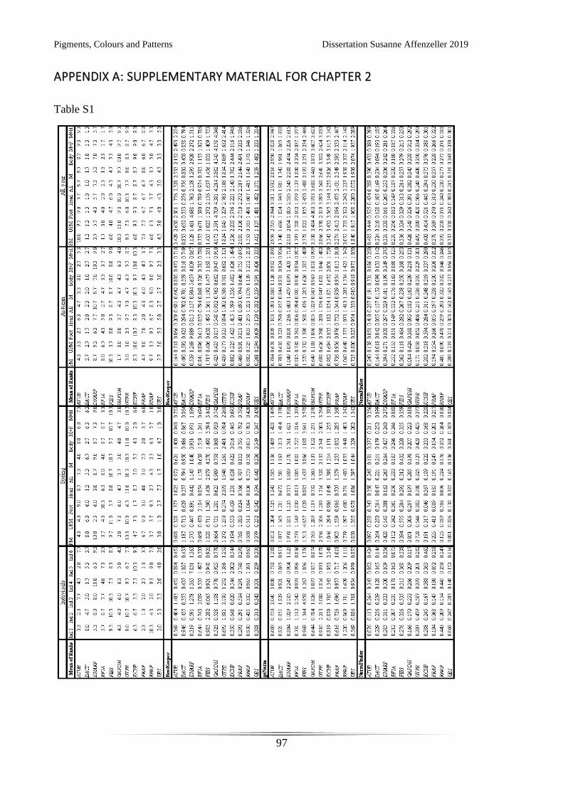
Pigments, Colours and Patterns Dissertation Susanne Affenzeller 2019
97
APPENDIX A: SUPPLEMENTARY MATERIAL FOR CHAPTER 2
Table S1
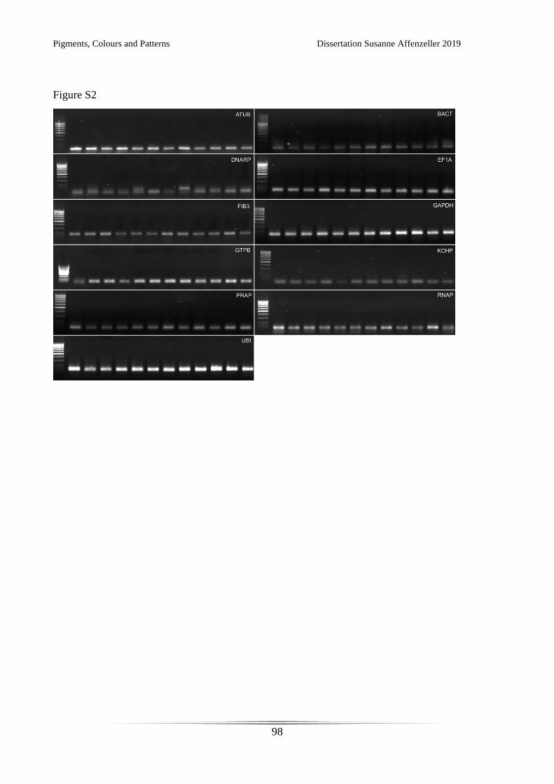
Pigments, Colours and Patterns Dissertation Susanne Affenzeller 2019
98
Figure S2
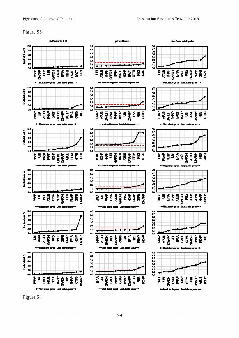
Pigments, Colours and Patterns Dissertation Susanne Affenzeller 2019
99
Figure S3
Figure S4
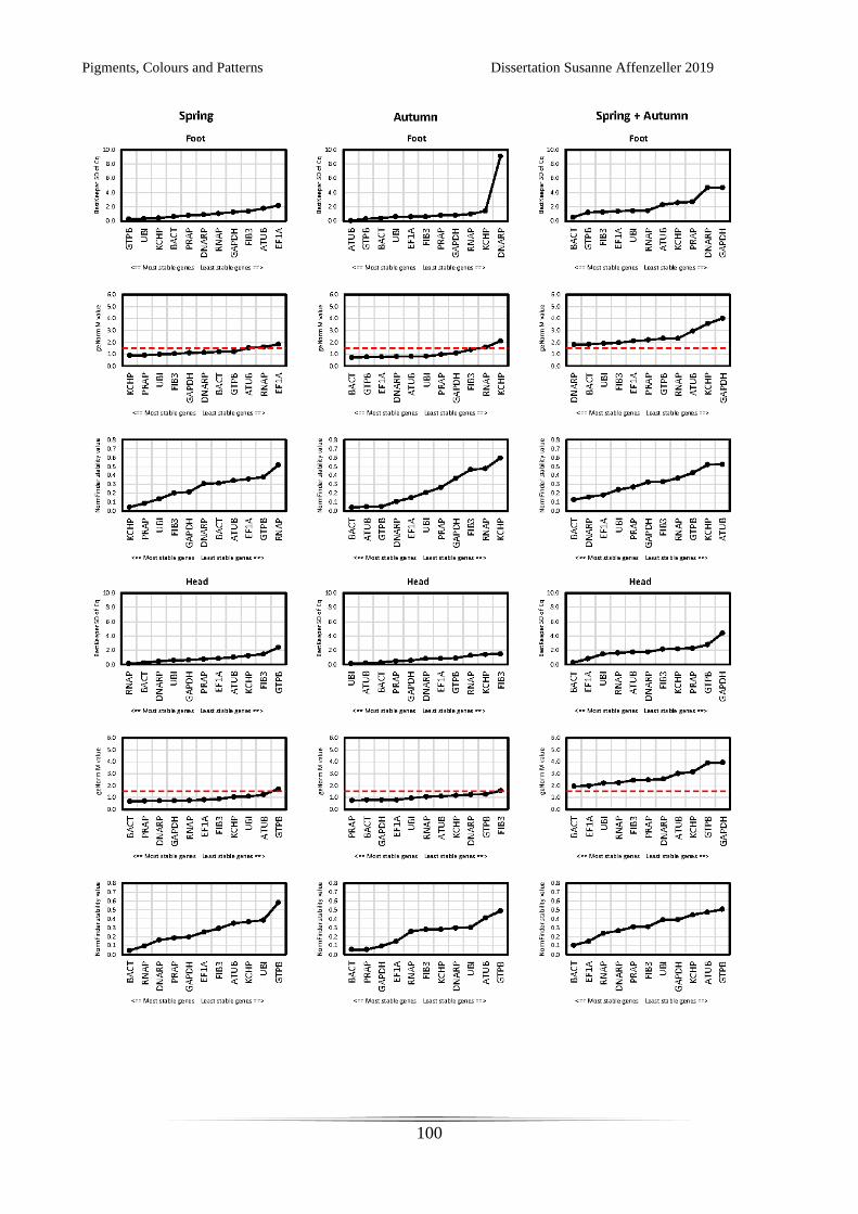
Pigments, Colours and Patterns Dissertation Susanne Affenzeller 2019
100
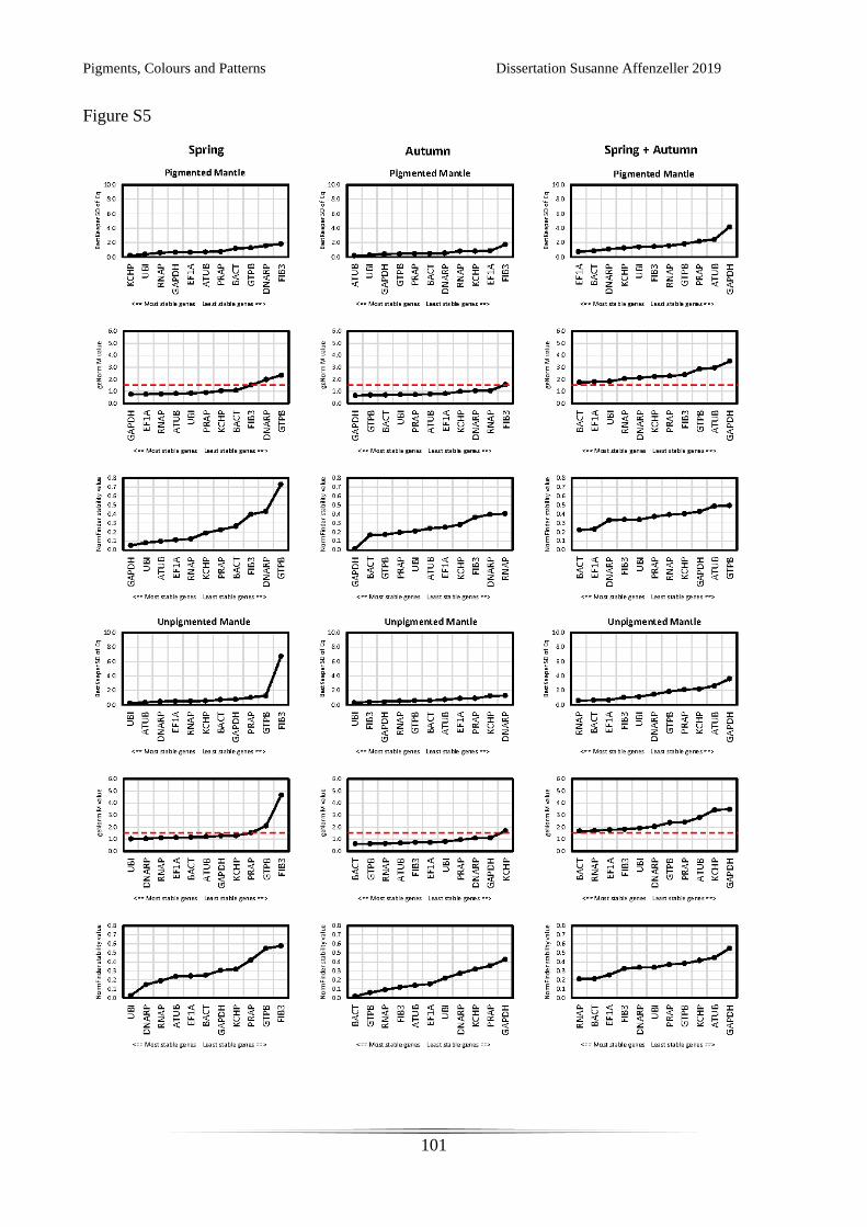
Pigments, Colours and Patterns Dissertation Susanne Affenzeller 2019
101
Figure S5
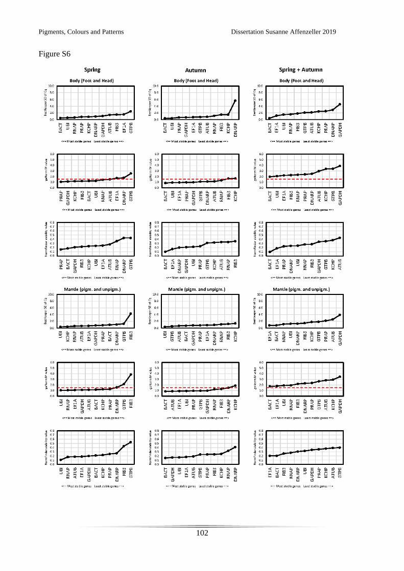
Pigments, Colours and Patterns Dissertation Susanne Affenzeller 2019
102
Figure S6
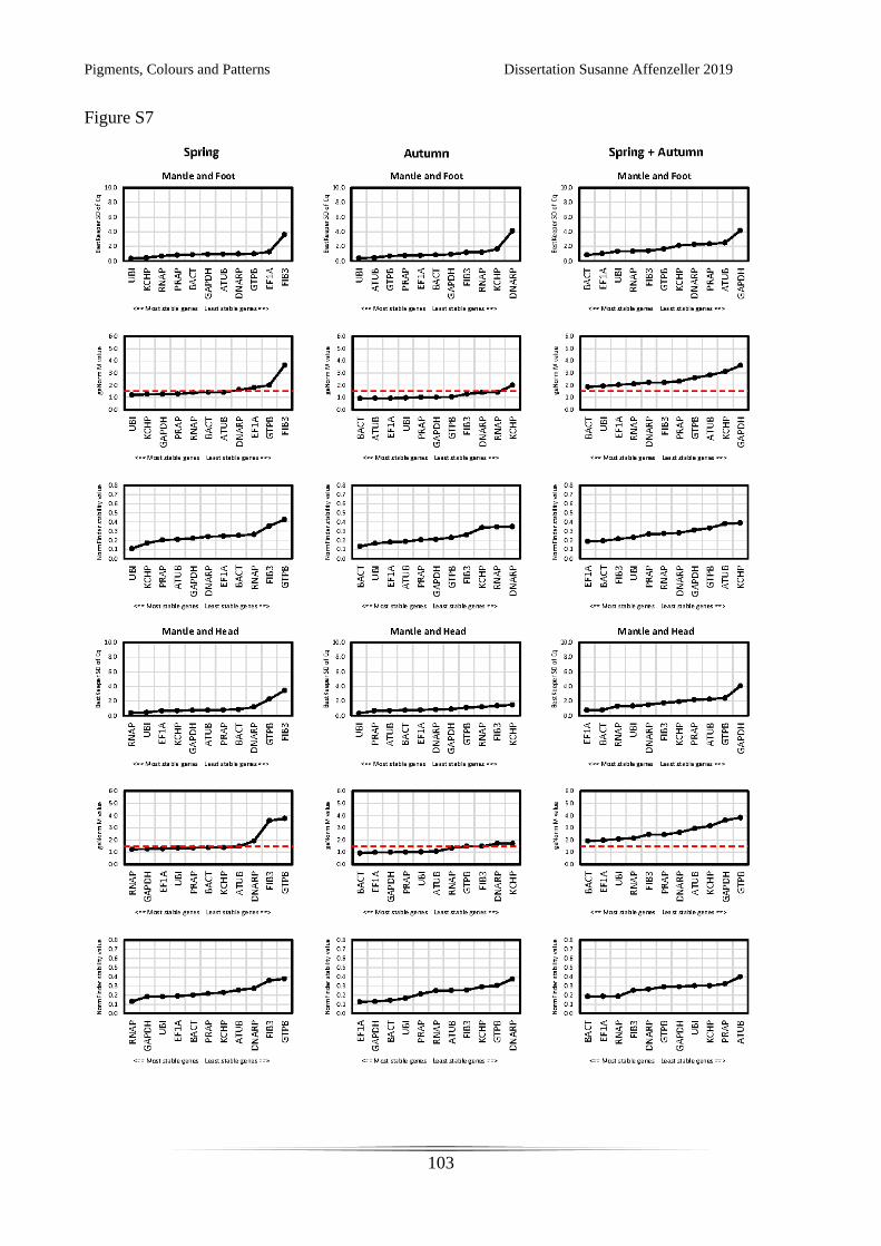
Pigments, Colours and Patterns Dissertation Susanne Affenzeller 2019
103
Figure S7
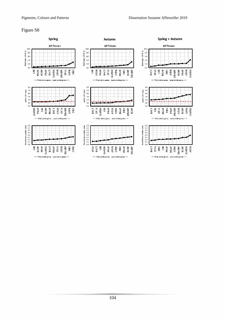
Pigments, Colours and Patterns Dissertation Susanne Affenzeller 2019
104
Figure S8
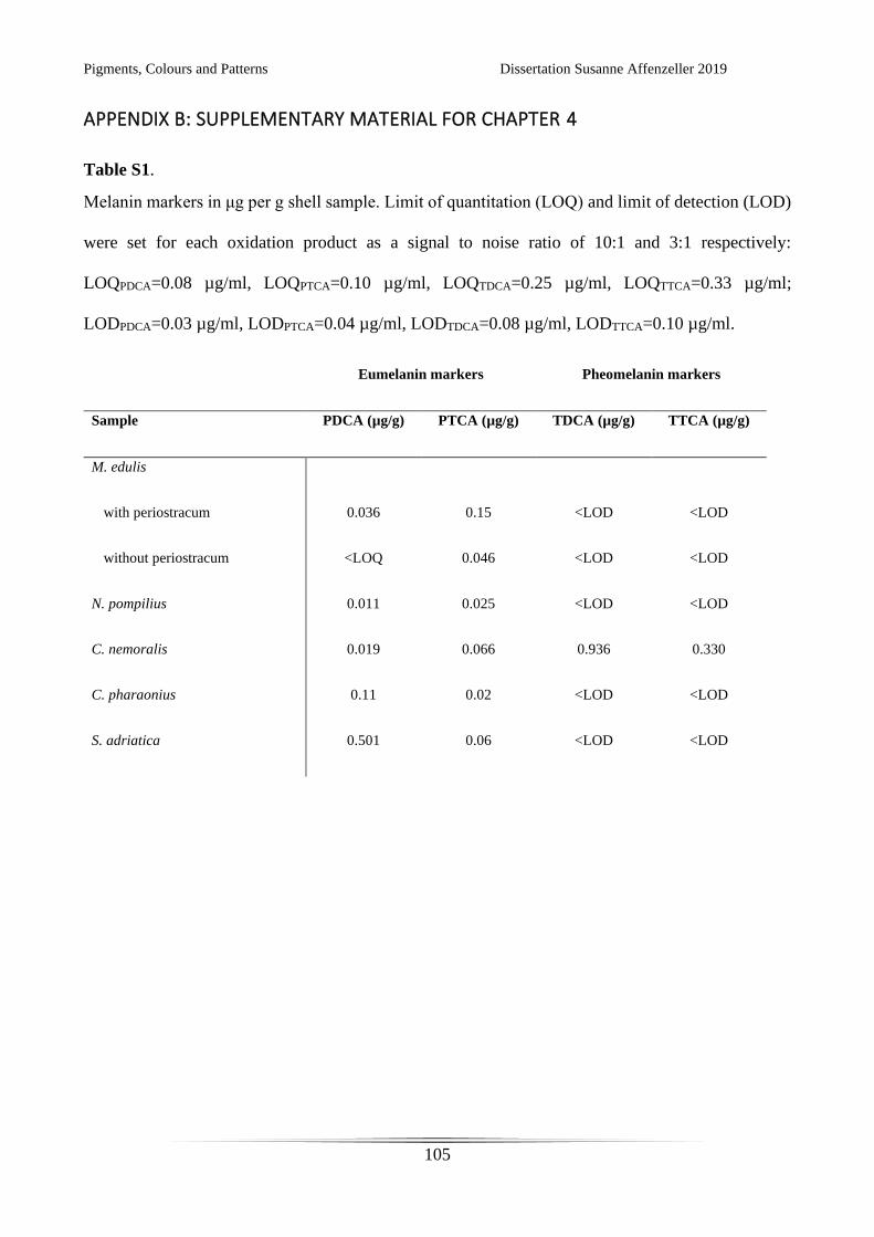
Pigments, Colours and Patterns Dissertation Susanne Affenzeller 2019
105
APPENDIX B: SUPPLEMENTARY MATERIAL FOR CHAPTER 4
Table S1.
Melanin markers in μg per g shell sample. Limit of quantitation (LOQ) and limit of detection (LOD)
were set for each oxidation product as a signal to noise ratio of 10:1 and 3:1 respectively:
LOQPDCA=0.08 µg/ml, LOQPTCA=0.10 µg/ml, LOQTDCA=0.25 µg/ml, LOQTTCA=0.33 µg/ml;
LODPDCA=0.03 µg/ml, LODPTCA=0.04 µg/ml, LODTDCA=0.08 µg/ml, LODTTCA=0.10 µg/ml.
Eumelanin markers Pheomelanin markers
Sample PDCA (µg/g) PTCA (µg/g) TDCA (µg/g) TTCA (µg/g)
M. edulis
with periostracum 0.036 0.15 <LOD <LOD
without periostracum <LOQ 0.046 <LOD <LOD
N. pompilius 0.011 0.025 <LOD <LOD
C. nemoralis 0.019 0.066 0.936 0.330
C. pharaonius 0.11 0.02 <LOD <LOD
S. adriatica 0.501 0.06 <LOD <LOD
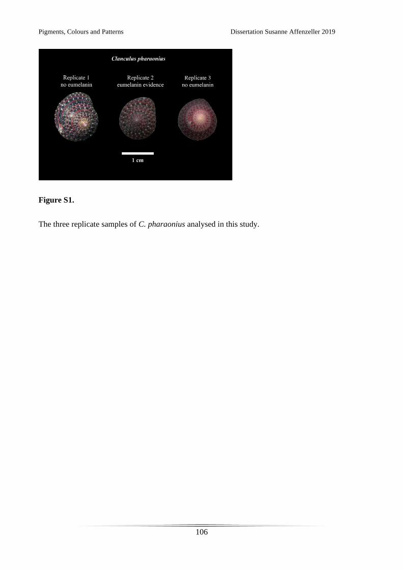
Pigments, Colours and Patterns Dissertation Susanne Affenzeller 2019
106
Figure S1.
The three replicate samples of C. pharaonius analysed in this study.

Pigments, Colours and Patterns Dissertation Susanne Affenzeller 2019
107
APPENDIX C: SUPPLEMENTARY MATERIAL FOR CHAPTER 5
Supplementary Table 1.
Melanin oxidation product markers in μg per g shell sample (n=3 for each sample, means ± standard
deviation (SD)). Limit of quantitation (LOQ) was set for each oxidation product as noise to signal
ration 1:10 by 9-point dilution series (LOQPDCA=0.08 µg/ml), LOQPTCA=0.10 µg/ml, LOQTDCA=0.25
µg/ml, LOQTTCA=0.33 µg/ml)
Sample PDCA
(µg/g)±SD
PTCA
(µg/g)±SD
TDCA
(µg/g)±SD
TTCA
(µg/g)±SD
pink morph
background 0.031±0.008 0.060±0.028 0.311±0.155 < LOQ
pink morph
band 0.051±0.027 0.083±0.037 0.356±0.124 < LOQ
yellow morph
background 0.063±0.037 0.168±0.025 1.384±0.635 0.469±0.243
yellow morph
band 0.081±0.003 0.148±0.028 1.653±0.714 0.736±0.412

Pigments, Colours and Patterns Dissertation Susanne Affenzeller 2019
108
STAR*Methods
Lead Contact and Materials Availability
Further information and requests for resources and reagents should be directed to and will be fulfilled
by the Lead Contact, Daniel John Jackson ([email protected]). Melanin oxidation product
standards must be requested from Prof. Shosuke Ito (Department of Chemistry, Fujita Health
University School of Health Sciences).
Experimental Model and Subject Details
Cepaea nemoralis
Six living animals and approximately 100 empty shells of C. nemoralis were collected at University
of Göttingen, Germany (51°33'24.0"N 9°57'27.3"E). Empty shells were cleaned and dried, then
crushed. Shell pieces were sorted according to replicate group and colour fraction. Tissue samples
were taken from fresh material by careful dissection of mantle and foot tissue.
Method Details
Shell Extraction, Melanin Oxidation and LC-MS Analyses
Two major morphs C. nemoralis (pink banded and yellow banded) were investigated by analysing
their colour sorted shell fragments (pink morph background, pink morph band, yellow morph
background, yellow morph band; three replicates each, each replicate is comprised of up to 8 shells).
Pigments, Colours and Patterns Dissertation Susanne Affenzeller 2019
109
Testing for eumelanin and pheomelanin oxidation products was carried out as previously described
(Affenzeller et al. in prep): Shells were cleaned in deionized water and weighed. Cleaned shell pieces
were dissolved in 6 M HCl and centrifuged at 13000 rpm for 15 min. Residue was washed twice in
HPLC grade water and remaining water was carefully removed by pipetting after centrifugation.
Samples were treated with Proteinase K in 1 M Tris-HCl buffer at 37°C for 2 h. Treatment was
stopped by acidification with 6 M HCl, samples were centrifuged and washed as described above.
Oxidation reactions for each sample contained 100 μL H2O, 375 μL 1 M K2CO3 and 25 μL 30% H2O2
and was carried out for 20h at 25°C under vigorous shaking. Remaining H2O2 was decomposed by
the addition of 50 μL 10% Na2SO3 and mixture was acidified with 140 μL 6 M HCl. The solution
was centrifuged at 13000rpm for 40min and supernatant was transferred to a fresh tube.
Samples were desalted via solid phase extraction on Strata-X 33μm Polymeric Reversed Phase
columns (200 mg/6 ml Phenomenex, ) under vacuum suction. Columns were conditioned with 5mL
methanol (MeOH) followed by 5 mL H2O. Shell extract was loaded onto the column diluted in 5 ml
0.3% formic acid and washed twice with 5 mL 0.3% formic acid. Column was dried for 30 min and
elution was carried out with 3 mL MeOH followed by 3 mL Ethyl acetate. Solvents were removed
under constant nitrogen stream at 40°C and samples dissolved in 200 μL H2O.
Conditions for chromatographic separation were as follows: An aliquote (10 μL) of oxidised sample
was directly injected into the LC-MS system run with a Gemini C18 column (5 μm particle size,
250x2 mm; Phenomenex, ) at a flow rate of 0.2 mL/min. The mobile phase of 0.3% formic acid in
H2O (eluent A) : MeOH (eluent B) (80:20) was run at 45°C over 20 min isocratically, followed by a
wash step of A : B (5:95) for 10 min and an equilibration phase to starting conditions for 10 min.
Mass spectrometric measurements were carried out on a Thermo Fisher Scientific (Waltham, USA)
Accela LC-MS system containing a Finnigan Surveyor PDA Detector and coupled to an LTQ
Pigments, Colours and Patterns Dissertation Susanne Affenzeller 2019
110
Orbitrap XL mass spectrometer. Ionisation was carried out with an electrospray ionisation (ESI) in
negative mode. Scan window was set to m/z = 120 - 220.
Reverse transcription quantitative PCR (qPCR) of melanin pathway genes in C. nemoralis
Four genes that are known to be involved in melanin synthesis and dark pigmentation were chosen
for qPCR testing: tyrosinase (Tyr), tyrosinase related protein (TyrRP), yellow-like gene (Yellow) and
laccase 2 (Lacc2). From C. nemoralis mantle tissue transcriptome data set (will be published
elsewhere) corresponding sequences were extracted based on their alignment score in tblastx
application of BLAST®. Primer for qPCR were designed with Primer3 (Untergasser et al., 2012).
Primer sequences and Genbank accession codes are listed in Data and Code Availability section.
Study design for qPCR experiments followed the protocol described in Affenzeller et al. (Affenzeller
et al., 2018): Six sub-adult individuals of C. nemoralis were collected at the University of Göttingen.
Total RNA from pigmented mantle (producing the band in the shell), unpigmented mantle (producing
background coloured shell) and foot tissue was extracted from each individual using Qiazol (Qiagen)
according to the manufacturer’s instructions resulting in a total of twelve RNA extractions. These
underwent a DNase treatment (RQ1 RNase-free DNase, Promega) according to the manufacturer’s
instructions. Nanodrop and agarose gel electrophoresis were employed to verify quality and integrity
of RNA. Synthesis of cDNA was carried out with 1 μg of total extracted RNA per sample using
Promega M-MLV reverse transcriptase and oligo dTs. Reaction was run at 42°C for 75 min, followed
by 15 min at 70°C to inactivate reverse transcriptase. The cDNA was stored at -20°C until further
use.
All qPCR runs followed a maximum sample layout, comply with the MIQE guidelines (Bustin et al.,
2009) and included no template controls (NTC) for each primer pair and three inter run calibrators
(IRC) EF1α, RNAP and UBI. Samples were run in triplicate, NTC and IRCs were run in duplicate.
Pigments, Colours and Patterns Dissertation Susanne Affenzeller 2019
111
Amplification reactions contained 5 μL 2x Rotor-Gene SYBR Green PCR Master Mix, 0.4 μL cDNA,
1μM final Primer concentration and 4.4 μL ddH2O to a final volume of 10 μL. Reactions were run on
a Rotor-Gene Q (Qiagen) using Rotor-Gene Q software (version 2.0.2) with the following
temperature profile: 5 min initial activation and denaturation at 95°C; 45 cycles of 5 sec denaturation
at 95°C, 10 sec annealing and extension at 60°C (data collection at this step); a final melt curve
analysis from 60°C to 95°C at a rate of 5 sec/1°C.
Quantification and Statistical Analyses
LC-MS of melanin oxidation products
Quantitation of melanin oxidation products was carried out by external calibration with standard
mixtures (obtained from S. Ito). External calibration was set with 9-point calibration curves. Limit of
quantitation (LOQ) was set as 1:10 noise to signal ratio. All manual peak integrations of
chromatograms and analyses of mass spectra were done in Xcalibur 2.2 Qual Browser (Thermo
Scientific, Waltham, USA). Quantitation was based on areas gained from peak integrations of UV
chromatograms in a range of 250-290 nm. Each colour fraction was run for three replicates
(comprised of up to eight shells each). Statistical analyses (mean and standard deviation calculations)
were carried out in Microsoft® Excel® for Office 365 MSO (16.0.11629.20192).
qPCR
Raw fluorescence data was baseline and amplification efficiency corrected in LinRegPCR (Ruijter et
al., 2009). Inter run correction was performed using Factor-qPCR (Ruijter et al., 2015). So gained
corrected cycle threshold (Cq) values were used to calculate the geometric means of technical
replicates. Normalisation and relative expression were calculated based on the Pfaffl method (Pfaffl,
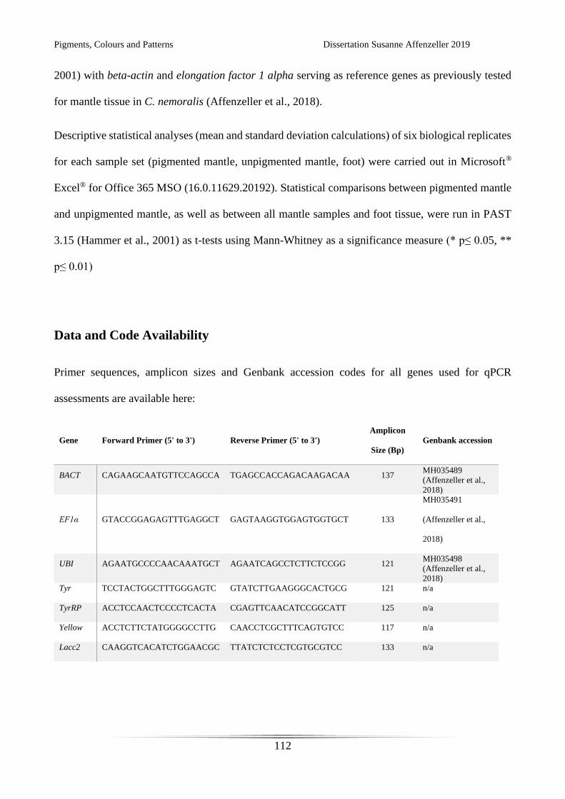
Pigments, Colours and Patterns Dissertation Susanne Affenzeller 2019
112
2001) with beta-actin and elongation factor 1 alpha serving as reference genes as previously tested
for mantle tissue in C. nemoralis (Affenzeller et al., 2018).
Descriptive statistical analyses (mean and standard deviation calculations) of six biological replicates
for each sample set (pigmented mantle, unpigmented mantle, foot) were carried out in Microsoft®
Excel® for Office 365 MSO (16.0.11629.20192). Statistical comparisons between pigmented mantle
and unpigmented mantle, as well as between all mantle samples and foot tissue, were run in PAST
3.15 (Hammer et al., 2001) as t-tests using Mann-Whitney as a significance measure (* p≤ 0.05, **
p≤ 0.01)
Data and Code Availability
Primer sequences, amplicon sizes and Genbank accession codes for all genes used for qPCR
assessments are available here:
Gene Forward Primer (5' to 3') Reverse Primer (5' to 3')
Amplicon
Size (Bp)
Genbank accession
BACT CAGAAGCAATGTTCCAGCCA TGAGCCACCAGACAAGACAA 137 MH035489
(Affenzeller et al.,
2018)
EF1α GTACCGGAGAGTTTGAGGCT GAGTAAGGTGGAGTGGTGCT 133
MH035491
(Affenzeller et al.,
2018)
UBI AGAATGCCCCAACAAATGCT AGAATCAGCCTCTTCTCCGG 121 MH035498
(Affenzeller et al.,
2018)
Tyr TCCTACTGGCTTTGGGAGTC GTATCTTGAAGGGCACTGCG 121 n/a
TyrRP ACCTCCAACTCCCCTCACTA CGAGTTCAACATCCGGCATT 125 n/a
Yellow ACCTCTTCTATGGGGCCTTG CAACCTCGCTTTCAGTGTCC 117 n/a
Lacc2 CAAGGTCACATCTGGAACGC TTATCTCTCCTCGTGCGTCC 133 n/a

Pigments, Colours and Patterns Dissertation Susanne Affenzeller 2019
113

Pigments, Colours and Patterns Dissertation Susanne Affenzeller 2019
114
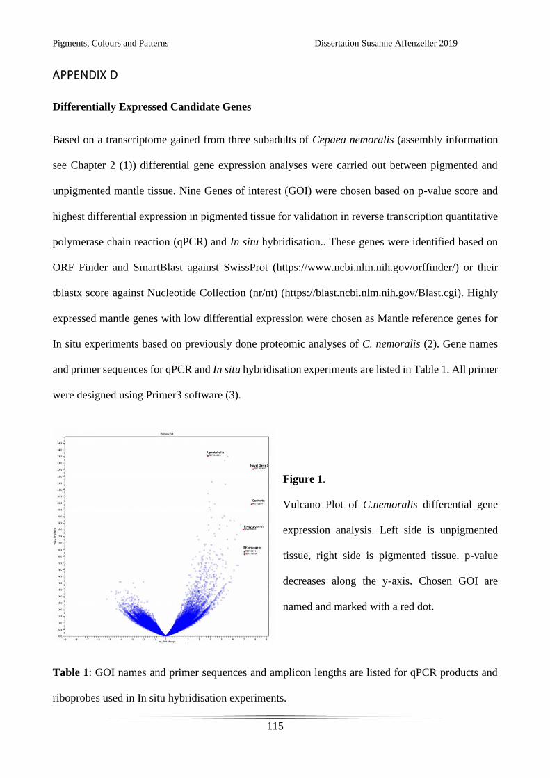
Pigments, Colours and Patterns Dissertation Susanne Affenzeller 2019
115
APPENDIX D
Differentially Expressed Candidate Genes
Based on a transcriptome gained from three subadults of Cepaea nemoralis (assembly information
see Chapter 2 (1)) differential gene expression analyses were carried out between pigmented and
unpigmented mantle tissue. Nine Genes of interest (GOI) were chosen based on p-value score and
highest differential expression in pigmented tissue for validation in reverse transcription quantitative
polymerase chain reaction (qPCR) and In situ hybridisation.. These genes were identified based on
ORF Finder and SmartBlast against SwissProt (https://www.ncbi.nlm.nih.gov/orffinder/) or their
tblastx score against Nucleotide Collection (nr/nt) (https://blast.ncbi.nlm.nih.gov/Blast.cgi). Highly
expressed mantle genes with low differential expression were chosen as Mantle reference genes for
In situ experiments based on previously done proteomic analyses of C. nemoralis (2). Gene names
and primer sequences for qPCR and In situ hybridisation experiments are listed in Table 1. All primer
were designed using Primer3 software (3).
Figure 1.
Vulcano Plot of C.nemoralis differential gene
expression analysis. Left side is unpigmented
tissue, right side is pigmented tissue. p-value
decreases along the y-axis. Chosen GOI are
named and marked with a red dot.
Table 1: GOI names and primer sequences and amplicon lengths are listed for qPCR products and
riboprobes used in In situ hybridisation experiments.
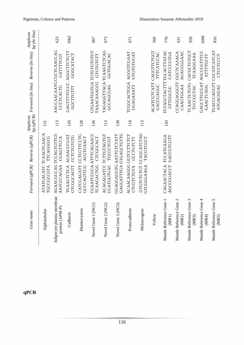
Pigments, Colours and Patterns Dissertation Susanne Affenzeller 2019
116
Am
pli
con
bp
(In
Sit
u)
62
3
10
62
66
7
67
3
67
1
56
0
77
9
63
1
95
9
10
00
81
0
Rev
erse
(In
Sit
u)
CG
CT
CA
AC
CA
G
GA
TT
TT
CG
T
AG
GC
TT
CT
GT
T
GG
GC
AT
AC
T
TG
TG
TG
TG
TG
T
GT
GT
GT
GT
T
TC
AA
GT
CT
CA
G
GG
TG
GA
CA
C
AG
GG
TC
GA
AT
GT
GT
GT
GG
AT
CA
GG
TT
CT
GG
T
TT
TC
AT
CC
AC
TG
CA
CT
TA
TA
C
CA
TC
CC
GT
GA
GG
CT
CA
AA
GA
AG
AC
GG
GA
AC
CA
CG
CT
GT
GC
T
TC
AA
GG
AA
A
AG
CC
CG
TT
TC
C
AT
TT
TG
CT
T
CG
CA
TC
AT
CA
T
CT
CC
TC
CC
T
Fo
rwa
rd (
In S
itu
)
AA
CC
AA
CA
AT
G
CC
CT
CA
CT
G
GA
GT
TT
TC
CC
C
GG
CT
TG
TT
T
GT
GA
AT
GG
TG
A
TA
AA
CA
AA
GC
G
G
TA
GA
GG
TT
AC
A
GC
CA
GC
GA
G
TC
GG
CA
CT
GG
A
GG
AG
AA
AT
T
AC
AT
CG
TC
AT
T
GA
CC
GA
GG
C
TG
TA
GC
TA
CT
TT
GT
TG
GC
GG
CC
AG
GA
GG
GT
T
CA
AG
TG
GA
A
TC
AA
CT
CT
GT
G
TC
CC
GG
TA
C
GA
GC
TT
GC
GA
T
GA
AC
TC
AG
G
TG
AC
CC
AG
GT
T
AT
GA
CG
GA
C
Am
pli
con
bp
(q
PC
R)
13
1
11
3
12
0
12
8
13
6
11
3
13
8
11
8
11
3
14
3
Rev
erse
(q
PC
R)
TC
AA
GT
CA
AC
A
TT
CA
GG
GC
G
TC
CA
GT
GT
TG
T
CG
AG
TT
CC
A
AG
AA
CC
CG
AT
CC
TC
TG
TG
TG
CC
TC
CT
TC
CT
G
AC
GT
GA
AC
T
AT
TT
CA
GA
AC
C
CT
CG
GC
AC
T
TC
TT
GT
AG
TG
T
TG
CC
CT
CG
T
AA
TT
GT
CT
AT
T
GT
GA
GC
TG
TT
G
G
GG
TC
CT
TC
TC
T
GC
CT
GT
CT
T
CC
AG
CA
TC
TA
G
TA
CG
TC
GC
T
TC
CA
TG
AA
GA
GA
CC
GT
CG
TT
Fo
rwa
rd (
qP
CR
)
AT
AT
GA
CA
TC
TG
CC
GC
CG
TA
AG
AA
CG
CC
AG
AA
TC
CC
AG
AA
TG
AA
CT
CT
CA
GT
CG
GC
AG
TT
CA
TC
GA
GA
TT
GC
CC
AG
TT
CG
CG
AA
TC
CG
CA
TC
AA
GA
CT
GG
AC
AG
AG
AT
CC
CG
AT
GG
TG
AC
GG
AG
GA
GG
TG
GA
GG
AT
TT
GA
AC
AA
CA
AG
GG
CT
CG
TC
TG
TA
GT
GT
CT
CT
GT
GT
CG
GG
AA
GA
CA
GA
AC
TA
CA
AG
CC
GG
AC
CT
Gen
e n
am
e
Alp
hat
ub
uli
n
Ad
ipo
cyte
pla
sma
mem
bra
n
pro
tein
(A
PM
-P)
Cad
her
in
Hae
mo
cyan
in
No
vel
Gen
e 1
(N
G1
)
No
vel
Gen
e 2
(N
G2
)
No
vel
Gen
e 3
(N
G3
)
Pro
toca
dh
erin
SK
Ion
cog
ene
Yel
low
Man
tle
Ref
eren
ce G
ene
1
(MR
1)
Man
tle
Ref
eren
ce G
ene
2
(MR
2)
Man
tle
Ref
eren
ce G
ene
3
(MR
3)
Man
tle
Ref
eren
ce G
ene
4
(MR
4)
Man
tle
Ref
eren
ce G
ene
5
(MR
5)
qPCR
Pigments, Colours and Patterns Dissertation Susanne Affenzeller 2019
117
Experimental comparison of GOI between Pigmented mantle tissue and Unpigmented mantle tissue
were run as described in Chapter 4. In short: 10 sub-adult individuals of C. nemoralis were collected
at the University of Göttingen. Total RNA from pigmented mantle (producing the band in the shell),
and unpigmented mantle (producing background coloured shell) was extracted from each individual
using Qiazol (Qiagen) according to the manufacturer’s instructions. All 20 RNA extractions
underwent a DNase treatment (RQ1 RNase-free DNase, Promega) according to the manufacturer’s
instructions. Quality and integrity of RNA were tested via Nanodrop and agarose gel electrophoresis..
cDNA synthesis was carried out with 1 μg of total extracted RNA per sample using Promega M-MLV
reverse transcriptase and oligo dTs. Each reaction was run at 42°C for 75 min, followed by 15 min at
70°C to inactivate reverse transcriptase. The cDNA was stored at -20°C until further use. Following
this protocol additional cDNAs were gained for four corresponding Foot tissue samples. Two
additional individuals were used to gain two Mantle edge and two Mantle back tissue samples.
Primer pairs used for qPCR are listed in Table 1. All qPCR runs followed a maximum sample layout
and the MIQE guidelines (4) and included no template controls (NTC) and three inter run calibrators
(IRC) EF1α, RNAP and UBI. Samples were run in triplicate, NTC and IRCs were run in duplicate.
Amplification reactions consisted of 5 μL 2x Rotor-Gene SYBR Green PCR Master Mix, 0.4 μL
cDNA, 1μM final Primer concentration and 4.4 μL ddH2O to a final volume of 10 μL. A Rotor-Gene
Q (Qiagen) using Rotor-Gene Q software (version 2.0.2) was used with the following temperature
profile: 5 min initial activation and denaturation at 95°C; 45 cycles of 5 sec denaturation at 95°C, 10
sec annealing and extension at 60°C (data collection at this step); a final melt curve analysis from
60°C to 95°C at a rate of 5 sec/1°C.
Baseline and amplification efficiency correction of raw fluorescence data was done in LinRegPCR
(5). Inter run correction was performed using Factor-qPCR (6). Geometric means of technical
replicates were used to gain final corrected cycle thresholds (Ct). Normalisation with beta-actin and
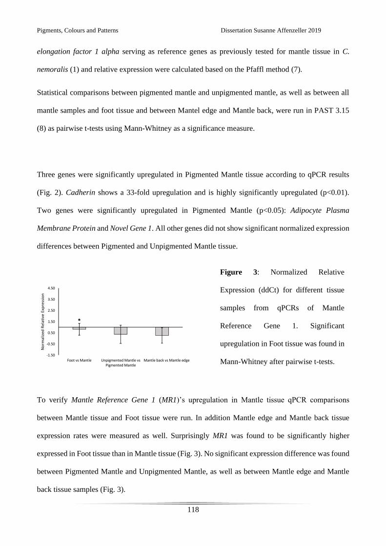
Pigments, Colours and Patterns Dissertation Susanne Affenzeller 2019
118
elongation factor 1 alpha serving as reference genes as previously tested for mantle tissue in C.
nemoralis (1) and relative expression were calculated based on the Pfaffl method (7).
Statistical comparisons between pigmented mantle and unpigmented mantle, as well as between all
mantle samples and foot tissue and between Mantel edge and Mantle back, were run in PAST 3.15
(8) as pairwise t-tests using Mann-Whitney as a significance measure.
Three genes were significantly upregulated in Pigmented Mantle tissue according to qPCR results
(Fig. 2). Cadherin shows a 33-fold upregulation and is highly significantly upregulated (p<0.01).
Two genes were significantly upregulated in Pigmented Mantle (p<0.05): Adipocyte Plasma
Membrane Protein and Novel Gene 1. All other genes did not show significant normalized expression
differences between Pigmented and Unpigmented Mantle tissue.
To verify Mantle Reference Gene 1 (MR1)’s upregulation in Mantle tissue qPCR comparisons
between Mantle tissue and Foot tissue were run. In addition Mantle edge and Mantle back tissue
expression rates were measured as well. Surprisingly MR1 was found to be significantly higher
expressed in Foot tissue than in Mantle tissue (Fig. 3). No significant expression difference was found
between Pigmented Mantle and Unpigmented Mantle, as well as between Mantle edge and Mantle
back tissue samples (Fig. 3).
Figure 2: Normalized Relative
Expression (ddCt) between Pigmented
Mantle tissue (top) and Unpigmented
Mantle Tissue (bottom). Significance
values according to Mann-Whitney
after pairwise t-test comparisons.
Figure 3: Normalized Relative
Expression (ddCt) for different tissue
samples from qPCRs of Mantle
Reference Gene 1. Significant
upregulation in Foot tissue was found in
Mann-Whitney after pairwise t-tests.
Pigments, Colours and Patterns Dissertation Susanne Affenzeller 2019
119
In situ hybridisation on C. nemoralis embryos and sub adult tissue section
Wholemount In situ hybridisation (WMISH) and tissue section In situ hybridisation (TSISH) for C.
nemoralis were based on protocols previously established for a limnic gastropod Lymnea stagnalis
(9–12).
Individuals of C. nemoralis were collected from the University of Göttingen in spring and summer.
Sub-adults were fixed for tissue sections as described in detail below. Adults were kept in glass
terraria on Cocos substrate mixed with sand and chalk. Terraria were kept humid and snails were fed
regularly with salad, cucumber and occasionally carrots. Egg clutches were carefully removed from
terraria once spotted and reared in plastic containers with soil under humid conditions.
Observations of mating behaviour and egg clutch development confirmed difficulties in keeping the
animals in conditions perfect for mating. Mating only occurred in first season after collection.
Keeping individuals over winter with hibernation phase was possible but no further mating occurred.
Clutch development seems to be asynchronous and egg capsules are opaque and easily broken when
handled, making clutch staging very difficult. WMISH were therefore conducted on young juveniles
(max. 5mm shell diameter) exhibiting first signs of shell banding.
Juvenile fixation was carried out in 3.7% paraformaldehyde (PFA) for 5 min. Samples were then
thoroughly washed in PBS (phosphate buffered saline solution) and shells were carefully taken of
with forceps and needles. Juveniles were treated in Proteinase K solution (500 μg/ml) for 10 min and
the digestion was stopped with two washes of 0.2% Glycine. All samples were thoroughly washed in
PBS and fixed for 30 min in 3.7% PFA. Fixative was washed of with PBS. Pre-hybridisation was
carried out at 55°C for 15 min. Riboprobes were synthesised from PCR products generated with In
situ primer pairs listed in Table 1 following the protocol in (9). Riboprobe concentrations were trialled
at 300-500 ng/ml. Hybridisation followed an initial denaturation step of 75°C for 15 min and was
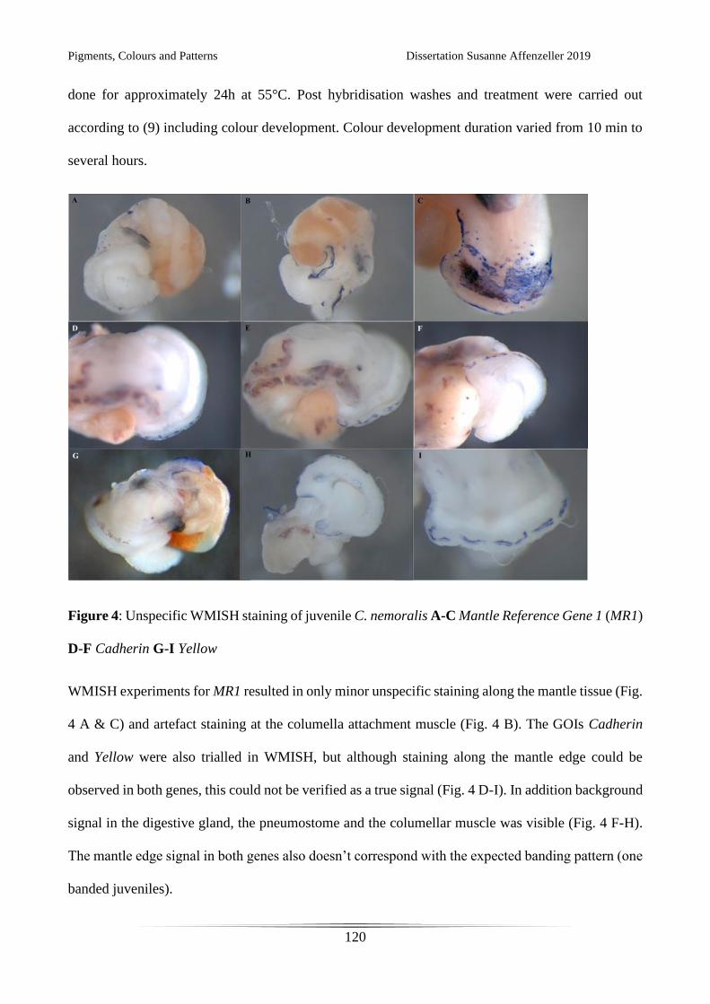
Pigments, Colours and Patterns Dissertation Susanne Affenzeller 2019
120
done for approximately 24h at 55°C. Post hybridisation washes and treatment were carried out
according to (9) including colour development. Colour development duration varied from 10 min to
several hours.
Figure 4: Unspecific WMISH staining of juvenile C. nemoralis A-C Mantle Reference Gene 1 (MR1)
D-F Cadherin G-I Yellow
WMISH experiments for MR1 resulted in only minor unspecific staining along the mantle tissue (Fig.
4 A & C) and artefact staining at the columella attachment muscle (Fig. 4 B). The GOIs Cadherin
and Yellow were also trialled in WMISH, but although staining along the mantle edge could be
observed in both genes, this could not be verified as a true signal (Fig. 4 D-I). In addition background
signal in the digestive gland, the pneumostome and the columellar muscle was visible (Fig. 4 F-H).
The mantle edge signal in both genes also doesn’t correspond with the expected banding pattern (one
banded juveniles).
Pigments, Colours and Patterns Dissertation Susanne Affenzeller 2019
121
Due to the limitations in individual numbers and difficulties in rearing juveniles further optimisation
of a WMISH protocol would be very time intensive and detrimental to population numbers. Finding
a good reference gene and overcoming the strengthened skin barrier of this terrestrial gastropod
should be the next steps to adjust published protocols for this species.
One approach was switching the experimental set up to In situ hybridisations on sub-adult tissue
sections. This allowed us to use larger animals with distinct banding patterns and at the same time
provides more experiments per individual. Optimisation of the protocol in (10) and (12) yielded the
following adjustments. Four different fixation schemes were trialled: 3.7% PFA, Carnoy’s (60%
ethanol, 30% chloroform, 10% glacial acetic acid), Methacarn (60% methanol, 30% chloroform, 10%
glacial acetic acid), 100% ethanol. Snails with slightly damaged shells were left in the respective
fixative for 60 min. After thorough washing and removal of the shell, samples were dehydrated
through an ethanol series. Each snail was dissected along its midline, allowing a smooth placing in
the cassette with a clear cutting start of the mantle edge. Samples were treated to three changes of
100% xylene and submerged in paraffin overnight. Setting in the cassette was carefully done with
forceps and the samples were cooled for at least three hours. Microtome cut sections of 8 μm were
adhesed to polylysine coated slides overnight at 60°C. Slides were treated with 100% xylene and
rehydrated before hybridisation treatment. Proteinase K treatment was carried out for 10 min with
concentrations of either 50 μg/ml or 100 μg/ml. Riboprobe synthesis from PCR products gained form
primer pairs in Table 1 was conducted as described above. Hybridisation with probe concentration of
800 ng/ml was done for 24 hours at 55°C, followed by washes and blocking as described in (12).
Samples were treated with anti DIG antibody conjugated to alkaline phosphatase (concentration
1/10000) for 12 hours. Final washes and colour development were carried out according to the
protocol in (12).
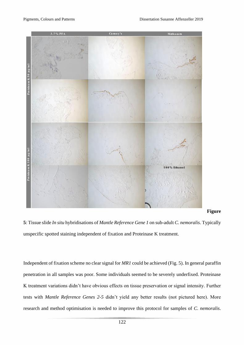
Pigments, Colours and Patterns Dissertation Susanne Affenzeller 2019
122
Figure
5: Tissue slide In situ hybridisations of Mantle Reference Gene 1 on sub-adult C. nemoralis. Typically
unspecific spotted staining independent of fixation and Proteinase K treatment.
Independent of fixation scheme no clear signal for MR1 could be achieved (Fig. 5). In general paraffin
penetration in all samples was poor. Some individuals seemed to be severely underfixed. Proteinase
K treatment variations didn’t have obvious effects on tissue preservation or signal intensity. Further
tests with Mantle Reference Genes 2-5 didn’t yield any better results (not pictured here). More
research and method optimisation is needed to improve this protocol for samples of C. nemoralis.
Pigments, Colours and Patterns Dissertation Susanne Affenzeller 2019
123
Especially tissue penetration should be further researched, as it seems these animals have a much
stronger skin barrier than other molluscs. This is unsurprising considering the habitat they are
commonly found in is dry and they have to adjust for water loss. It can also not be said with certainty
that there are no inhibiting factors influencing these In situ experiments.
References
1. Affenzeller S, Cerveau N, Jackson DJ. Identification and validation of reference genes for qPCR in the
terrestrial gastropod Cepaea nemoralis. PLoS ONE. 2018;13(8):e0201396.
2. Mann K, Jackson D. Characterization of the pigmented shell-forming proteome of the common grove snail
Cepaea nemoralis. BMC Genomics. 2014;15(1):249.
3. Untergasser A, Cutcutache I, Koressaar T, Ye J, Faircloth BC, Remm M, u.a. Primer3—new capabilities and
interfaces. Nucleic Acids Research. 2012;40(15):e115–e115.
4. Bustin SA, Benes V, Garson JA, Hellemans J, Huggett J, Kubista M, u.a. The MIQE Guidelines: Minimum
Information for Publication of Quantitative Real-Time PCR Experiments. Clinical Chemistry. 2009;55(4):611–
22.
5. Ruijter JM, Ramakers C, Hoogaars WMH, Karlen Y, Bakker O, van den Hoff MJB, u.a. Amplification
efficiency: linking baseline and bias in the analysis of quantitative PCR data. Nucleic Acids Research.
2009;37(6):e45–e45.
6. Ruijter JM, Ruiz Villalba A, Hellemans J, Untergasser A, van den Hoff MJB. Removal of between-run variation
in a multi-plate qPCR experiment. Biomolecular Detection and Quantification. 2015;5:10–4.
7. Pfaffl MW. A new mathematical model for relative quantification in real-time RT-PCR. Nucleic Acids
Research. 2001;29(9):2002–7.
8. Hammer Ø, Harper DAT, Ryan PD. PAST: PAlaeontological STatistics softwarre package for education and
data analysis. Paleontologia Electronica. 2001;4(1):9.
9. Hohagen J, Herlitze I, Jackson DJ. An optimised whole mount in situ hybridisation protocol for the mollusc
Lymnaea stagnalis. BMC Developmental Biology. 2015;15(19):13.
10. Herlitze I, Marie B, Marin F, Jackson DJ. Molecular modularity and asymmetry of the molluscan mantle
revealed by a gene expression atlas. Gigascience. 2018;7(6):1–15.
11. Jackson DJ, Herlitze I, Hohagen J. A Whole Mount In Situ Hybridization Method for the Gastropod Mollusc
Lymnaea stagnalis. J Vis Exp. 2016;(109).
12. Herlitze I. Molecular characterisation of biomineralising genes in the freshwater pond snail Lymnea stagnalis.
Georg-August-Universität Göttingen; Göttingen. 2017.

Pigments, Colours and Patterns Dissertation Susanne Affenzeller 2019
124
ACKNOWLEDGEMENTS
My gratitude is due to Daniel John Jackson for affording me the opportunity to keep working on my
passion for molluscs during my PhD research.
I would like to thank Gregor Bucher and Klaus Wolkenstein for their supervision and support.
Furthermore I would like to thank Volker Thiel, Christoph Bleidorn, Sven Bradler and Nico Posnien
for spending time and effort on my committee.
Nicholas Cerveau deserves thanks for fruitful discussions, explanations when needed and inspiring
coffee breaks. I thank Anne Geissler for her support, knowledge on snails and laughs when I most
needed them.
I would like to thank Ines Herlitze, Juliane Germer and Eva-Maria Sadowski for their encouragements
and sharing their wisdom on PhD life. Alexander Schmidt I thank for mentoring and allowing me to
develop my teaching skills.
For sound advise and a push of motivation when needed I thank Elisabeth Haring and Gerhard
Steiner, as well as Lisa-Maria Schmidt and Hannah Schmidbaur.
Without the unwavering support, repeated reminders not to give up and a steady tea supply my thesis
would probably not be what it is now: Many thanks are due to Leyla Seyfullah!
Regardless of distance and minor and major catastrophes my family always supported me. I am
deeply grateful to them for their understanding and encouragements.
I dedicate this thesis to Thomas: You take my crazy phases with a laugh and my desperate ones with
a warm hug. Thank you for always reminding me that “Aufgebn tuat ma nur an Briaf”

Pigments, Colours and Patterns Dissertation Susanne Affenzeller 2019
125
Pigments, Colours and Patterns Dissertation Susanne Affenzeller 2019
126
CURRICULUM VITAE
Susanne Affenzeller
Date of Birth: 3.8.1990
Work experience:
2016 – 2019 Research associate, Georg-August-University Göttingen, Geobiology: Evolution of the
Metazoa group
2014 – 2016 Scientific Collection Management, University of Vienna: Mollusca (Collection Salvini-
Plawen)
2014 – 2016 University Lecturer, University of Vienna: Basic Course in Phylogenetics
2012 – 2014 University Tutor, University of Vienna: Basic Practical Course in Zoology (Baupläne)
Formal Education:
2016 – 2019 PhD (Geoscience) Georg-August-University Göttingen Supervisor: Daniel J. Jackson
2013 – 2015 M.Sc. (Zoology) University of Vienna Supervisor: Gerhard Steiner
2009 – 2013 B.Sc. (Biology) University of Vienna
Nonformal Education:
2009 – 2018 Scout Leadership Training to Woodbadge Level at Pfadfinder und Pfadfinderinnen
Österreichs
2017 Workshop “Business Competence and Basic Accounting for Scientists”
2016 Lehrgang Gene Technology, Biosafety and Biosecurity nach § 15 GenTSV,
Medizinische Hochschule Hannover
2016 Workshops “Grant Writing for Scientists” & “Higher Education for Natural Scientists”
Languages: German, English, French, Spanish
Publications:
Pigments, Colours and Patterns Dissertation Susanne Affenzeller 2019
127
Affenzeller, S., Cerveau, N., Jackson, D.J. 2018. Identification and validation of reference genes for qPCR in
the terrestrial gastropod Cepaea nemoralis. PloS One, 13(8), e0201396.
Affenzeller, S., & Steiner, G. 2017. Catalog of taxa introduced by Luitfried Salvini-Plawen (1939–2014).
Zootaxa, 4337(1), 73-90.
Affenzeller, S., Haar, N., & Steiner, G. 2017. Revision of the genus complex Gibbula: an integrative approach
to delineating the Eastern Mediterranean genera Gibbula Risso, 1826, Steromphala Gray, 1847, and Phorcus Risso,
1826 using DNA-barcoding and geometric morphometrics (Vetigastropoda, Trochoidea). Organisms Diversity &
Evolution, 17 (4), 789-812.
Conference Contributions and Prizes:
Affenzeller, S., Wolkenstein, K., Cerveau, N., Jackson, D.J. 2018. A Colourful mystery: Insights into the
Cepaea nemoralis banding pattern mechanism. Abstracts Abstracts 111th Annual Meeting German Zoological Society,
Sept. 10th-15th 2018 Greifswald, Germany
Affenzeller, S., Haar, N. Steiner, G. 2016. Species delineation: Comparing the powers of shell morphometrics
and DNA-barcoding in the trochid gastropod genus Gibbula. Abstracts 19th International Congress of UNITAS
MALACOLOGIA, July 18th-24th 2016, Penang, Malaysia
Affenzeller, S. 2015. Morphometric approaches to species delineation and characterisation in the trochid
Genus Gibbula (Gastropoda). Abstracts Biosystematics from Past to Present 9. Jahrestagung NOBIS, Nov. 27th-28th
2015 Eggenburg, Austria
Affenzeller, S., Haar, N., Steiner, G. 2015. Species delineation: Comparing the powers of DNA-barcoding and
shell morphometrics in the trochid gastropod genus Gibbula. Abstracts 108th Annual Meeting German Zoological
Society, Sept. 9th-12th 2015 Graz, Austria
Frontiers of Zoology Prize 2015 for Best Student Short Talk in Systematics Symposium: Affenzeller, S., Haar,
N., Steiner, G. 2015. Species delineation: Comparing the powers of DNA-barcoding and shell morphometrics in the
trochid gastropod genus Gibbula. 108th Annual Meeting German Zoological Society, Sept. 9th-12th 2015 Graz
NOBIS-Preis für herrausragende Forschung 2015: Affenzeller, S. 2015. Morphometric approaches to species
delineation and characterisation in the trochid Genus Gibbula (Gastropoda). Abstracts Biosystematics from Past to
Present 9. Jahrestagung NOBIS, Nov. 27th-28th 2015 Eggenburg, Austria

Pigments, Colours and Patterns Dissertation Susanne Affenzeller 2019
128

Pigments, Colours and Patterns Dissertation Susanne Affenzeller 2019
129