Embed Size (px)
Citation preview
"Pilado add Value" - valorisation of non-traditional
marine resources
Francisco Pires Avelelas
A thesis submitted to the School of Tourism and Maritime Technology,
Polytechnic Institute of Leiria in partial fulfilment of the requirements for the
Degree of Master of Science in Biotechnology on Marine Resources, held
under the scientific supervision of Prof. Sérgio Miguel Martins Leandro Franco
(School of Tourism and Maritime Technology, Polytechnic Institute of Leiria -
Portugal), and Ph.D. Sónia Cotrim Marques (Centre for Functional Ecology,
University of Coimbra – Portugal).
2013

ii
iii
Title: "Pilado add Value" - valorisation of non-traditional marine resources
Copyright© Francisco Pires Avelelas
School of Tourism and Maritime Technology – Peniche Polytechnic Institute of Leiria
2013
The School of Tourism and Maritime Technology and the Polytechnic Institute of Leiria are
entitled, perpetual and without geographical boundaries, archive and publish this project
work through printed copies reproduced on paper or digital form, or by any other known
means or to be invented, and to disseminate through scientific repositories and admit your
copying and distributing educational purposes or research, non-commercial, as long as
credit is given to the author and publisher

iv
v
Acknowledgments
First of all, I would like to thank colleagues Rui Albuquerque and Pedro Sá (GIRM – IP
Leiria) for their help during the preparation of the raw material, and Purse seine fishing
vessel “Mestre Comboio – Peniche” and Pedro Murraças, for providing the swimming
crab.
Also, I have to thank GAC Oeste (PROMAR – European Fisheries Fund) that sponsor the
project Pilado add value, and the Polytechnic Institute of Leiria for logistical and finnancial
support. To Altakitin, for all the support and interest on developing this project
Furthermore I would like to thank André Horta for introducing me to new topics and
laboratorial procedures, as well for the support on the way.
Also, I like to thank everyone who have willingly shared their precious time during the
process of laboratorial experiments, such as the technicians Pedro Ramalho, Vera
Severiano, Cristina Salas and professor Paulo Nunes.
I would like to express my gratitude towards my coordinator and mentor, professor Sérgio
Leandro for all the support, orientation and fellowship through the learning process of this
master thesis. To Ph.D. Sónia Cotrim Marques, for all the precious help and guidance
during the past months of this project.
My sincere thanks to Cátia Velez for being always there when I most needed.
Last but not the least, I would like to thank my loved ones (parents and grandparents),
once everything I am today, as a person, professional and human been, I owe to them.

vi
vii
Resumo
Os subprodutos da pesca têm recebido maior atenção devido à perceção dos seus
impactos negativos na economia e no ambiente. Contudo, os subprodutos de crustáceos
contêm vários compostos que podem ser usados como fonte de biopolímeros, como por
exemplo a quitina, tendo esta uma grande variedade de aplicações biotecnológicas.
O caranguejo Polybius henslowii, é um recurso marinho extremamente abundante na
costa oeste Portuguesa durante os meses de Verão, não sendo contudo aproveitada para
fins comerciais. Com este estudo procura-se assim contribuir para a valorização
económica deste recurso, como fonte de matéria-prima para a extração de polímeros
visando aplicações biotecnológicos.
Para avaliar o seu potencial, a quitina foi extraída e quitosano produzido, a partir de
distintas partes do corpo de Polybius henslowii: pereópodes e carapaça. O quitosano
obtido, serviu, por sua vez, de matéria-prima para a produção de quitosano solúvel em
água (WSC) e oligomeros de quitosano (COS), tendo todas as amostras sido
caracterizadas e testadas quanto às suas propriedades antibacteriana, antifúngica e
antioxidante, redução do radical 1, 1-Difenil-2-picrilhidrazil (DPPH).
A caracterização das amostras de quitosano obtidas a partir de ambos os segmentos,
demonstraram diferenças quanto ao seu rendimento, peso molecular e viscosidade. Os
oligomeros de quitosano (COS) revelaram atividade antibacteriana contra todas as
bactérias Gram-negativas testadas e a Gram-positiva, Lactobacillus planctarum
(ATCC8014). COS mostraram melhores resultados na concentração mínima inibitória do
que WSC contra todos os microrganismos testados, com maior inibição de crescimento
demonstrada para Escherichia coli (ATCC10536) entre as concentrações de 0.125-0.0625
mg/mL.
Os oligomeros demonstraram também maior atividade na redução do radical DPPH e na
atividade antifúngica contra quatro espécies de fungo. A maior capacidade de redução foi
obtida pelas amostras de COS obtidos a partir de pereópodes e carapaças (40%) com
1mg/mL de amostra.
viii
A capacidade de inibição do crescimento das espécies de fungos testados provou ser
maior também para as amostras de oligomeros, do que para o quitosano solúvel em
água. A maior atividade observada foi conseguida pelas amostras de pCOS (oligomeros
de quitosano de pereópodes) e cCOS (oligomeros de quitosano de carapaças)
para Cryphonectria parasitica (DSMZ 62626) com 84.5±3.14% e 85.6±2.27%,
respetivamente.
Tendo por base os resultados obtidos, é possível concluir pelas características
bioquímicas e propriedades biológicas deste recurso marinho não tradicional, Polybius
henslowii, suportam a sua utilização por indústrias biotecnológicas, promovendo assim a
sua valorização económica.
A exploração desta espécie vítima de captura acidental pode também aumentar desta
forma a competitividade da atividade pesqueira através da reorientação e diversificação
das espécies alvo, com potencial impacto no desenvolvimento sustentável nas
comunidades costeiras.
Palavras-chave: Polybius henslowii; quitosano; oligomeros de quitosano; quitosano
solúvel em água.
ix
Abstract
In recent years, the valorisation of by-products resulted from fisheries discards has
received much attention due to the awareness of economic and environmental negative
impacts. However, the crustacean shellfish by-products contain several compounds that
can be processed to yield chitin which has a myriad of industrial and biotechnological
applications.
The swimming crab, Polybius henslowii, despite being an extremely abundant marine
resource, it is not presently subject to commercial use. Given the negative impact on the
fishing nets fisherman´s see this marine resource as a plague and not as a potential
source of economic incomes. Thereby, this work aims to be a contribution for the
economic valorisation of Polybius henslowii as a raw material for polymer extraction,
aiming for biotechnological purposes.
Chitin and chitosan were extracted and produced from segmented body parts of Polybius
henslowii: pereopods and carapace. Chitosan served then as raw material for the
production of water-soluble chitosans (WSC) and chitooligosaccharides (COS), being all
products characterized and their biological properties tested for DPPH radical scavenging,
antibacterial and antifungal activity.
Chitosan samples were obtained from segmented body parts, proved some differences
regarding yield, molecular weight and intrinsic viscosity properties. The obtained
chitooligosaccharides (COS), revealed antibacterial activity against all the tested Gram-
negative and Gram-positive Lactobacillu planctarum (ATCC8014). COS showed better
results in minimal inhibitory concentrations than WSC against all tested microorganisms,
proving higher growth inhibition for Escherichia coli (ATCC10536) with MIC values
ranging between 0.125 - 0.0625 mg/mL.
Chitooligosaccharides also showed higher ability than water soluble chitosans regarding
scavenging and antifungal activities. The highest DPPH radical scavenging activity was
obtained by pCOS and cCOS at the highest concentration of 10mg/mL, being 83.1% and
84.74%, respectively.
The capability to inhibit fungus growth was clearly higher for the COS samples, from both
segmented body parts than for WSC. The highest inhibition was achieved by pCOS and
x
cCOS samples for Cryphonectria parasitica (DSMZ 62626) with 84.5±3.14% and
85.6±2.27%, respectively.
In conclusion, the biochemical characteristics of this non-traditional marine resource,
Polybius henslowii and its biological properties, support its use as raw material for
biotechnology industries enhancing this way its economic value. The promotion of this
bycatch specie, also increases competitiveness of the fishing activities through
reorientation of fisheries and diversification of targeted species, with potential implication
on the sustainable development of coastal communities.
Palavras-chave: Polybius henslowii; chitosan; chitooligosaccharides; water soluble
chitosan.
xi
Table of Contents
Acknowledgments .......................................................................................................... v
Resumo ..........................................................................................................................vii
Abstract ...........................................................................................................................ix
1. Introduction ................................................................................................................. 1
1.1 Marine resources ........................................................................................................ 1
1.1.1 A non-traditional marine resource ........................................................................... 1
1.2 Chitin .......................................................................................................................... 3
1.2.1 Chemical structure ................................................................................................... 3
1.2.2 Industrial applications ................................................................................................... 6
1.3 Chitosan ..................................................................................................................... 7
1.3.1 Definition and structure ................................................................................................ 7
1.3.2 Physicochemical properties ..................................................................................... 9
1.3.2.1 Degree of deacetylation (DD) .............................................................................. 9
1.3.2.2 Molecular-weight ................................................................................................. 10
1.3.2.3 Viscosity ................................................................................................................ 10
1.3.2.4 Solubility ................................................................................................................ 11
1.3.3 Biological properties ................................................................................................12
1.3.3.1 Biocompatibility, toxicity and biodegradability ................................................. 12
1.3.3.2 Antioxidant activity ............................................................................................... 12
1.3.3.3 Antibacterial activity ............................................................................................ 13
1.3.3.4 Antifungal activity ................................................................................................. 14
1.3.4 Potential Applications ..............................................................................................14
1.4 Chitooligosaccharide ..................................................................................................17
1.5 The project "Pilado add value" ...................................................................................17
2. Objectives ..................................................................................................................21
3. Materials and Methods ..............................................................................................23
3.1 Samples collection and processing ............................................................................23
3.2 Biochemical characterization of raw material .............................................................23
xii
3.3 Chitin extraction and chitosan production ...................................................................23
3.3.1 Chitin extraction .......................................................................................................23
3.3.2 Chitosan production ................................................................................................24
3.3.3 Reuse of reagents for extraction and production procedures ...........................24
3.4 Water-soluble chitosan production .............................................................................25
3.5 Physicochemical properties .......................................................................................26
3.5.1 Viscosity ....................................................................................................................26
3.5.2 Molecular weight ......................................................................................................26
3.5.3 Degree of Deacetylation (DD%) ............................................................................26
3.6 Biological properties ...................................................................................................27
3.6.1 Scavenging ability on 1,1-diphenyl-2-picrylhydrazyl radicals............................27
3.6.2 Antibacterial activity .................................................................................................27
3.6.2.1 Minimal inhibitory concentration (MIC) ..............................................................28
3.6.3 Antifungal activity .....................................................................................................28
4. Results & Discussion ................................................................................................31
4.1 Raw material characterization ....................................................................................31
4.2 Chitin extraction procedure optimization ....................................................................32
4.3 Chitosan and chitooligosaccharides characterization .................................................34
4.4 Reuse of reagents for extraction and production procedures .....................................40
4.5 DPPH radical scavenging activity ...............................................................................41
4.6 Antibacterial activity ...................................................................................................43
4.6.1 Minimal inhibitory concentration ............................................................................45
4.7 Antifungal assay ........................................................................................................46
5. Conclusion .................................................................................................................49
6. Future perspectives ...................................................................................................51
References .....................................................................................................................53
Attachments ...................................................................................................................63
xiii
List of Figures
Figure 1 - Adult female of Polybius henslowii. Source:
www.flickr.com/photos/oceanaeurope/. ............................................................................. 2
Figure 2 - Chemical structure of chitin and cellulose. Source: Elsabeea et al., 2009……..3
Figure 3 - Hierarchical levels in the chitin-protein matrix in crustacean cuticles. (a) chitin
crystals surrounded by proteins. (b) Chitin-protein fibrils. (c) Schematic representation of
fibrils lying horizontal and parallel in successive planes (Einbu, 2007). ............................. 4
Figure 4 - Arrangement of the polymer chains in the three forms of chitin (Einbu, 2007). . 5
Figure 5 - Chemical structure of a chitin (a) and partially de-N-acetylated chitosan (b).
Source: Vårum and Smidsrød, 2005. ................................................................................ 8
Figure 6 - Altakitn logo. Source: www.altakitin.com. .......................................................18
Figure 7 - Examples of Altakitin products available for biomedical applications. Source:
www.pofc.qren.pt/media/noticias/entity/altakitin. ..............................................................18
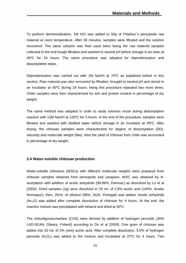
Figure 8 - Fourier Transform Infrared spectra for chitin samples from carapace (DD%,
27.1±0.1) and pereopods (DD%, 27.2±0.2) body parts. ...................................................34
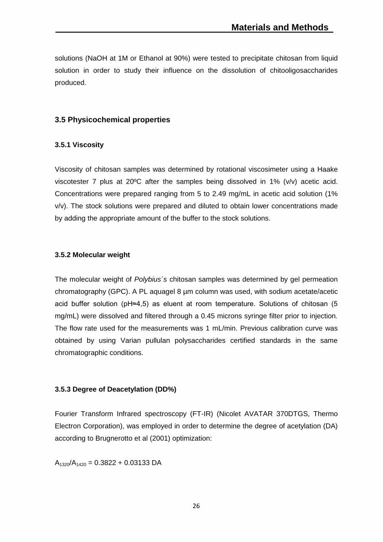
Figure 9 - Fourier Transform Infrared spectra for chitosan samples from carapace (DD%,
95.1±0.01) and pereopods (DD%, 94.3±0.04) body parts. ...............................................35
Figure 10 - Fourier Transform Infrared spectra for water soluble chitosan (WSC) samples
from carapace (DD%, 55±3.21) and pereopods (DD%, 72±0.86) body parts. ..................37
Figure 11 - Fourier Transform Infrared spectra for chitooligosaccharides (COS) samples
from carapace (DD%, 95±0.62) and pereopods (DD%, 93.3±0.04) body parts. ...............39
Figure 12 - Fourier Transform Infrared spectra for chitosan samples subjected to reuse of
solvents, after treatment 1 (DD%, 93±0.9), treatment 2 (DD%, 93±1.6) and treatment 3
(DD%, 92±3.0). Values are means of three replicates ± standard errors. .........................41
xiv
Figure 13 - Scavenging ability of water-soluble chitosan (WSC) and
chitooligosaccharides (COS) on 1,1-diphenyl-2-picrylhydrazyl radicals. Values are means
of eight replicates ± standard errors. ................................................................................42
Figure 14 - Effect of LWSC and WSC samples on the growth of six different species of
bacteria: two Gram-positive bacteria (Staphylococcus aureus ATCC12600 and
Lactobacillus planctarum ATCC 8014) and four Gram-negative bacteria (Escherichia coli
ATCC25922, Escherichia coli ATCC10536, Bacillus subtilis ATCC6633, Salmonella
enteritidis ATCC13076). Values are means of eight replicates ± standard error.. .............44
Figure 15 - Effect of LWSC and WSC samples (concentration ranging from 0,0125 to 0.1
mg/mL) on the growth of four different fungal species: Heterobasidion annosum,
Phytophthora cinnamomi, Cryphonectria parasitica and Botrytis cinerea. Values are
means of eight replicates ± standard error. ......................................................................48
Figure 16 - Flowchat of "Pilado add value" laboratorial activities. ....................................64
Figure 17 - Poster presented at International Conference of the European Chitin Society
(Euchis Congress), Porto (Portugal) - May 2013…............................................................65
Figure 18 - Poster presented at European Biotechnology Congress (Eurobiotech
Congress), Bratislava (Slovakia) - May 2013………………………………………….……...65
xv
List of Tables
Table 1 - Resume of chitin derivatives and respective application. Source: Kumar et al.,
2000…………………………………………………………………………………...…………....7
Table 2 - Influence of degree of deacetylation (DD) and molecular weight (Mw) on
antimicrobial activity. Source: Aranaz et al., 2009…………………….……...………….…13
Table 3 - General recommendations for the use of chitosan in several applications. (DD -
degree of deacetylation; Mw - molecular weight). Source: Aranaz et al., 2009………....…15
Table 4 - Characterization of dried samples of Polybius henslowii expressed as the mass
percentage of the initial dried material (% of dry weight). Values are means of three
replicates ± standard errors. ............................................................................................31
Tabela 5 - Ash and protein content of segmented body parts of Polybius henslowii after
treatment with three different concentrations of HCl and NaOH (1M, 0.75M and 0.5M).
Also percentage of removal from raw material was evaluated for both treatments. Values
are means of three replicates ± standard errors.…………………………………….……..33
Table 6 - Characterization of chitosan in terms of yield (%), viscosity ([ɳ]), deacetylation
degree (DD%) and molecular weight (Mw) obtained from both body parts of the swimming
crab Polybius henslowii. pWSC - Pereopods water soluble chitosan; pCOS - pereopods
chitooligosaccharides; sWSC - shells water soluble chitosan; sCOS - shells
chitooligosaccharides. Values are means of three replicates ± standard errors. ..............36
Table 7 - Characterization of chitin and chitosan samples from pereopods body part
expressed as the mass percentage of the initial dried material (% of dry weight). Values
are means of three replicates ± standard errors. ..............................................................40
Table 8 - MIC values (mg/mL) of WCS and LWCS samples against five different species
of bacteria (four gram-negative and one gram-positive) ...................................................45
Table 9 - Contents of chitin in different organisms. ..........................................................63

xvi

Introduction
1
1. Introduction
1.1 Marine resources
The ocean covers more than 70% of the Earth’s surface and therefore is a huge source
of biological diversity (Targett et al., 2002), as well as resources. Since ancient times,
the sea has been viewed as a source of food, minerals and natural products with a
significant impact on human societies. However, with the increase of population and its
needs, it became evident the need to found new sustainable resources assuming the
sea as a potential one.
Marine organisms represent a huge resource with a wide range of benefits for several
areas (Yen et al., 2007). In addition of being an excellent food source (if correctly
managed), it assumes a significant importance for emerging areas focused on life
quality increase, such as biotechnology and pharmaceutical industry.
One way to achieve such future is to extract several biomolecules from discards and
by-products resulting from fisheries processing industry, turning wastes as key
resources. Once extracted, such compounds could be used as raw material for high-
value products, with direct applications on pharmaceutical, medical or food technology
areas.
1.1.1 A non-traditional marine resource
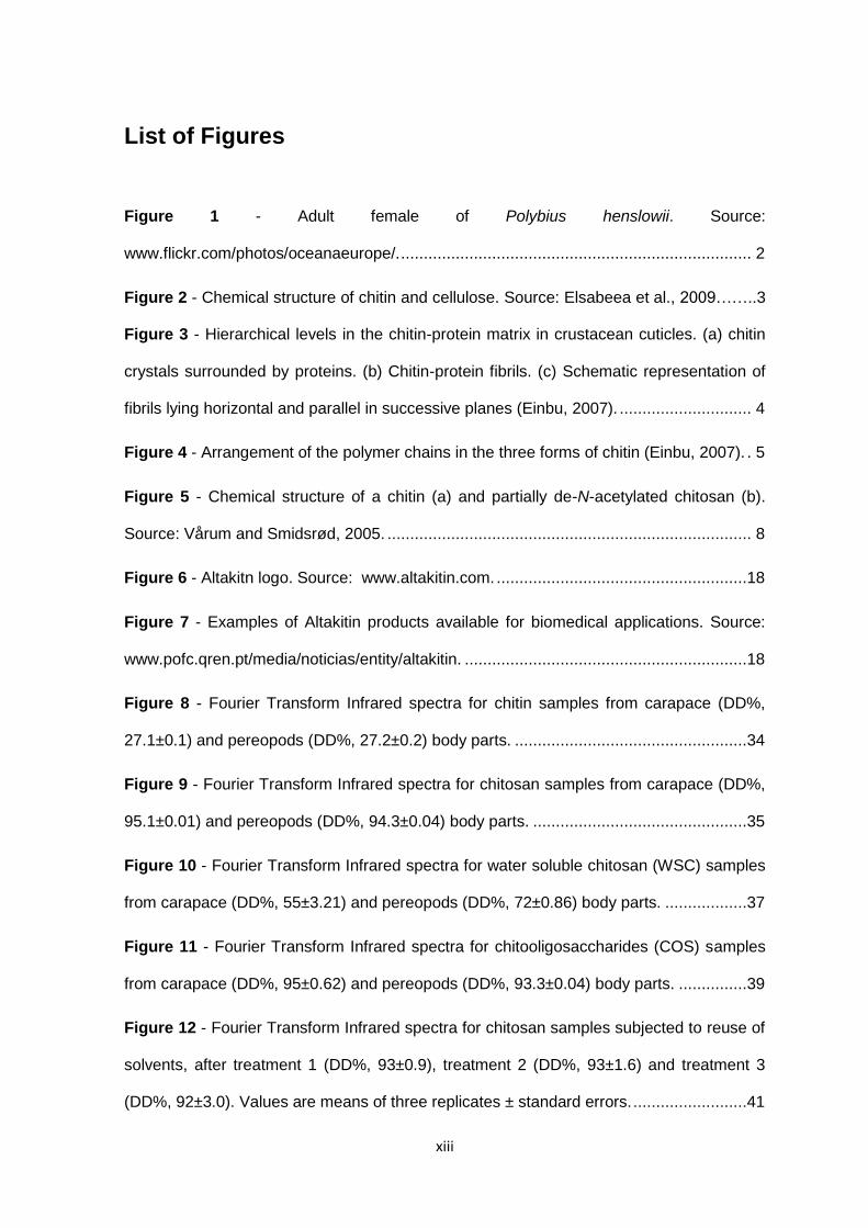
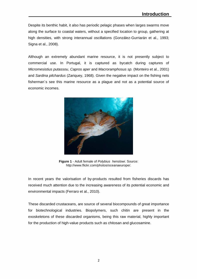
Polybius henslowii (Figure 1) is a reddish-brown crab with a roughly circular carapace
up to 4.5 cm across with broad spines along the front edge of the rim (Saldanha, 2003).
The last pereopod is paddle-shaped terminally obtuse and rounded (Hayward & Ryland
1995).
Henslow’s swimming crab is a benthopelagic specie, distributed between 80 and 650m
of depth and is found along the eastern Atlantic coasts, from Ireland and Britain to the
Alborán Sea (Cartes et al., 2002; Serrano et al., 2011) and Morocco (Hayward and
Ryland, 1995).
Introduction
2
Despite its benthic habit, it also has periodic pelagic phases when larges swarms move
along the surface to coastal waters, without a specified location to group, gathering at
high densities, with strong interannual oscillations (González-Gurriarán et al., 1993;
Signa et al., 2008).
Although an extremely abundant marine resource, it is not presently subject to
commercial use. In Portugal, it is captured as bycatch during captures of
Micromesistius putassou, Capros aper and Macroramphosus sp. (Monteiro et al., 2001)
and Sardina pilchardus (Zariquey, 1968). Given the negative impact on the fishing nets
fisherman´s see this marine resource as a plague and not as a potential source of
economic incomes.
Figure 1 - Adult female of Polybius henslowi. Source:
http://www.flickr.com/photos/oceanaeurope/.
In recent years the valorisation of by-products resulted from fisheries discards has
received much attention due to the increasing awareness of its potential economic and
environmental impacts (Ferraro et al., 2010).
These discarded crustaceans, are source of several biocompounds of great importance
for biotechnological industries. Biopolymers, such chitin are present in the
exoskeletons of these discarded organisms, being this raw material, highly important
for the production of high-value products such as chitosan and glucosamine.
Introduction
3
1.2 Chitin
1.2.1 Chemical structure
Chitin is one of the most abundant polymers on earth and the most abundant amino-
polysaccharide. First identified in 1884, chitin (poly[b-(1-4)-2-acetoamido-2-deoxy-D-
glucopyranose]) is a polymer of N-acetyl-D-glucosamine, widely distributed in nature,
especially in the exoskeletons of marine invertebrates such as prawn, crab and lobster
(Al Sagheer et al, 2009).
It is also found in microorganisms, e.g. in the cell walls and structural membranes of
mycelia of fungi, yeast and green algae (Mathur and Narang, 1990). Chitin has a
resemblance to cellulose both in chemical structure and in biological function as a
structural polysaccharide and may be regarded as a cellulose derivative with an
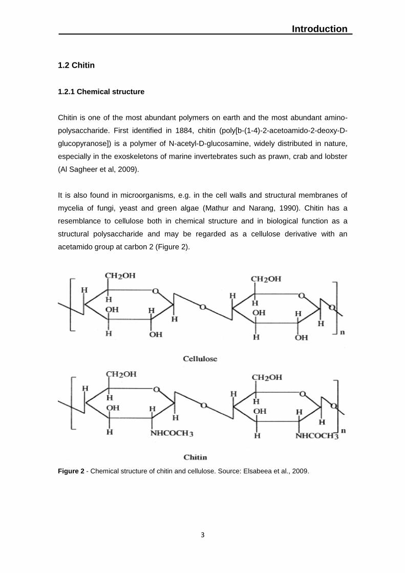
acetamido group at carbon 2 (Figure 2).
Figure 2 - Chemical structure of chitin and cellulose. Source: Elsabeea et al., 2009.
Introduction
4
Both polymers mainly serve as structural components supporting cell and body
surfaces: cellulose strengthens the cell wall of plant cells whereas chitin contributes to
the mechanical strength of fungal cell walls and exoskeletons of arthropods (Gooday,
1990).
In the chitin crystal structure, the chains form hydrogen-bonded sheets linked by C=O
and H-N-groups. In addition, each chain has intramolecular hydrogen bonds between
the neighbouring sugar rings: the carbonyl group bonds to the hydroxyl group on C-6.
There is also a second hydrogen bond between the OH-group on C-3 and the ring
oxygen, similar to that in cellulose (Minke and Blackwel, 1978). This extensive
hydrogen bonding enhances the stiffness of the chitin chain. Because chitin has a
compact structure, it is insoluble in most solvents. Also, chitin is closely associated with
protein, minerals, lipids and pigments.
The chitin molecules are known to be ordered into helicoidally microfibrillar structures,
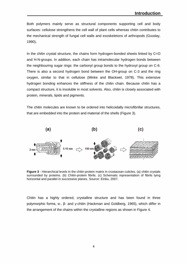
that are embedded into the protein and material of the shells (Figure 3).
Figure 3 - Hierarchical levels in the chitin-protein matrix in crustacean cuticles. (a) chitin crystals surrounded by proteins. (b) Chitin-protein fibrils. (c) Schematic representation of fibrils lying horizontal and parallel in successive planes. Source: Einbu, 2007.
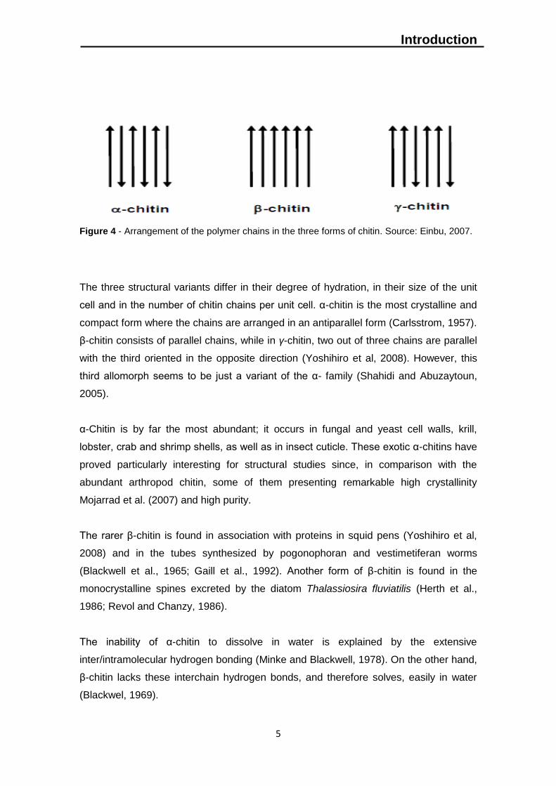
Chitin has a highly ordered, crystalline structure and has been found in three
polymorphic forms, α-, β- and γ-chitin (Hackman and Goldberg, 1965), which differ in
the arrangement of the chains within the crystalline regions as shown in Figure 4.
Introduction
5
Figure 4 - Arrangement of the polymer chains in the three forms of chitin. Source: Einbu, 2007.
The three structural variants differ in their degree of hydration, in their size of the unit
cell and in the number of chitin chains per unit cell. α-chitin is the most crystalline and
compact form where the chains are arranged in an antiparallel form (Carlsstrom, 1957).
β-chitin consists of parallel chains, while in γ-chitin, two out of three chains are parallel
with the third oriented in the opposite direction (Yoshihiro et al, 2008). However, this
third allomorph seems to be just a variant of the α- family (Shahidi and Abuzaytoun,
2005).
α-Chitin is by far the most abundant; it occurs in fungal and yeast cell walls, krill,
lobster, crab and shrimp shells, as well as in insect cuticle. These exotic α-chitins have
proved particularly interesting for structural studies since, in comparison with the
abundant arthropod chitin, some of them presenting remarkable high crystallinity
Mojarrad et al. (2007) and high purity.
The rarer β-chitin is found in association with proteins in squid pens (Yoshihiro et al,
2008) and in the tubes synthesized by pogonophoran and vestimetiferan worms
(Blackwell et al., 1965; Gaill et al., 1992). Another form of β-chitin is found in the
monocrystalline spines excreted by the diatom Thalassiosira fluviatilis (Herth et al.,
1986; Revol and Chanzy, 1986).
The inability of α-chitin to dissolve in water is explained by the extensive
inter/intramolecular hydrogen bonding (Minke and Blackwell, 1978). On the other hand,
β-chitin lacks these interchain hydrogen bonds, and therefore solves, easily in water
(Blackwel, 1969).
Introduction
6
1.2.2 Industrial applications
Chitin is mainly used as a raw material to produce chitin-derived products, such as
chitosan, chitin/chitosan derivatives, oligosaccharides and glucosamine. An increasing
number of useful products derived from chitin continue to attract commercial
development (Table 1).
The large number of patents filed involving chitin-derived products reflects the
commercial expectations for these products (US Patent and Trademark Office, 2006).
An estimated 75% of produced chitin is used to manufacture products for the
nutraceutical market.
Currently the major driving force in the market is the increasing sales of glucosamine
as a dietary supplement (Sandford, 2002). Approximately 65% of the produced chitin is
converted into glucosamine, ≈25% is converted into chitosans, ≈9% is used to produce
oligosaccharides and approximately 1% goes to the production of N-acetylglucosamine
(Mustaparta, 2006).
The main industrial sources of raw material for the production of chitin today are
cuticles of various crustaceans, mainly from crab and shrimp (Kim and Rajapakse,
2005) and the market price is approximately 5-8 US$ for average quality chitin
(Mustaparta, 2006).
Introduction
7
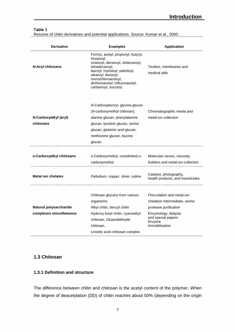
Table 1 Resume of chitin derivatives and potential applications. Source: Kumar et al., 2000.
1.3 Chitosan
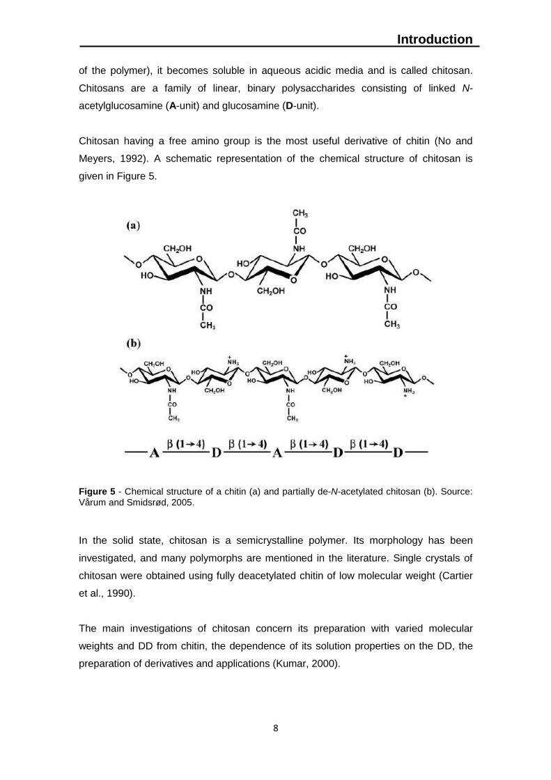
1.3.1 Definition and structure
The difference between chitin and chitosan is the acetyl content of the polymer. When
the degree of deacetylation (DD) of chitin reaches about 50% (depending on the origin
Derivative
Examples
Application
N-Acyl chitosans
Formyl, acetyl, propionyl, butyryl, hexanoyl, octanoyl, decanoyl, dodecanoyl, tetradecanoyl, lauroyl, myristoyl, palmitoyl, stearoyl, benzoyl, monochloroacetoyl, dichloroacetyl, trifluoroacetyl, carbamoyl, succinyl,
Textiles, membranes and
medical aids
N-Carboxyalkyl (aryl)
chitosans
N-Carboxybenzyl, glycine-glucan
(N-carboxymethyl chitosan),
alanine glucan, phenylalanine
glucan, tyrosine glucan, serine
glucan, glutamic acid glucan,
methionine glucan, leucine
glucan
Chromatographic media and
metal ion collection
o-Carboxyalkyl chitosans
o-Carboxymethyl, crosslinked o-
carboxymethyl
Molecular sieves, viscosity
builders and metal ion collection
Metal ion chelates
Palladium, copper, silver, iodine
Catalyst, photography, health products, and insecticides
Natural polysaccharide
complexes miscellaneous
Chitosan glucans from various
organisms
Alkyl chitin, benzyl chitin
Hydroxy butyl chitin, cyanoethyl
chitosan, Glutaraldehyde
chitosan,
Linoelic acid–chitosan complex
Flocculation and metal ion
chelation Intermediate, serine
protease purification
Enzymology, dialysis and special papers Enzyme Immobilization
Introduction
8
of the polymer), it becomes soluble in aqueous acidic media and is called chitosan.
Chitosans are a family of linear, binary polysaccharides consisting of linked N-
acetylglucosamine (A-unit) and glucosamine (D-unit).
Chitosan having a free amino group is the most useful derivative of chitin (No and
Meyers, 1992). A schematic representation of the chemical structure of chitosan is
given in Figure 5.
Figure 5 - Chemical structure of a chitin (a) and partially de-N-acetylated chitosan (b). Source:
Vårum and Smidsrød, 2005.
In the solid state, chitosan is a semicrystalline polymer. Its morphology has been
investigated, and many polymorphs are mentioned in the literature. Single crystals of
chitosan were obtained using fully deacetylated chitin of low molecular weight (Cartier
et al., 1990).
The main investigations of chitosan concern its preparation with varied molecular
weights and DD from chitin, the dependence of its solution properties on the DD, the
preparation of derivatives and applications (Kumar, 2000).
Introduction
9
1.3.2 Physicochemical properties
1.3.2.1 Degree of deacetylation (DD)
Chitosan production from chitin deacetylation involves the removal of acetyl groups
from its molecular chain, leaving behind a polysaccharide with a high degree of
chemical reactive amino group (-NH2).
Being the degree of deacetylation (DD) an important property in chitosan production, it
affects the physicochemical properties as well as its possible applications (Rout, 2001).
Deacetylation also affects the biodegradability and immunological activity (Tolaimate et
al., 2000). A sharp nomenclature border has not been defined between chitin and
chitosan based on the degree of N-deacetylation (Rout, 2001).
The degree of deacetylation of chitosan ranges from 56% to 99% with an average of
80%, depending on the crustacean species and the preparation methods (No, 2000;
No and Meyers, 1995). In any case, the degree of deacetylation can be employed to
differentiate between chitin and chitosan because it determines the content of free
amino groups in the polysaccharides.
In fact there are two advantages of chitosan over chitin. In order to dissolve chitin,
highly toxic solvents such as lithium chloride and dimethylacetamide are used whereas
chitosan is readily dissolved in diluted acetic acid. The second advantage is that
chitosan possesses free amine groups which are an active site in many chemical
reactions.
Various methods have been reported for the determination of the degree of
deacetylation of chitosan. These included ninhydrin test, linear potentiometric titration,
near-infrared spectroscopy, nuclear magnetic resonance spectroscopy, hydrogen
bromide titrimetry, infrared spectroscopy, and first derivative UV-spectrophotometry
(Yuan et al., 2011).
Introduction
10
1.3.2.2 Molecular-weight
Like its composition, the molecular weight of chitosan varies with the raw material
sources and the method of preparation. Molecular weight of native chitin is usually
larger than one million Daltons while commercial chitosan products have the molecular
weight range of 100,000 – 1,200,000 Daltons, depending on the process and grades of
the product (Li et al., 1992). Due to this range of molecular weights, chitosan can be
categorized into low molecular weight chitosans (LMWC, 5-20kDa) (Lin et al., 2009;
Yang et al., 2009), medium molecular weights chitosan (MMWC, ~100kDa) and high
molecular weight chitosan (HMWC > 300 kDa) (Chien et al., 2007). Like most
polysaccharides, concerning to molecular weight, chitosan is polydisperse.
In general, high temperature, dissolved oxygen, and shear stress can cause
degradation of chitosan. For instance at a temperature over 280ºC, thermal
degradation of chitosan occurs and polymer chains rapidly break down, thereby
lowering molecular weight (Rout, 2001).
Also, maximal depolymerization caused by utilization of high temperature or
concentrated acids, such as hydrochloric acid followed by acetic acid and sulfurous
acid, results in molecular weight changes with minimal degradation with the use of
EDTA.
The molecular weight (Mw) of chitosan depends on the degree of deacetylation (DD). A
higher DD would therefore imply a smaller Mw as the acetamido groups are replaced by
amino groups of lower molecular weight. However, this relationship between Mw and
DD is not very predictable (Kofuji et al., 2005).
The molecular weight of chitosan can be determined by different methods such as
chromatography (Bough et al., 1978), light scattering (Muzzarelli, 1977), and
viscometry (Maghami and Roberts, 1988).
1.3.2.3 Viscosity
Viscosity is an important factor in the conventional determination of molecular weight of
chitosan and in determining its commercial applications in complex biological
environments (e.g. food industry). Higher molecular weight chitosan polymers often
render highly viscous solutions, which may not be desirable for industrial handling.
Introduction
11
Some factors during chitin processing affect the production of chitosan and its
properties such as the degree of deacetylation, molecular weight, concentration of
solution, ionic strength, pH, and temperature. For instance, chitosan viscosity
decreases with an increased time of demineralization (Moorjani et al., 1975).
Viscosity of chitosan in acetic acid tends to increase with decreasing pH but decrease
with decreasing pH in HCl, giving rise to the definition of ‘Intrinsic Viscosity’ of chitosan
which is a function of the degree of ionization as well as ion strength. Moorjani et al.
(1975) also stated that it is not desirable to bleach the material (i.e., bleaching with
acetone or sodium hypochlorite) at any stage since bleaching considerably reduces the
viscosity of the final chitosan product.
Similarly, No et al. (1999), demonstrated that chitosan viscosity is considerably affected
by physical (grinding, heating, autoclaving, ultrasonication) and chemical (ozone)
treatments, except for freezing, and decreases with an increase in treatment time and
temperature.
1.3.2.4 Solubility
The solubility, however, is mainly controlled by the degree of deacetylation. It is
estimated that deacetylation must be at least 85% complete in order to achieve the
desired solubility (No et al., 1995).
The solubility is related with ionic concentration, pH, strength of the acid used for
protonation, distribution of acetyl groups along the chain, as well as the conditions of
polysaccharide isolation and drying. It is also important to consider the intra-chain H
bonds involving the hydroxyl groups. As a result, it can be stated that the solubility of
chitosan is quite difficult to control as it depends on so many parameters.
A number of solvents for chitin and chitosan can be found in the literature. Generally,
the solubility decreases with molecular weight increasing (Rathke and Hodson, 1994).
Few attempts have been made to enhance chitosan’s solubility in organic solvents
(Nishimura et al., 1991). However, significant efforts have been made to enhance its
solubility in water.
Introduction
12
One major reason for that is because most of the biological applications of chemical
substances require that the material can be easily processed and functional at neutral
pH. Thus, obtaining water-soluble derivative of chitosan is an important step towards its
further application as a biofunctional material (Jia et al., 2001).
1.3.3 Biological properties
1.3.3.1 Biocompatibility, toxicity and biodegradability
Chitosan is considered biocompatible both in vitro (with epithelial and myocardial cells)
and in vivo (with fibroblasts, chondrocytes, hepatocytes and keratinocytes). This
property is somewhat attributed to factors such as the natural source, molecular
weight, DD and production method (Aranaz et al., 2009).
The lethal dose (LD50) for oral administration of chitosan has been reported to be 16
g/kg body weight in rabbits (Ilium, 1998) and 10g/kg body weight in mice (Aranaz et al.,
2009; Domard and Domard, 2002). Chitosan also proved haemocompatibility and
coagulating action both in vitro and in vivo. Even in severe anticoagulation conditions
and in the presence of abnormal activity by platelets, in vitro, chitosan maintains its
coagulation properties.
Regarding biodegradability, chitosan can be hydrolyzed by enzymes such as
chitosanase, glucosaminidase, chitobiase and N-acetyl-glucosaminidase (Domard and
Domard, 2002). In mammals, this activity produces non-toxic chitosan oligosaccharides
of different lengths that can be used in glycosaminoglycans and glycoproteins.
1.3.3.2 Antioxidant activity
Antioxidant properties of chitosan produced from crab species have been studied in
recent years (Yen et al., 2008; Youn et al., 2009). The obtained results showed that all
chitosan polymers derived from crab, have significant antioxidant properties, such as
scavenging ability on hydroxyl radicals and chelating ability on ferrous ions. In addition,
the prolonged N-deacetylation resulted on chitosan with more effective antioxidant
properties.
Introduction
13
All crab chitosans exhibited comparable antioxidant properties. Therefore, chitosan
with significant antioxidant properties can be used in several uses, such as, food
supplement or in pharmaceutical applications.
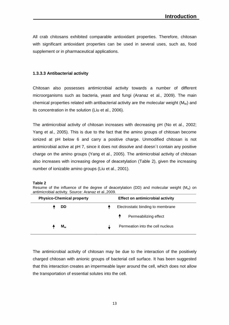
1.3.3.3 Antibacterial activity
Chitosan also possesses antimicrobial activity towards a number of different
microorganisms such as bacteria, yeast and fungi (Aranaz et al., 2009). The main
chemical properties related with antibacterial activity are the molecular weight (MW) and
its concentration in the solution (Liu et al., 2006).
The antimicrobial activity of chitosan increases with decreasing pH (No et al., 2002;
Yang et al., 2005). This is due to the fact that the amino groups of chitosan become
ionized at pH below 6 and carry a positive charge. Unmodified chitosan is not
antimicrobial active at pH 7, since it does not dissolve and doesn´t contain any positive
charge on the amino groups (Yang et al., 2005). The antimicrobial activity of chitosan
also increases with increasing degree of deacetylation (Table 2), given the increasing
number of ionizable amino groups (Liu et al., 2001).
Table 2 Resume of the influence of the degree of deacetylation (DD) and molecular weight (Mw) on antimicrobial activity. Source: Aranaz et al.,2009.
Physico-Chemical property Effect on antimicrobial activity
DD Electrostatic binding to membrane
Permeabilizing effect
Mw Permeation into the cell nucleus
The antimicrobial activity of chitosan may be due to the interaction of the positively
charged chitosan with anionic groups of bacterial cell surface. It has been suggested
that this interaction creates an impermeable layer around the cell, which does not allow
the transportation of essential solutes into the cell.
Introduction
14
Another antimicrobial mechanism is related to the inhibition of RNA and protein
synthesis by permeation of chitosan into the cell nucleus. This mechanism is related to
low molecular weight chitosan´s.
Chitosan may also act as a chelating agent rendering trace elements, metals, and
essential nutrients from the microorganism (Prashanth and Tharanathan, 2007).
Despite being insoluble in aqueous media at neutral and basic conditions, chitosan is
soluble in aqueous diluted acids.
Other water soluble chitosan derivates have been studied for antimicrobial activity (Liu
et al., 2006). This way, acid solutions are no longer a problem for chitosan antibacterial
activity tests.
1.3.3.4 Antifungal activity
Chitosan and its derivatives have been reported as a promising alternative to control
postharvest diseases (Fisk et al., 2008). Chitosan and chitooligosaccharides also
proved already broad-spectrum regarding antifungal properties (Jeon, 2001; No, 2002;
Liu et al., 2006).
Indeed, chitosan is an ideal preservative coating for fresh fruit and vegetables because
of its film-forming and biochemical properties (Muzzarelli, 1986) that enhances shelf life
and control fungal contamination of several fruit crops (Romanazzi et al., 2002).
Recent studies has indicated that pre-harvest sprays of chitosan on strawberry plants
reduced gray mold during fruit storage (Reddy et al., 2000). Coating of citrus fruit with
chitosan was effective in controlling fruit decay caused by Penicillium digitatum and
Penicillium expansum (Chien et al., 2007). Gray mold and blue mold rots caused by B.
cinerea and P. expansum, respectively, in sweet cherry fruit were reduced by pre-
harvest spraying or postharvest dipping of chitosan (Romanazzi et al., 2003).
1.3.4 Potential Applications
Increasingly over the last decade, chitosan-based materials have been examined and a
relatively high number of potential products have been developed for several areas

Introduction
15
such as wastewater treatment (removal of heavy metal ions, flocculation/coagulation
of dyes and proteins, membrane purification processes), food industry (anti-cholesterol
and fat binding, shelf-life increasing, packaging material, animal feed additive),
agriculture (seed and fertilizer coating, controlled agrochemical release), pulp and
paper industry (surface treatment, photographic paper), cosmetics and toiletries
(moisturizer, body creams, bath lotion) (Felse, 1999; Kurita, 2001; Shahidi et al.,1999;
No and Meyers, 2000).
However, owing to the unparalleled biological properties, the most exciting and
promising uses of chitosan-based materials are those related with medicine and
biotechnology (Krajewska, 1991; Paul and Sharma, 2000; Singla and Chawla, 2001).
In medicine they may be employed as bacteriostatic and antifungal agents, drug
delivery vehicles, drug controlled release systems, artificial cells, wound healing
ointments/dressings, haemodialysis membranes, contact lenses, artificial skin, surgical
structures and on tissue engineering (Kumar, 2000).
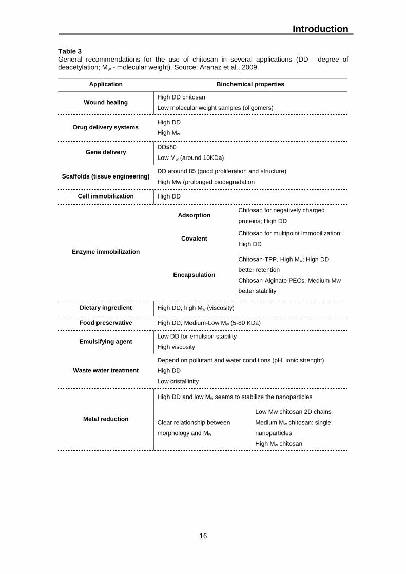
Chitosan applications are highly determined by the biochemical properties of the
polymer (Table 3). The degree of deacetylation (DD), defined as the molar fraction of
deacetylated units in the polymorph chain (Zhang et al., 2005), is one of the most
important factors influencing the properties of chitosan (Kurita, 2006), such as
solubility, flexibility, polymer conformation and viscosity (Dash et al., 2011).
Indeed, several efforts have been performed on the preparation of functional
derivatives of chitosan through chemical modifications (Kumar et al., 2004; Hirano et
al., 1996). In its linear polyglucosamine chains of high molecular weight, chitosan has
reactive amino and hydroxyl groups, amenable to chemical modifications. For example,
chemically modified chitosan structures allow solubility improvement, increasing this
way their range of possible applications.
In resume, during the last years it has been observed a large e increase in chitosan
studies due to its biological properties such as biocompatibility, biodegradability, non-
toxicity, high affinity towards proteins, availability of reactive functional groups,
adsorption and antimicrobial activity (Kumar, 2000). Owing to these characteristics,
chitosan-based materials, are predicted to be widely exploited in the near future
especially on environmentally friendly applications. Chitosan is assumed to be the
polymer of 21st century.
Introduction
16
Table 3 General recommendations for the use of chitosan in several applications (DD - degree of deacetylation; Mw - molecular weight). Source: Aranaz et al., 2009.
Application Biochemical properties
Wound healing High DD chitosan
Low molecular weight samples (oligomers)
Drug delivery systems High DD
High Mw
Gene delivery DD≤80
Low Mw (around 10KDa)
Scaffolds (tissue engineering) DD around 85 (good proliferation and structure)
High Mw (prolonged biodegradation
Cell immobilization High DD
Enzyme immobilization
Adsorption Chitosan for negatively charged
proteins; High DD
Covalent Chitosan for multipoint immobilization;
High DD
Encapsulation
Chitosan-TPP, High Mw; High DD
better retention
Chitosan-Alginate PECs; Medium Mw
better stability
Dietary ingredient High DD; high Mw (viscosity)
Food preservative High DD; Medium-Low Mw (5-80 KDa)
Emulsifying agent Low DD for emulsion stability
High viscosity
Waste water treatment
Depend on pollutant and water conditions (pH, ionic strenght)
High DD
Low cristallinity
Metal reduction
High DD and low Mw seems to stabilize the nanoparticles
Clear relationship between
morphology and Mw
Low Mw chitosan 2D chains
Medium Mw chitosan: single
nanoparticles
High Mw chitosan
Introduction
17
1.4 Chitooligosaccharide
Chitooligosaccharides (COS) are β-1,4 linked homo- or heterooligomers of N-
acetylglucosamine and/or glucosamine. Unlike chitosan, chitooligosaccharides are
readily soluble in water due to their shorter chain lengths and free amino groups in
glucosamine units.
The low viscosity and the greater solubility of chitooligosaccharides at neutral pH have
attracted the interest of many researchers to use chitosan in its oligosaccharide form.
In the case of crude chitosan, the solution viscosity is usually quite high, which makes it
difficult to prepare high concentration solutions that can be advantageously used, for
example, in pharmaceuticals. Also the solubilisation with acids, impose some
restrictions in its range of applications. However, being soluble in water, COS allows
high concentration solution with small viscosities, and removes the acidic solution
problem increasing its potential applications.
Also, chitosan oligosaccharides have been reported to have biological activities such
as antimicrobial, antifungal, antioxidant, and immunostimulant effects (Kim and
Rajapakse, 2005). The biological activities of chitooligosaccharides depend on both
chain length and fraction of acetylated units (Bahrke et al., 2002) together with charge
distribution and the nature of the chemical modification (Muzzarelli, 1996).
There are a number of preparation methods for chitooligosacharides. Recent studies
have focused on the preparation mainly by two distinct strategies: derivatization (e.g.
N-acetylation (Lu et al., 2004) or molecular-weight degradation (e.g. acid hydrolysis
(Cabrera and Cutsem, 2005), or hydrogen peroxide (Du et al., 2009). A microwave-
based preparation technique has recently been patent. Also enzymatic hydrolysis have
been performed. However, the high costs of enzymatic hydrolyses restricts its use in
COS production.
1.5 The project "Pilado add value"
The fishing activity is mainly directed towards animal protein production for human
consumption, not aiming to provide raw material for the extraction and purification of
biopolymers with application in a wide range of areas such as agriculture,
biotechnology, pharmaceutical industry and biomedicine.
Introduction
18
Among a wide variety of marine resources, the crustaceans are assumed as one of the
main sources of biopolymers as a result of their high abundance, reproductive aspects,
life cycle and biochemical composition. As a result of its natural properties, it has been
verified in recent years a strong global prospecting for biopolymers such as chitin and
chitosan.
"Pilado add value" emerged as a promising approach to answer such demand through
diversification of fisheries aiming biotechnology purposes. The project, financed by
GAC-Oeste (Grupo de Ação Costeira do Oeste), under Axis 4 of PROMAR (EFF -
European fisheries fund), accounted also with the collaboration of the purse seine
fishing vessel “Mestre Comboio – Peniche”, OP Centro and Altakitin (Figure 6).
Figure 6 - Altakitn logo. Source: www.altakitin.com.
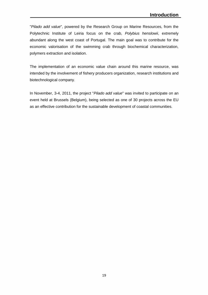
Altakitin is a Portuguese company dedicated to the research, development and
manufacturing of raw material for medical applications, such as chitin, chitosan,
chitosan derivates, injectable bone substitutes and wound dressings (Figure 7).
Figure 7 - Examples of Altakitin products available for biomedical applications. Source:
www.pofc.qren.pt/media/noticias/entity/altakitin.
Introduction
19
"Pilado add value", powered by the Research Group on Marine Resources, from the
Polytechnic Institute of Leiria focus on the crab, Polybius henslowii, extremely
abundant along the west coast of Portugal. The main goal was to contribute for the
economic valorisation of the swimming crab through biochemical characterization,
polymers extraction and isolation.
The implementation of an economic value chain around this marine resource, was
intended by the involvement of fishery producers organization, research institutions and
biotechnological company.
In November, 3-4, 2011, the project "Pilado add value" was invited to participate on an
event held at Brussels (Belgium), being selected as one of 30 projects across the EU
as an effective contribution for the sustainable development of coastal communities.
Introduction
20
Objectives
21
2. Objectives
The present study aims to be a contribution for the economic valorisation of Polybius
henslowii as a raw material for polymer extraction, with potential biotechnological
applications. In order to study this non-traditional marine resource and its biotechnological
applications, it was performed the following experiments;
(1) Biochemical characterization of pereopods and carapace from Polybius
henslowii (raw material) in terms of ash, protein, free fat and chitin content;
(2) Optimization of laboratorial procedures for chitin chemical extraction and
chitosan production.
(3) Characterization of the products obtained from the segmented crab body parts,
namely: (a) Chitin, (b) Chitosan, (c) Water soluble chitosan (WSC) and (d)
Chitooligosaccharides (COS);
(4) Evaluation of the biological properties of chitosan products:
(a) Antioxidant activity:
- Scavenging activity on 1,1-diphenyl-2-picrylhydrazyl radicals;
(b) Antibacterial activity against six different species of bacteria:
- Two Gram-positive bacteria (Staphylococcus aureus ATCC12600 and
Lactobacillus planctarum ATCC 8014);
- Four Gram-negative bacteria (Escherichia coli ATCC25922, Escherichia
coli ATCC10536, Bacillus subtilis ATCC6633, Salmonella enteritidis
ATCC13076).
(c) Antifungal activity against four different fungi:
- Cryphonectria parasitica (DSMZ 62626);
- Phytophthora cinnamomi (DSMZ 62654);
- Botrytis cinerea (DSMZ 4709);
- Heterobasidion annosum (DSMZ 1531).

Objectives
22
Materials and Methods
23
3. Materials and Methods
3.1 Samples collection and processing
Polybius henslowii swimming crabs were captured along the West coast of Peniche
(Portugal) by purse seine fishing vessels during the capture of Sardina pilchardus through
summer months, at 2012. Organisms were boiled, dried in an incubator (Binder, Bohemia,
USA) at 100ºC for 2 days and segmented into carapace and pereopods. Then, raw
material was powdered and sieved into particles between 150 and 500 µm (diameter).
3.2 Biochemical characterization of raw material
Samples were biochemically characterized in terms of protein, ash, lipids and chitin
content. The ash content was determined by initially drying the raw material in an
incubator at 100ºC for 6 hours measuring its dry weight. Then, dried samples were placed
in a furnace at 530ºC, during 20 hours and the remaining material was weighed after
cooling in a desiccator.
Protein assay was carried by means of microbiuret method (Johnson, 1978) and
compared to a standard curve of bovine serum albumin (Sigma-Aldrich, Steinheim,
Germany).
Free fat was determined through lipids extraction by Soxlet method according to ISO 1444
(1996).
Chitin content was evaluated through dried weight method (24 hours at 5ºC), after
submitting the raw material to demineralization with 1M HCl (Sigma Aldrich, Steinheim,
Germany) and deproteinization with 1M NaOH (Scharlau, Barcelona, Spain) solutions.
3.3 Chitin extraction and chitosan production
3.3.1 Chitin extraction
Materials and Methods
24
Chitin isolation involved two basic steps: demineralization and deproteinization. Different
concentrations of HCl and NaOH were employed in order to optimize the extraction
process.
Raw samples were subject to three different concentrations of HCl to carry out
demineralization; 0.5, 0.75 and 1M at 21ºC, during 30 minutes (ratio of 1:30, w/v). Raw
material demineralized was then washed with distilled water until neutral pH.
After oven-drying at 50ºC, the samples were subjected to three different concentrations of
NaOH (0.5M, 0.75M and 1M) at a ratio of 1:15 (w/v), in order to optimize the protein
removal from samples.
These treatments involved exposure for 2 hours at 70ºC in a shaking water-bath (Julabo
SW22, Allentown, USA). Then, samples were washed with distilled water until the solution
became colourless with neutral pH.
Demineralization and deproteinization efficiency were respectively determined through
ash content determination and microbiuret assay as described above.
3.3.2 Chitosan production
Chitosan was obtained through chitin treatment with 12M NaOH at 120ºC during 7
(pereopods chitin) and 3 hours (carapace chitin) according to a ratio of 1:15 (w/v). In order
to collect the chitosans produced, samples were filtrated, washed with distilled water and
stored in an incubator during 24h at 45ºC.
These conditions yielded chitosan samples from both body parts which exhibited complete
dissolution in acetic acid (1%, v/v).
3.3.3 Reuse of reagents for extraction and production procedures
In order to optimize the amount of reagents necessary to performed chitin extraction and
chitosan production, all steps - demineralization, deproteization and deacetylation, were
carried consecutively. The same chemical solutions were employed three consecutive
times at each step.
Materials and Methods
25
To perform demineralization, 1M HCl was added to 50g of Polybius´s pereopods raw
material at room temperature. After 30 minutes, samples were filtrated and the solution
recovered. The same solution was then used twice being the raw material samples
collected in the end trough filtration and washed to neutral pH before storage in an oven at
45ºC for 24 hours. The same procedure was adopted for deproteinization and
deacetylation steps.
Deproteinization was carried out with 1M NaOH at 70ºC as explained before in this
section. Raw material was also recovered by filtration, brought to neutral pH and stored in
an incubator at 45ºC during 24 hours, being this procedure repeated two more times.
Chitin samples were then characterized for ash and protein content in percentage of dry
weight.
The same method was adopted in order to study solvents reuse during deacetylation
reaction with 12M NaOH at 120ºC for 3 hours. In the end of the procedure, samples were
filtrated and washed with distilled water before storage in an incubator at 45ºC. After
drying, the chitosan samples were characterized for degree of deacetylation (DD),
viscosity and molecular weight (Mw). Also the yield of chitosan from chitin was accounted
in percentage of dry weight.
3.4 Water-soluble chitosan production
Water-soluble chitosans (WSCs) with different molecular weights were prepared from
chitosan samples obtained from pereopods and carapace. WSC was obtained by N-
acetylation with addition of acetic anhydride (99,99%, Panreac) as described by Lu et al
(2004). Dried samples (1g) were dissolved in 25 mL of 2.8% acetic acid (100%, Analar
Normapur); then, 25mL of ethanol (96%, AGA, Portugal) was added. Acetic anhydride
(Ac2O) was added after complete dissolution of chitosan for 4 hours. At the end, the
reaction mixture was precipitated with ethanol and dried at 55ºC.
The chitooligosaccharides (COS) were derived by addition of hydrogen peroxide (30%
LAD-SCAN, Gliwice, Poland) according to Du et al (2009). One gram of chitosan was
added into 20 mL of 2% (w/w) acetic acid. After complete dissolution, 5.5% of hydrogen
peroxide (H2O2) was added to the mixture and incubated at 37ºC for 4 hours. Two
Materials and Methods
26
solutions (NaOH at 1M or Ethanol at 90%) were tested to precipitate chitosan from liquid
solution in order to study their influence on the dissolution of chitooligosaccharides
produced.
3.5 Physicochemical properties
3.5.1 Viscosity
Viscosity of chitosan samples was determined by rotational viscosimeter using a Haake
viscotester 7 plus at 20ºC after the samples being dissolved in 1% (v/v) acetic acid.
Concentrations were prepared ranging from 5 to 2.49 mg/mL in acetic acid solution (1%
v/v). The stock solutions were prepared and diluted to obtain lower concentrations made
by adding the appropriate amount of the buffer to the stock solutions.
3.5.2 Molecular weight
The molecular weight of Polybius´s chitosan samples was determined by gel permeation
chromatography (GPC). A PL aquagel 8 µm column was used, with sodium acetate/acetic
acid buffer solution (pH≈4,5) as eluent at room temperature. Solutions of chitosan (5
mg/mL) were dissolved and filtered through a 0.45 microns syringe filter prior to injection.
The flow rate used for the measurements was 1 mL/min. Previous calibration curve was
obtained by using Varian pullulan polysaccharides certified standards in the same
chromatographic conditions.
3.5.3 Degree of Deacetylation (DD%)
Fourier Transform Infrared spectroscopy (FT-IR) (Nicolet AVATAR 370DTGS, Thermo
Electron Corporation), was employed in order to determine the degree of acetylation (DA)
according to Brugnerotto et al (2001) optimization:
A1320/A1420 = 0.3822 + 0.03133 DA
Materials and Methods
27
Spectra for all samples were recorded in Kbr pellets (Sigma Aldrich, Steinheim, Germany)
by accumulation of at least 32 scans, with a resolution of 1 cm-1.
3.6 Biological properties
3.6.1 Scavenging ability on 1,1-diphenyl-2-picrylhydrazyl radicals
Chitosan scavenging activity on 1,1-diphenyl-2-picrylhydrazyl radicals (DPPH) was
determined according to the method described by Duan et al (2006). DPPH (Sigma
Aldrich, Steinheim, Germany) solution was previously prepared at 0.1mM with methanol.
At each microtube, 10 μl of each sample was added to 990 μl of DPPH solution. Samples
concentrations varied from 0.0625 to 10mg/mL. The reaction mixture was shaken
vigorously and stored in the dark at room temperature for 30 min. The absorbance was
then read at 517nm in a microplate reader (Biotek, Vermont, USA). All samples were run
in triplicate. Ascorbic acid was used for comparison.
The free radical scavenging activity was calculated by the following equation:
Scavenging activity (%) = [1-(absorbancesample/absorbancecontrol)] x 100.
With:
Abscontrol = DPPH solution absorbance +10 μl of DMSO;
Abssample = DPPH solution absorbance + 10 μl of sample.
3.6.2 Antibacterial activity
Antibacterial activity of water-soluble chitosan and chitooligosaccharides (WSC and COS,
respectively) was examined as the inhibitory effects against the growth of two Gram-
positive bacteria (Staphylococcus aureus ATCC12600 and Lactobacillus planctarum
ATCC 8014) and four Gram-negative bacteria (Escherichia coli ATCC25922, Escherichia
coli ATCC10536, Bacillus subtilis ATCC6633, Salmonella enteritidis ATCC13076).
Chitosan stock solutions at 10 mg/mL were prepared by adding 1 g of water-soluble
chitosan or chitooligosaccharide to 100 mL liquid broth (pH6). The antibacterial assay
was carried out in 96-well microplates, where 20µl of each bacteria aliquot was added to
180 µl chitosan stock solution previously described at a concentration of 10 mg/mL, at
37ºC. The incubation period depended on the growth curve of each microorganism.
Materials and Methods
28
Chloramphenicol (reference antibiotic) was added at 1 µg/mL as a positive control for
growth inhibition. Distilled water was added to each broth solution as a negative control
allowing the development of all bacteria tested.
Percentage of inhibition was determined by spectrophotometric optical density (O.D.)
recorded between the incubation period. For each assay 8 replicates were performed.
3.6.2.1 Minimal inhibitory concentration (MIC)
The minimum inhibitory concentration (MIC) was defined as the lowest concentration
water-soluble chitosan required to completely inhibit bacterial growth after incubation at
37ºC. The incubation period was carried until the end of the exponential phase of each
microorganism. For the determination of MIC, water-soluble chitosans and
chitooligosaccharides samples were added to LB (Merck, Whitehouse Station, EUA), NA
(Merck, Whitehouse Station, EUA) or MRS (Merck, Whitehouse Station, EUA) broth at
final chitosan concentrations of 1, 0.5, 0.25, 0.125, 0.0625 and 0.03125 mg/mL.
Antibacterial activity was tested against one Gram-positive bacteria (Lactobacillus
planctarum ATCC 8014) and four Gram-negative bacteria (Escherichia coli ATCC25922,
Escherichia coli ATCC10536, Bacillus subtilis ATCC6633, Salmonella enteritidis
ATCC13076).
3.6.3 Antifungal activity
The antifungal assessment of chitosan compounds was conducted using a mycelial radial
growth inhibition technique against four plants pathogens, Cryphonectria parasitica
(DSMZ 62626), Phytophthora cinnamomi (DSMZ 62654), Botrytis cinerea (DSMZ 4709)
and Heterobasidion annosum (DSMZ 1531) grown on PDA medium (El Ghaouth et al.,
1992).
The chestnut blight fungus, Cryphonectria parasitica, is an endemic specie in Asia,
Europe, and North America, where it destroyed billions of mature American chestnut trees
during the first half of the 20th century (Nuss, 1992).
Materials and Methods
29
Phytophthora cinnamomi (Podger, 1972) is known for being responsible by the ‘‘jarrah
dieback’’ disease causing serious damage to the jarrah forest, being the world´s most
invasive species of plant pathogen, present in 70 countries.
Botrytis cinerea, a ubiquitous fungal pathogen, causes gray mold rot on a large number of
economically important agricultural and horticultural crops (Keller et al., 2003). It is the
most common postharvest pathogen of table grapes in most regions of the world, leading
to severe losses of table grapes after harvest (Cappellini et al., 1986).
The H. annosum complex has a global distribution and includes some of the most
destructive forest pathogens in the boreal forest region responsible for an abnormal
change in roots structure, ultimately causing death of the tree (Woodward et al., 1998).
All these species affect several different plant trees in Portugal, such as pines (H.
annosum), chestnuts (P. cinnamomi and C. parasitica) and eucalyptus (B. cinerea).
For a mycelial radial growth assay, water-soluble chitosan and chitooligosaccharides were
added into PDA medium at concentrations ranging from 0.0125 to 0.1 mg/mL in sterile
culture plates (60mm diameter) and contaminated with 4mm diameter mycelial plugs
taken from fresh cultures. The pH of the PDA plates was left unadjusted at 6. Control
plates of PDA were prepared without chitosan samples. The mycelial radial growth
measurements were determined when the control had grown to the edge of the plate.
Each fungal colony prepared in triplicate, was measured in mm.
Materials and Methods
30
Results and Discussion
31
4. Results & Discussion
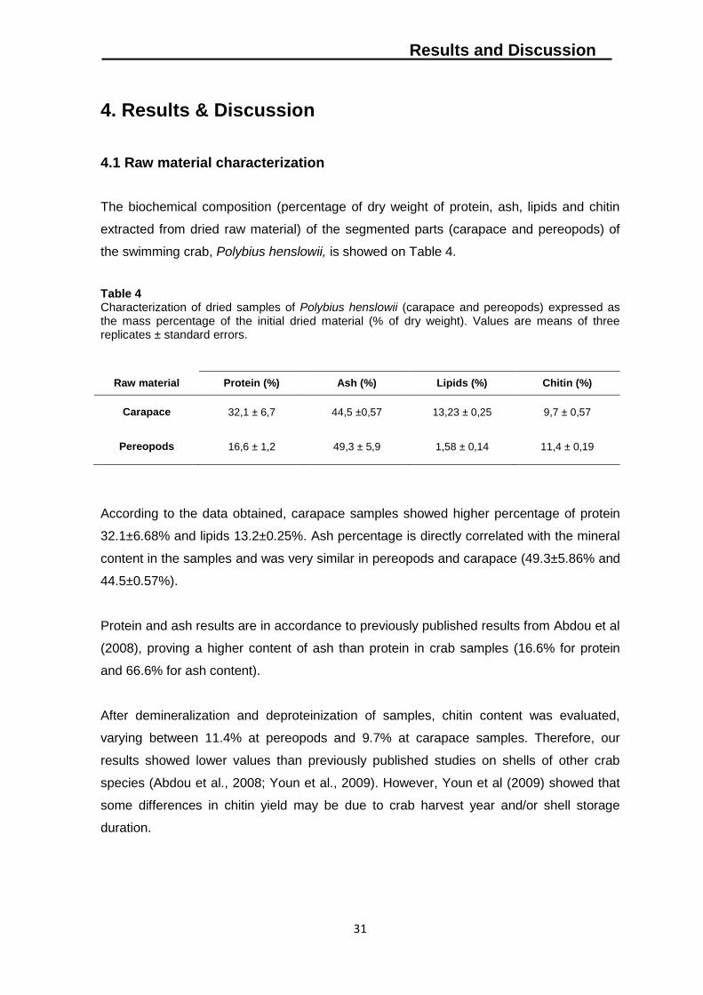
4.1 Raw material characterization
The biochemical composition (percentage of dry weight of protein, ash, lipids and chitin
extracted from dried raw material) of the segmented parts (carapace and pereopods) of
the swimming crab, Polybius henslowii, is showed on Table 4.
Table 4 Characterization of dried samples of Polybius henslowii (carapace and pereopods) expressed as the mass percentage of the initial dried material (% of dry weight). Values are means of three replicates ± standard errors.
Raw material Protein (%) Ash (%) Lipids (%) Chitin (%)
Carapace 32,1 ± 6,7 44,5 ±0,57 13,23 ± 0,25 9,7 ± 0,57
Pereopods 16,6 ± 1,2 49,3 ± 5,9 1,58 ± 0,14 11,4 ± 0,19
According to the data obtained, carapace samples showed higher percentage of protein
32.1±6.68% and lipids 13.2±0.25%. Ash percentage is directly correlated with the mineral
content in the samples and was very similar in pereopods and carapace (49.3±5.86% and
44.5±0.57%).
Protein and ash results are in accordance to previously published results from Abdou et al
(2008), proving a higher content of ash than protein in crab samples (16.6% for protein
and 66.6% for ash content).
After demineralization and deproteinization of samples, chitin content was evaluated,
varying between 11.4% at pereopods and 9.7% at carapace samples. Therefore, our
results showed lower values than previously published studies on shells of other crab
species (Abdou et al., 2008; Youn et al., 2009). However, Youn et al (2009) showed that
some differences in chitin yield may be due to crab harvest year and/or shell storage
duration.
Results and Discussion
32
By comparing the results in table 4, it may be concluded that the segment parts
(pereopods or carapace) defined as chitin raw material source may have influence on
extraction procedures given the observed differences concerning biochemical
composition.
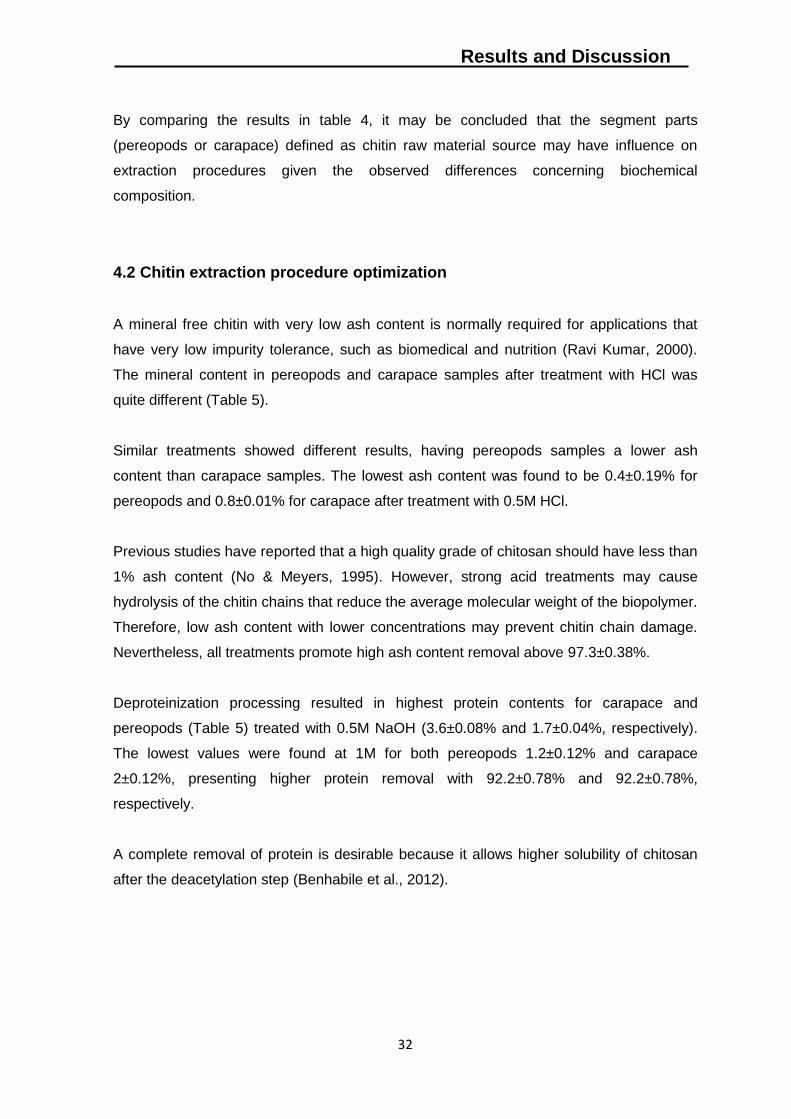
4.2 Chitin extraction procedure optimization
A mineral free chitin with very low ash content is normally required for applications that
have very low impurity tolerance, such as biomedical and nutrition (Ravi Kumar, 2000).
The mineral content in pereopods and carapace samples after treatment with HCl was
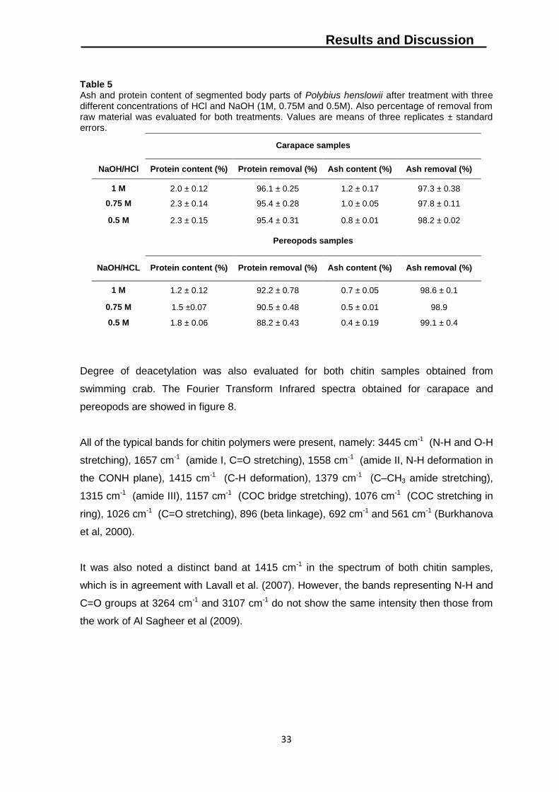
quite different (Table 5).
Similar treatments showed different results, having pereopods samples a lower ash
content than carapace samples. The lowest ash content was found to be 0.4±0.19% for
pereopods and 0.8±0.01% for carapace after treatment with 0.5M HCl.
Previous studies have reported that a high quality grade of chitosan should have less than
1% ash content (No & Meyers, 1995). However, strong acid treatments may cause
hydrolysis of the chitin chains that reduce the average molecular weight of the biopolymer.
Therefore, low ash content with lower concentrations may prevent chitin chain damage.
Nevertheless, all treatments promote high ash content removal above 97.3±0.38%.
Deproteinization processing resulted in highest protein contents for carapace and
pereopods (Table 5) treated with 0.5M NaOH (3.6±0.08% and 1.7±0.04%, respectively).
The lowest values were found at 1M for both pereopods 1.2±0.12% and carapace
2±0.12%, presenting higher protein removal with 92.2±0.78% and 92.2±0.78%,
respectively.
A complete removal of protein is desirable because it allows higher solubility of chitosan
after the deacetylation step (Benhabile et al., 2012).
Results and Discussion
33
Table 5 Ash and protein content of segmented body parts of Polybius henslowii after treatment with three different concentrations of HCl and NaOH (1M, 0.75M and 0.5M). Also percentage of removal from raw material was evaluated for both treatments. Values are means of three replicates ± standard errors.
Carapace samples
NaOH/HCl Protein content (%) Protein removal (%) Ash content (%) Ash removal (%)
1 M 2.0 ± 0.12 96.1 ± 0.25 1.2 ± 0.17 97.3 ± 0.38
0.75 M 2.3 ± 0.14 95.4 ± 0.28 1.0 ± 0.05 97.8 ± 0.11
0.5 M 2.3 ± 0.15 95.4 ± 0.31 0.8 ± 0.01 98.2 ± 0.02
Pereopods samples
NaOH/HCL Protein content (%) Protein removal (%) Ash content (%) Ash removal (%)
1 M 1.2 ± 0.12 92.2 ± 0.78 0.7 ± 0.05 98.6 ± 0.1
0.75 M 1.5 ±0.07 90.5 ± 0.48 0.5 ± 0.01 98.9
0.5 M 1.8 ± 0.06 88.2 ± 0.43 0.4 ± 0.19 99.1 ± 0.4
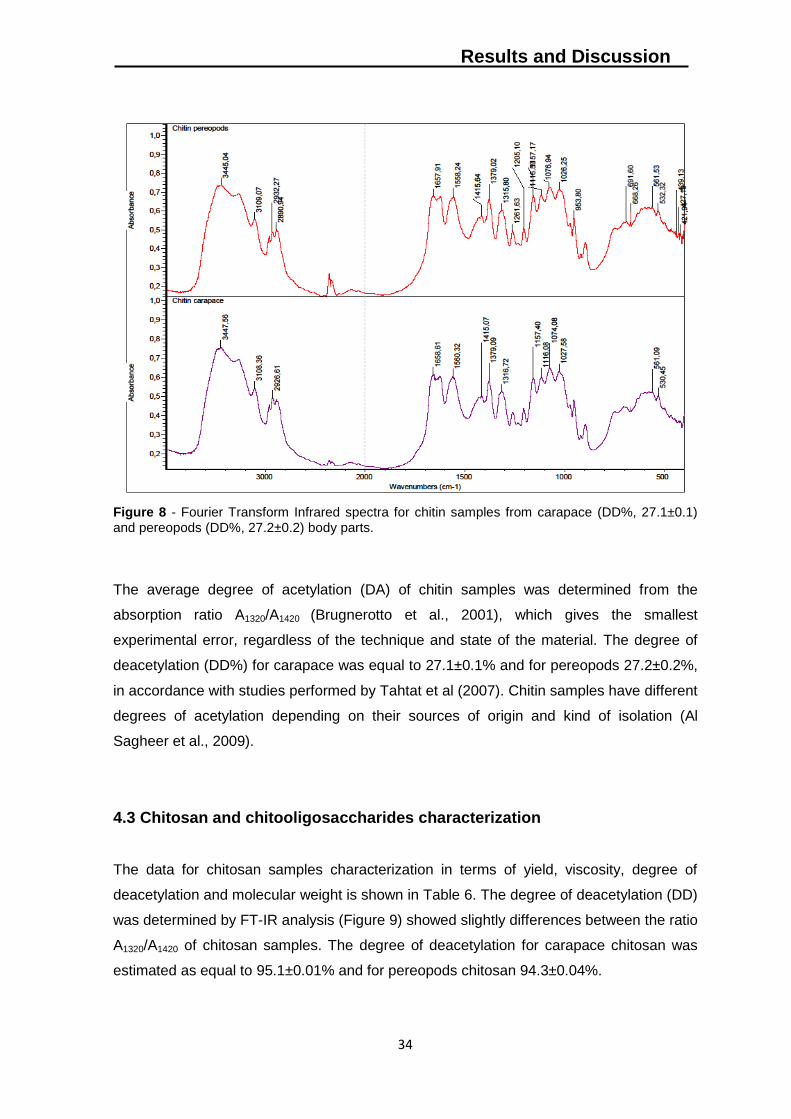
Degree of deacetylation was also evaluated for both chitin samples obtained from
swimming crab. The Fourier Transform Infrared spectra obtained for carapace and
pereopods are showed in figure 8.
All of the typical bands for chitin polymers were present, namely: 3445 cm-1 (N-H and O-H
stretching), 1657 cm-1 (amide I, C=O stretching), 1558 cm-1 (amide II, N-H deformation in
the CONH plane), 1415 cm-1 (C-H deformation), 1379 cm-1 (C–CH3 amide stretching),
1315 cm-1 (amide III), 1157 cm-1 (COC bridge stretching), 1076 cm-1 (COC stretching in
ring), 1026 cm-1 (C=O stretching), 896 (beta linkage), 692 cm-1 and 561 cm-1 (Burkhanova
et al, 2000).
It was also noted a distinct band at 1415 cm-1 in the spectrum of both chitin samples,
which is in agreement with Lavall et al. (2007). However, the bands representing N-H and
C=O groups at 3264 cm-1 and 3107 cm-1 do not show the same intensity then those from
the work of Al Sagheer et al (2009).
Results and Discussion
34
Figure 8 - Fourier Transform Infrared spectra for chitin samples from carapace (DD%, 27.1±0.1) and pereopods (DD%, 27.2±0.2) body parts.
The average degree of acetylation (DA) of chitin samples was determined from the
absorption ratio A1320/A1420 (Brugnerotto et al., 2001), which gives the smallest
experimental error, regardless of the technique and state of the material. The degree of
deacetylation (DD%) for carapace was equal to 27.1±0.1% and for pereopods 27.2±0.2%,
in accordance with studies performed by Tahtat et al (2007). Chitin samples have different
degrees of acetylation depending on their sources of origin and kind of isolation (Al
Sagheer et al., 2009).
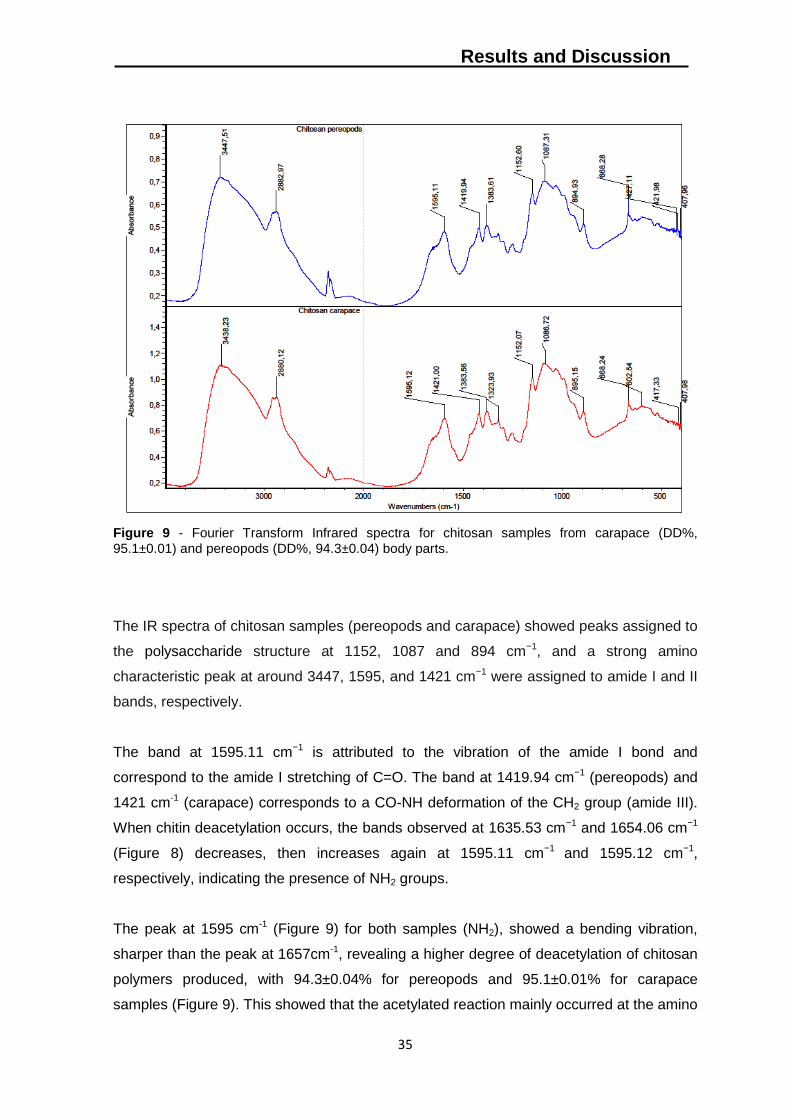
4.3 Chitosan and chitooligosaccharides characterization
The data for chitosan samples characterization in terms of yield, viscosity, degree of
deacetylation and molecular weight is shown in Table 6. The degree of deacetylation (DD)
was determined by FT-IR analysis (Figure 9) showed slightly differences between the ratio
A1320/A1420 of chitosan samples. The degree of deacetylation for carapace chitosan was
estimated as equal to 95.1±0.01% and for pereopods chitosan 94.3±0.04%.
Results and Discussion
35
Figure 9 - Fourier Transform Infrared spectra for chitosan samples from carapace (DD%,
95.1±0.01) and pereopods (DD%, 94.3±0.04) body parts.
The IR spectra of chitosan samples (pereopods and carapace) showed peaks assigned to
the polysaccharide structure at 1152, 1087 and 894 cm−1, and a strong amino
characteristic peak at around 3447, 1595, and 1421 cm−1 were assigned to amide I and II
bands, respectively.
The band at 1595.11 cm−1 is attributed to the vibration of the amide I bond and
correspond to the amide I stretching of C=O. The band at 1419.94 cm−1 (pereopods) and
1421 cm-1 (carapace) corresponds to a CO-NH deformation of the CH2 group (amide III).
When chitin deacetylation occurs, the bands observed at 1635.53 cm−1 and 1654.06 cm−1
(Figure 8) decreases, then increases again at 1595.11 cm−1 and 1595.12 cm−1,
respectively, indicating the presence of NH2 groups.
The peak at 1595 cm-1 (Figure 9) for both samples (NH2), showed a bending vibration,
sharper than the peak at 1657cm-1, revealing a higher degree of deacetylation of chitosan
polymers produced, with 94.3±0.04% for pereopods and 95.1±0.01% for carapace
samples (Figure 9). This showed that the acetylated reaction mainly occurred at the amino
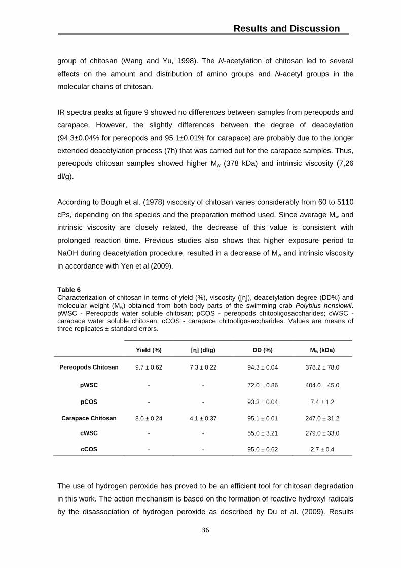
Results and Discussion
36
group of chitosan (Wang and Yu, 1998). The N-acetylation of chitosan led to several
effects on the amount and distribution of amino groups and N-acetyl groups in the
molecular chains of chitosan.
IR spectra peaks at figure 9 showed no differences between samples from pereopods and
carapace. However, the slightly differences between the degree of deaceylation
(94.3±0.04% for pereopods and 95.1±0.01% for carapace) are probably due to the longer
extended deacetylation process (7h) that was carried out for the carapace samples. Thus,
pereopods chitosan samples showed higher Mw (378 kDa) and intrinsic viscosity (7,26
dl/g).
According to Bough et al. (1978) viscosity of chitosan varies considerably from 60 to 5110
cPs, depending on the species and the preparation method used. Since average Mw and
intrinsic viscosity are closely related, the decrease of this value is consistent with
prolonged reaction time. Previous studies also shows that higher exposure period to
NaOH during deacetylation procedure, resulted in a decrease of Mw and intrinsic viscosity
in accordance with Yen et al (2009).
Table 6 Characterization of chitosan in terms of yield (%), viscosity ([ɳ]), deacetylation degree (DD%) and molecular weight (Mw) obtained from both body parts of the swimming crab Polybius henslowii. pWSC - Pereopods water soluble chitosan; pCOS - pereopods chitooligosaccharides; cWSC - carapace water soluble chitosan; cCOS - carapace chitooligosaccharides. Values are means of three replicates ± standard errors.
Yield (%) [ɳ] (dl/g) DD (%) Mw (kDa)
Pereopods Chitosan 9.7 ± 0.62 7.3 ± 0.22 94.3 ± 0.04 378.2 ± 78.0
pWSC - - 72.0 ± 0.86 404.0 ± 45.0
pCOS - - 93.3 ± 0.04 7.4 ± 1.2
Carapace Chitosan 8.0 ± 0.24 4.1 ± 0.37 95.1 ± 0.01 247.0 ± 31.2
cWSC - - 55.0 ± 3.21 279.0 ± 33.0
cCOS - - 95.0 ± 0.62 2.7 ± 0.4
The use of hydrogen peroxide has proved to be an efficient tool for chitosan degradation
in this work. The action mechanism is based on the formation of reactive hydroxyl radicals
by the disassociation of hydrogen peroxide as described by Du et al. (2009). Results
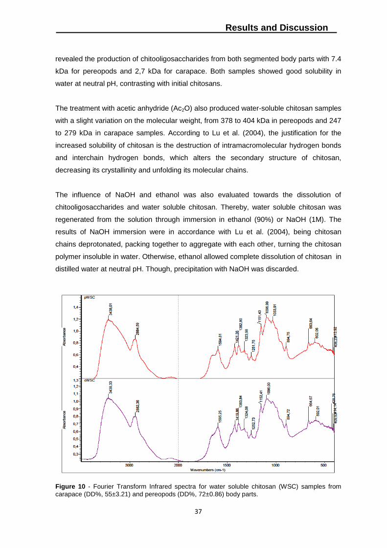
Results and Discussion
37
revealed the production of chitooligosaccharides from both segmented body parts with 7.4
kDa for pereopods and 2,7 kDa for carapace. Both samples showed good solubility in
water at neutral pH, contrasting with initial chitosans.
The treatment with acetic anhydride (Ac2O) also produced water-soluble chitosan samples
with a slight variation on the molecular weight, from 378 to 404 kDa in pereopods and 247
to 279 kDa in carapace samples. According to Lu et al. (2004), the justification for the
increased solubility of chitosan is the destruction of intramacromolecular hydrogen bonds
and interchain hydrogen bonds, which alters the secondary structure of chitosan,
decreasing its crystallinity and unfolding its molecular chains.
The influence of NaOH and ethanol was also evaluated towards the dissolution of
chitooligosaccharides and water soluble chitosan. Thereby, water soluble chitosan was
regenerated from the solution through immersion in ethanol (90%) or NaOH (1M). The
results of NaOH immersion were in accordance with Lu et al. (2004), being chitosan
chains deprotonated, packing together to aggregate with each other, turning the chitosan
polymer insoluble in water. Otherwise, ethanol allowed complete dissolution of chitosan in
distilled water at neutral pH. Though, precipitation with NaOH was discarded.
Figure 10 - Fourier Transform Infrared spectra for water soluble chitosan (WSC) samples from
carapace (DD%, 55±3.21) and pereopods (DD%, 72±0.86) body parts.
Results and Discussion
38
Structural changes between chitosan samples (Figure 9) and water soluble chitosan
(WSC) are also confirmed by FT-IR spectra (Figure 10). The peak at 1595 cm-1 assigned
to the bending vibration of -NH2 group (amide II) decreased intensity in the s of figure 10
when compared with chitosan´s spectra at figure 9.
Also the appearance of a new peak at 1654 cm-1 and the increasing intensity at 1316 cm-1
was due to the distortion vibration of acetamido I, and III, indicating that the degree of
deacetylation (DD) decreased. Therefore, according to Hu et al. (2008), the present
results indicate that the WCS was synthesized by N-acetyl substitution, supported by
structural changes.
Chitosan with high degree of deaceylation (DD) is water insoluble because of its rigid
crystalline structure. The N-acetylation of chitosan leads to several effects on the amount
and distribution of amino groups and N-acetyl groups in the molecular chains of chitosan.
By N-acetylation of chitosan, WSC can be soluble in water. Meanwhile, the crystallinity of
WSC decreased, indicating that the crystalline structure was modify by N-acetylation of
chitosan (Lu et al, 2004).
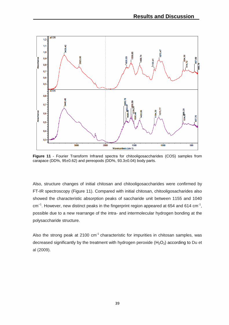
Results and Discussion
39
Figure 11 - Fourier Transform Infrared spectra for chitooligosaccharides (COS) samples from
carapace (DD%, 95±0.62) and pereopods (DD%, 93.3±0.04) body parts.
Also, structure changes of initial chitosan and chitooligosaccharides were confirmed by
FT-IR spectroscopy (Figure 11). Compared with initial chitosan, chitooligosacharides also
showed the characteristic absorption peaks of saccharide unit between 1155 and 1040
cm−1. However, new distinct peaks in the fingerprint region appeared at 654 and 614 cm-1,
possible due to a new rearrange of the intra- and intermolecular hydrogen bonding at the
polysaccharide structure.
Also the strong peak at 2100 cm-1 characteristic for impurities in chitosan samples, was
decreased significantly by the treatment with hydrogen peroxide (H2O2) according to Du et
al (2009).
Results and Discussion
40
4.4 Reuse of reagents for extraction and production procedures
In order to decrease the amount of reagents by its reuse needed to process Polybius
henslowii raw material to obtain chitosan products, all the chemical reactions for a given
procedure (demineralization, deproteinization and deacetylation) were employed
consecutively using the same solution (Table 7).
Regarding demineralization step, the reuse of acid solutions (HCl 0.5M) proved low
efficiency towards mineral removal after the first treatment. The acid solution didn´t
allowed the removal of mineral contents below 1%, with 6.9±2.26% (at 2nd treatment) and
44.4±0.54 (at 3rd treatment).
On the other hand, the deproteinization step, using 1M NaOH, showed no significant
differences (t-test, p>0.05) between the first, second and third treatments with 2.5±0.41%,
2.8±0.28% and 3.1±0.44% respectively.
Table 7 Characterization of chitin and chitosan samples from pereopods body part expressed as the mass percentage of the initial dried material (% of dry weight). Values are means of three replicates ± standard errors.
Demineralization/Deproteinization Deacetylation
Ash% Protein % Yield % DD % Viscosity Mw
Treatment 1 0.5±0.04 2.5±0.41 82.2±1.20 93±0.9 6,3±0.12 333.8±34
Treatment 2 6.9±2.26 2.8±0.28 81.8±0.31 93±1.6 6,9±0.80 305.8±23
Treatment 3 44.4±0.54 3.1±0.44 82.7±2.21 92±3.0 6,4±0.76 328.2±9
Regarding chitosan samples, no significant differences (t-test, p>0.05) were observed in
terms of degree of deacetylation, viscosity and molecular weight, being all treatments
effective, allowing chitosan production soluble in acidic media.
IR spectra of the chitosan samples produced are showed in figure 12. The same peaks at
1646 cm-1 assigned for amide I band are present in all three spectra. Also the peaks at
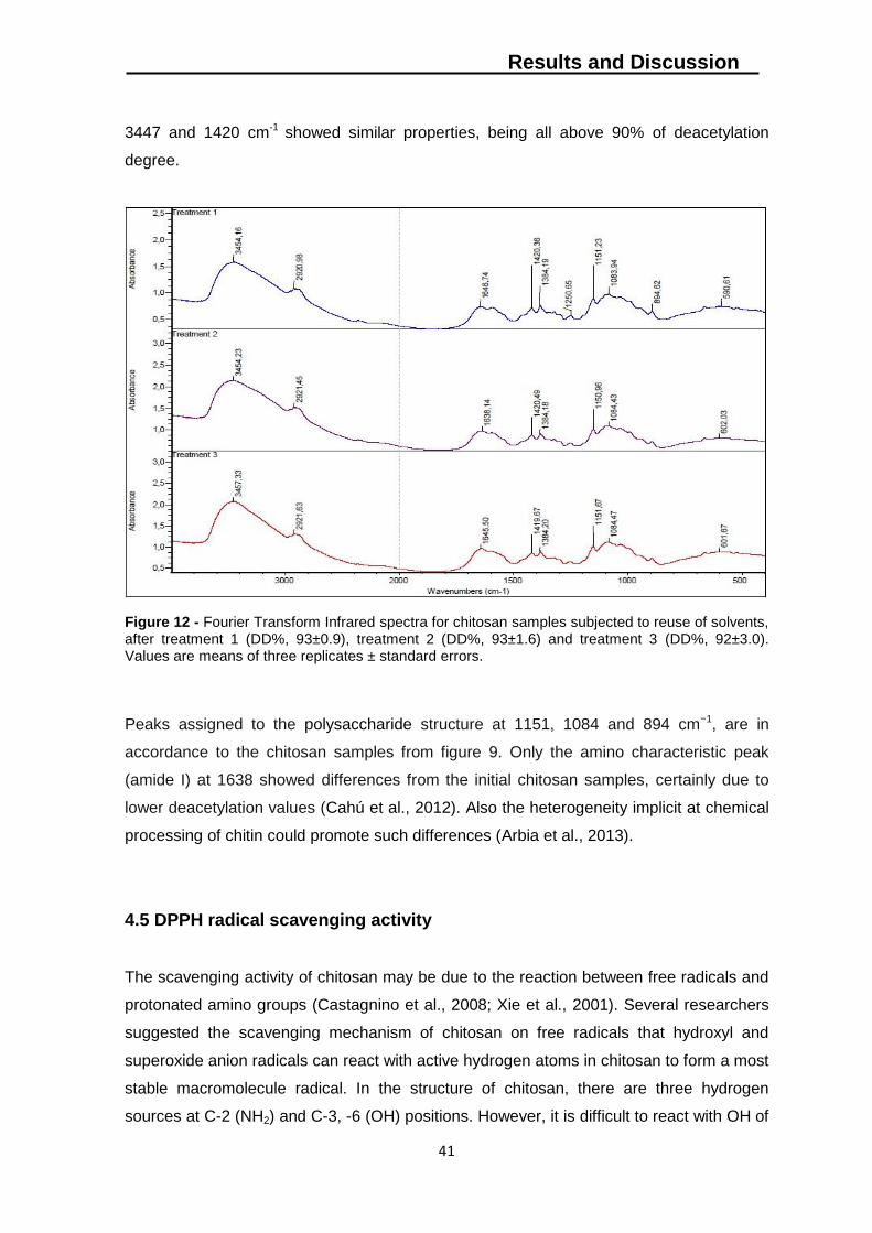
Results and Discussion
41
3447 and 1420 cm-1 showed similar properties, being all above 90% of deacetylation
degree.
Figure 12 - Fourier Transform Infrared spectra for chitosan samples subjected to reuse of solvents, after treatment 1 (DD%, 93±0.9), treatment 2 (DD%, 93±1.6) and treatment 3 (DD%, 92±3.0). Values are means of three replicates ± standard errors.
Peaks assigned to the polysaccharide structure at 1151, 1084 and 894 cm−1, are in
accordance to the chitosan samples from figure 9. Only the amino characteristic peak
(amide I) at 1638 showed differences from the initial chitosan samples, certainly due to
lower deacetylation values (Cahú et al., 2012). Also the heterogeneity implicit at chemical
processing of chitin could promote such differences (Arbia et al., 2013).
4.5 DPPH radical scavenging activity
The scavenging activity of chitosan may be due to the reaction between free radicals and
protonated amino groups (Castagnino et al., 2008; Xie et al., 2001). Several researchers
suggested the scavenging mechanism of chitosan on free radicals that hydroxyl and
superoxide anion radicals can react with active hydrogen atoms in chitosan to form a most
stable macromolecule radical. In the structure of chitosan, there are three hydrogen
sources at C-2 (NH2) and C-3, -6 (OH) positions. However, it is difficult to react with OH of
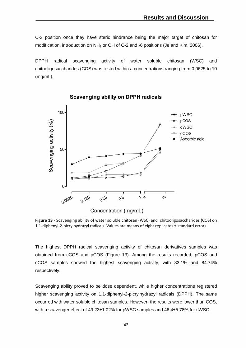
Results and Discussion
42
C-3 position once they have steric hindrance being the major target of chitosan for
modification, introduction on NH2 or OH of C-2 and -6 positions (Je and Kim, 2006).
DPPH radical scavenging activity of water soluble chitosan (WSC) and
chitooligosaccharides (COS) was tested within a concentrations ranging from 0.0625 to 10
(mg/mL).
Figure 13 - Scavenging ability of water soluble chitosan (WSC) and chitooligosaccharides (COS) on 1,1-diphenyl-2-picrylhydrazyl radicals. Values are means of eight replicates ± standard errors.
The highest DPPH radical scavenging activity of chitosan derivatives samples was
obtained from cCOS and pCOS (Figure 13). Among the results recorded, pCOS and
cCOS samples showed the highest scavenging activity, with 83.1% and 84.74%
respectively.
Scavenging ability proved to be dose dependent, while higher concentrations registered
higher scavenging activity on 1,1-diphenyl-2-picrylhydrazyl radicals (DPPH). The same
occurred with water soluble chitosan samples. However, the results were lower than COS,
with a scavenger effect of 49.23±1.02% for pWSC samples and 46.4±5.78% for cWSC.
Results and Discussion
43
Comparing to the scavenger ability of ascorbic acid (one of the forms of vitamin C), COS
exhibited much higher activity at 10mg/mL, proving its potential use as a an antioxidant
source.
Previous investigators have revealed that the DPPH radical scavenging activity of
chitosan increased with decreasing molecular weight (MW) (Chien et al., 2007; Kim and
Thomas, 2007; Yen et al., 2007). This is in accordance with our results, showing lower
molecular weight chitooligosaccharides higher activity than water soluble chitosan.
According to Kim and Thomas (2007), the higher MW chitosan (WSC) would have lower
mobility than the lower MW chitosan (COS). Consequently, this would increase the
possibility of inter- and intramolecular bonding of the high MW chitosan molecules, and,
thus, the chance of exposure of their amine groups might be restricted.
Although, the present results for water soluble chitosan are in accordance with the work of
Yen et al (2008) with approximately 20% scavenging ability by crab chitosan samples with
1mg/mL.
4.6 Antibacterial activity
Antibacterial activity of chitosan and its derivatives against several bacterial species has
been recognized and is considered as one of the most important properties linked directly
to their possible biological applications (Du et al., 2009). The antibacterial activity of these
compounds is influenced by a number of factors such as degree of polymerization (Park
et al., 2004; Park et al., 2002), level of deacetylation (Chung et al., 2004), type of
microorganism (Gerasimenko et al., 2004).
In order to study the effect of molecular size of chitosan obtained from different body parts
of Polybius, water soluble chitosan samples with different molecular weights were
prepared with a concentration of 10 mg/mL. Antimicrobial activity of water soluble chitosan
and chitooligosaccharidess were evaluated by spectroscopy analyses in the microplate
reader.
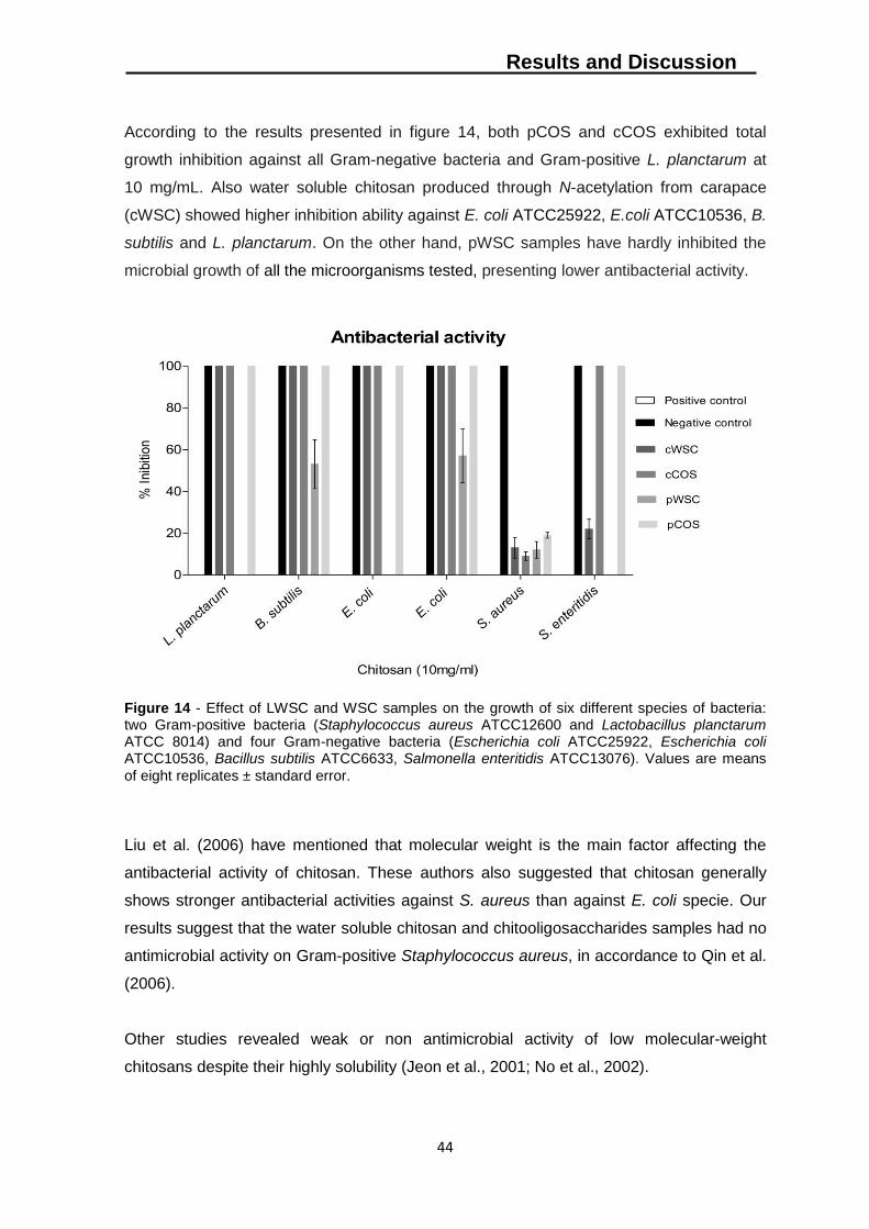
Results and Discussion
44
According to the results presented in figure 14, both pCOS and cCOS exhibited total
growth inhibition against all Gram-negative bacteria and Gram-positive L. planctarum at
10 mg/mL. Also water soluble chitosan produced through N-acetylation from carapace
(cWSC) showed higher inhibition ability against E. coli ATCC25922, E.coli ATCC10536, B.
subtilis and L. planctarum. On the other hand, pWSC samples have hardly inhibited the
microbial growth of all the microorganisms tested, presenting lower antibacterial activity.
Figure 14 - Effect of LWSC and WSC samples on the growth of six different species of bacteria: two Gram-positive bacteria (Staphylococcus aureus ATCC12600 and Lactobacillus planctarum ATCC 8014) and four Gram-negative bacteria (Escherichia coli ATCC25922, Escherichia coli ATCC10536, Bacillus subtilis ATCC6633, Salmonella enteritidis ATCC13076). Values are means
of eight replicates ± standard error.
Liu et al. (2006) have mentioned that molecular weight is the main factor affecting the
antibacterial activity of chitosan. These authors also suggested that chitosan generally
shows stronger antibacterial activities against S. aureus than against E. coli specie. Our
results suggest that the water soluble chitosan and chitooligosaccharides samples had no
antimicrobial activity on Gram-positive Staphylococcus aureus, in accordance to Qin et al.
(2006).
Other studies revealed weak or non antimicrobial activity of low molecular-weight
chitosans despite their highly solubility (Jeon et al., 2001; No et al., 2002).
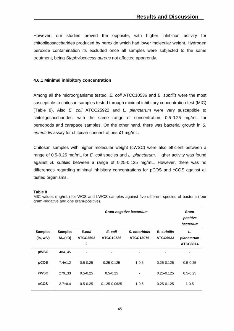
Results and Discussion
45
However, our studies proved the opposite, with higher inhibition activity for
chitooligosaccharides produced by peroxide which had lower molecular weight. Hydrogen
peroxide contamination its excluded once all samples were subjected to the same
treatment, being Staphylococcus aureus not affected apparently.
4.6.1 Minimal inhibitory concentration
Among all the microorganisms tested, E. coli ATCC10536 and B. subtilis were the most
susceptible to chitosan samples tested through minimal inhibitory concentration test (MIC)
(Table 8). Also E. coli ATCC25922 and L. planctarum were very susceptible to
chitoligosaccharides, with the same range of concentration, 0.5-0.25 mg/mL for
pereopods and carapace samples. On the other hand, there was bacterial growth in S.
enteritidis assay for chitosan concentrations ≤1 mg/mL.
Chitosan samples with higher molecular weight (cWSC) were also efficient between a
range of 0.5-0.25 mg/mL for E. coli species and L. planctarum. Higher activity was found
against B. subtilis between a range of 0.25-0.125 mg/mL. However, there was no
differences regarding minimal inhibitory concentrations for pCOS and cCOS against all
tested organisms.
Table 8 MIC values (mg/mL) for WCS and LWCS samples against five different species of bacteria (four gram-negative and one gram-positive).
Gram-negative bacterium Gram-
positive
bacterium
Samples
(%, w/v)
Samples
Mw (kD)
E.coli
ATCC2592
2
E. coli
ATCC10536
S. enteritidis
ATCC13076
B. subtilis
ATCC6633
L.
planctarum
ATCC8014
pWSC 404±45 - - - - -
pCOS 7.4±1.2 0.5-0.25 0.25-0.125 1-0.5 0.25-0.125 0.5-0.25
cWSC 279±33 0.5-0.25 0.5-0.25 - 0.25-0.125 0.5-0.25
cCOS 2.7±0.4 0.5-0.25 0.125-0.0625 1-0.5 0.25-0.125 1-0.5
Results and Discussion
46
The mostly accepted mechanism explains that COS can alter permeability characteristics
of microbial cell membrane and further prevent the entry of materials or cause leakage of
cell constituents that finally leads to death of bacteria (Sudharshan et al., 1992). Chung et
al. (2004) studied the cell surface characteristics of a number of Gram-positive and Gram-
negative bacteria species and revealed that charge distribution on the cell surfaces is
apparently an important factor regarding antibacterial activity increasing or decreasing its
susceptibility towards COS. Apparently there is a higher distribution of negative charge on
the surface of gram-negative than in gram-positive. However, this was not supported by
our results.
According to our findings (Table 8), Chitooligosaccharides from carapace (cCOS) and
pereopods (pCOS) showed higher antibacterial activity against all tested organisms than
WSC. It confirmed that relevant decrease of molecular weight could improve the
antibacterial activity of chitosan in accordance to Du et al. (2009). The molecular weight
dependence of the antimicrobial activity of chitosan was more pronounced at lower
concentrations.
4.7 Antifungal assay
To prevent and treat cultures against fungi, chemical products are apply. Although, most
of them, such as sulfur dioxide fumigation (Droby and Lichter, 2004), present serious
consequences for the environment. In order to avoid this source of pollution, chitosan
appears as a possible way to prevent the growth of several fungi on crop fields or gardens
(Badawy and Rabea, 2009; Yong-cai et al., 2009).
The results of water soluble chitosan (WSC) and chitooligosaccharides (COS) applied
against four different species of fungi are present in Figure 15. The concentration range
varied from 0.0125 to 0.1 mg/mL and inhibition percentages were determined by
differences in radial growth on the agar plates for all species. From four fungal species
tested in this study, non was completely inhibited by chitosan samples (WSC and COS) at
the highest concentration tested, 0.1mg/mL.
The results obtained in microbiological assays showed that the capability to inhibit fungal
growth was clearly higher for the COS samples from both segmented body parts than for
Results and Discussion
47
WSC. Despite not all species tested exhibited the same vulnerability towards
concentrations, antifungal activity of chitosan samples proved to be dependent, increasing
the inhibitory capacity with higher concentrations.
The highest inhibition was achieved by pCOS and cCOS samples for Cryphonectria
parasitica with 84.5±3.14% and 85.6±2.27%, respectively. Phytophthora cinnamomi
showed lower vulnerability to chitosan samples, with higher inhibition by pCOS
58.9±1.8%. This way, chitooligosaccharides from pereopods (pCOS) showed higher
activity against Heteobasidion annosum and Phytophthora cinnamomi. Regarding Botrytis
cinerea and Cryphonectria parasitica, the higher activity showed by pCOS and cCOS,
presented no differences.
Though, water soluble chitosan (WSC) revealed lower influence on fungal inhibition than
COS samples. Only Heterobasidion annosum showed equal performances towards
inhibition ability between cWSC and cCOS with 20.5±8.11% and 28.8±4.64%,
respectively.
The results proved that lower molecular weight samples (COS), showed better ability
towards fungal growth inhibition than the chitosan with higher molecular weight (water
soluble chitosan) (Xu et al., 2007; Badawy and Rabea, 2009).
Several mechanisms for the antimicrobial action of chitosan have been proposed. For
example, it has been suggested that chitosan may inhibit microbial growth by acting as a
chelating agent rendering metals, trace elements or essential nutrients unavailable for the
organism to grow at the normal rate (Skjak-Braek et al., 1989).
Another hypothesis explains the activity of chitosan as being based on the electrostatic
interaction of the charged amino groups of chitosan with negatively charged cell wall
surface of the targeted microorganisms, which can lead to the disruption of the cell wall
and therefore to the death of the cell. Also it is possible for antifungal activity of chitosan
accounts for its chains to cross the cell membrane inhibiting the cell growing from inside
(Guo et al. 2007).
In another study (Liu et al., 2007), low molecular weight chitosan at different
concentrations (0.01–1%) markedly inhibited mycelial growth of B. cinerea.
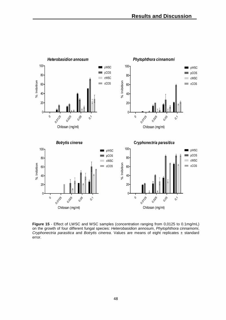
Results and Discussion
48
Figure 15 - Effect of LWSC and WSC samples (concentration ranging from 0,0125 to 0.1mg/mL) on the growth of four different fungal species: Heterobasidion annosum, Phytophthora cinnamomi, Cryphonectria parasitica and Botrytis cinerea. Values are means of eight replicates ± standard
error.
Conclusion
49
5. Conclusion
The present work aimed to study the biotechnological potential of Polybius henslowii, an
extremely abundant marine resource not subject to any commercial use in Portugal.
Despite the lower yields of polymers isolated (chitin/chitosan) from both segmented body
parts, when compared with other crab sources, the results support the economical
sustainability of chemical processing resulting from the reuse of solvents. Reutilization of
solvents showed interesting results towards a more eco-friendly approach, allowing
reproducing the same conditions and generating similar polymers after three following
treatments, saving by this way a huge amount of reagents. These findings lower
considerably the cost for production of chitosan.
Chitosans samples obtained from segmented pereopods and carapace, showed
differences regarding yield, molecular weight and intrinsic viscosity properties. However,
only chitosan derivatives revealed higher interest for biotechnological applications based
on their biological properties.
Chitooligosaccharides with low molecular weight demonstrated relative stronger
scavenging activity. This result demonstrated that the scavenging ativity on DPPH was
closely related to molecular weight and concentration in solution. Compared with water
soluble chitosan, chitooligosaccharides have a shorter chain with ability to form
intramolecular hydroxyl bonds, which means that the hydroxyl and amino groups were
available for the radical scavenging process.
Also the antibacterial and antifungal activities proved to be dose dependent and clearly
related to molecular weight. The obtained COS from both segmented body parts revealed
excellent antibacterial activity against all the tested Gram-negative bacteria and Gram-
positive Lactobacillus planctarum.
Regarding antifungal activity, Botrytis cinerea and Cryphonectria parasitica were highly
inhibited by COS without differences between samples. COS showed better results in
minimal inhibitory concentrations than WSC against all tested microorganisms, proving
higher growth inhibition for Escherichia coli (ATCC10536) with MIC values ranging
between 0.125 - 0.0625 mg/mL.
Conclusion
50
The capability to inhibit fungus growth was clearly higher for the COS samples, from both
segmented body parts than for WSC. The highest inhibition was achieved by pCOS and
cCOS samples for Cryphonectria parasitica (DSMZ 62626) with 84.5±3.14% and
85.6±2.27%, respectively.
In conclusion, the biochemical characteristics of this non-tradiotional marine resource and
its biological properties, support the hypothesis of using it as raw material for
biotechnology industries enhancing by that way its economic value.
The promotion of this bycatch specie, also increases competitiveness on fishing activities
through reorientation of fisheries and diversification of targeted species for biotechnology
purposes, with potential implication on the sustainable development of coastal
communities.
Future perspectives
51
6. Future perspectives
Through this study, different protocols were evaluated in order to extract chitin, produce
chitosan and derivatives through fast, simple and less expensive methods. It was
achieved the final goal of knowledge transfer from I&D activities to the industry.
Despite the proved properties of chitosan and its derivatives produced, chemical
processing of chitin still has several disadvantages. First, the amount and cost of
reagents. Despite the promissing results through reuse of solvents, chemicals are still
necessary, being disposal at the end of the process. Therefore, more eco-friendly
strategies have to be apply in order to generate a more sustainable protocol. One
possibility could the use of bacteria and enzymes for demineralization, deproteinization
and deacetylation.
Also the problem of heterogeneity during processing needs to be solved. The differences
obtained at the end of the deacetylation process occurred mainly due to the
hetererogeous deacetylation conditions, given that the reaction was carried out on an oil
bath without agitation. This fact increases disparities between temperatures inside the
reaction flasks and produces polymers with different biochemical/structural properties.
Though, it is important to improve homogeneity during the process from raw material in
order to obtain a low polydisperse polymer. One solution could be the use of a close
bioreactor in order to ensure that all samples are subject to the same conditions.
Three biological properties were tested in this study. Nevertheless, more experiments
have to be carried out in order to evaluate the full potential of Polybius henslowii as raw
material source. Other antioxidant activities should be performed such as scavenging
ability on hydroxyl radicals, chelating ability on ferrous ions and reducing power.
In addition, rheological properties should be tested allowing the determination of the
degree of crystalinity for all products. Also, to confirm the polymers physical structure,
they should be analysed through Scanning Electron Microscopy (SEM).
It´s also of great importance to screan Polybius henslowii raw material for heavy metals,
once its presence could decrease considerably the range of applications.
Future perspectives
52
Therefore, this raw material still possess some questions regarding its biological and
physical properties, if the final goal is to be used by biotecnological industry. However, this
study gives helpful indications, supporting its potential as a raw material for
biotechnological purposes.
References
53
References Abdou, E.S., Nagy, K.S.A., Elsabee, M.Z. (2008). Extraction and characterization of chitin and chitosan from local sources. Bioresource Technology. 99, 1359-1367. Al Sagheer, F.A., Al-Sughayer, M.A., Muslim, S., Elsabee, M.Z. (2009). Extraction and characterization of chitin and chitosan from marine sources in Arabian Gulf. Carbohydrate Polymers. 77, 410–419. Aranaz, I., Mengíbar, M., Harris, R., Paños, I., Miralles, B., Acosta, N., Galed, G., Heras A. (2009). Functional Characterization of Chitin and Chitosan. Current Chemical Biology. 3, 203-230. Arbia, W., Arbia, L., Adour, L., Amrane, A. (2013). Chitin Extraction from Crustacean Shells Using Biological Methods – A Review. Food Technol. Biotechnol. 51(1), 12-25. Badawy, M.E.I., Rabea, E.I. (2009). Potential of the biopolymer chitosan with different molecular weights to control postharvest gray mold of tomato fruit. Postharvest Biology and Technology. 51, 10–117. Bahrke, S., Einarsson, J.M., Gislason, J., Haebel, S., Peter-Katalinc, J., Peter, M.G. (2002). Characterization of chitooligosaccharides by mass spectrometry. Advances in Chitin Science, vol 6. (Ed. Vårum K.M.). Trondheim, Norway. Bajaj, M., Winter, J., Gallert, C. (2011). Effect of deproteinization and decalcification conditions on viscosity of chitin and chitosan extracted from Crangon crangon. Biochem. Eng. J. 56, 51-62. Bautista, J., Jover, M., Guttierrez, J.F., Corpas, R., Cremades, O., Fontiveros, E., Iglesias, F., Veja, J. (2001). Preparation of crayfish chitin by in situ lactic acid production. Process Biochem. 37, 229-234. Benhabile, M.S., Salah, R., Lounici, H., Drouiche, N., Goosen, M.F.A., Mameri, N. (2012). Antibacterial activity of chitin, chitosan and its oligomers prepared from shrimp shell waste. Food Hydrocolloids. 29, 48-56. Blackwel, J. (1969). Structure of Beta-Chitin or Parallel Chain Systems of Poly-Beta-(1-]4)-N-Acetyl-D-Glucosamine. Biopolymers. 7(3),281-286. Blackwell, J., Parker, K.D., Rudall, K.M. (1965). Chitin in pogonophore tubes. J Mar Biol Assoc UK. 45, 659-61. Bough, W.A., Salter, W.L., Wu, A.C.M., Perkins, B.E. (1978). Influence of manufacturing variables on the characteristics and effectiveness of chitosan products. Chemical composition, viscosity, and molecular weight distribution of chitosan products. Biotechnol. Bioeng. 20, 1931. Brugnerotto, J., Lizardi J., Goycoolea, F.M., Argüelles-Monal, W., Desbrières, J., Rinaudo, M. (2001). An infrared investigation in relation with chitin and chitosan characterization. Polymer. 42, 3569-3580.
References
54
Burkhanova, N.D. , Yugai, S.M. , Pulatova, K.P., Nikononvich, G.V., Milusheva, R.Y., Voropaeva N.L., Rashidova S.S. (2000). Structural investigations of chitin and its deacetylation products. Chemistry of Natural Compounds. 36, 4. Cabreraa, J.C., Cutsemb, P.V. (2005). Preparation of chitooligosaccharides with degree of polymerization higher than 6 by acid or enzymatic degradation of chitosan. Biochemical Engineering Journal. 25, 165-172. Cahú, T.B., Santos, S.D., Mendes,A., Córdula, C.R., Chavante, S.F., Carvalho L.B., Nader, H.B., Bezerra, R.S. (2012). Recovery of protein, chitin, carotenoids and glycosaminoglycans from Pacific white shrimp (Litopenaeus vannamei) processing waste. Process Biochemistry. 47, 570-577. Cappellini, R.A., Ceponis, M.J., Lightner, G.W. (1986). Disorders in table grape shipments to the New York market, 1972–1984. Plant Dis. 70, 1075-1079. Carlstrom, D. (1957). The Crystal Structure of Alpha-Chitin (Poly-N-Acetyl D-Glucosamine). Journal of Biophysical and Biochemical Cytology 3(5), 669-683. Cartes, J.E., Abelló, P., Lloris, D., Carbonell, A., Torres, P., Maynou, F., Gil de Sola, L. (2002). Feeding guilds of western Mediterranean demersal fish and crustaceans: an analysis based on a spring survey. Scientia Marina. 66, 209-220. Cartier, N., Domard, A., Chanzy, H. (1990). Single crystals of chitosan. Int J Biol Macromol. 12, 289-94. Castagnino, E., Ottaviani, M. F., Cangiotti, M., Morelli, M., Casettari, L., & Muzzarelli, R. A. A. (2008). Radical scavenging activity of 5-methylpyrrolidinone chitosan and dibutyryl chitin. Carbohydrate Polymers. 74, 640–647. Crini, G., Guibal, E., Morcellet, M., Torri, G., Badot., P.M. (2009). Chitine et chitosane. Préparation, propriétés et principales applications. In: chitine et chitosane. Du biopolymère à l’application, 1st Ed., Presses universitaires de Franche-Comté, France. 19-54. Chien, P. J., Sheu, F., Huang, W.T., Su, M.S. (2007). Effect of molecular weight of chitosan on their antioxidative activities in apple juice. Food Chemistry. 102, 1192–1198. Choorit, W., Patthanamanee, W., Manurakchinakorn, S. (2008). Use of response surface method for the determination of demineralization efficiency in fermented shrimp shells. Bioresour. Technol. 99, 6168-6173. Chung, Y.C., Su, Y. P., Chen, C.C., Jia, G., Wang, H.L., Wu, J. C. (2004). Relationship between antibacterial activity of chitosan and surface characteristics of cell wall. Acta Pharmacologica Sinica, 25, 932–936. Dash, M., Chiellini, F., Ottenbrite, R.M., Chiellini, E. (2011). A versatile semi-synthetic polymer in biomedical applications. Progress in Polymer Science. 36, 981-1014. Domard, A., Domard, M. (2002). Chitosan: structure-properties relationship and biomedical applications, in Polymeric Biomaterials (ed S. Dumitriu), Dekker, New York.

References
55
Droby, S., Lichter, A. (2004). Post-harvest Botrytis infection: etiology, development and management. In: Elad, Y., Williamson, B., Tudzynski, P., Delen, N. (Eds.), Botrytis: Biology. Pathology and Control. Kluwer Academic Publishers, London, UK. 349-367. Du, Y., Zhao, Y., Dai, S., Yang, B. (2009). Preparation of water-soluble chitosan from shrimp shell and its antibacterial activity. Innovative Food Science and Emerging Technologies. 10, 103-107. Duan, X.J., Zhang, W.W., Li, X.M., Wang, B.G. (2006). Evaluation of antioxidant property of extract and fractions obtained from a red alga, Polysiphonia urceolata. Food Chemistry. 95, 37-43. El Ghaouth, A., J. Arul, A. Asselin, and N. Benhamou. (1992). Antifungal activity of chitosan on post harvest pathogens: Induction of morphological and cytological alterations in Rhizopus stolonifer. Mycol. 96, 769-779. Elsabeea, M.Z., Morsib, R.E., Al-Sabaghc, A.M. (2009). Surface active properties of chitosan and its derivatives. Colloids and Surfaces B: Biointerfaces. 74, 1-16. Fagbenro, O. (1996). Preparation, properties and preservation of lactic acid fermented shrimp heads. Food R. Int. 29, 595-599. Felse, P.A. (1999). Panda T. Studies on applications of chitin and its derivatives. Bioprocess Eng. 20, 505-12. Ferraro, V., Cruz, I.B., Jorge, R.F., Malcata, F.X., Pintado, M.E., Castro, P.M.L. (2010). Valorisation of natural extracts from marine source focused on marine by-products: A review. Food Research International. 43, 2221-2233. Fisk, C.L., Silver, A.M., Strik, B.C., Zhao, Y. (2008). Postharvest quality of hardy kiwifruit (Actinidia arguta "Ananasnaya") associated with packaging and storage conditions. Postharvest Biology and Technology. 47(3), 338-345. Gaill, F., Persson, J., Sugiyama, P., Vuong, R., Chanzy, H. (1992). The chitin system in the tubes of deep sea hydrothermal vent worms. J Struct Biol. 109, 116-28. Gerasimenko, D.V., Avdienko, I.D., Bannikova, G.E., Zueva, O.Y., Varlamov, V.P. (2004). Antibacterial effects of water-soluble low molecular-weight chitosans on different microorganisms. Applied Biochemistry and Microbiology. 40, 253-257. González-Gurriarán, E.F., Freire, J., Fernández, L. (1993). Geostatistical analysis of spatial distribution of Liocarcinus depurator, Macropipus tuberculatus and Polybius henslowii (Crustacea: Brachyura) over the Galician continental shelf (NW Spain). Marine Biology. 115, 453-461. Gooday, G. W., (1990). The Ecology of Chitin Degradation. Advances in Microbial Ecology. 11, 387-430. Guo, Z., Xing, R., Liu, S., Zhong, Z., Ji, X., Wang, L., Li, P. (2007). Antifungal properties of Schiff bases of chitosan, N-substituted chitosan and quaternized chitosan. Carbohydr. 342, 1329-1332.
References
56
Guo, Z.Y., Xing, R., Liu, S., Zhong, Z., Ji, X., Wang, L. (2007). The influence of the cationic of quaternized chitosan on antifungal activity. Int J Food Microbiol. 118(2),214–7. Hackman, R.H., Goldberg, M. (1965). Studies on Chitin (6), Nature of Alpha and Beta-Chitins. Australian Journal of Biological Sciences. 18(4), 935-941. Hayward, P.J., Ryland, J.S. (1995). Handbook of the Marine Fauna of North–West Europe. Oxford University Press, Oxford. 454. Healy, M., Green, A., Healy, A. (2003). Bioprocessing of marine crustacean shell waste. Acta Biotechnol. 23, 151-160. Herth, W., Mulisch, M., Zugenmaier, P. (1986). Comparison of chitin fibril structure and assembly in three unicellular organisms. In: Muzzarelli R, Jeuniaux C, Gooday GW, editors. Chitin in nature and technology. New York: Plenum Publishing Corporation. 107-20. Hirano, S. (1996). Chitin biotechnology applications. Biotechnol Annu Rev. 2, 237–58. Ilium, L. (1998). Chitosan and its use as a pharmaceutical excipient. Pharm Res. 15(9), 1326-1331. ISO 1444 (1996). International Organization for Standardization. Je, J.Y., Kim, S.K. (2006). Reactive oxygen species scavenging activity of amino derivatized chitosan with different degree of deacetylation. Bioorganic & Medicinal Chemistry. 14, 5989-5994. Jeon, Y.J., Kim, S.K. (2000). Production of chitooligosaccharides using an ultrafiltration membrane reactor and their antibacterial activity. Carbohydrate Polymers, 41, 133–144. Jia, Z., Shen, D., Xu, W. (2001). Synthesis and antibacterial activities of quaternary ammonium salt of chitosan. Carbohydrate Research. 333(1), 1-6. Johnson, M. K. (1978). Variable sensitivity in the micro biuret assay of protein. Analytical Biochemistry. 86(1), 320-323. Jung, W.J., Jo, G.H., Kuk, J.H., Kim, Y.J., Oh, K.T., Park, R.D. (2007). Production of chitin from red crab shell waste by successive fermentation with Lactobacillus paracasei KCTC-3074 and Serratia marcescens FS-3. Carbohydr. Polym. 68, 746-750. Jung, W.J., Kuk, J.H., Kim, K.Y., Park, R.D. (2005). Demineralization of red crab shell waste by lactic acid Fermentation. Appl. Microbiol. Biotechnol. 67, 851-854. Kandra, P,. Mohan, C.M., Smitha, P.V., Hemalatha, K.P.J. (2010). Bioremediation of shrimp biowaste by using natural probiotic for chitin and carotenoid production an alternative method to hazardous chemical method. Int. J. App. Pharma. Technol. I, 903-910. Keller, M., Viret, O., Cole, F.M. (2003). Botrytis cinerea infection in grape flowers: defense reaction, latency, and disease expression. Phytopathology. 93, 316-322.
References
57
Khanafari, A., Marandi, R., Sanatei, S. (2008). Recovery of chitin and chitosan from shrimp waste by chemical and microbial methods. Iran J. Environ. Health Sci. Eng. 5, 19-24. Kim, K.W., Thomas, R.L. (2007). Antioxidative activity of chitosans with varying molecular weights. Food Chemistry. 101, 308-313. Kim, S.K., Rajapakse, N. (2005). Enzymatic production and biological activities of chitosan oligosaccharides (COS): A review. Carbohydrate Polymers. 62(4), 357-368. Kjartansson, G.T., Zivanovic, S., Kristberg, K., Weiss, J. (2006). Sonication-assisted extraction of chitin from north Atlantic shrimps (Pandalus borealis). J. Agric. Food Chem. 54, 5894-5902. Kofuji, K., Qian, C., Nishimura, M., Sugiyama, I., Murata, Y., Kawashima, S. (2005). Relationship between physicochemical characteristics and functional properties of chitosan. European Polymer Journal. 41(11), 2784-2791. Krajewska, B. (1991). Chitin and its derivatives as supports for immobilization of enzymes. Acta Biotechnol. 11, 269-77. Kumar, M.N, Muzzarelli, R.A, Muzzarelli, C., Sashiwa, H, Domb, A.J. (2004). Chitosan chemistry and pharmaceutical perspectives. Chem Rev. 104, 6017-84. Kumar, M.N.V.R. (2000). A review of chitin and chitosan applications. Reactive & Functional Polymers. 46(1), 1-27. Kurita, K. (2001). Controlled functionalization of the polysaccharide chitin. Prog Polym Sci. 26, 1921-71. Kurita, K. (2006). Chitin and Chitosan: Functional biopolymers from marine crustaceans. Marine Biotechnol. 8, 203-226. Li, Q., Dunn, E.T., Grandmaison, E.W., Goosen, M.F.A. (1992). Applications and properties of chitosan. J. Bioactive and Compatible Polym. 7, 370-397. Lin, S., Lin, Y., Chen, H. (2009). Low molecular weight chitosan prepared with the aid of cellulase, lysozyme and chitinase: Characterization and antibacterial activity. Food Chemestry. 116(1), 47-53. Liu, L., Tian, S. Meng, X.G., Xu, Y. (2007). Effects of chitosan on control of postharvest diseases and physiological responses of tomato fruits. Postharvest Biology and Technology. 44, 300-306. Liu, N., Chen, X.G., Park, H.J., Liu, C.G., Liu, C.S., Meng, X.H., Yu, L.J. (2006). Effect of MW and concentration of chitosan on antibacterial activity of Escherichia coli. Carbohydrate Polymers. 64, 60-65. Lu, S., Song, X., Cao, D., Chen, Y., Yao, K. (2004). Preparation of Water-Soluble Chitosan. Journal of Applied Polymer Science. 91, 3497-3503. Luis, C.A., Huerta, S., Hall, G.M., Shirai, K. (2002). Pilot scale lactic acid fermentation of shrimp wastes for chitin recovery. Process Biochem. 37, 1359–1366.
References
58
Maghami, G.G., Roberts, G.A.F. (1988). Evaluation of the viscometric constants for chitosan. Makromol. Chem. 189, 195-200. Mathur, N.K., Narang, C.K. (1990). Chitin and Chitosan, Versatile Polysaccharides from Marine Animals. Journal of Chemical Education. 67(11), 938-942. Minke, R., Blackwell, J. (1978). Structure of Alpha-Chitin. Journal of Molecular Biology. 120(2), 167-181. Mojarrad, J.S., Nemati, N., Valizadeh, H., Ansarin, M., Bourbour, S. (2007). Preparation of glucosamine from exoskeleton of shrimp and predicting production yield by response surface methodology. J. Agric. Food. Chem. 55, 2246-2250. Monteiro, P., Araújo, A., Erzini, K., Castro, M. (2001). Discards of the Algarve (southern Portugal) crustacean trawl fishery. Hydrobiologia. 44, 267-277. Moorjani, M.N., Achutha, V., Khasim, D.I. (1975). Parameters affecting the viscosity of chitosan from prawn waste. J. Food Sci. Technol. 12, 187-189. Mustaparta, E. (2006). Prices and market information on chitin products. Oral communication. Muzzarelli, R.A.A. (1996). Chitosan-based dietary foods. Carbohydrate Polymers. 29, 309-316. Muzzarelli, R.A.A. (1997). Chitin. Pergamom: Oxford. Muzzarelli, R.A.A. (1998). Colorimetric determination of chitosan. Anal. Biochem. 260, 255-257. Nishimura, S.I., Kohgo, O., Kurita, K., Kuzuhara, H. (1991). Chemospecific manipulations of a rigid polysaccharide: syntheses of novel chitosan derivatives with excellent solubility in common organic solvents by region selective chemical modifications. Macromolecules. 24(17), 4745-4748. No, H. K., Meyers, S. P. (1995). Preparation and characterization of chitin and chitosan: a review. Journal of Aquatic Food Product Technology. 4, 27-52. No, H.K, Meyers, S.P. (1999). Method for Rapid and Accurate Measurement of Chitosan Viscosity. J. Food Sci. 4(2), 85-87. No, H.K., Meyers, S.P. (1995). Preparation and Characterization of Chitin and Chitosan-A Review. Journal of Aquatic Food Product Technology. 4(2), 27-52. No, H.K., Meyers, S.P. (2000). Application of chitosan for treatment of wastewaters. Rev Environ Contam Toxicol. 163, 1-28. No, H.K., Park, N.Y., Lee, S.H., Hwang, H.J., Meyers, S.P. (2002). Antibacterial Activities of Chitosans and Chitosan Oligomers with Different Molecular Weights on Spoilage Bacteria Isolated from Tofu. Journal of Food Science. 67(4), 1511-1514. Nuss, D.L. (1992). Biological control of chestnut blight: an example of virus-mediated attenuation of fungal pathogenesis. Microbiol. Rev. 56 (4), 561-576.
References
59
Oh, Y.S., Shih, I.L., Tzeng, Y.M., Wang, S.L. (2000). Protease produced by Pseudomonas aeruginosa K-187 and its application in the deproteinization of shrimp and crab shell wastes. Enzyme Microb. Technol. 27, 3-10. Park, P.J., Je, J.Y., Byun, H.G., Moon, S.H., Kim, S.K. (2004). Antimicrobial activity of hetero-chitosans and their oligosaccharides with different molecular weights. Journal of Microbiology and Biotechnology. 14, 317-323. Park, P.J., Kim, S.K., Lee, H.K. (2002). Antimicrobial activity of chitooligosaccharides on Vibrio parahaemolyticus. Journal of Chitin and Chitosan. 7, 225-230. Paul, W., Sharma, C.P. (2000). Chitosan, a drug carrier for the 21st century: a review. STP Pharmaceut Sci.10, 5-22. Percot, A., Viton, C., Domard, A. (2003). Optimization of chitin extraction from shrimp shells. Biomacromolecules. 4, 12-18. Peter, M.G. (1995). Applications and environmental aspects of chitin and chitosan. Journal of Macromolecular Science, Part A: Pure Applied Chemistry. A32, 629-640. Podger, F.D. (1972). Phytophthora cinnamomi, a cause of lethal disease in indigenous plant communities in Western Australia. Phytopathology. 62, 972-981. Prashanth, K.V.H., Tharanathan, R.N. (2007). Chitin/chitosan: modifications and their unlimited application potential - an overview. Trends in Food Science and Technology. 18 (3), 117-131. Qin, C., Li, H., Xiao, Q., Liu, Y., Zhu J., Du, Y. (2006). Water-solubility of chitosan and its microbial activity. Carbohydrate Polymers. 63, 367-374. Rao, M.S., Muňoz, J., Stevens, W.F. (2000). Critical factors in chitin production by fermentation of shrimp biowaste. Appl. Microbiol. Biotechnol. 54, 808-813. Rathke, T.D., Hodson, S.M. (1994). Review of chitin and chitosan as fibre and film formers. Journal of Molecular Science. Reviews in Macromolecular Chemistry. C34, 375. Reddy, M.V.B., Belkacemi, K., Corcuff, R., Castaigne, F., Arul, J. (2000). Effect of pre–harvest chitosan sprays on post–harvest infection by Botrytis cinerea and quality of strawberry fruit. Postharvest Biology and Technology, Amsterdan. 20, 39-51. Revol, J-F., Chanzy, H. (1986). High-resolution electron microscopy of b-chitin microfibrils. Biopolymers. 25, 1599-601. Romanazzi, G., Nigro, F., Ippolito, A. (2003). Short hypobaric treatments potentiate the effect of chitosan in reducing storage decay of sweet cherries. Postharvest Biology and Technology. 29, 73-80. Ronanazzi, G., Nigro, F., Ippolito, A., Venere, Di., Salerno, M. (2002). Effects of pre- and postharvest chitosan treatments to control storage gray mould of table grapes. Journal of Food Science. 67, 1862-1867. Rout, S. K. (2001). Physicochemical, Functional, and Spectroscopic analysis of crawfish chitin and chitosan as affected by process modification. Dissertation.
References
60
Saldanha, L. (2003). Fauna Submarina Atlântica. Lisboa: Publicações Europa-América. Sandford, P.A. (2002). Commercial sources of chitin & chitosan and their utilization. Advances in Chitin Science, vol 6. (Ed. Vårum K.M.). Trondheim, Norway. Serrano, A., Sánchez, F., Punzón, A., Velasco, F., Olaso, I. (2011). Deep sea megafaunal assemblages off the northern Iberian slope related to environmental factors. Scientia Marina. 75, 425-437. Shahidi, F., Abuzaytoun, R. (2005). Chitin, chitosan, and co-products: chemistry, production, applications, and health effects. Adv. Food Nutr. Res. 49, 93-35. Shahidi, F., Arachchi, J.K.V., Jeon, Y.J. (1999). Food applications of chitin and chitosan. Food Sci Technol.10, 37-51. Shirai, K., Guerrero, I., Huerta, S., Saucedo, G., Casillo, A., Obdulia, G.R., Hall, M.G. (2001). Effect of initial glucose and inoculation level of lactic acid bacteria in shrimp waste ensilation. Enzyme Microb. Technol. 28, 446-452. Signa, G., Cartes, J.E., Solé, M., Serrano, A., Sánchez, F. (2008). Trophic ecology of the swimming crab Polybius henslowii Leach, 1820 in Galician and Cantabrian Seas: Influences of natural variability and the Prestige oil spill. Continental Shelf Research. 28, 2659-2667. Singla, A.K., Chawla, M., (2001). Chitosan: some pharmaceutical and biological aspects- an update. Pharm Pharmacol. 53, 1047-67. Skjak-Braek, G., Anthonsen, T., Sandford, P. (1989). Chitin and chitosan, Elsevier Applied Science, London. 560. Sudarshan, N.R., Hoover, D.G., Knorr, D. (1992). Antibacterial Action of Chitosan Food Biotechnol. 6, 257-272. Tahtat, D., Uzun, C., Mahlous, M., Güven O. (2007). Beneficial effect of gamma irradiation on the N-deacetylation of chitin to form chitosan. Nuclear Instruments and Methods in Physics Research B. 265, 425-428. Targett, N., Baier, R., Gerwick, W., Grimes, D.J., Heidelberg, J., Pomponi, S., Prince, R. (2002). Marine Biotechnology in the Twenty-First Century - Problems, Promise and Products. National Academy Press, Washington, D.C. Tolaimate, A., Desbrieres, J., Rhazi, M., Alagui, A., Vincendon, M., Vottero, P. (2000). On the influence of deacetylation process on the physiochemical characteristics of chitosan from squid chitin. Polym. 41, 2463-2469. US Patent and Trademark Office (2006). Homepage: http://www.uspto.gov/patft/ index.htmL. Valdez-Peña, A.U. , Espinoza-Perez, J.D., Sandoval-Fabian, G.C., Balagurusamy, N., Hernandez-Rivera, A., De-la-Garza-Rodriguez, Contreras-Esquivel, J.C. (2010). Screening of industrial enzymes for deproteinization of shrimp head for chitin recovery. Food Sci. Biotechnol. 19, 553-557.
References
61
Vårum, K.M., Smidsrød, O. (2005). Structure-property relationship in chitosan. In: Dumitriu, S., editor. Polysaccharides: Structural diversity and Functional Versatility. 2nd ed. New York, U.S.A., Marcel Dekker. pp. 625-642. Wang, A.Q., Yu, X.D. (1998). Study on the preparation and properties of alkyl-chitosan derivatives. Journal Functional Polymer. 11, 83-6. Wang, S.L., Chio, S.H. (1998). Deproteinization of shrimp and crab shell with the protease of Pseudomonas aeruginosa K-187. Enzyme Microb. Technol. 22, 629-633. Woodward, S., Stenlid, J., Karjalainen, R., Hutterman A. (1998). Heterobasidion annosum: Biology, Ecology, Impact and Control CAB International, Wallingford, UK. Woodward, S., Stenlid, J., Karjalainen, R., Hüttermann, A., (1998). Preface. In: Woodward S, Stenlid J, Karjalainen R, Hüttermann A, eds. Heterobasidion annosum biology, ecology, impact and control. London, England: CAB International. xi–xii. Xie, W., Xu, P., Liu, Q. (2001). Antioxidant activity of water-soluble chitosan derivatives. Bioorganic & Medicinal Chemistry Letters. 11, 1699-1701. Xu, J., Zhao, X., Han, X., Du, Y. (2007). Antifungal activity of oligochitosan against Phytophthora capsici and other plant pathogenic fungi in vitro. Pesticide Biochem Physiol. 87, 220-228. Xu, Y., Gallert, C., Winter, J. (2008). Chitin purification from shrimp wastes by microbial deproteinization and decalcification. Appl Microbiol Biotechnol. 79, 687-697. Yang, J.K., Shih, I.L., Tzeng, Y.M., Wang, S.L. (2000). Production and purification of protease from a Bacillus subtilis that can deproteinize crustacean wastes. Enzyme Microb. Technol. 26, 406-413. Yang, T.C., Chou, C.C., Li, C.F. (2005). Antibacterial activity of N-alkylated disaccharide chitosan derivatives. Int J Food Microbiol. 97(3), 237-45. Yang, X., Yuan, X., Cai, D., Wang, S., Zong, L. (2009). Low molecular weight chitosan in DNA vaccine delivery via mucosa. Intenational journal of pharmaceutics. 375(1-2), 123-132. Yen, M.T., Tseng, Y.H., Li, R.C., Mau, J.L. (2007). Antioxidant properties of fungal chitosan from shiitake stipes. LWT-Food Science and Technology. 40, 255-261. Yen, M.T., Yang, J.H, Mau, J.L. (2009). Physicochemical characterization of chitin and chitosan from crab shells. Carbohydrate Polymers. 75, 15-21. Yen, M.T., Yang, J.H., Mau, J.L. (2008). Antioxidant properties of chitosan from crab shells. Carbohydrate Polymers. 74, 840-844. Yong-cai, L., Xiao-juan, S., Yang, B., Yong-hong G.E., Yi, W. (2009). Antifungal Activity of Chitosan on Fusarium sulphureum in relation to dry root of potato tuber. Agricultural Sciences in China. 8, 597-604. Yoshihiro, K., Hideo, T., Hajime, S., Tomohiro, H., Hirohiko, H., Masatoshi, K., Tadayuki, I., Takeshi, T. (2008). Interaction force of chitin-binding domains onto chitin surface. Biomacromolecules. 9, 2126-2131.
References
62
Youn, D.K., No, H.K., Prinyawiwatkul, W. (2009). Physicochemical and functional properties of chitosans prepared from shells of crabs harvested in three different years. Carbohydrate Polymers. 78, 41-45. Yuan, Y., Chesnutt, B.M., Haggard, W.O., Bumgardner, J.D. (2011). Deacetylation of Chitosan: material characterization and in vitro evaluation via albumin adsorption and pre-osteoblastic cell cultures materials. 4, 1399-1416. Zariquey, R. (1968). Crustáceos decápodos ibéricos. Investigación Pesquera 32-310. Zhang, Y., Xue, C., Xue, Y., Gao, R., Zhang, X. (2005). Determination of the degree of deacetylation of chitin and chitosan by X-ray powder diffraction. Carbohydrate. Res. 340, 1914-1917.
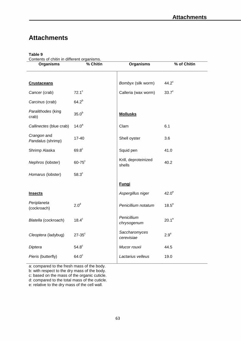
Attachments
63
Attachments
Table 9 Contents of chitin in different organisms.
Organisms % Chitin Organisms % of Chitin
Crustaceans
Bombyx (silk worm)
44.2c
Cancer (crab) 72.1c Calleria (wax worm) 33.7
c
Carcinus (crab) 64.2b
Paralithodes (king
crab) 35.0
b Mollusks
Callinectes (blue crab) 14.0a Clam 6.1
Crangon and
Pandalus (shrimp) 17-40 Shell oyster 3.6
Shrimp Alaska 69.8c Squid pen 41.0
Nephros (lobster) 60-75c
Krill, deproteinized
shells 40.2
Homarus (lobster) 58.3c
Fungi
Insects Aspergillus niger 42.0e
Periplaneta
(cockroach) 2.0
d Penicillium notatum 18.5
e
Blatella (cockroach) 18.4c
Penicillium
chrysogenum 20.1
e
Cleoptera (ladybug) 27-35c
Saccharomyces
cerevisiae 2.9
e
Diptera 54.8c Mucor rouxii 44.5
Pieris (butterfly) 64.0c Lactarius velleus 19.0
a: compared to the fresh mass of the body. b: with respect to the dry mass of the body. c: based on the mass of the organic cuticle. d: compared to the total mass of the cuticle. e: relative to the dry mass of the cell wall.
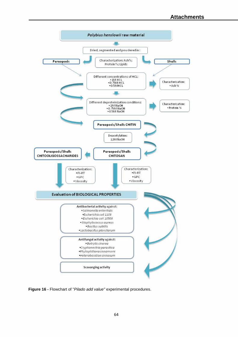
Attachments
64
Figure 16 - Flowchart of "Pilado add value" experimental procedures.
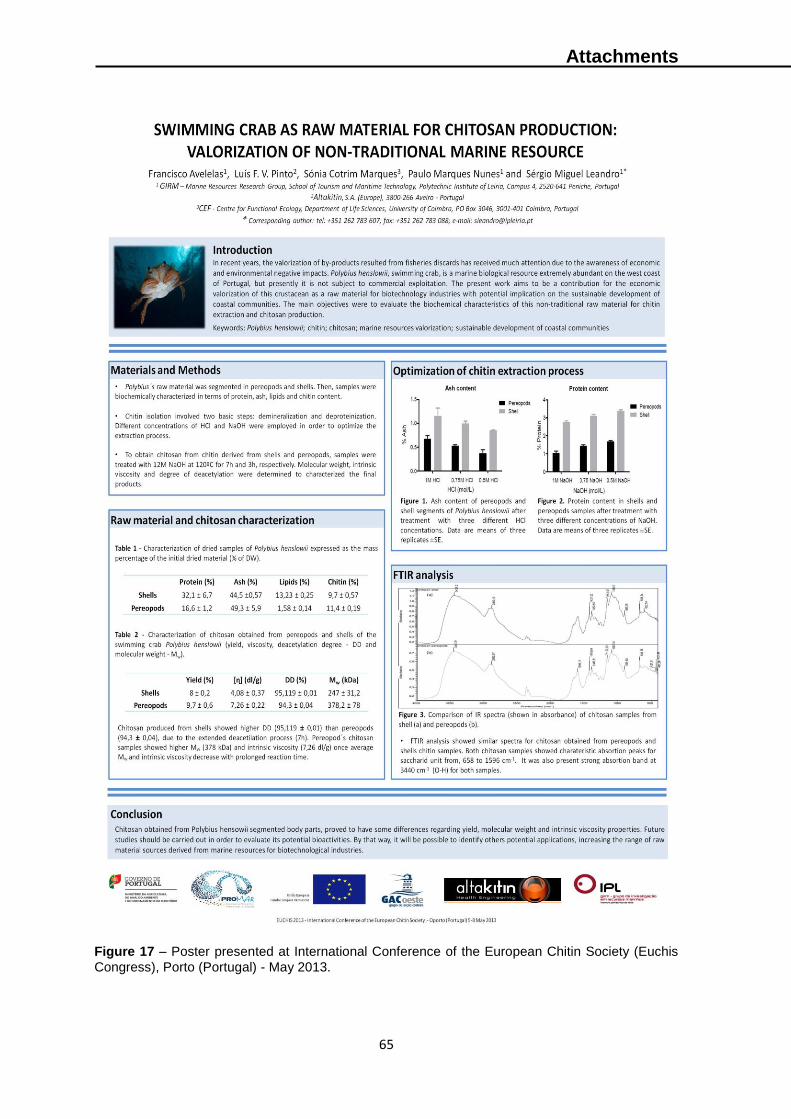
Attachments
65
Figure 17 – Poster presented at International Conference of the European Chitin Society (Euchis
Congress), Porto (Portugal) - May 2013.

Attachments
66
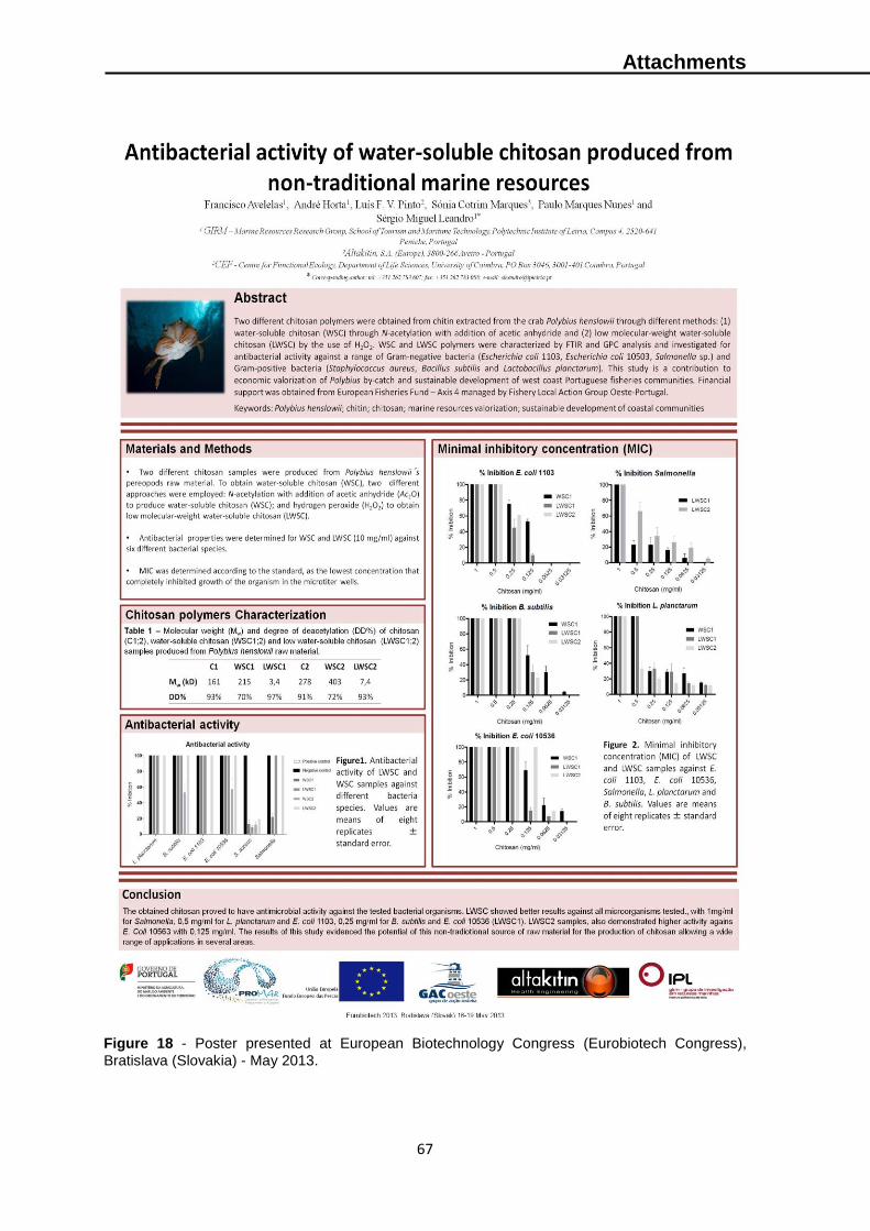
Attachments
67
Figure 18 - Poster presented at European Biotechnology Congress (Eurobiotech Congress),
Bratislava (Slovakia) - May 2013.
Attachments
68