Embed Size (px)
Citation preview
Place Cells and Place RecognitionMaintained by Direct
Entorhinal-Hippocampal Circuitry
Vegard H. Burn, Mona K. Otnaess, Sturla Molden, Hill-Aina Steffenach, Menno P. Witter, May-Britt Moser, Ed
vard I. Moser
Science VOL 296 , 21 June 2002
Presented by Min-Yu Sun
Department of Life Science
Outline• Background Introduction• Material and Methods• Hypothesis• Material and Methods• Conclusion I• Material and Methods• Conclusion II• Material and Methods• Conclusion III• Summary
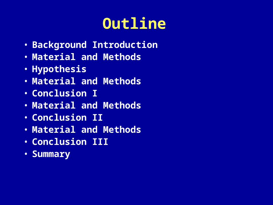
Background Introduction
• Hippocampus : a cognitive ( 認知 ) map
• Place cell: Hippocampal principal neurons,
exhibit location-specific firing.
• Place field
• CA1
• CA3
• Entorhinal cortex
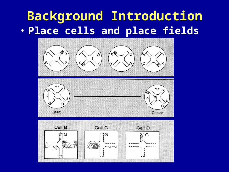
Background Introduction• Place cells and place fields
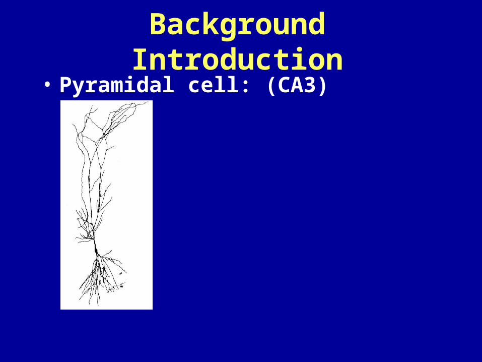
Background Introduction• Pyramidal cell: (CA3)
Background Introduction
• Hippocampus : a cognitive ( 認知 ) map
• Place cell: Hippocampal principal neurons,
exhibit location-specific firing.
• Place field
• CA1
• CA3
• Entorhinal cortex
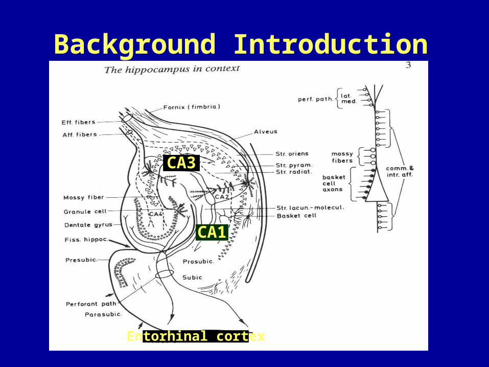
Background Introduction
CA1
CA3
Entorhinal cortex
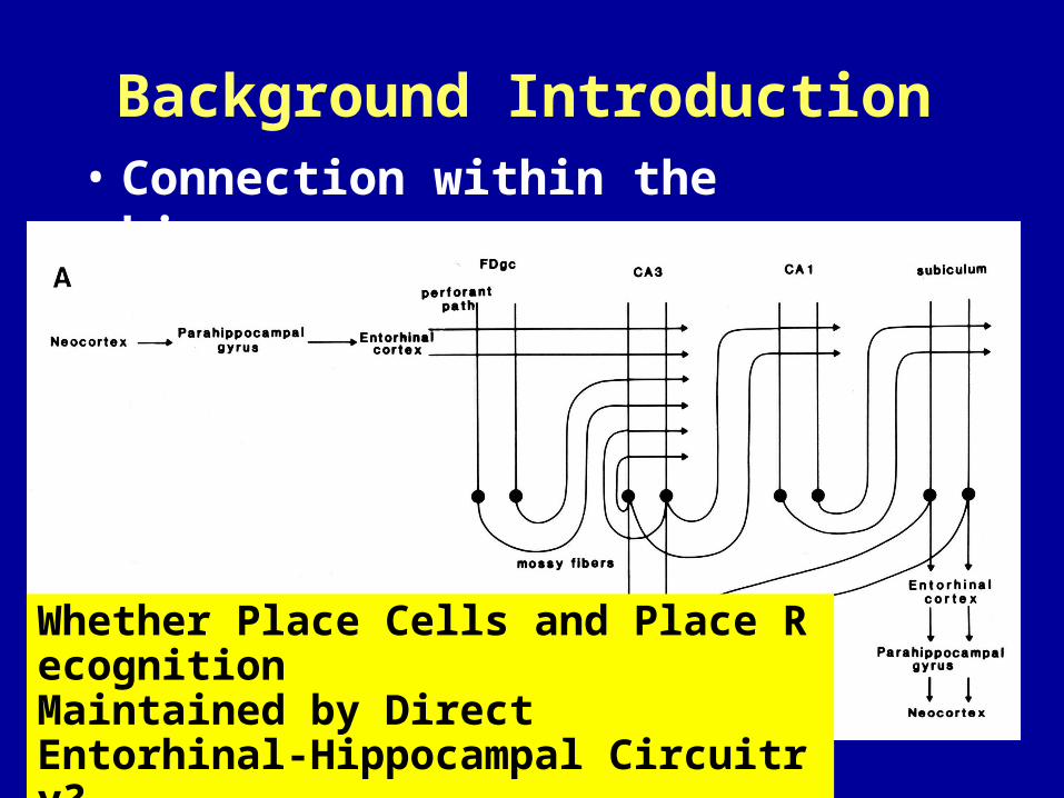
Background Introduction• Connection within the hippocampus
Whether Place Cells and Place RecognitionMaintained by DirectEntorhinal-Hippocampal Circuitry?
Material and Methods
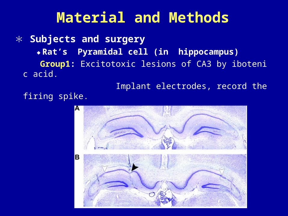
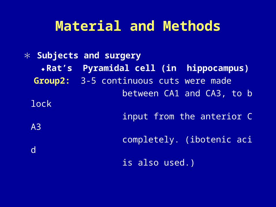
* Subjects and surgery ◆Rat’s Pyramidal cell (in hippocampus)
Group1: Excitotoxic lesions of CA3 by ibotenic acid.
Implant electrodes, record the firing spike.
Material and Methods
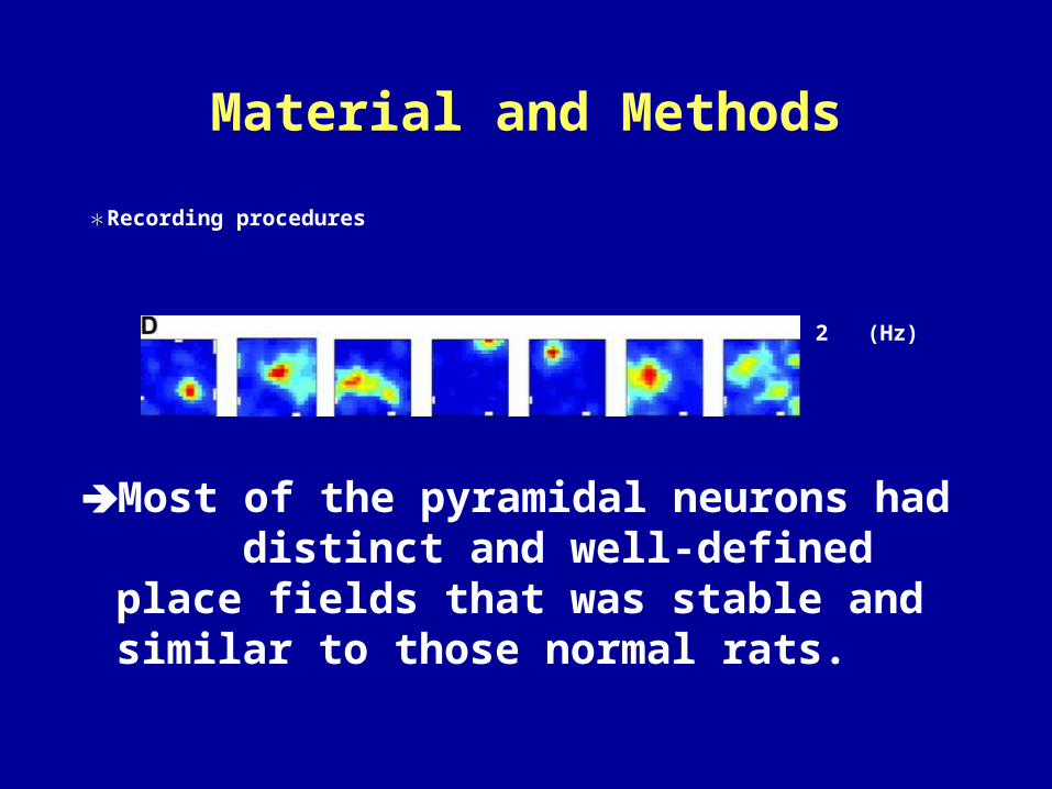
*Recording procedures
9 11 3 12 6 11 2 (Hz)
Most of the pyramidal neurons had distinct and well-defined place fields that was stable and similar to those normal rats.
Hypothesis
• Area CA3 may not be necessary for establishing and maintaining place fields in area CA1
• That spatial information from the neocortex may reach the hippocampus primarily through the alternative route: the direct pathway from layer III of the entorhinal cortex.
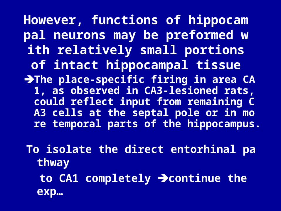
However, functions of hippocampal neurons may be preformed with relatively small
portions of intact hippocampal tissue
The place-specific firing in area CA1, as observed in CA3-lesioned rats, could reflect input from remaining CA3 cells at the septal pole or in more temporal parts of the hippocampus.
To isolate the direct entorhinal pathway
to CA1 completely continue the exp…
Material and Methods
* Subjects and surgery
◆Rat’s Pyramidal cell (in hippocampus)
Group2: 3-5 continuous cuts were made
between CA1 and CA3, to block
input from the anterior CA3
completely. (ibotenic acid
is also used.)
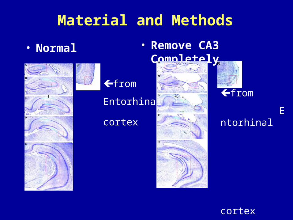
Material and Methods
• Remove CA3 Completely• Normal
from
Entorhinal
cortex
from
Entorhinal
cortex
Material and Methods
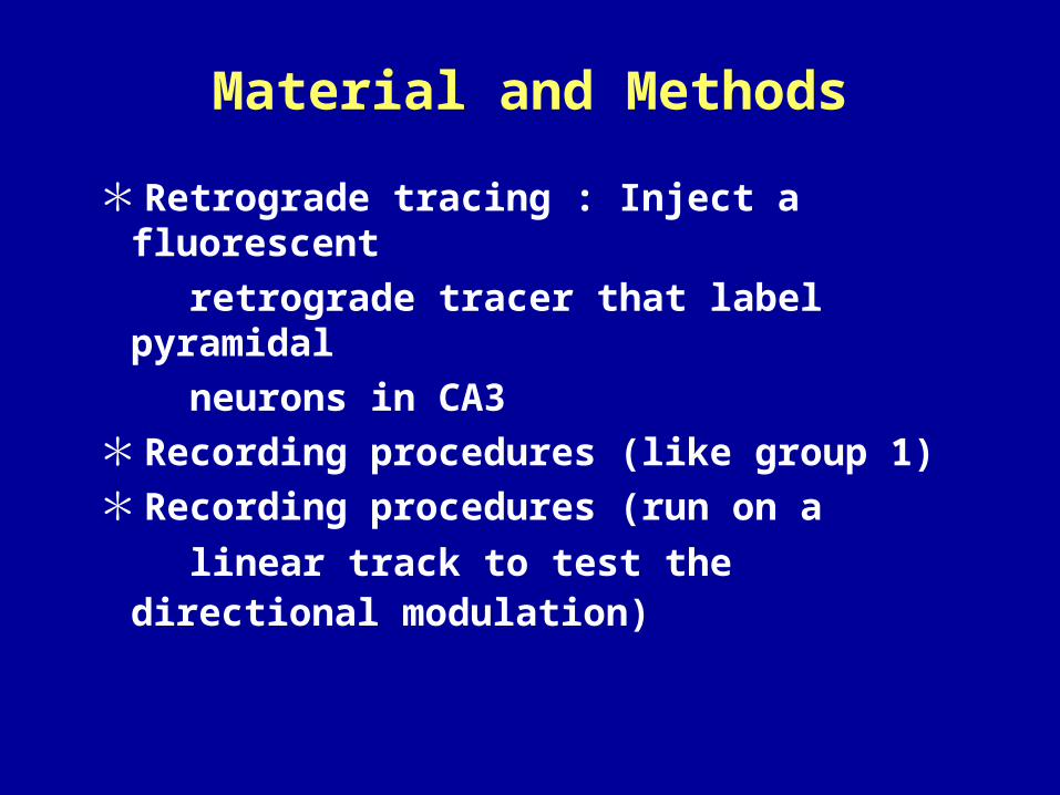
* Retrograde tracing : Inject a fluorescent
retrograde tracer that label pyramidal
neurons in CA3
* Recording procedures (like group 1)
* Recording procedures (run on a
linear track to test the directional modulation)
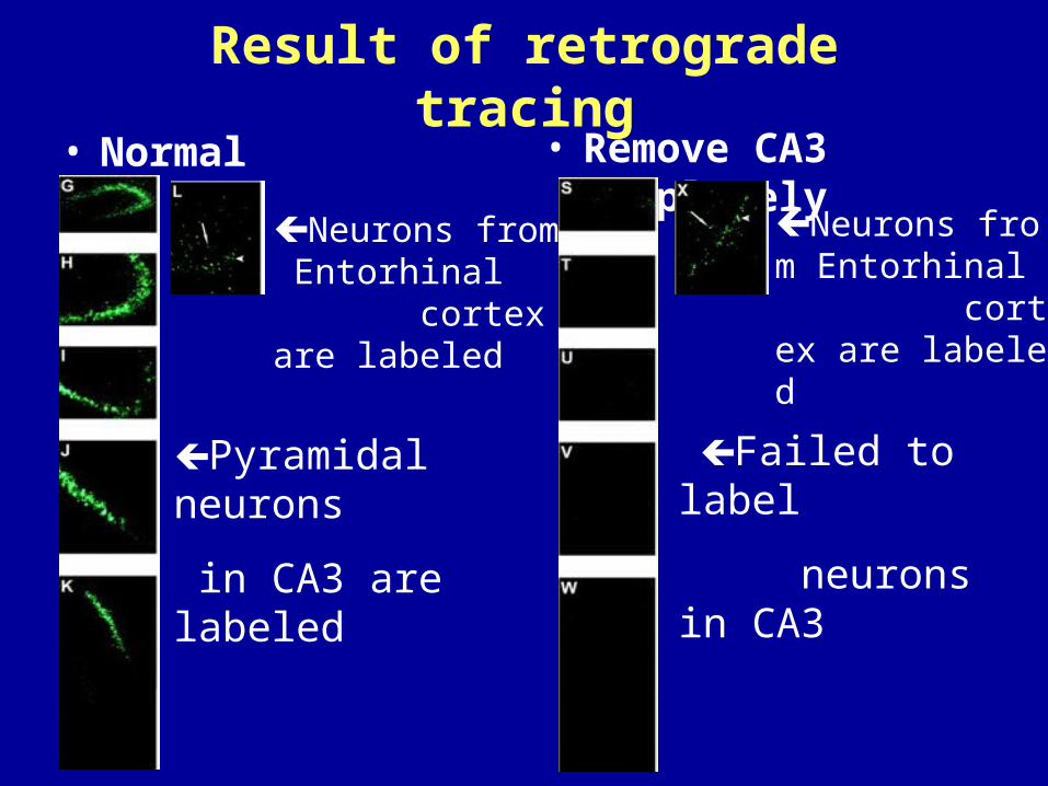
Result of retrograde tracing• Normal • Remove CA3 Completely
Neurons from Entorhinal cortex are labeled
Pyramidal neurons
in CA3 are labeled
Failed to label
neurons in CA3
Neurons from Entorhinal cortex are labeled
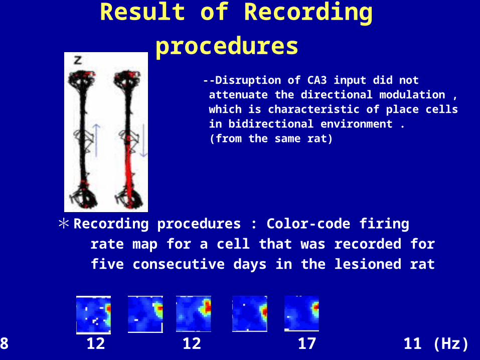
Result of Recording procedures
--Disruption of CA3 input did not attenuate the directional modulation , which is characteristic of place cells in bidirectional environment . (from the same rat)
* Recording procedures : Color-code firing
rate map for a cell that was recorded for
five consecutive days in the lesioned rat
8 12 12 17 11 (Hz)
Conclusion I
• Area CA3 may not be necessary for establishing and maintaining place fields in area CA1
• That spatial information from the neocortex may reach the hippocampus primarily through the alternative route: the direct pathway from layer III of the entorhinal cortex.
Discussion
• Whether removal of CA3 input had more subtle effects on place cells in area CA1?
Continue the exp to “Quantitative
description of place fields”…..
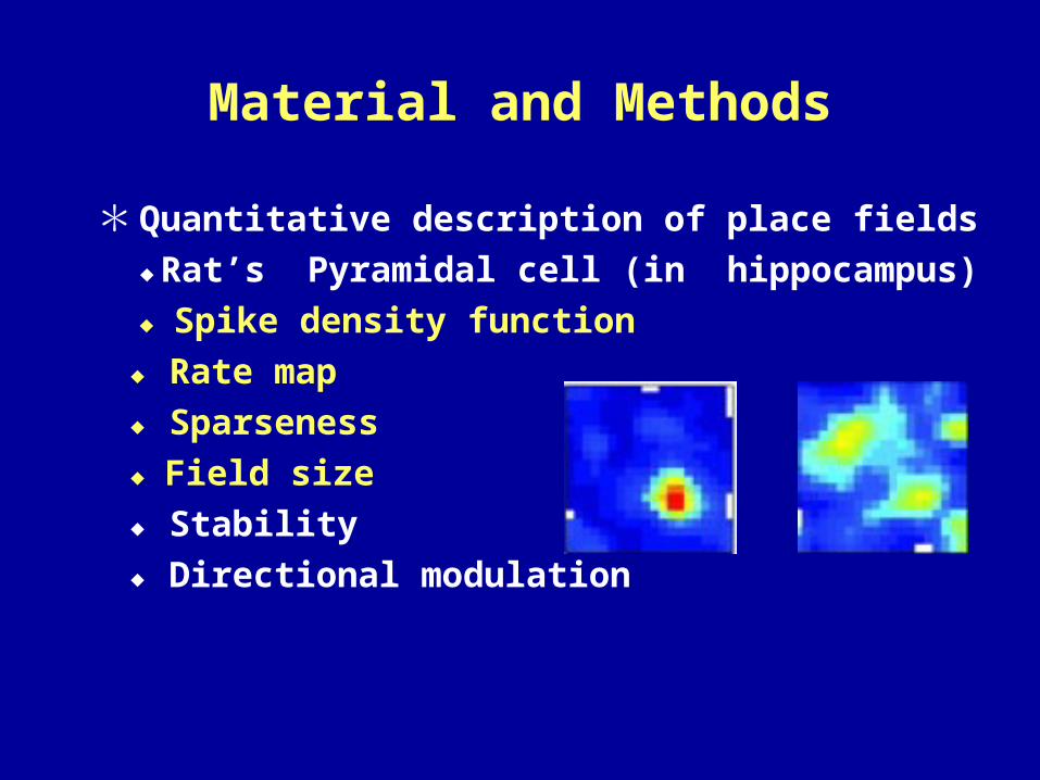
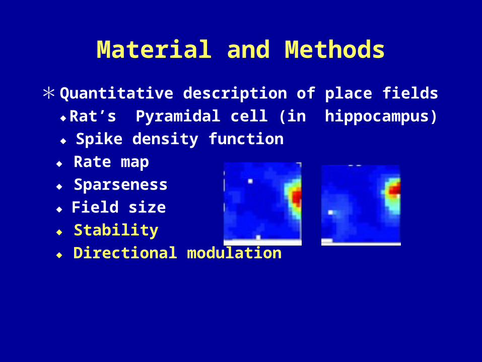
Material and Methods
* Quantitative description of place fields
◆Rat’s Pyramidal cell (in hippocampus)
◆ Spike density function
◆ Rate map
◆ Sparseness
◆ Field size
◆ Stability
◆ Directional modulation
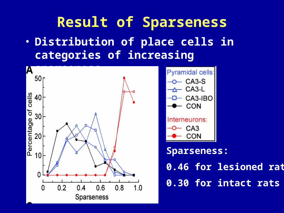
Result of Sparseness• Distribution of place cells in categories of
increasing sparseness
Sparseness:
0.46 for lesioned rats
0.30 for intact rats
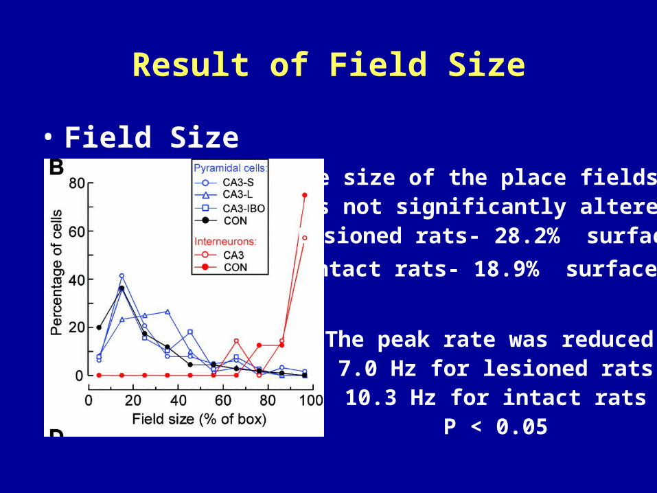
Result of Field Size
• Field SizeThe size of the place fields
was not significantly altered:Lesioned rats- 28.2% surface
Intact rats- 18.9% surface
The peak rate was reduced:7.0 Hz for lesioned rats10.3 Hz for intact rats
P < 0.05
Results of Sparseness and Field Size
• The result was independent of the type of CA3 lesion.
• These effects were small compared to the differences between the firing fields of pyramidal cells and interneurons.
Material and Methods
* Quantitative description of place fields
◆Rat’s Pyramidal cell (in hippocampus)
◆ Spike density function
◆ Rate map
◆ Sparseness
◆ Field size
◆ Stability
◆ Directional modulation
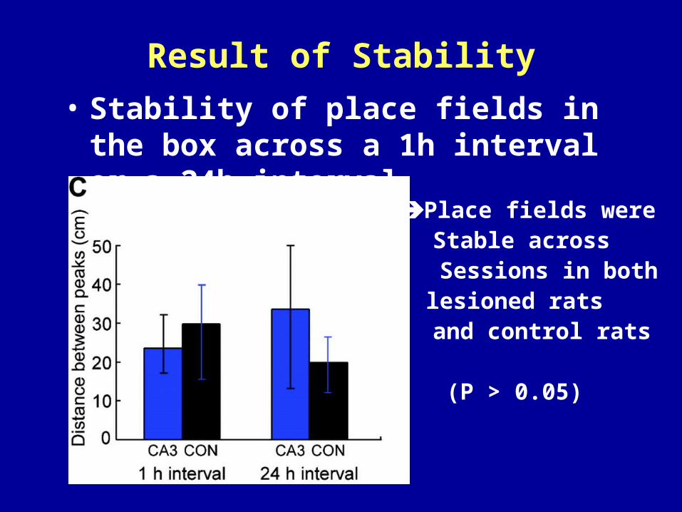
Result of Stability
• Stability of place fields in the box across a 1h interval or a 24h interval
Place fields were Stable across
Sessions in both lesioned rats
and control rats
(P > 0.05)
Result of Stability
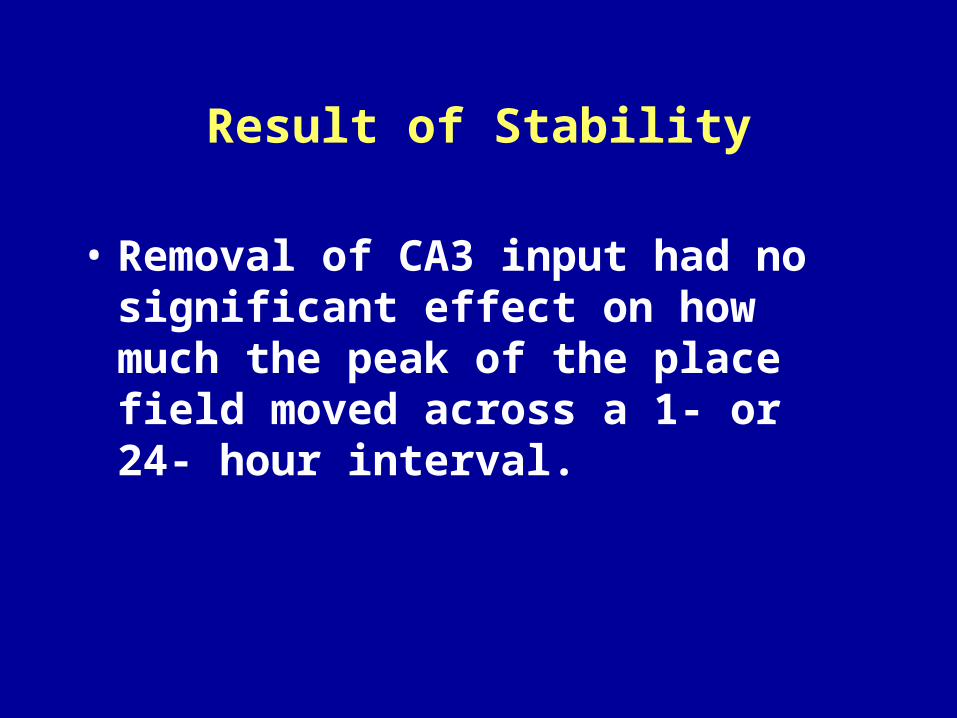
• Removal of CA3 input had no significant effect on how much the peak of the place field moved across a 1- or 24- hour interval.
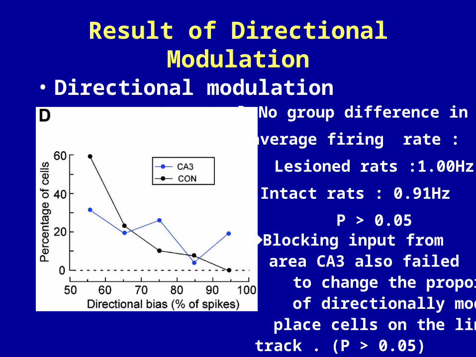
Result of Directional Modulation
• Directional modulation
Blocking input from area CA3 also failed
to change the proportion of directionally modulated
place cells on the linear track . (P > 0.05)
No group difference in
average firing rate :
Lesioned rats :1.00Hz
Intact rats : 0.91Hz
P > 0.05
Conclusion II• The direct pathway from the entorhinal corte
x thus seems to be sufficient for establishing and maintaining fundamental properties of place cells in area CA1
Discussion: Whether the reduced circuitry also
supported memory?
…… continue the exp
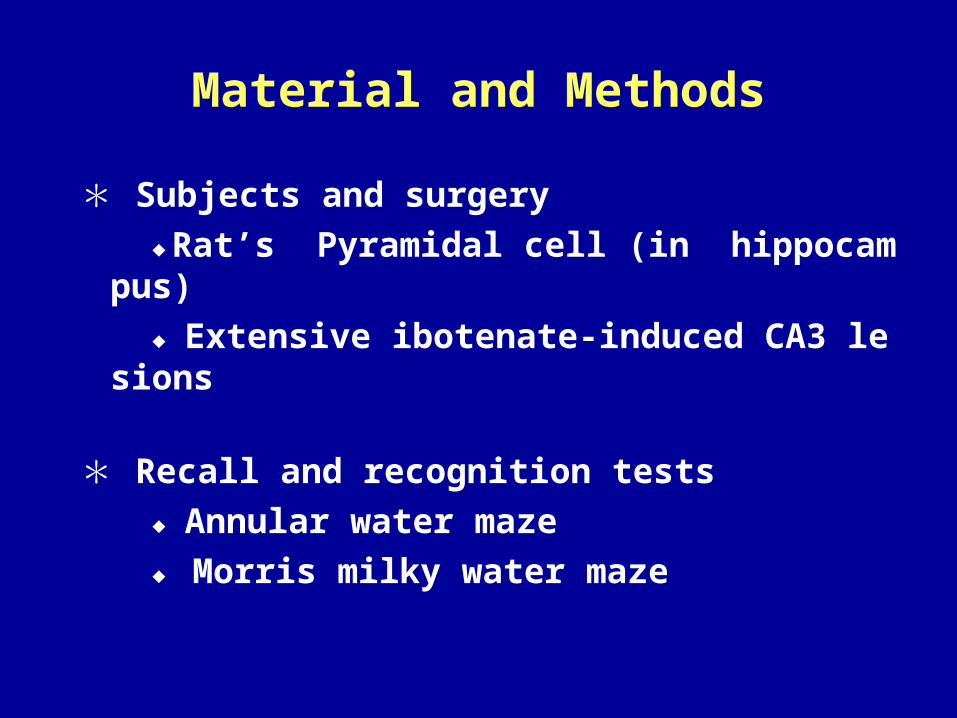
Material and Methods
* Subjects and surgery
◆Rat’s Pyramidal cell (in hippocampus)
◆Extensive ibotenate-induced CA3 lesions
* Recall and recognition tests
◆Annular water maze
◆ Morris milky water maze
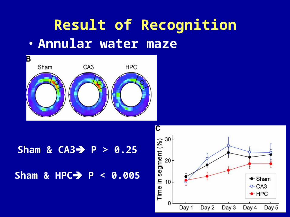
Result of Recognition• Annular water maze
Sham & CA3 P > 0.25
Sham & HPC P < 0.005
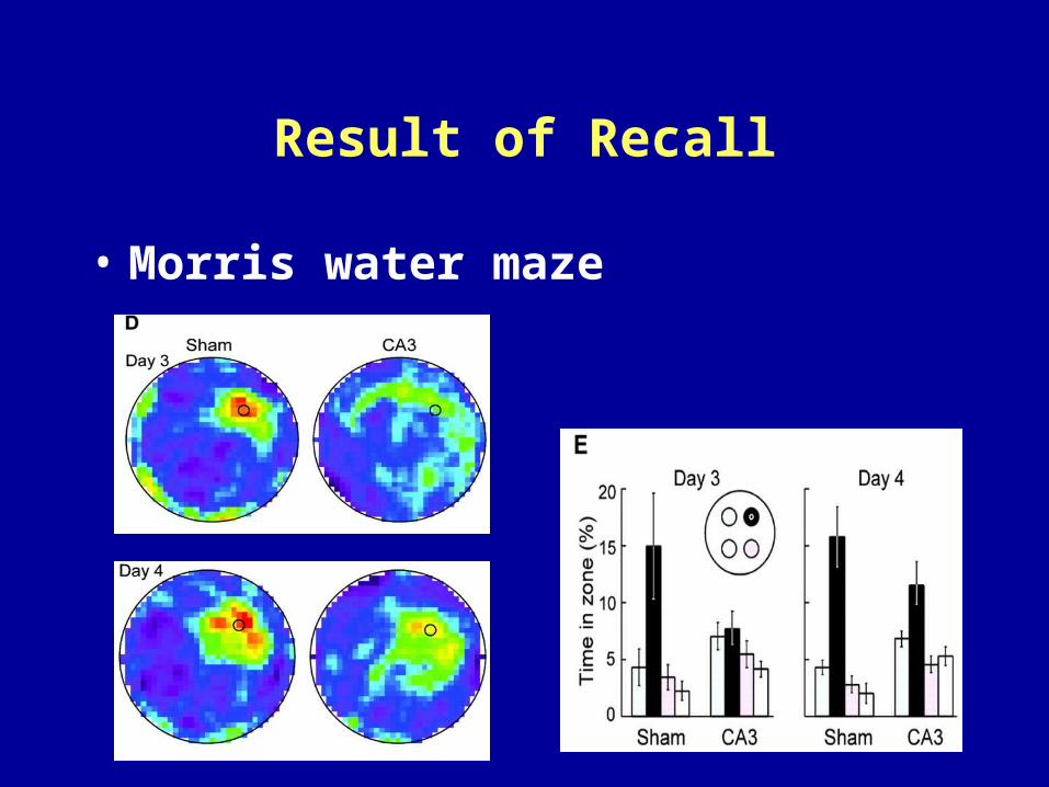
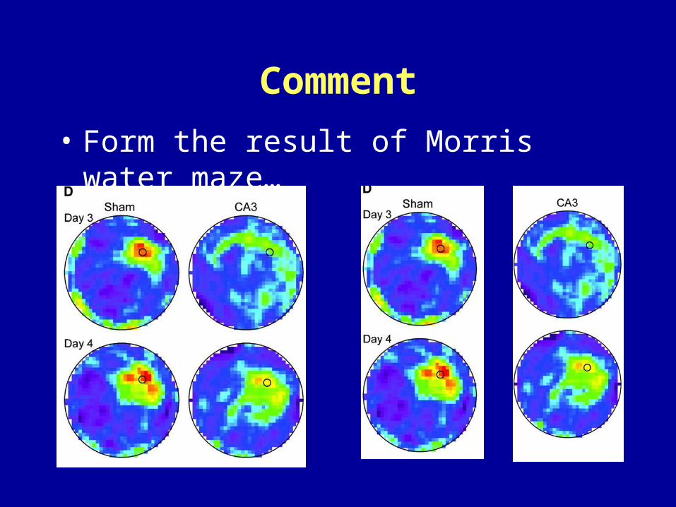
Result of Recall
• Morris water maze
Conclusion III
• Spatial recognition memory is fully achievable with an isolated entorhinal-CA1 network.
• The isolated entorhinal-CA1 circuit does not support recall of remote locations or trajectories toward these locations.
Summary
• Direct entorhinal-hippocampal connections have significant capacity for transforming weak location-modulated signals.
• The isolated entorhinal-CA1 circuit does not support recall of remote locations or trajectories toward these locations.

Summary• These results suggest that the hippocampus
contains two functionally separable memory circuits:The direct entorhinal-CA1 system is sufficient for recollection-based recognition memory, but recall depends on intact CA3-CA1 connectivity.
Comment
• Form the result of Morris water maze…


![Molden 2.0: quantum chemistry meets proteins · 2017. 10. 9. · In his landmark article on the free radical catalysis by galactose oxidase, Whittaker [30] used Molden to calculate](https://img.pdfslide.net/doc/110x75/60a8a1e8d1e5314e770e31b0/molden-20-quantum-chemistry-meets-proteins-2017-10-9-in-his-landmark-article.jpg)