Embed Size (px)
Citation preview
Plant cyclotides disrupt epithelial cells in the midgutof lepidopteran larvaeBarbara L. Barbeta*, Alan T. Marshall†‡, Amanda D. Gillon*, David J. Craik§, and Marilyn A. Anderson*¶
Departments of *Biochemistry and †Zoology, and ‡Analytical Electron Microscopy Laboratory, La Trobe University, Melbourne, Victoria 3086, Australia;and §Institute for Molecular Bioscience, University of Queensland, Brisbane, Queensland 4072, Australia
Communicated by Adrienne Clarke, University of Melbourne, Victoria, Australia, October 30, 2007 (received for review February 1, 2007)
Several members of the Rubiaceae and Violaceae plant familiesproduce a series of cyclotides or macrocyclic peptides of 28–37 aawith an embedded cystine knot. The cyclic peptide backbonetogether with the knotted and strongly braced structure confersexceptional chemical and biological stability that has attractedattention for potential pharmaceutical applications. Cyclotidesdisplay a diverse range of biological activities, such as uterotonicaction, anti-HIV activity, and neurotensin antagonism. In plants,their primary role is probably protection from insect attack. Inges-tion of the cyclotide kalata B1 severely retards the growth of larvaefrom the Lepidopteran species Helicoverpa armigera. We examinedthe gut of these larvae after consumption of kalata B1 by light,scanning, and transmission electron microscopy. We establishedthat kalata B1 induces disruption of the microvilli, blebbing, swell-ing, and ultimately rupture of the cells of the gut epithelium. Thehistology of this response is similar to the response of H. armigeralarvae to the Bacillus thuringiensis delta-endotoxin, which iswidely used to control these insect pests of crops such as cotton.
circular peptides � insecticidal � kalata B1 � Lepidoptera � microscopy
Cyclotides are a series of cyclic minipeptides of 28–37 aa thatare expressed at high levels in the leaves, stems, and roots of
several plant species (1, 2). They have an unusual topology basedon a cyclic peptide backbone together with a cystine knot inwhich two disulfide bonds are threaded by a third disulfide bond(2, 3). This structure forces hydrophobic amino acids onto thesurface of the molecule, creating a hydrophobic face on theotherwise hydrophilic surface. The cyclotides are thus soluble inboth organic and aqueous solvents and are very stable atextremes of pH and temperature (4). The absence of free N andC termini, together with the tightly packed cystine knot, alsorenders the cyclotides resistant to the activity of proteases (4).The cyclotides are the largest family of circular proteins, al-though others have been described in bacteria, plants, andanimals (5).
Cyclotides were first isolated from the African plant Olden-landia affinis. Two peptides, kalata B1 and B2, were recognizedas the active components in a traditional medicine used toaccelerate childbirth (6). Additional members of the cyclotidefamily were subsequently identified in screening studies directedtoward discovery of bioactive molecules such as neurotensinantagonists (7), inhibitors of HIV replication (8), and hemolyticagents (9). More than 100 cyclotides have since been isolatedfrom various members of the Rubiaceae, Violaceae, and Cucur-bitaceae plant families (10, 11). A single plant may have at least12 cyclotide genes and produce dozens of different cyclotides(12–14). Although an earlier study reported antimicrobial activ-ity (15), it appears that their predominant activity in plants isinsecticidal (13, 16). Jennings et al. (13) demonstrated thatHelicoverpa punctigera failed to develop past the second instarstage when raised on artificial diets containing the cyclotidekalata B1 (13). Similar observations of severe growth retardationwere also observed with Helicoverpa armigera after ingestion ofthe kalata B1, B2, and B5 cyclotides (16). Those studies estab-lished that the cyclotides did not affect the activity of larval
digestive enzymes such as �-amylase, trypsin, or chymotrypsin,but did not establish whether the growth retardant effect wascaused by an antifeedant or toxic effect (13).
In the current study, we describe the effect of the cyclotidekalata B1 on the morphology of midgut epithelial cells of H.armigera larvae. Swelling and lysis of the columnar and gobletcells was evident at both light and electron microscopic levels,providing an explanation for the insecticidal activity of cyclotidesagainst lepidopteran pests. The changes in morphology observedresemble the substantial changes to the insect midgut that areinduced by the delta-endotoxins from Bacillus thuringiensis (17).
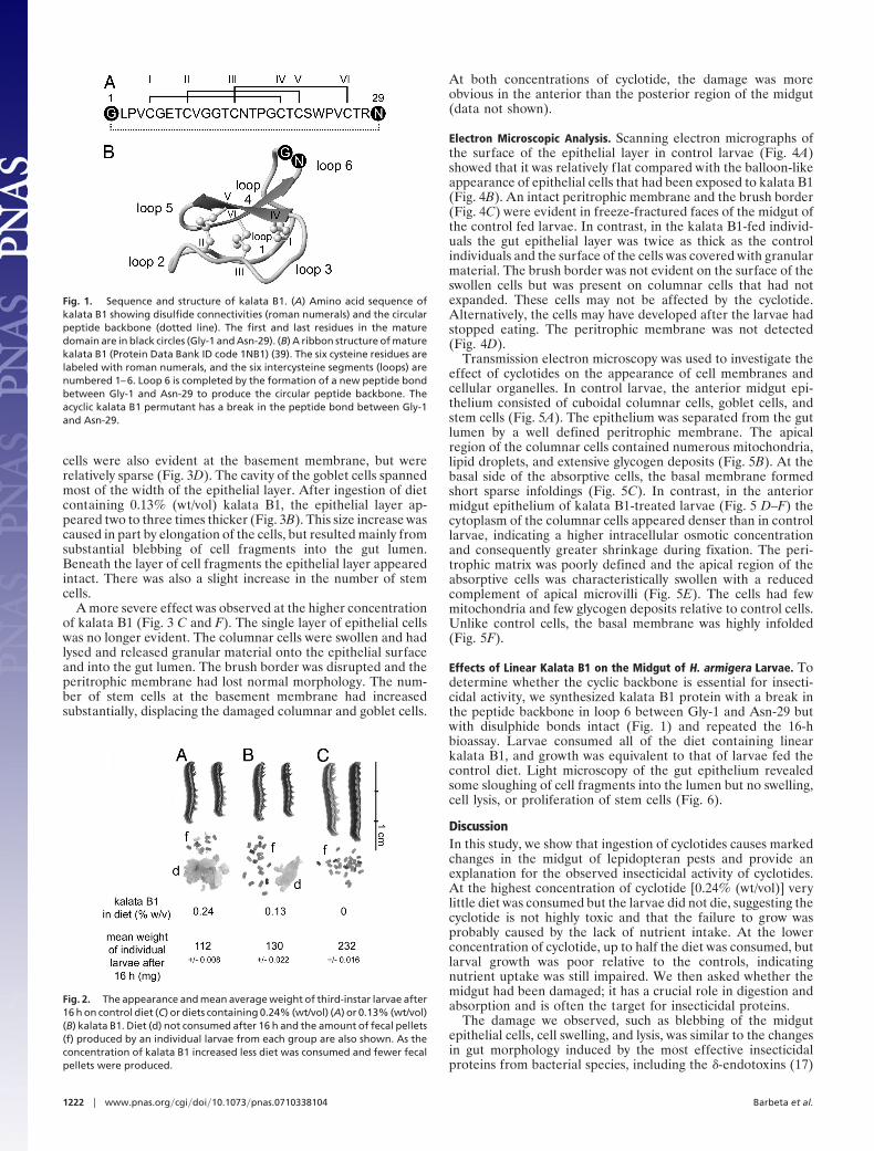
ResultsThe sequence and structure of kalata B1 are illustrated in Fig.1. The 29-aa peptide is excised from the 11-kDa precursorprotein Oak1 (13) and ligation of Gly-1 and Asn-29 results incyclization, as indicated in Fig. 1. Because of this processingmechanism, it was of interest to compare the insecticidal activityof the corresponding synthetic linear derivative of kalata B1 withthat of the natural cyclic molecule. The linear derivative ishereafter referred to as linear B1.
Effects of Kalata B1 on Growth of H. armigera. Third-instar larvaewere starved for 6 h before they were placed on an artificial dietcontaining kalata B1 [0.13% (wt/vol) and 0.24% (wt/vol)] orcasein as a control. Consumption of diet and production of fecalpellets were monitored. During the 16-h bioassay, individualswithin the control group fed continuously, consuming all 330 mgof diet. In contrast, larvae on the kalata B1 diets fed for a shortperiod and moved away from the diet. Fig. 2 shows the meanweight of individuals from each of the three diet groups and theamount of diet remaining at the end of the bioassay and the fecalpellets produced. Control larvae doubled in size over the 16-hbioassay. Larvae on the diet with the highest concentration ofkalata B1 [0.24% (wt/vol)] did not grow, consumed very little,and produced few fecal pellets. Larvae exposed to the lowerconcentration of kalata B1 were only 16% larger than larvae fedon the highest concentration of kalata B1 even though they hadconsumed about half the diet and produced an intermediateamount of fecal pellets relative to the other two groups.
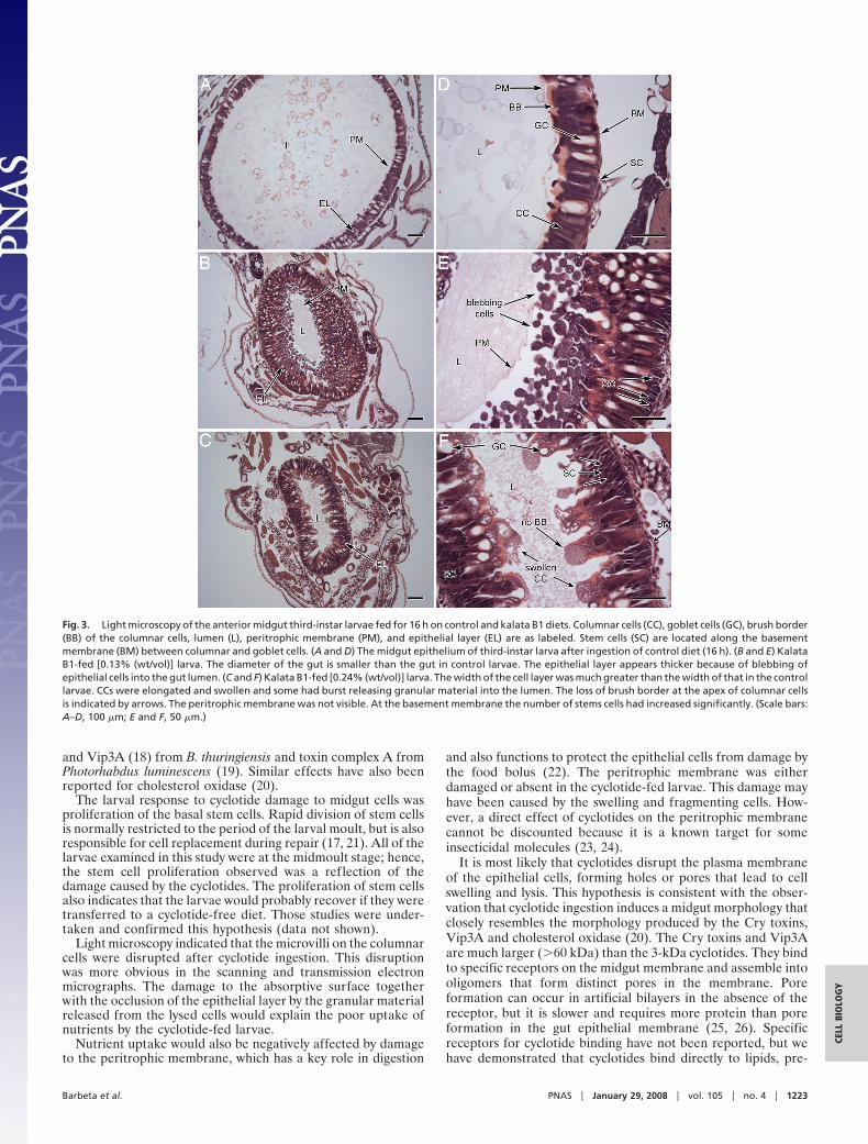
Effects of Kalata B1 on the Midgut of H. armigera Larvae. Thediameter of the gut in the control larvae (Fig. 3A) was approx-imately three times larger than the gut of larvae that had ingestedkalata B1 [0.13% (wt/vol) or 0.24% (wt/vol)], reflecting theimpact of kalata B1 on larval growth during the 16-h bioassay.Exposure to kalata B1 also led to substantial thickening of theepithelial layer (Fig. 3 B and C). In control larvae, this layerconsisted of a single layer of columnar and goblet cells. Stem
Author contributions: B.L.B. and M.A.A. designed research; B.L.B. and A.T.M. performedresearch; D.J.C. contributed new reagents/analytic tools; B.L.B., A.T.M., A.D.G., and M.A.A.analyzed data; and B.L.B., A.D.G., and M.A.A. wrote the paper.
The authors declare no conflict of interest.
¶To whom correspondence should be addressed. E-mail: [email protected].
© 2008 by The National Academy of Sciences of the USA
www.pnas.org�cgi�doi�10.1073�pnas.0710338104 PNAS � January 29, 2008 � vol. 105 � no. 4 � 1221–1225
CELL
BIO
LOG
Y
cells were also evident at the basement membrane, but wererelatively sparse (Fig. 3D). The cavity of the goblet cells spannedmost of the width of the epithelial layer. After ingestion of dietcontaining 0.13% (wt/vol) kalata B1, the epithelial layer ap-peared two to three times thicker (Fig. 3B). This size increase wascaused in part by elongation of the cells, but resulted mainly fromsubstantial blebbing of cell fragments into the gut lumen.Beneath the layer of cell fragments the epithelial layer appearedintact. There was also a slight increase in the number of stemcells.
A more severe effect was observed at the higher concentrationof kalata B1 (Fig. 3 C and F). The single layer of epithelial cellswas no longer evident. The columnar cells were swollen and hadlysed and released granular material onto the epithelial surfaceand into the gut lumen. The brush border was disrupted and theperitrophic membrane had lost normal morphology. The num-ber of stem cells at the basement membrane had increasedsubstantially, displacing the damaged columnar and goblet cells.
At both concentrations of cyclotide, the damage was moreobvious in the anterior than the posterior region of the midgut(data not shown).
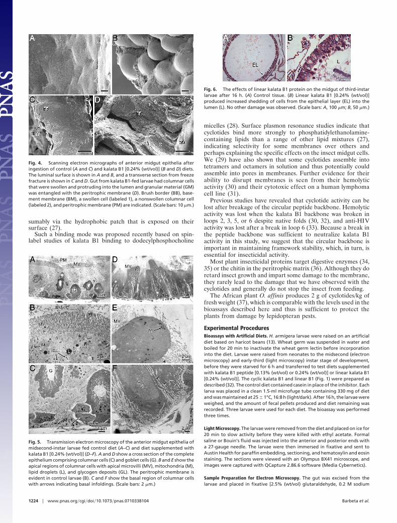
Electron Microscopic Analysis. Scanning electron micrographs ofthe surface of the epithelial layer in control larvae (Fig. 4A)showed that it was relatively flat compared with the balloon-likeappearance of epithelial cells that had been exposed to kalata B1(Fig. 4B). An intact peritrophic membrane and the brush border(Fig. 4C) were evident in freeze-fractured faces of the midgut ofthe control fed larvae. In contrast, in the kalata B1-fed individ-uals the gut epithelial layer was twice as thick as the controlindividuals and the surface of the cells was covered with granularmaterial. The brush border was not evident on the surface of theswollen cells but was present on columnar cells that had notexpanded. These cells may not be affected by the cyclotide.Alternatively, the cells may have developed after the larvae hadstopped eating. The peritrophic membrane was not detected(Fig. 4D).
Transmission electron microscopy was used to investigate theeffect of cyclotides on the appearance of cell membranes andcellular organelles. In control larvae, the anterior midgut epi-thelium consisted of cuboidal columnar cells, goblet cells, andstem cells (Fig. 5A). The epithelium was separated from the gutlumen by a well defined peritrophic membrane. The apicalregion of the columnar cells contained numerous mitochondria,lipid droplets, and extensive glycogen deposits (Fig. 5B). At thebasal side of the absorptive cells, the basal membrane formedshort sparse infoldings (Fig. 5C). In contrast, in the anteriormidgut epithelium of kalata B1-treated larvae (Fig. 5 D–F) thecytoplasm of the columnar cells appeared denser than in controllarvae, indicating a higher intracellular osmotic concentrationand consequently greater shrinkage during fixation. The peri-trophic matrix was poorly defined and the apical region of theabsorptive cells was characteristically swollen with a reducedcomplement of apical microvilli (Fig. 5E). The cells had fewmitochondria and few glycogen deposits relative to control cells.Unlike control cells, the basal membrane was highly infolded(Fig. 5F).
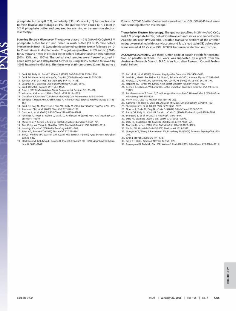
Effects of Linear Kalata B1 on the Midgut of H. armigera Larvae. Todetermine whether the cyclic backbone is essential for insecti-cidal activity, we synthesized kalata B1 protein with a break inthe peptide backbone in loop 6 between Gly-1 and Asn-29 butwith disulphide bonds intact (Fig. 1) and repeated the 16-hbioassay. Larvae consumed all of the diet containing linearkalata B1, and growth was equivalent to that of larvae fed thecontrol diet. Light microscopy of the gut epithelium revealedsome sloughing of cell fragments into the lumen but no swelling,cell lysis, or proliferation of stem cells (Fig. 6).
DiscussionIn this study, we show that ingestion of cyclotides causes markedchanges in the midgut of lepidopteran pests and provide anexplanation for the observed insecticidal activity of cyclotides.At the highest concentration of cyclotide [0.24% (wt/vol)] verylittle diet was consumed but the larvae did not die, suggesting thecyclotide is not highly toxic and that the failure to grow wasprobably caused by the lack of nutrient intake. At the lowerconcentration of cyclotide, up to half the diet was consumed, butlarval growth was poor relative to the controls, indicatingnutrient uptake was still impaired. We then asked whether themidgut had been damaged; it has a crucial role in digestion andabsorption and is often the target for insecticidal proteins.
The damage we observed, such as blebbing of the midgutepithelial cells, cell swelling, and lysis, was similar to the changesin gut morphology induced by the most effective insecticidalproteins from bacterial species, including the �-endotoxins (17)
Fig. 1. Sequence and structure of kalata B1. (A) Amino acid sequence ofkalata B1 showing disulfide connectivities (roman numerals) and the circularpeptide backbone (dotted line). The first and last residues in the maturedomain are in black circles (Gly-1 and Asn-29). (B) A ribbon structure of maturekalata B1 (Protein Data Bank ID code 1NB1) (39). The six cysteine residues arelabeled with roman numerals, and the six intercysteine segments (loops) arenumbered 1–6. Loop 6 is completed by the formation of a new peptide bondbetween Gly-1 and Asn-29 to produce the circular peptide backbone. Theacyclic kalata B1 permutant has a break in the peptide bond between Gly-1and Asn-29.
Fig. 2. The appearance and mean average weight of third-instar larvae after16 h on control diet (C) or diets containing 0.24% (wt/vol) (A) or 0.13% (wt/vol)(B) kalata B1. Diet (d) not consumed after 16 h and the amount of fecal pellets(f) produced by an individual larvae from each group are also shown. As theconcentration of kalata B1 increased less diet was consumed and fewer fecalpellets were produced.
1222 � www.pnas.org�cgi�doi�10.1073�pnas.0710338104 Barbeta et al.
and Vip3A (18) from B. thuringiensis and toxin complex A fromPhotorhabdus luminescens (19). Similar effects have also beenreported for cholesterol oxidase (20).
The larval response to cyclotide damage to midgut cells wasproliferation of the basal stem cells. Rapid division of stem cellsis normally restricted to the period of the larval moult, but is alsoresponsible for cell replacement during repair (17, 21). All of thelarvae examined in this study were at the midmoult stage; hence,the stem cell proliferation observed was a reflection of thedamage caused by the cyclotides. The proliferation of stem cellsalso indicates that the larvae would probably recover if they weretransferred to a cyclotide-free diet. Those studies were under-taken and confirmed this hypothesis (data not shown).
Light microscopy indicated that the microvilli on the columnarcells were disrupted after cyclotide ingestion. This disruptionwas more obvious in the scanning and transmission electronmicrographs. The damage to the absorptive surface togetherwith the occlusion of the epithelial layer by the granular materialreleased from the lysed cells would explain the poor uptake ofnutrients by the cyclotide-fed larvae.
Nutrient uptake would also be negatively affected by damageto the peritrophic membrane, which has a key role in digestion
and also functions to protect the epithelial cells from damage bythe food bolus (22). The peritrophic membrane was eitherdamaged or absent in the cyclotide-fed larvae. This damage mayhave been caused by the swelling and fragmenting cells. How-ever, a direct effect of cyclotides on the peritrophic membranecannot be discounted because it is a known target for someinsecticidal molecules (23, 24).
It is most likely that cyclotides disrupt the plasma membraneof the epithelial cells, forming holes or pores that lead to cellswelling and lysis. This hypothesis is consistent with the obser-vation that cyclotide ingestion induces a midgut morphology thatclosely resembles the morphology produced by the Cry toxins,Vip3A and cholesterol oxidase (20). The Cry toxins and Vip3Aare much larger (�60 kDa) than the 3-kDa cyclotides. They bindto specific receptors on the midgut membrane and assemble intooligomers that form distinct pores in the membrane. Poreformation can occur in artificial bilayers in the absence of thereceptor, but it is slower and requires more protein than poreformation in the gut epithelial membrane (25, 26). Specificreceptors for cyclotide binding have not been reported, but wehave demonstrated that cyclotides bind directly to lipids, pre-
Fig. 3. Light microscopy of the anterior midgut third-instar larvae fed for 16 h on control and kalata B1 diets. Columnar cells (CC), goblet cells (GC), brush border(BB) of the columnar cells, lumen (L), peritrophic membrane (PM), and epithelial layer (EL) are as labeled. Stem cells (SC) are located along the basementmembrane (BM) between columnar and goblet cells. (A and D) The midgut epithelium of third-instar larva after ingestion of control diet (16 h). (B and E) KalataB1-fed [0.13% (wt/vol)] larva. The diameter of the gut is smaller than the gut in control larvae. The epithelial layer appears thicker because of blebbing ofepithelial cells into the gut lumen. (C and F) Kalata B1-fed [0.24% (wt/vol)] larva. The width of the cell layer was much greater than the width of that in the controllarvae. CCs were elongated and swollen and some had burst releasing granular material into the lumen. The loss of brush border at the apex of columnar cellsis indicated by arrows. The peritrophic membrane was not visible. At the basement membrane the number of stems cells had increased significantly. (Scale bars:A–D, 100 �m; E and F, 50 �m.)
Barbeta et al. PNAS � January 29, 2008 � vol. 105 � no. 4 � 1223
CELL
BIO
LOG
Y
sumably via the hydrophobic patch that is exposed on theirsurface (27).
Such a binding mode was proposed recently based on spin-label studies of kalata B1 binding to dodecylphosphocholine
micelles (28). Surface plasmon resonance studies indicate thatcyclotides bind more strongly to phosphatidylethanolamine-containing lipids than a range of other lipid mixtures (27),indicating selectivity for some membranes over others andperhaps explaining the specific effects on the insect midgut cells.We (29) have also shown that some cyclotides assemble intotetramers and octamers in solution and thus potentially couldassemble into pores in membranes. Further evidence for theirability to disrupt membranes is seen from their hemolyticactivity (30) and their cytotoxic effect on a human lymphomacell line (31).
Previous studies have revealed that cyclotide activity can belost after breakage of the circular peptide backbone. Hemolyticactivity was lost when the kalata B1 backbone was broken inloops 2, 3, 5, or 6 despite native folds (30, 32), and anti-HIVactivity was lost after a break in loop 6 (33). Because a break inthe peptide backbone was sufficient to neutralize kalata B1activity in this study, we suggest that the circular backbone isimportant in maintaining framework stability, which, in turn, isessential for insecticidal activity.
Most plant insecticidal proteins target digestive enzymes (34,35) or the chitin in the peritrophic matrix (36). Although they doretard insect growth and impart some damage to the membrane,they rarely lead to the damage that we have observed with thecyclotides and generally do not stop the insect from feeding.
The African plant O. affinis produces 2 g of cyclotides/kg offresh weight (37), which is comparable with the levels used in thebioassays described here and thus is sufficient to protect theplants from damage by lepidopteran pests.
Experimental ProceduresBioassays with Artificial Diets. H. armigera larvae were raised on an artificialdiet based on haricot beans (13). Wheat germ was suspended in water andboiled for 20 min to inactivate the wheat germ lectin before incorporationinto the diet. Larvae were raised from neonates to the midsecond (electronmicroscopy) and early-third (light microscopy) instar stage of development,before they were starved for 6 h and transferred to test diets supplementedwith kalata B1 peptide [0.13% (wt/vol) or 0.24% (wt/vol)] or linear kalata B1[0.24% (wt/vol)]. The cyclic kalata B1 and linear B1 (Fig. 1) were prepared asdescribed (32). The control diet contained casein in place of the inhibitor. Eachlarva was placed in a clean 1.5-ml microfuge tube containing 330 mg of dietand was maintained at 25 � 1°C, 16:8 h (light/dark). After 16 h, the larvae wereweighed, and the amount of fecal pellets produced and diet remaining wasrecorded. Three larvae were used for each diet. The bioassay was performedthree times.
Light Microscopy. The larvae were removed from the diet and placed on ice for20 min to slow activity before they were killed with ethyl acetate. Formalsaline or Bouin’s fluid was injected into the anterior and posterior ends witha 27-gauge needle. The larvae were then immersed in fixative and sent toAustin Health for paraffin embedding, sectioning, and hematoxylin and eosinstaining. The sections were viewed with an Olympus BX41 microscope, andimages were captured with QCapture 2.86.6 software (Media Cybernetics).
Sample Preparation for Electron Microscopy. The gut was excised from thelarvae and placed in fixative [2.5% (wt/vol) glutaraldehyde, 0.2 M sodium
Fig. 4. Scanning electron micrographs of anterior midgut epithelia afteringestion of control (A and C) and kalata B1 [0.24% (wt/vol)] (B and D) diets.The luminal surface is shown in A and B, and a transverse section from freezefracture is shown in C and D. Gut from kalata B1-fed larvae had columnar cellsthat were swollen and protruding into the lumen and granular material (GM)was entangled with the peritrophic membrane (D). Brush border (BB), base-ment membrane (BM), a swollen cell (labeled 1), a nonswollen columnar cell(labeled 2), and peritrophic membrane (PM) are indicated. (Scale bars: 10 �m.)
Fig. 5. Transmission electron microscopy of the anterior midgut epithelia ofmidsecond-instar larvae fed control diet (A–C) and diet supplemented withkalata B1 [0.24% (wt/vol)] (D–F). A and D show a cross section of the completeepithelium comprising columnar cells (C) and goblet cells (G). B and E show theapical regions of columnar cells with apical microvilli (MV), mitochondria (M),lipid droplets (L), and glycogen deposits (GL). The peritrophic membrane isevident in control larvae (B). C and F show the basal region of columnar cellswith arrows indicating basal infoldings. (Scale bars: 2 �m.)
Fig. 6. The effects of linear kalata B1 protein on the midgut of third-instarlarvae after 16 h. (A) Control tissue. (B) Linear kalata B1 [0.24% (wt/vol)]produced increased shedding of cells from the epithelial layer (EL) into thelumen (L). No other damage was observed. (Scale bars: A, 100 �m; B, 50 �m.)
1224 � www.pnas.org�cgi�doi�10.1073�pnas.0710338104 Barbeta et al.
phosphate buffer (pH 7.2), osmolarity 333 mOsmols�kg�1] before transferto fresh fixative and storage at 4°C. The gut was then rinsed (3 � 5 min) in0.2 M phosphate buffer and prepared for scanning or transmission electronmicroscopy.
Scanning Electron Microscopy. The gut was placed in 2% (wt/vol) OsO4 in 0.2 Mphosphate buffer for 2 h and rinsed in wash buffer (10 � 15 min) beforeimmersion in fresh 1% (wt/vol) thiocarbohydrazide for 10 min followed by 10-to 15-min rinses in distilled water. The gut was postfixed in 2% (wt/vol) OsO4
for 30 min and rinsed in distilled water before dehydration in an ethanol series(70%, 95%, and 100%). The dehydrated samples were freeze-fractured inliquid nitrogen and dehydrated further by using 100% acetone followed by100% hexamethyldisilane. The tissue was platinum-coated (2 nm) by using a
Polaron SC7640 Sputter Coater and viewed with a JOEL JSM-6340 field emis-sion scanning electron microscope.
Transmission Electron Microscopy. The gut was postfixed in 2% (wt/vol) OsO4
in 0.2 M phosphate buffer, dehydrated in an ethanol series, and embedded inAraldite 502 resin (ProSciTech). Ultrathin transverse sections of the anteriormidgut were stained with uranyl acetate and Sato’s lead stain (38) before theywere viewed at 80 kV in a JOEL 1200EX transmission electron microscope.
ACKNOWLEDGMENTS. We thank Simon Eade at Austin Health for prepara-tion of paraffin sections. This work was supported by a grant from theAustralian Research Council. D.J.C. is an Australian Research Council Profes-sorial Fellow.
1. Craik DJ, Daly NL, Bond T, Waine C (1999) J Mol Biol 294:1327–1336.2. Craik DJ, Cemazar M, Wang CK, Daly NL (2006) Biopolymers 84:250–266.3. Saether O, et al. (1995) Biochemistry 34:4147–4158.4. Colgrave ML, Craik DJ (2004) Biochemistry 43:5965–5975.5. Craik DJ (2006) Science 311:1563–1564.6. Gran L (1970) Meddelelser Norsk Farmaceutisk Selskap 32:173–180.7. Witherup KM, et al. (1994) J Nat Prod 57:1619–1625.8. Gustafson KR, McKee TC, Bokesch HR (2004) Curr Protein Pept Sci 5:331–340.9. Schopke T, Hasan AMI, Kraft R, Otto A, Hiller K (1993) Scientia Pharmaceutica 61:145–
153.10. Craik DJ, Daly NL, Mulvenna J, Plan MR, Trabi M (2004) Curr Protein Pept Sci 5:297–315.11. Simonsen SM, et al. (2005) Plant Cell 17:3176–3189.12. Dutton JL, et al. (2004) J Biol Chem 279:46858–46867.13. Jennings C, West J, Waine C, Craik D, Anderson M (2001) Proc Natl Acad Sci USA
98:10614–10619.14. Mulvenna JP, Sando L, Craik DJ (2005) Structure (London) 13:691–701.15. Tam JP, Lu YA, Yang JL, Chiu KW (1999) Proc Natl Acad Sci USA 96:8913–8918.16. Jennings CV, et al. (2005) Biochemistry 44:851–860.17. Spies AG, Spence KD (1985) Tissue Cell 17:379–394.18. Yu CG, Mullins MA, Warren GW, Koziel MG, Estruch JJ (1997) Appl Environ Microbiol
63:532–536.19. Blackburn M, Golubeva E, Bowen D, Ffrench-Constant RH (1998) Appl Environ Micro-
biol 64:3036–3041.
20. Purcell JP, et al. (1993) Biochem Biophys Res Commun 196:1406–1413.21. Loeb MJ, Martin PA, Hakim RS, Goto S, Takeda M (2001) J Insect Physiol 47:599–606.22. Ryerse, JS., Purcell, JP., Sammons, RD., Lavrik, PB (1992) Tissue Cell 24:751–771.23. Hopkins TL, Harper MS (2001) Arch Insect Biochem Physiol 47:100–109.24. Pechan T, Cohen A, Williams WP, Luthe DS (2002) Proc Natl Acad Sci USA 99:13319–
13323.25. Puntheeranurak T, Stroh C, Zhu R, Angsuthanasombat C, Hinterdorfer P (2005) Ultra-
microscopy 105:115–124.26. Vie V, et al. (2001) J Membr Biol 180:195–203.27. Kamimori H, Hall K, Craik DJ, Aguilar MI (2005) Anal Biochem 337:149 –153.28. Shenkarev ZO, et al. (2006) FEBS J 273:2658–2672.29. Nourse A, Trabi M, Daly NL, Craik DJ (2004) J Biol Chem 279:562–570.30. Barry DG, Daly NL, Clark RJ, Sando L, Craik DJ (2003) Biochemistry 42:6688–6695.31. Svangard E, et al. U (2007) J Nat Prod 70:643–647.32. Daly NL, Craik DJ (2000) J Biol Chem 275:19068–19075.33. Daly NL, Gustafson KR, Craik DJ (2004) FEBS Lett 574:69–72.34. Morton RL, et al. (2000) Proc Natl Acad Sci USA 97:3820–3825.35. Carlini CR, Grossi-de-Sa MF (2002) Toxicon 40:1515–1539.36. Gongora CE, Wang S, Barbehenn RV, Broadway RM (2001) Entomol Exp Appl 99:193–
204.37. Gran L (1973) Lloydia 36:174–178.38. Sato T (1968) J Electron Microsc 17:158–159.39. Rosengren KJ, Daly NL, Plan MR, Waine C, Craik DJ (2003) J Biol Chem 278:8606–8616.
Barbeta et al. PNAS � January 29, 2008 � vol. 105 � no. 4 � 1225
CELL
BIO
LOG
Y