Embed Size (px)
Citation preview
61© 2017 Journal of Indian Academy of Dental Specialist Researchers | Published by Wolters Kluwer ‑ Medknow
Pleomorphic adenoma, the most common salivary gland tumor, consists of epithelial and mesenchymal components. Its morphologic complexity results from differentiation of tumor cells into fibrous, hyalinized, myxoid, chondroid, and osseous areas. The diagnosis is made by the clinical and histopathological examination. The occurrence of pleomorphic adenoma in upper lip is very rare. Kroll and Hick reviewed 4042 cases of pleomorphic adenomas of the salivary glands. Of these, 445 originated in the minor salivary glands, of which only 16.9% were located in the upper lip and 2.9% in the lower lip. Pleomorphic adenomas of the minor salivary glands generally present as painless, submucosal swellings. We report a case of pleomorphic adenoma which presents as a swelling of upper lip for a 72‑year‑old female patient.
Keywords: Minor salivary gland, mixed tumor, upper lip
Pleomorphic Adenoma: Case Report and Review of LiteratureN. Balan, M. Sudhaa Mani, S. Yasmeen Ahamed, D. A. Divya
Access this article onlineQuick Response Code:
Website: www.jiadsr.org
DOI: 10.4103/jiadsr.jiadsr_17_17
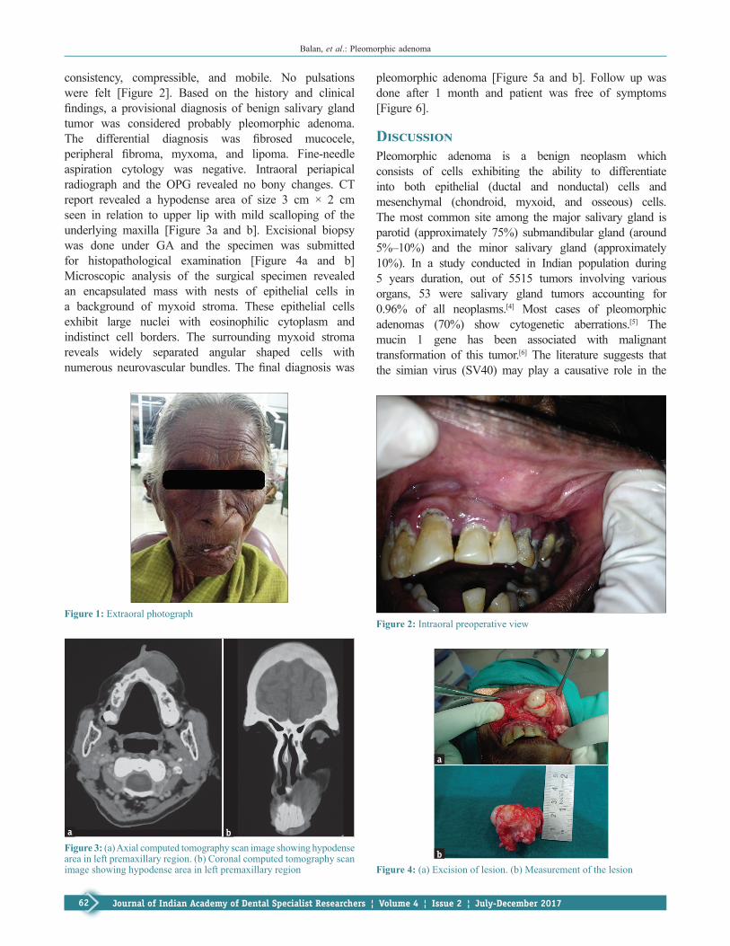
3 cm × 2 cm in size. It extended superiorly 3 cm from inner canthus of the eye, inferiorly to vermillion border of the upper lip, medially from infranasal depression laterally to nasolabial fold. There was deviation of the nasal septum to the right side, elevation of left ala of nose and obliteration of nasolabial fold. The surface appeared smooth with no secondary changes. On palpation, the swelling had no local rise in temperature, nontender, firm in consistency, and freely mobile. No pulsations were felt [Figure 1]. Bilateral submandibular lymph nodes were palpable, firm in consistency and mobile.
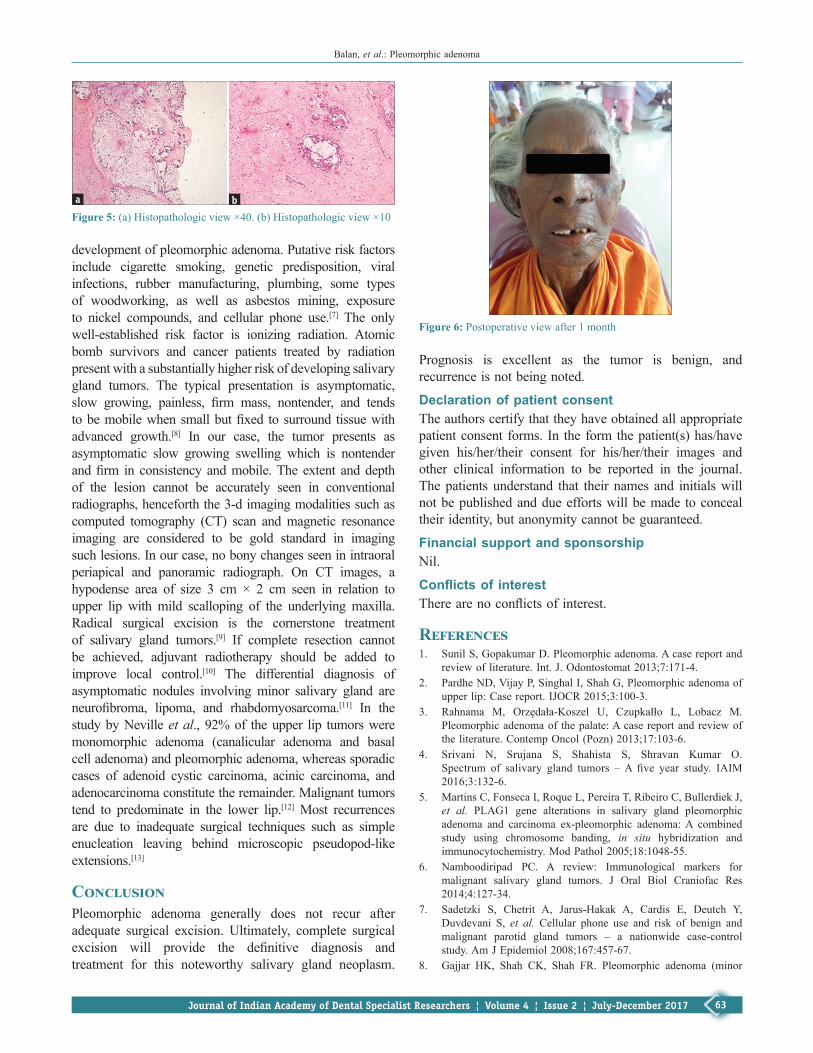
On intraoral examination, a well‑defined swelling was present on the left maxillary labial mucosa, ovoid, 3 cm × 2 cm in size, surface was smooth and mucosa over the swelling appeared pale pink, extends anteriorly from maxillary labial frenum, posteriorly 1 cm away from buccal frenum, superiorly 0.5 cm from vermillion border of upper lip, inferiorly to attached gingiva. There were no secondary changes such as sinus opening or pus discharge or ulceration. On palpation, it was nontender, firm in
Case Report
Introduction
T he term pleomorphic adenoma was suggested by Willis. In earlier years, it was referred by names
such as enclavoma, branchioma, endothelioma, and enchondroma.[1] It is the commonest of the salivary gland tumors, accounting for 50%–70% of cases of parotid tumors, 40%–60% submandibular tumors, and 10% minor salivary gland tumors, with palate (60%) being the most common followed by upper lip (20% of cases).[2] The most commonly affected age group are in the fourth, fifth, and sixth decades; 60% of them are female.[3] It has been suggested that 25% of benign mixed tumors undergo malignant transformation.
Case ReportA 72‑year‑old female patient reported to the Department of Oral Medicine and Radiology with the complaint of swelling in the left middle‑third of face for the past 1 year. History revealed that the swelling was gradual in onset and slowly increased in size for the past 1 year. The swelling was asymptomatic. Patient reported of the nasal stiffness of the left nostril and associated difficulty in breathing. There was no history of trauma, loss of appetite, and loss of weight.
On extraoral examination, a well‑defined swelling was present on left middle‑third of the face, oval,
Department of Oral Medicine and Radiology, Vivekanandha Dental College for Women, Namakkal, Tamil Nadu, India
Abs
trac
t
Address for correspondence: Dr. M. Sudhaa Mani, Department of Oral Medicine and Radiology, Vivekanandha
Dental College for Women, Tiruchengode, Namakkal, Tamil Nadu, India.
E‑mail: [email protected]
How to cite this article: Balan N, Mani MS, Ahamed SY, Divya DA. Pleomorphic adenoma: Case report and review of literature. J Indian Acad Dent Spec Res 2017;4:61-4.
This is an open access article distributed under the terms of the Creative Commons Attribution-NonCommercial-ShareAlike 3.0 License, which allows others to remix, tweak, and build upon the work non-commercially, as long as the author is credited and the new creations are licensed under the identical terms.
For reprints contact: [email protected]
Balan, et al.: Pleomorphic adenoma
62 Journal of Indian Academy of Dental Specialist Researchers ¦ Volume 4 ¦ Issue 2 ¦ July‑December 2017
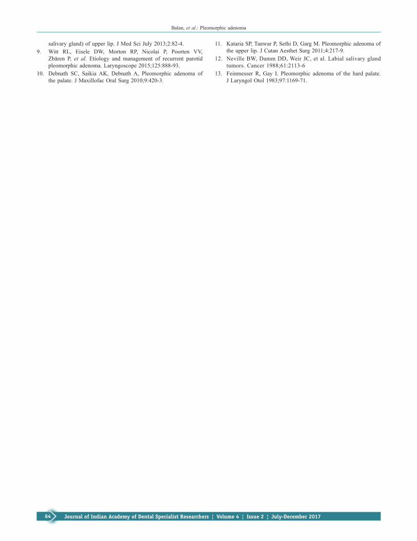
consistency, compressible, and mobile. No pulsations were felt [Figure 2]. Based on the history and clinical findings, a provisional diagnosis of benign salivary gland tumor was considered probably pleomorphic adenoma. The differential diagnosis was fibrosed mucocele, peripheral fibroma, myxoma, and lipoma. Fine‑needle aspiration cytology was negative. Intraoral periapical radiograph and the OPG revealed no bony changes. CT report revealed a hypodense area of size 3 cm × 2 cm seen in relation to upper lip with mild scalloping of the underlying maxilla [Figure 3a and b]. Excisional biopsy was done under GA and the specimen was submitted for histopathological examination [Figure 4a and b] Microscopic analysis of the surgical specimen revealed an encapsulated mass with nests of epithelial cells in a background of myxoid stroma. These epithelial cells exhibit large nuclei with eosinophilic cytoplasm and indistinct cell borders. The surrounding myxoid stroma reveals widely separated angular shaped cells with numerous neurovascular bundles. The final diagnosis was
pleomorphic adenoma [Figure 5a and b]. Follow up was done after 1 month and patient was free of symptoms [Figure 6].
DiscussionPleomorphic adenoma is a benign neoplasm which consists of cells exhibiting the ability to differentiate into both epithelial (ductal and nonductal) cells and mesenchymal (chondroid, myxoid, and osseous) cells. The most common site among the major salivary gland is parotid (approximately 75%) submandibular gland (around 5%–10%) and the minor salivary gland (approximately 10%). In a study conducted in Indian population during 5 years duration, out of 5515 tumors involving various organs, 53 were salivary gland tumors accounting for 0.96% of all neoplasms.[4] Most cases of pleomorphic adenomas (70%) show cytogenetic aberrations.[5] The mucin 1 gene has been associated with malignant transformation of this tumor.[6] The literature suggests that the simian virus (SV40) may play a causative role in the
Figure 2: Intraoral preoperative viewFigure 1: Extraoral photograph
Figure 4: (a) Excision of lesion. (b) Measurement of the lesionb
a
Figure 3: (a) Axial computed tomography scan image showing hypodense area in left premaxillary region. (b) Coronal computed tomography scan image showing hypodense area in left premaxillary region
ba
Balan, et al.: Pleomorphic adenoma
63Journal of Indian Academy of Dental Specialist Researchers ¦ Volume 4 ¦ Issue 2 ¦ July‑December 2017
development of pleomorphic adenoma. Putative risk factors include cigarette smoking, genetic predisposition, viral infections, rubber manufacturing, plumbing, some types of woodworking, as well as asbestos mining, exposure to nickel compounds, and cellular phone use.[7] The only well‑established risk factor is ionizing radiation. Atomic bomb survivors and cancer patients treated by radiation present with a substantially higher risk of developing salivary gland tumors. The typical presentation is asymptomatic, slow growing, painless, firm mass, nontender, and tends to be mobile when small but fixed to surround tissue with advanced growth.[8] In our case, the tumor presents as asymptomatic slow growing swelling which is nontender and firm in consistency and mobile. The extent and depth of the lesion cannot be accurately seen in conventional radiographs, henceforth the 3‑d imaging modalities such as computed tomography (CT) scan and magnetic resonance imaging are considered to be gold standard in imaging such lesions. In our case, no bony changes seen in intraoral periapical and panoramic radiograph. On CT images, a hypodense area of size 3 cm × 2 cm seen in relation to upper lip with mild scalloping of the underlying maxilla. Radical surgical excision is the cornerstone treatment of salivary gland tumors.[9] If complete resection cannot be achieved, adjuvant radiotherapy should be added to improve local control.[10] The differential diagnosis of asymptomatic nodules involving minor salivary gland are neurofibroma, lipoma, and rhabdomyosarcoma.[11] In the study by Neville et al., 92% of the upper lip tumors were monomorphic adenoma (canalicular adenoma and basal cell adenoma) and pleomorphic adenoma, whereas sporadic cases of adenoid cystic carcinoma, acinic carcinoma, and adenocarcinoma constitute the remainder. Malignant tumors tend to predominate in the lower lip.[12] Most recurrences are due to inadequate surgical techniques such as simple enucleation leaving behind microscopic pseudopod‑like extensions.[13]
ConclusionPleomorphic adenoma generally does not recur after adequate surgical excision. Ultimately, complete surgical excision will provide the definitive diagnosis and treatment for this noteworthy salivary gland neoplasm.
Prognosis is excellent as the tumor is benign, and recurrence is not being noted.
Declaration of patient consentThe authors certify that they have obtained all appropriate patient consent forms. In the form the patient(s) has/have given his/her/their consent for his/her/their images and other clinical information to be reported in the journal. The patients understand that their names and initials will not be published and due efforts will be made to conceal their identity, but anonymity cannot be guaranteed.
Financial support and sponsorshipNil.
Conflicts of interestThere are no conflicts of interest.
References1. Sunil S, Gopakumar D. Pleomorphic adenoma. A case report and
review of literature. Int. J. Odontostomat 2013;7:171‑4.2. Pardhe ND, Vijay P, Singhal I, Shah G, Pleomorphic adenoma of
upper lip: Case report. IJOCR 2015;3:100‑3.3. Rahnama M, Orzędała‑Koszel U, Czupkałło L, Lobacz M.
Pleomorphic adenoma of the palate: A case report and review of the literature. Contemp Oncol (Pozn) 2013;17:103‑6.
4. Srivani N, Srujana S, Shahista S, Shravan Kumar O. Spectrum of salivary gland tumors – A five year study. IAIM 2016;3:132‑6.
5. Martins C, Fonseca I, Roque L, Pereira T, Ribeiro C, Bullerdiek J, et al. PLAG1 gene alterations in salivary gland pleomorphic adenoma and carcinoma ex‑pleomorphic adenoma: A combined study using chromosome banding, in situ hybridization and immunocytochemistry. Mod Pathol 2005;18:1048‑55.
6. Namboodiripad PC. A review: Immunological markers for malignant salivary gland tumors. J Oral Biol Craniofac Res 2014;4:127‑34.
7. Sadetzki S, Chetrit A, Jarus‑Hakak A, Cardis E, Deutch Y, Duvdevani S, et al. Cellular phone use and risk of benign and malignant parotid gland tumors – a nationwide case‑control study. Am J Epidemiol 2008;167:457‑67.
8. Gajjar HK, Shah CK, Shah FR. Pleomorphic adenoma (minor
Figure 6: Postoperative view after 1 month
Figure 5: (a) Histopathologic view ×40. (b) Histopathologic view ×10ba
Balan, et al.: Pleomorphic adenoma
64 Journal of Indian Academy of Dental Specialist Researchers ¦ Volume 4 ¦ Issue 2 ¦ July‑December 2017
salivary gland) of upper lip. J Med Sci July 2013;2:82‑4.9. Witt RL, Eisele DW, Morton RP, Nicolai P, Poorten VV,
Zbären P, et al. Etiology and management of recurrent parotid pleomorphic adenoma. Laryngoscope 2015;125:888‑93.
10. Debnath SC, Saikia AK, Debnath A, Pleomorphic adenoma of the palate. J Maxillofac Oral Surg 2010;9:420‑3.
11. Kataria SP, Tanwar P, Sethi D, Garg M. Pleomorphic adenoma of the upper lip. J Cutan Aesthet Surg 2011;4:217‑9.
12. Neville BW, Damm DD, Weir JC, et al. Labial salivary gland tumors. Cancer 1988;61:2113‑6
13. Feinmesser R, Gay I. Pleomorphic adenoma of the hard palate. J Laryngol Otol 1983;97:1169‑71.


![Ductal Adenocarcinoma Ex Pleomorphic Adenoma of the ... · lesions [2, 5]. Carcinoma ex pleomorphic adenoma (Ca ex PA) is a rare transformation of a benign primary PA to a malignant](https://img.pdfslide.net/doc/110x75/60bd399bb7acaf776f026cd1/ductal-adenocarcinoma-ex-pleomorphic-adenoma-of-the-lesions-2-5-carcinoma.jpg)