Embed Size (px)
Citation preview
1 | P a g e
PMB definition guideline for Early Stage Pancreatic Cancer
PMB definition guideline for early stage pancreatic cancer
2 | P a g e
PMB definition guideline for Early Stage Pancreatic Cancer
Disclaimer:
The early stage pancreatic cancer benefit definition has been developed for the majority of standard
patients. These benefits may not be sufficient for outlier patients. Therefore Regulation 15(h) and 15(I) may
be applied for patients who are inadequately managed by the stated benefits. The benefit definition does
not describe specific in-hospital management such as theatre, anaesthetists, anaesthetist drugs and
nursing care. However, these interventions form part of care and are prescribed minimum benefits.
3 | P a g e
PMB definition guideline for Early Stage Pancreatic Cancer
Table of Contents
1. Introduction ............................................................................................................................................... 5
2. Scope and Purpose .................................................................................................................................. 5
3. Epidemiology and burden of Disease ...................................................................................................... 6
4. Diagnosis and Staging .............................................................................................................................. 6
5. Treatment options for early stage pancreatic cancer ................................................................................ 8
6. Follow-Up care .......................................................................................................................................... 9
7. References ……………………………………………………………………………………………….……....11
4 | P a g e
PMB definition guideline for Early Stage Pancreatic Cancer
Abbreviations
CMS Council for Medical Schemes
CT Computed tomographic
DTPs Diagnosis treatment pairs
ERCP Endoscopic retrograde cholangiopancreatography
ESPAC European Study Group on Pancreatic Cancer
FBC Full Blood Count
MRCP Magnetic resonance cholangiopancreatography
MRI Magnetic resonance imaging
OS Overall survival
PMB Prescribed minimum benefit
5 | P a g e
PMB definition guideline for Early Stage Pancreatic Cancer
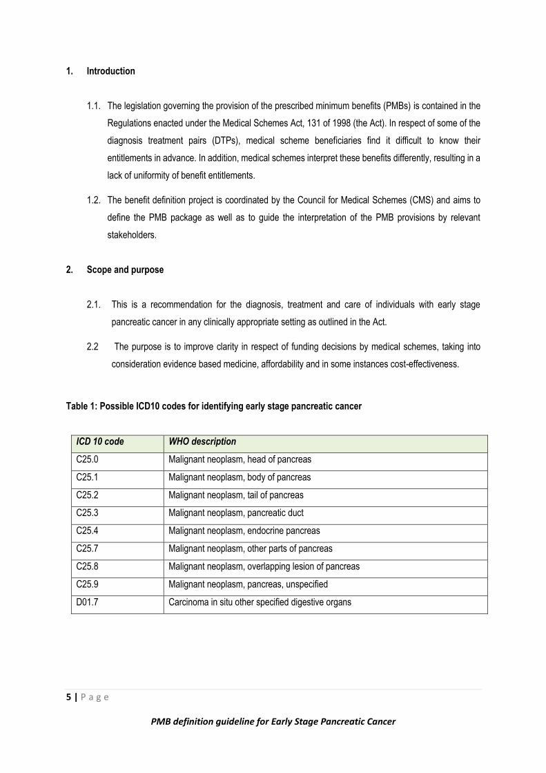
1. Introduction
1.1. The legislation governing the provision of the prescribed minimum benefits (PMBs) is contained in the
Regulations enacted under the Medical Schemes Act, 131 of 1998 (the Act). In respect of some of the
diagnosis treatment pairs (DTPs), medical scheme beneficiaries find it difficult to know their
entitlements in advance. In addition, medical schemes interpret these benefits differently, resulting in a
lack of uniformity of benefit entitlements.
1.2. The benefit definition project is coordinated by the Council for Medical Schemes (CMS) and aims to
define the PMB package as well as to guide the interpretation of the PMB provisions by relevant
stakeholders.
2. Scope and purpose
2.1. This is a recommendation for the diagnosis, treatment and care of individuals with early stage
pancreatic cancer in any clinically appropriate setting as outlined in the Act.
2.2 The purpose is to improve clarity in respect of funding decisions by medical schemes, taking into
consideration evidence based medicine, affordability and in some instances cost-effectiveness.
Table 1: Possible ICD10 codes for identifying early stage pancreatic cancer
ICD 10 code WHO description
C25.0 Malignant neoplasm, head of pancreas
C25.1 Malignant neoplasm, body of pancreas
C25.2 Malignant neoplasm, tail of pancreas
C25.3 Malignant neoplasm, pancreatic duct
C25.4 Malignant neoplasm, endocrine pancreas
C25.7 Malignant neoplasm, other parts of pancreas
C25.8 Malignant neoplasm, overlapping lesion of pancreas
C25.9 Malignant neoplasm, pancreas, unspecified
D01.7 Carcinoma in situ other specified digestive organs
6 | P a g e
PMB definition guideline for Early Stage Pancreatic Cancer
3. Epidemiology and burden of disease
Pancreatic cancer is one of the leading causes of cancer mortality in developed countries and one of the most
lethal malignant neoplasms across the world (Ferlay, Soerjomataram & Dikshit, 2015). Globally it is the seventh
leading cause of cancer mortality in men and women, causing more than 300 000 deaths annually (Torre, Bray &
Siegel, 2015. In South Africa, cancer of pancreas is the 12th most frequent cancer, with breast and cervical cancer
ranked first and second respectively. The National Cancer Registry (2012) estimates the lifetime risk of developing
pancreatic cancer at about 1 in 698 for males and 1 in 1211 for females (National Cancer Registry, 2012).
4. Diagnosis and Staging:
4.1. Pre-diagnostic work up
4.1.1. The work up of a suspected pancreatic cancer patient ideally should not only focus on the
establishment of the diagnosis, but also on the potential for fitness to undergo curative treatment.
The nature of pancreatic cancer is complex and thus, evaluation of all patients with pancreatic
cancer should be managed by a multidisciplinary team, including gastroenterologists, radiologists,
oncologists, surgeons, pathologists and palliative care specialists.
4.1.2. There is a number of investigations that should be conducted as part of a pre-diagnosis work up
for early stage pancreatic cancer (see table 2).
4.1.3. Imaging remains the primary means through which the stage of pancreatic cancer is determined.
For many patients presenting with the common symptoms of pancreatic cancer, ultrasound of the
abdomen should be the first imaging test to be conducted. With a reported sensitivity of 80 – 95%,
ultrasound of the abdomen can identify the pancreatic tumour as well as dilated bile ducts.
Sensitivity is however reduced in the evaluation of the body and tail of the pancreas and provides
less accurate staging information (Cotton, Lees & Vallon, 1980; Taylor, Buchin & Viscomi, 1981).
4.1.4. Tumour marker CA19-9 is a sialylated Lewis A blood group antigen. As the CA19-9 biomarker is
commonly expressed and shed in pancreatic and hepatobiliary disease in many malignancies, it is
therefore considered not tumour specific. Individuals who are jaundiced with cholestasis will induce
false positive results, as CA19-9 levels correlate with high levels of bilirubin levels and do not
necessarily indicate cancer or advanced disease (Kim, Y., Kim, H. & Park, 2009).
4.1.5. The degree of increase in CA 19-9 levels, however, may be useful in differentiating pancreatic
adenocarcinoma from inflammatory conditions of the pancreas and CA 19-9 therefore remains a
good marker, with sensitivity of 79 to 81% and specificity of 82 – 90% in symptomatic patients
(Kondo, Murakami & Uemura, 2010). Preoperative CA 19-9 levels correlate with both AJCC
staging and resectability and thus provide additional information for staging and determining
7 | P a g e
PMB definition guideline for Early Stage Pancreatic Cancer
resectability (Oettle, Post & Neuhaus, 2007). The timing of preoperative measurement of CA 19-9
levels should be after biliary decompression is complete and bilirubin levels are normal.
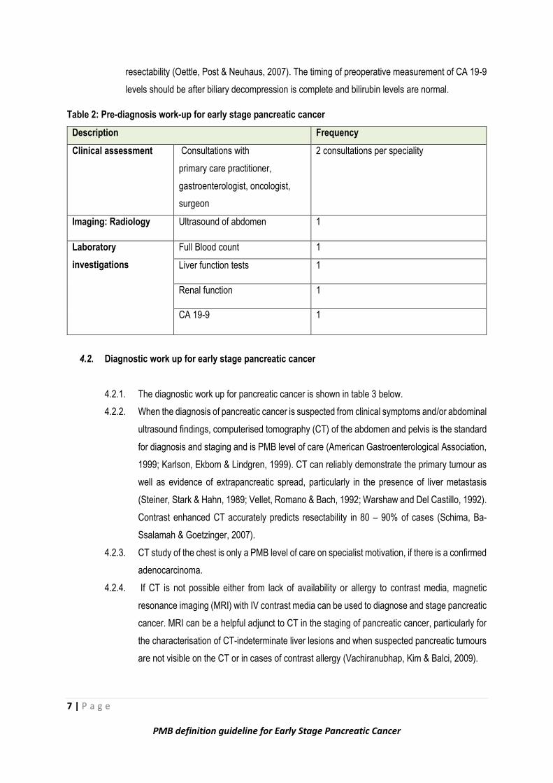
Table 2: Pre-diagnosis work-up for early stage pancreatic cancer
Description Frequency
Clinical assessment Consultations with
primary care practitioner,
gastroenterologist, oncologist,
surgeon
2 consultations per speciality
Imaging: Radiology Ultrasound of abdomen 1
Laboratory
investigations
Full Blood count 1
Liver function tests 1
Renal function 1
CA 19-9 1
4.2. Diagnostic work up for early stage pancreatic cancer
4.2.1. The diagnostic work up for pancreatic cancer is shown in table 3 below.
4.2.2. When the diagnosis of pancreatic cancer is suspected from clinical symptoms and/or abdominal
ultrasound findings, computerised tomography (CT) of the abdomen and pelvis is the standard
for diagnosis and staging and is PMB level of care (American Gastroenterological Association,
1999; Karlson, Ekbom & Lindgren, 1999). CT can reliably demonstrate the primary tumour as
well as evidence of extrapancreatic spread, particularly in the presence of liver metastasis
(Steiner, Stark & Hahn, 1989; Vellet, Romano & Bach, 1992; Warshaw and Del Castillo, 1992).
Contrast enhanced CT accurately predicts resectability in 80 – 90% of cases (Schima, Ba-
Ssalamah & Goetzinger, 2007).
4.2.3. CT study of the chest is only a PMB level of care on specialist motivation, if there is a confirmed
adenocarcinoma.
4.2.4. If CT is not possible either from lack of availability or allergy to contrast media, magnetic
resonance imaging (MRI) with IV contrast media can be used to diagnose and stage pancreatic
cancer. MRI can be a helpful adjunct to CT in the staging of pancreatic cancer, particularly for
the characterisation of CT-indeterminate liver lesions and when suspected pancreatic tumours
are not visible on the CT or in cases of contrast allergy (Vachiranubhap, Kim & Balci, 2009).
8 | P a g e
PMB definition guideline for Early Stage Pancreatic Cancer
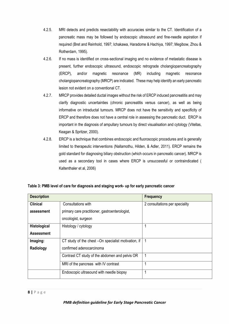
4.2.5. MRI detects and predicts resectability with accuracies similar to the CT. Identification of a
pancreatic mass may be followed by endoscopic ultrasound and fine-needle aspiration if
required (Bret and Reinhold, 1997; Ichakawa, Haradome & Hachiya, 1997; Megibow, Zhou &
Rotherdam, 1995).
4.2.6. If no mass is identified on cross-sectional imaging and no evidence of metastatic disease is
present, further endoscopic ultrasound, endoscopic retrograde cholangiopancreatography
(ERCP), and/or magnetic resonance (MR) including magnetic resonance
cholangiopancreatography (MRCP) are indicated. These may help identify an early pancreatic
lesion not evident on a conventional CT.
4.2.7. MRCP provides detailed ductal images without the risk of ERCP induced pancreatitis and may
clarify diagnostic uncertainties (chronic pancreatitis versus cancer), as well as being
informative on intraductal tumours. MRCP does not have the sensitivity and specificity of
ERCP and therefore does not have a central role in assessing the pancreatic duct. ERCP is
important in the diagnosis of ampullary tumours by direct visualisation and cytology (Vitellas,
Keagan & Spritzer, 2000).
4.2.8. ERCP is a technique that combines endoscopic and fluoroscopic procedures and is generally
limited to therapeutic interventions (Nallamothu, Hilden, & Adler, 2011). ERCP remains the
gold standard for diagnosing biliary obstruction (which occurs in pancreatic cancer). MRCP is
used as a secondary tool in cases where ERCP is unsuccessful or contraindicated (
Kaltenthaler et al, 2006)
Table 3: PMB level of care for diagnosis and staging work- up for early pancreatic cancer
Description Frequency
Clinical
assessment
Consultations with
primary care practitioner, gastroenterologist,
oncologist, surgeon
2 consultations per speciality
Histological
Assessment
Histology / cytology 1
Imaging:
Radiology
CT study of the chest –On specialist motivation, if
confirmed adenocarcinoma
1
Contrast CT study of the abdomen and pelvis OR 1
MRI of the pancreas with IV contrast 1
Endoscopic ultrasound with needle biopsy 1
9 | P a g e
PMB definition guideline for Early Stage Pancreatic Cancer
Imaging:
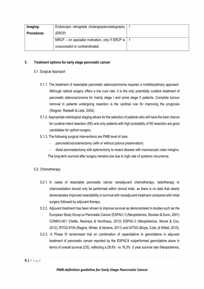
Procedures
Endoscopic retrograde cholangiopancreatography
(ERCP)
1
MRCP – on specialist motivation, only if ERCP is
unsuccessful or contraindicated.
1
5. Treatment options for early stage pancreatic cancer
5.1. Surgical Approach
5.1.1. The treatment of resectable pancreatic adenocarcinoma requires a multidisciplinary approach.
Although radical surgery offers a low cure rate, it is the only potentially curative treatment of
pancreatic adenocarcinoma for mainly stage I and some stage II patients. Complete tumour
removal in patients undergoing resection is the cardinal rule for improving the prognosis
(Wagner, Redaelli & Lietz, 2004).
5.1.2. Appropriate radiological staging allows for the selection of patients who will have the best chance
for curative intent resection (R0) and only patients with high probability of R0 resection are good
candidates for upfront surgery.
5.1.3. The following surgical interventions are PMB level of care:
- pancreaticoduodenectomy (with or without pylorus preservation)
- distal pancreatectomy with splenectomy to resect disease with macroscopic clear margins.
The long-term survival after surgery remains low due to high rate of systemic recurrence.
5.2. Chemotherapy
5.2.1. In cases of resectable pancreatic cancer neoadjuvant chemotherapy, radiotherapy or
chemoradiation should only be performed within clinical trials, as there is no data that clearly
demonstrates improved resectability or survival with neoadjuvant treatment compared with initial
surgery followed by adjuvant therapy.
5.2.2. Adjuvant treatment has been shown to improve survival as demonstrated in studies such as the
European Study Group on Pancreatic Cancer (ESPAC-1) (Neoptolemos, Stocken & Dunn, 2001)
CONKO-001 (Oettle, Neuhaus & Hochhaus, 2013) ESPAC-3 (Neoptolemos, Moore & Cox,
2012), RTOG 9704 (Regine, Winter, & Abrams, 2011) and GITSG (Boyle, Czito, & Willett, 2015).
5.2.3. A Phase III randomised trial on combination of capecitabine to gemcitabine in adjuvant
treatment of pancreatic cancer reported by the ESPAC4 outperformed gemcitabine alone in
terms of overall survival (OS), reflecting a 28.8% vs 16.3% 5 year survival rate (Neoptolemos,
10 | P a g e
PMB definition guideline for Early Stage Pancreatic Cancer
Palmer, Ghaneh, Valle, Cunningham, Wadsley, Meyer, Anthoney, Glimelius, Falk, Segersvard,
Izbicki, Middleton, Ross, Wasan, Mcdonald, Crosby, Psarelli, Hammel & Buchler, 2016).
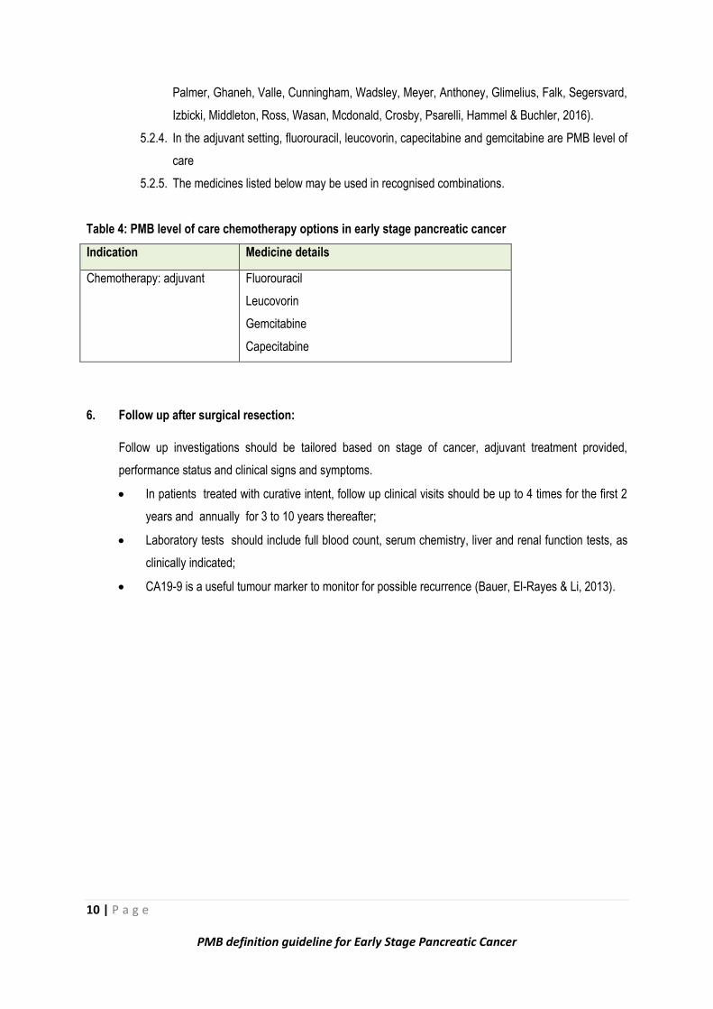
5.2.4. In the adjuvant setting, fluorouracil, leucovorin, capecitabine and gemcitabine are PMB level of
care
5.2.5. The medicines listed below may be used in recognised combinations.
Table 4: PMB level of care chemotherapy options in early stage pancreatic cancer
Indication Medicine details
Chemotherapy: adjuvant Fluorouracil
Leucovorin
Gemcitabine
Capecitabine
6. Follow up after surgical resection:
Follow up investigations should be tailored based on stage of cancer, adjuvant treatment provided,
performance status and clinical signs and symptoms.
In patients treated with curative intent, follow up clinical visits should be up to 4 times for the first 2
years and annually for 3 to 10 years thereafter;
Laboratory tests should include full blood count, serum chemistry, liver and renal function tests, as
clinically indicated;
CA19-9 is a useful tumour marker to monitor for possible recurrence (Bauer, El-Rayes & Li, 2013).
11 | P a g e
PMB definition guideline for Early Stage Pancreatic Cancer
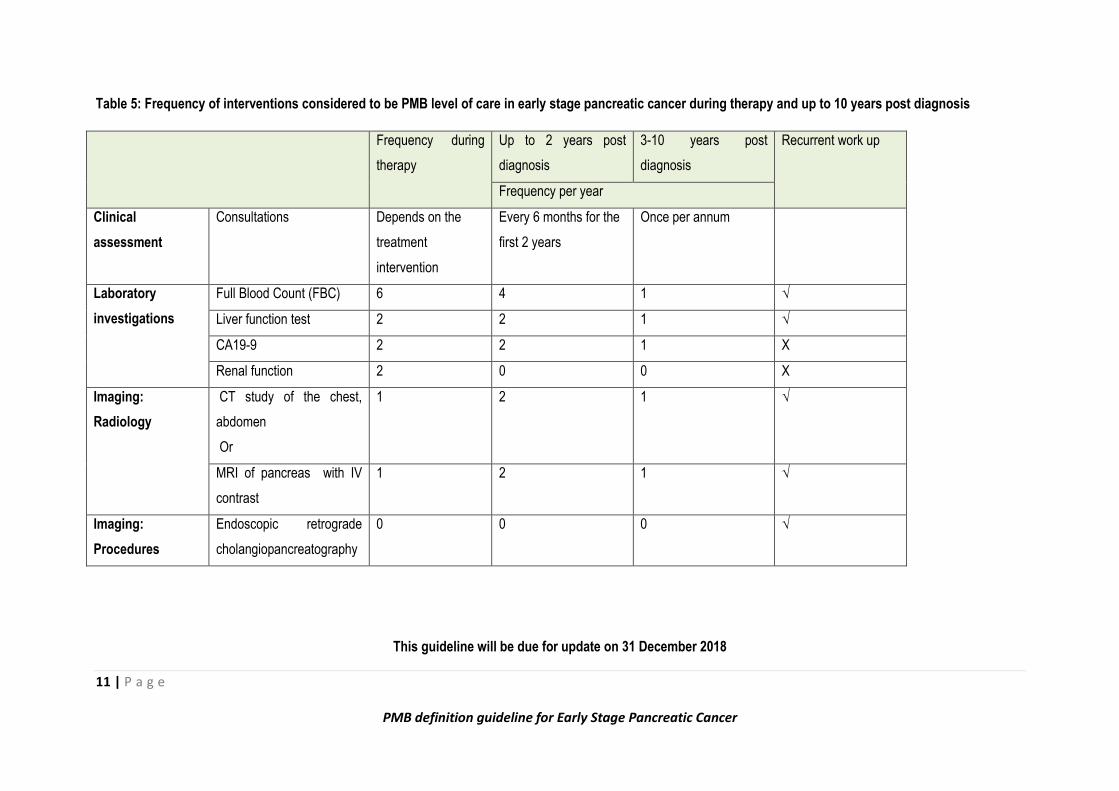
Table 5: Frequency of interventions considered to be PMB level of care in early stage pancreatic cancer during therapy and up to 10 years post diagnosis
Frequency during
therapy
Up to 2 years post
diagnosis
3-10 years post
diagnosis
Recurrent work up
Frequency per year
Clinical
assessment
Consultations Depends on the
treatment
intervention
Every 6 months for the
first 2 years
Once per annum
Laboratory
investigations
Full Blood Count (FBC) 6 4 1 √
Liver function test 2 2 1 √
CA19-9 2 2 1 X
Renal function 2 0 0 X
Imaging:
Radiology
CT study of the chest,
abdomen
Or
1 2 1 √
MRI of pancreas with IV
contrast
1 2 1 √
Imaging:
Procedures
Endoscopic retrograde
cholangiopancreatography
0 0 0 √
This guideline will be due for update on 31 December 2018
12 | P a g e
PMB definition guideline for Early Stage Pancreatic Cancer
7. References
American Gastroenterological Association medical position statement. 1999. Epidemiology, diagnosis and
treatment of pancreatic ductal adenocarcinoma. Gastroenterology, 117(6):1463-1484.
Bauer, T.M., El-Rayes, B.F. & Li X. 2013. Carbohydrate antigen 19-9 is a prognostic and predictive
biomarker in patients with advanced pancreatic cancer who receive gemcitabine-containing chemotherapy:
a pooled analysis of 6 prospective trials. Cancer, 119: 285-292.
Boyle, J., Czito, B. & Willett, C. 2015. Adjuvant radiation therapy for pancreatic cancer: a review of the old
and the new. Journal of Gastrointestinal Oncology, 6 (4):436-444.
Bret, P.M. and Reinhold, C. 1997. Magnetic resonance cholangiopancreatography. Endoscopy, 29:472-
486.
Cotton, P.B., Lees, W.R. & Vallon, A.G. 1980. Grey-scale Ultrasonography an endoscopic
pancreaotography in pancreatic diagnosis. Radiology; 134; 453-9.
Ferlay, J., Soerjomataram, I. & Dikshit, R. 2015. Cancer incidence and mortality worldwide: sources,
methods and major patterns in GLOBOCAN, International Journal of Cancer; 136: E359–E386.
Ichakawa, T., Haradome, H. & Hachiya, J. 1997. Pancreatic ductal adenocarcinoma: preoperative
assessment with helical CT versus dynamic gadolinium MR imaging. Radiology, 202:665-62.
Karlson, B.M., Ekbom, A. & Lindgren, P.G. 1999. Abdominal US for diagnosis of pancreatic tumour:
prospective cohort analysis. Radiology; 213:107-11.
Kaltenthaler E.C., Walters, S.J., Chilcott, J., Blakeborough, A., Bravo Vergel, Y. & Thomas, S., 2006, MRCP
compared to diagnostic ERCP for diagnosis when biliary obstruction is suspected: a systematic review.
Biomed Central: Medical Imaging: 6:9.
Kim, Y.C., Kim, H.J. & Park, J.H. 2009. Can preoperative CA19-9 and CEA levels predict the resectability
of patients with pancreatic adenocarcinoma? Journal of Gastroenterology and Hepatology, 24: 1869-1875.
Kondo, N., Murakami, Y. & Uemura, K. 2010. Prognostic impact of perioperative serum CA 19-9 levels in
patients with resectable pancreatic cancer. Annals of Surgical Oncology, 17: 2321-2329.
13 | P a g e
PMB definition guideline for Early Stage Pancreatic Cancer
Megibow, A.J., Zhou, X.H. & Rotherdam, H. 1995. Pancreatic adenocarcinoma: CT versus MR imaging in
evaluation of resectability – report of the Radiology Diagnostic Oncology Group. Radiology, 195:327-32.
Nallamothu, G., Hilden, K. & Adler, D.G. 2011. Endoscopic retrograde cholangiopancreatography for non-
gastroenterologists: What you need to know. Hospital practice (Minneap), 39: 70-80.
National Cancer Registry. 2012. Cancer in South Africa. 2012. Full Report: National Institute for
Occupational Health, 2011. Available from http://www.nioh.ac.za/assets/files/NCR%202012%20results.pdf
[Accessed 1 March 2017]
Neoptolemos, J.P., Moore, M.J. & Cox, T.F. 2012. Effect of adjuvant chemotherapy with fluorouracil plus
folinic acid or gemcitabine vs observation on survival in patient with resected periampullary
adenocarcinoma: the ESPAC-3 periampullary cancer randomised trial. Journal of the American Medical
Association, 308: 147-156.
Neoptolemos, J.P., Palmer, D., Ghaneh, P., Valle, J.W., Cunningham, D., Wadsley, J., Meyer, T., Anthoney,
A., Glimelius, G., Falk, S., Segersvard, R., Izbicki, J.R. , Middleton, G.W., Ross, P.J., Wasan, H.,
Mcdonald, A., Crosby, T.D.L., Psarelli, E.E., Hammel, P. & Buchler, M.W. 2016. ESPAC-4: A multicenter,
international, open-label randomized controlled phase III trial of adjuvant combination chemotherapy of
gemcitabine and capecitabine versus monotherapy gemcitabine in patients with resected pancreatic ductal
adenocarcinoma. Presented at: the 2016 Annual Meeting of the American Society of Clinical Oncology,
Abstract LBA4006.
Neoptolemos, J.P., Stocken, D.D. & Dunn, J.A. 2001. Influence of resection margins on survival for patients
with pancreatic cancer treated by adjuvant chemoradiation and/or chemotherapy in the ESPAC-1
randomized controlled trial. Annals of Surgery, 234: 758–768.
Oettle, H., Neuhaus, P. & Hochhaus, A. 2013. Adjuvant chemotherapy with gemcitabine and long-term
outcomes among patients with resected pancreatic cancer: the CONKO-001 randomized trial. Journal of
the American Medical Association. 310 (14): 1473 –1481.
Regine, W.F., Winter, K.A. & Abrams, R. 2011. Fluorouracil-based chemoradiation with either gemcitabine
or fluorouracil chemotherapy after resection of pancreatic adenocarcinoma: 5-year analysis of the U.S.
Intergroup/RTOG 9704 phase III trial. Annals of Surgical Oncology, 18:1319-26.
Schima, W., Ba-Ssalamah, A. & Goetzinger, P. 2007. State of the art magnetic resonance imaging of
pancreatic cancer. Topics in Magnetic Resonance Imaging, 421-429.
14 | P a g e
PMB definition guideline for Early Stage Pancreatic Cancer
Steiner, E., Stark, D.D. & Hahn, P.F. 1989. Imaging of pancreatic neoplasms: comparison of MRI and CT.
American Journal of Roentgenology, 152: 487-91.
Torre, L.A., Bray, F. & Siegel, R.L. 2015. Global cancer statistics. CA: A cancer journal for clinicians; 65:87–
108.
Vachiranubhap, B., Kim, Y.H. & Balci, N.C. 2009. Magnetic resonance imaging of adenocarcinoma of the
pancreas. Topics in Magnetic Resonance Imaging, 20:3-9.
Vellet, A.D., Romano, W. & Bach, D.B. 1992. Adenocarcinoma of the pancreatic ducts: comparative
evaluation with CT and MRI imaging at 1.5T. Radiology, 183:87-95.
Vitellas, K.M., Keagan, M.T. & Spritzer, C.E. 2000. MR Cholangiopancreatography of bile and pancreatic
duct abnormalities with emphasis of single shot fast spin-echo technique. Radiographics, 20:1108-12.
Wagner, M., Redaelli, C. & Lietz, M. 2004. Curative resection is the single most important factor determining
outcome in patients with pancreatic adenocarcinoma. British Journal of Surgery, 91:586–94.
Warshaw, A.L. and Del Castillo, C.F. 1992. Pancreatic carcinoma. The New England Journal of Medicine,
326:455-65.