Embed Size (px)
Citation preview
Pneumonia
Community acquired pneumonia
(CAP)
Definition • Pneumonia is acute infection leads to
inflammation of the parenchyma of the lung (the alveoli) (consolidation and exudation)
• The histologically1. Fibrinopurulent alveolar exudate seen in acute
bacterial pneumonias.2. Mononuclear interstitial infiltrates in viral and
other atypical pneumonias3. Granulomas and cavitation seen in chronic
pneumonias
• It may present as acute, fulminant clinical disease or as chronic disease with a more protracted course
Epidemiology
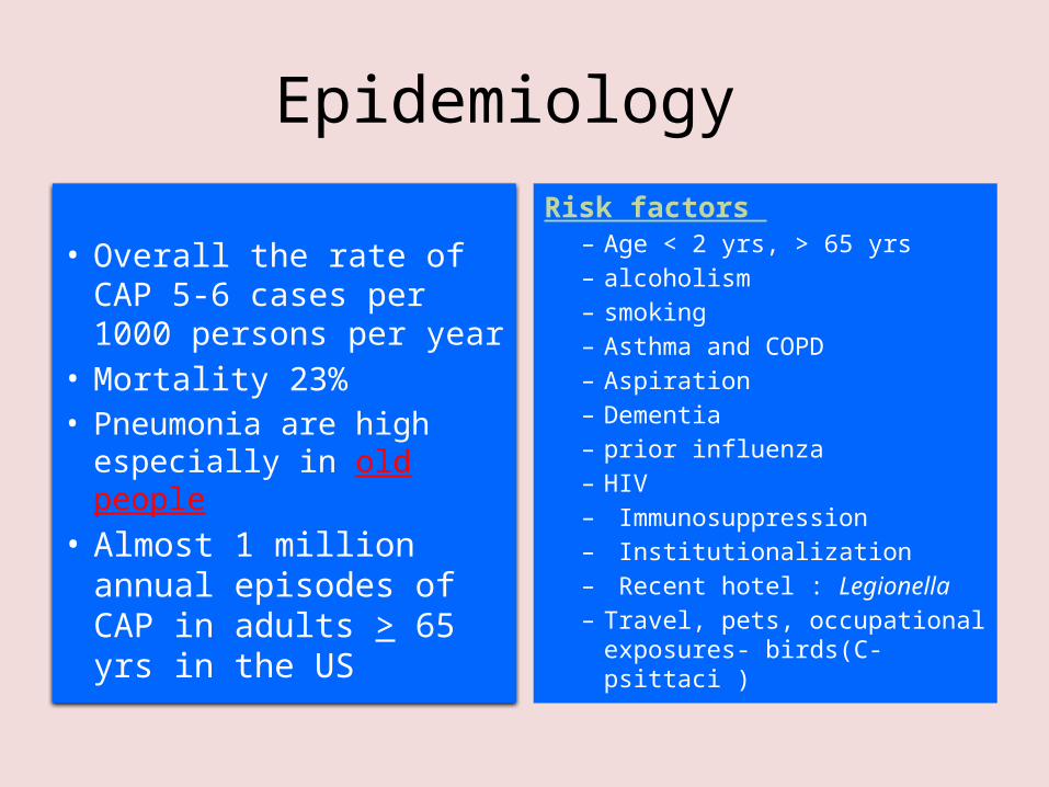
• Overall the rate of CAP 5-6 cases per 1000 persons per year
• Mortality 23%• Pneumonia are high
especially in old people• Almost 1 million
annual episodes of CAP in adults > 65 yrs in the US
Risk factors – Age < 2 yrs, > 65 yrs– alcoholism – smoking – Asthma and COPD– Aspiration– Dementia– prior influenza– HIV– Immunosuppression– Institutionalization– Recent hotel : Legionella– Travel, pets, occupational
exposures- birds(C- psittaci )

Etiological agents
• Bacterial• Fungal• Viral • Parasitic • Other non-
infectious factors like – Chemical– Allergen

Pathogenesis
Two factors involved in the formation of pneumonia– Pathogens– Host defenses.

Defense mechanism of respiratory tract
• Filtration and deposition of environmental pathogens in the upper airways
• Cough reflux• Mucociliary clearance • Alveolar macrophages• Humoral and cellular immunity• Oxidative metabolism of neutrophils

Pathophysiology :
1. Inhalation or aspiration of pulmonary pathogenic organisms into a lung segment or lobe.
2. Results from secondary bacteraemia from a distant source, such as Escherichia coli urinary tract infection and/or bacteraemia(less commonly).
3. Aspiration of Oropharyngeal contents (multiple pathogens).

Classification
• Bacterial pneumonia classified according to:1. Pathogen-(most useful-choose antimicrobial
agents)2. Anatomy3. Acquired environment
1. Gram-positive bacteria as - Streptococcus pneumoniae is the most
common cause of typical pneumonia - Staphylococcus aureus- Group A hemolytic streptococci
2. Gram-negative bacteria - Klebsiella pneumoniae - Hemophilus influenzae
- Moraxella catarrhal - Escherichia coli
3. Anaerobic bacteria
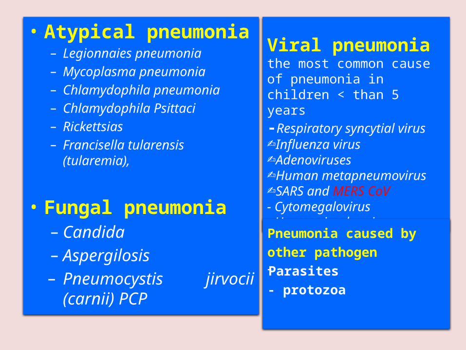
• Atypical pneumonia– Legionnaies pneumonia – Mycoplasma pneumonia – Chlamydophila pneumonia– Chlamydophila Psittaci– Rickettsias– Francisella tularensis (tularemia),
• Fungal pneumonia– Candida– Aspergilosis– Pneumocystis jirvocii
(carnii) PCP
Viral pneumoniathe most common cause of pneumonia in children < than 5 years-Respiratory syncytial virus -Influenza virus -Adenoviruses -Human metapneumovirus-SARS and MERS CoV- Cytomegalovirus- Herpes simplex virus Pneumonia caused by other pathogen-Parasites- protozoa

CAP and bioterrorism agents
• Bacillus anthracis (anthrax)• Yersinia pestis (plague) • Francisella tularensis (tularemia)• Coxialla . burnetii (Q fever)
• Level three agents

Classification by anatomy
1. Lobar: entire lobe2. Lobular:
(bronchopneumonia).3. Interstitial
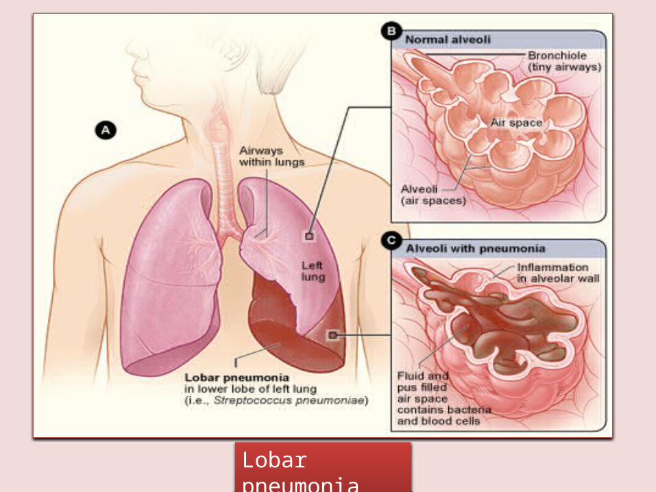
Lobar pneumonia

Classification by acquired environment
Community acquired pneumonia (CAP) Hospital acquired pneumonia (HAP) Nursing home acquired pneumonia (NHAP)
Immunocompromised host pneumonia (ICAP)

Outpatient Streptococcus pneumoniaeMycoplasma / Chlamydophila H. influenzae, Staph aureusRespiratory viruses
Inpatient, non-ICU Streptococcus pneumoniaeMycoplasma / ChlamydophilaH. influenzae, Staph aureusLegionellaRespiratory viruses
ICU Streptococcus pneumoniaeStaph aureus, LegionellaGram neg bacilli(Enterobacteriaceae, and Pseudomonas aeruginosa), H. influenzae

CAP- Cough/fever/sputum production + infiltrate
• CAP : pneumonia acquired outside of hospitals or extended-care facilities for > 14 days before onset of symptoms.– Streptococcus pneumoniae (most
common)– Haemophilus influenzae–mycoplasma pneumoniae– Chlamydia pneumoniae–Moraxella catarrhalis– Staph.aureus
• Drug resistance streptococcus pneumoniae(DRSP) is a major concern.
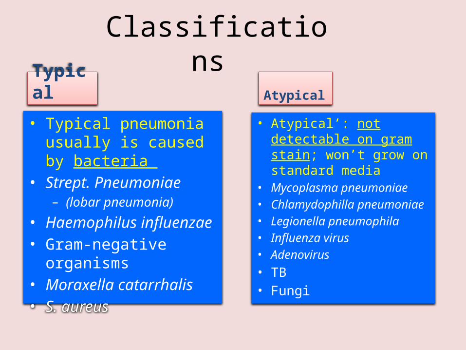
Classifications Typical
• Typical pneumonia usually is caused by bacteria
• Strept. Pneumoniae– (lobar pneumonia)
• Haemophilus influenzae
• Gram-negative organisms
• Moraxella catarrhalis• S. aureus
Atypical
• Atypical’: not detectable on gram stain; won’t grow on standard media
• Mycoplasma pneumoniae• Chlamydophilla pneumoniae• Legionella pneumophila• Influenza virus• Adenovirus
• TB • Fungi

Community acquired pneumonia
• Strep pneumonia 48%
• Viral 23%
• Atypical orgs(MP,LG,CP) 22%
• Haemophilus influenza 7%
• Moraxella catharralis 2%
• Staph aureus 1.5%
• Gram –ive orgs 1.4%
• Anaerobes
Clinical manifestationlobar pneumonia
• The onset is acute• Prior viral upper respiratory infection
• Respiratory symptoms– Fever– Shaking chills– Cough with sputum production (rusty-
sputum)– Chest pain- or pleurisy– Shortness of breath

Diagnosis • Clinical
– History & physical
• X-ray examination• Laboratory
– CBC- leukocytosis– Sputum Gram stain- 15%– Blood culture- 5-14% – Pleural effusion culture
Pneumococcal pneumonia
Drug Resistant Strep Pneumoniae• 40% of U.S. Strep pneumo CAP has some
antibiotic resistance:– PCN, cephalosporins, macrolides,
tetracyclines, clindamycin, bactrim, quinolones
• All MDR strains are sensitive to vancomycin or linezolid; most are sensitive to respiratory quinolones
• For Pneumonia, pneumococcal resistance to β-lactams is relative and can usually be overcome by increasing β-lactam doses (not for meningitis!)
Atypical pneumonia • Chlamydia pneumonia
• Mycoplasma pneumonia
• Legionella spp
• Psittacosis (parrots)
• Q fever (Coxiella burnettii)
• Viral (Influenza, Adenovirus)
• AIDS– PCP– TB (M. intracellulare)
• Approximately 15% of all CAP• Not detectable on gram stain• Won’t grow on standard media• Often extrapulmonary
manifestations:– Mycoplasma: otitis, nonexudative
pharyngitis, watery diarrhea, erythema multiforme, increased cold agglutinin titre
– Chlamydophilla: laryngitis• Most don’t have a bacterial cell
wall Don’t respond to β-lactams
• Therapy: macrolides, tetracyclines, quinolones (intracellular penetration, interfere with bacterial protein synthesis)
Mycoplasma pneumonia• Eaton agent (1944)• No cell wall• Common• Rare in children and in
> 65• People younger than
40.• Crowded places like
schools, homeless
shelters, prisons.• Mortality rate 1.4%
• Usually mild and responds well to antibiotics.
• Can be very serious • May be associated
with a skin rash, hemolysis, myocarditis or pancreatitis

Mycoplasmapneumonia
Cx-ray

Chlamydia pneumonia• Obligate intracellular organism
• 50% of adults sero-positive
• Mild disease
• Sub clinical infections common
• 5-10% of community acquired pneumonia

Psittacosis
• Chlamydophila psittaci
• Exposure to birds• Bird owners, pet
shop employees, vets
• Parrots, pigeons and poultry
• Birds often asymptomatic
• 1st: Tetracycline• Alt: Macrolide
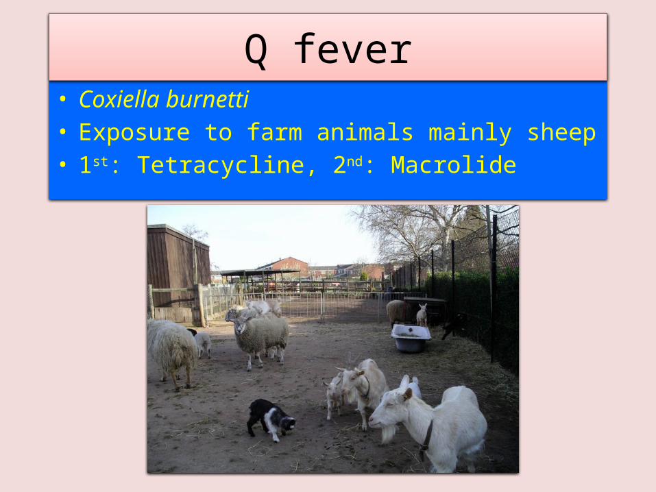
• Coxiella burnetti• Exposure to farm animals mainly sheep• 1st: Tetracycline, 2nd: Macrolide
Q fever

Legionella pneumophila
• Hyponatraemia common – (<130mMol)
• Bradycardia
• WBC < 15,000
• Abnormal LFTs
• Raised CPK
• Acute Renal failure
• Positive urinary antigen
• Legionnaire's disease.
• Serious outbreaks linked to exposure to cooling towers
• ICU admissions.

Legionnaires on ICU
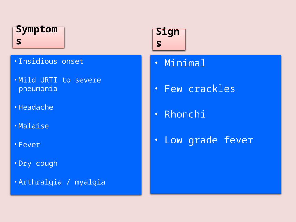
Symptoms
• Insidious onset
• Mild URTI to severe pneumonia
• Headache
• Malaise
• Fever
• Dry cough
• Arthralgia / myalgia
Signs
• Minimal
• Few crackles
• Rhonchi
• Low grade fever

Diagnosis & Treatment • CBC
• Mild elevation WBC
• U&Es
• Low serum Na (Legionalla)
• Deranged LFTS
• ↑ ALT
• ↑ Alk Phos• Culture on special media BCYE
• Cold agglutinins (Mycoplasma)
• Serology
• DNA detection
• Macrolide
• Rifampicicn
• Quinolones
• Tetracycline
• Treat for 10-14 days
• (21 in immunosupressed)
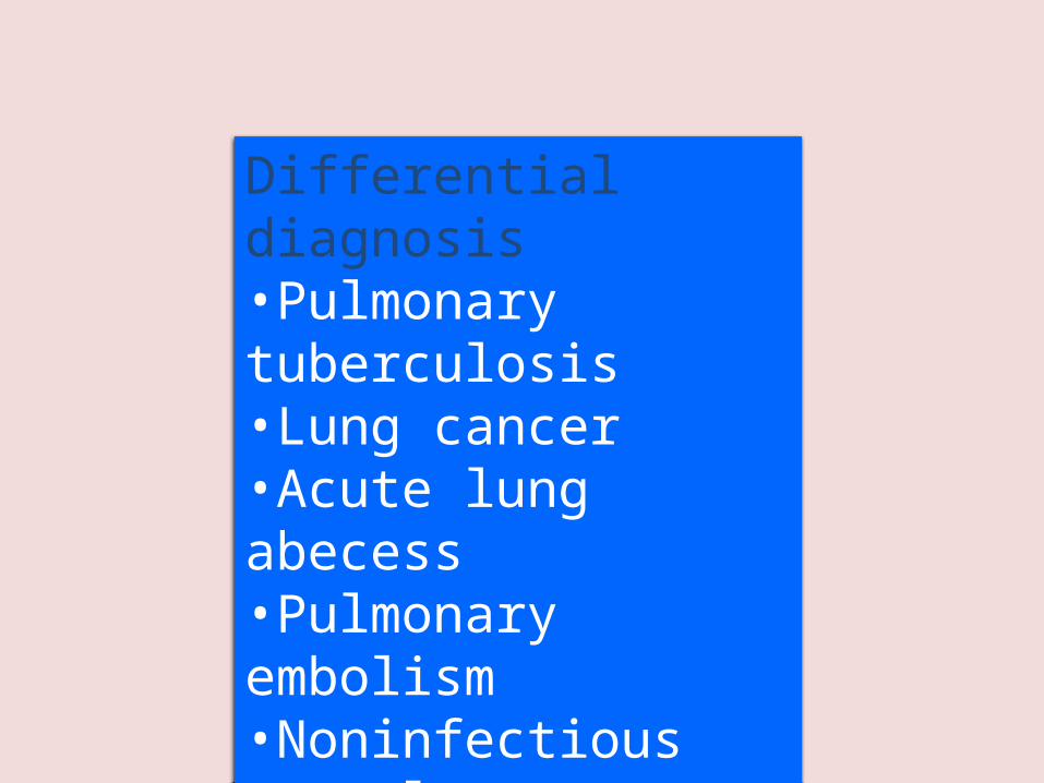
Differential diagnosis •Pulmonary tuberculosis•Lung cancer•Acute lung abecess•Pulmonary embolism•Noninfectious pulmonary infiltration

Evaluate the severity & degree of pneumonia
Is the patient will require hospital admission? – Patient characteristics– Co-morbid illness– Physical examinations– Basic laboratory findings

The diagnostic standard of sever pneumonia (Do not memorize)
• Altered mental status• Pa02<60mmHg. PaO2/FiO2<300,
needing MV• Respiratory rate>30/min• Blood pressure<90/60mmHg• Chest X-ray shows that bilateral
infiltration, multilobar infiltration and the infiltrations enlarge more than 50% within 48h.
• Renal function: U<20ml/h, and <80ml/4h

• Outpatient, healthy patient with no exposure to antibiotics in the last 3 months
• Outpatient, patient with comorbidity or exposure to antibiotics in the last 3 months
• Inpatient : Not ICU• Inpatient : ICU
Patient Management

• Macrolide: Azithromycin, Clarithromycin• Doxycycline• Beta Lactam :Amoxicillin/clavulinic acid,
Cefuroxime• Respiratory Flouroquinolone:Gatifloxacin,
Levofloxacin or Moxifloxacin• Antipeudomonas Beta lactam: Cetazidime• Antipneumococcal Beta lactam :Cefotaxime
Antibiotic Treatment
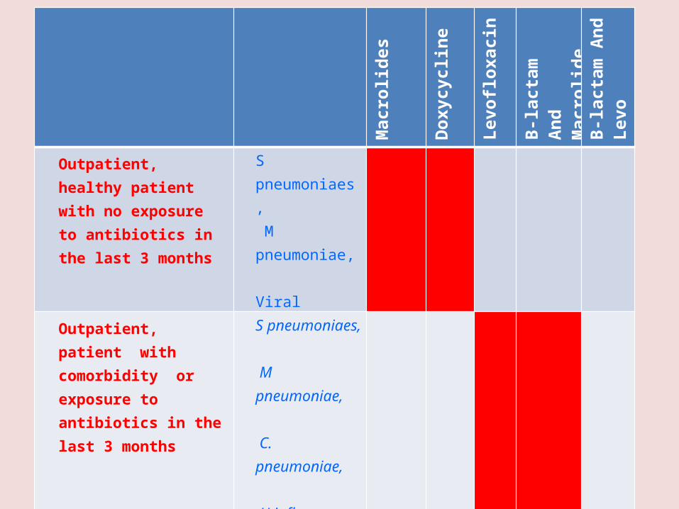
Macrolides
Doxycycline
Levofloxacin
B-lactamAnd Macrolide
B-lactam And Levo
Outpatient, healthy patient with no exposure to antibiotics in the last 3 months
S pneumoniaes, M pneumoniae, Viral
Outpatient, patient with comorbidity or exposure to antibiotics in the last 3 months
S pneumoniaes, M pneumoniae, C. pneumoniae, H influenzae M.catarrhalis anaerobesS aureus
Inpatient : Not ICU Same as above +legionella
Inpatient : ICU Same as above + Pseudomonas