Embed Size (px)
Citation preview
RESEARCH ARTICLE Open Access
Positron emission tomography in the diagnosticpathway for intracystic infection in adpkd and“cystic” kidneys. a case seriesGiorgina B Piccoli1*, Vincenzo Arena3, Valentina Consiglio1, Maria Chiara Deagostini1, Ettore Pelosi3,Anastasios Douroukas3, Daniele Penna3 and Giancarlo Cortese2
Abstract
Background: Intracystic infection, in Autosomal Dominant Polycystic Kidney Disease (ADPKD) and in kidneys withmultiple cysts, is a diagnostic and therapeutic challenge, as conventional imaging techniques may not discriminateamong “complicated” cysts (infection, bleeding, neoplasia), and as the clinical picture may be attenuated, inparticular in early phases. Positron Emission Tomography with fluorodeoxyglucose (FDG-PET) was recentlysuggested as a tool to detect infection in ADPKD, in single cases and small series.The aim of the study was to report on the role of FDG-PET in the work-up of 10 cases of suspected cysticinfections, affected by ADPKD or with multiple kidney cysts.
Methods: Observational study. Review of clinical charts and of the imaging data since the use of FDG-PET fordetecting cystic infections (2008-2010).
Results: In 2008-2010, 6 patients with ADPKD and 4 with multiple kidney cysts were referred for suspectedintracystic infections (3 males, 7 females, aged 55-83 years, in all CKD stages); in one case the imaging was done inthe work-up of a complicated “uremic” cyst. The clinical picture, the usual inflammatory markers and/or theconventional imaging techniques did not allow conclusive diagnosis at referral or during follow-up (ultrasounds inall, CT in 8/10). Nine patients displayed inflammatory signs (increase in C-reactive protein and other biochemicalmarkers) and constitutional symptoms (fever in 9/10).FDG-PET was positive in 6 cases (5 kidney and 1 liver cyst), was repeated during follow-up in 4 patients and wasnegative in 4 cases. In the positive cases, FDG-PET guided the therapeutic choices; in particular, the duration oftherapy was supported by imaging data in the 4 cases with multiple scans. No relapse was recorded afterdiscontinuation of antibiotic therapy in the treated patients. The negative cases did not develop clinical signs ofcystic infection over follow-up.
Conclusion: In this case series, the largest prospective one so far published and the only one including differenttypes of renal cysts, FDG-PET is confirmed as a promising diagnostic tool for detecting intracystic infection inADPKD and in multiple kidney cysts, and a potential guide for tailoring therapy. Further larger and multicenterstudies are needed to evaluate the cost-benefit ratio and the limits of this imaging technique in the clinical setting.
Keywords: Positron emission tomography, polycystic kidney disease, infection, kidney cysts, long-term antibiotictherapy
* Correspondence: [email protected] of Clinical and Biological Sciences of the University of Turin,San Luigi Gonzaga Hospital, regione Gonzole 10, Orbassano (TO), 10043 ItalyFull list of author information is available at the end of the article
Piccoli et al. BMC Nephrology 2011, 12:48http://www.biomedcentral.com/1471-2369/12/48
© 2011 Piccoli et al; licensee BioMed Central Ltd. This is an Open Access article distributed under the terms of the Creative CommonsAttribution License (http://creativecommons.org/licenses/by/2.0), which permits unrestricted use, distribution, and reproduction inany medium, provided the original work is properly cited.
BackgroundAutosomal Dominant Polycystic Kidney Disease(ADPKD) is the most common monogenic severe kidneydisease, with an average incidence of 1 in 800 live births[1,2]. In spite of its high frequency, its clinical manage-ment is still controversial and even some of the mostcommon clinical threats, such as intracystic infection,may represent an unmet challenge [3-7]. The clinicalspectrum of infection in the kidney and liver cysts isprotean: the disease ranges from mild abdominal dis-comfort, with a moderate increase of acute phase reac-tants, to severe life-threatening disease [2-7]. Diagnosticproblems arise from the absence of a diagnostic goldstandard in imaging and from the various, often non-specific clinical manifestations [4-7]. The need for long-term antibiotic therapy has to be weighed against therisk of side effects of prolonged treatment; the presenceof kidney functional impairment may limit the use ofmany antibiotics [1-3].The more vaguely defined “cystic kidneys”, or multiple
renal cysts, share with ADPKD the problem of definitionand treatment of intracystic infections. The issue is alsocrucial in the case of acquired cystic disease of uremicpatients [8,9].Conventional imaging methods, including ultrasounds,
CT (Computerized Tomography) and MR (MagneticResonance) scans, are valuable in discriminatingbetween non-complicated and complicated cysts, but areoften unable to clearly discriminate between bleedingand infection or, in particular in acquired cystic disease,neoplasia [3,5-9]. The concomitance of renal functionimpairment limits the potential of CT or MR scans, dueto the relative or absolute contraindications for contrastmedia. Furthermore, the presence of several “compli-cated” cysts is common in severely enlarged liver or kid-neys in ADPKD, and a complex structural derangementis usual in “acquired uremic cysts”, often impairing theprecise localization of the infectious process [1-3].The use of scintigraphy with leucocytes labelled with
indium or gallium has been reported as a promisingdiagnostic tool [10-12]. The limits of this technique arethe lack of prompt availability, the high costs and therelatively poor spatial discrimination [13]. The first twolimits are partly shared by Positron Emission Tomogra-phy (FDG-PET), able to identify metabolically active tis-sues, including infection, vasculitis and several types ofneoplasia [14-18]. However, when associated with CTscanning, FDG-PET has good spatial discrimination,which may allow the guiding of percutaneous proce-dures or the study of the adjacent tissues [14-22]. Thetracer, a glucose analogue, has strong avidity for mostmetabolically active tissues and is not toxic for the kid-neys. These characteristics have led several Authors to
consider FDG-PET/CT a technique of choice for thediagnostic work-up of fever of unknown origin [19-22].Several recent case reports and two larger case series
have suggested that FDG-PET is a very promising toolin the diagnosis of the infected kidney and liver cysts inADPKD, probably superior to scintigraphy on accountof its better spatial discrimination, particularly whencombined with CT scanning [3,14-18]. In spite of thehigh potential interest, few studies have investigated theuse of FDG-PET/CT in the diagnosis and follow-up ofinfected kidney cysts.Using a search strategy on Medline in March 2010,
including the key words “Postitron Emission tomogra-phy” and “kidney cysts”, we were able to retrieve 5 casereports and small series, dealing with overall 7 patients.The widest experience was recently reported by aFrench group, describing promising results in 8 furthercases studied with PET scan, within one of the largestrecent retrospective analysis on cyst infections (41 epi-sodes in 33 patients over a 10 year period) [3,14-18].Here we report on 10 consecutive patients with sus-
pected cystic infection (6 with ADPKD and 4 affectedby multiple kidney cysts), in which the diagnosis wasbased and the clinical management was tailored uponthe results of FDG-PET. This is, to our knowledge, thelargest prospective series on the use of FDG-PET/CT incystic kidney diseases, including both ADPKD and othercystic diseases of the kidney.
MethodsStudy setting and patient selectionThe present study regards a consecutive series ofpatients with suspected infection of a kidney or livercyst, either in ADPKD or in kidney with multiple cysts.One case was referred within the work-up started forthe presence of a non-symptomatic “complicated cyst”.Patients were either referred to the Nephrology Unit
of San Luigi Gonzaga University Hospital, Orbassano,Turin, Italy (8 cases) or studied in cooperation with thecaregivers of the Chair of Nephrology of the Universityof Turin (2 cases). The latter two cases were on renalreplacement therapy (one on dialysis and one with akidney graft).The diagnosis of ADPKD was based upon the typical
family history and shared criteria [1-3,23], consisting or,according to Pei et al [23], in the absence of family his-tory, the presence of three or more renal cysts for indi-viduals aged between 15 to 39, two or more cysts ineach kidney for individuals aged 40 to 59 years, andfour or more cysts in each kidney for individuals agedover 60 years. The definition of “isolated” kidney cystswas posed in the presence of a lower number of cysts aspreviously defined [23] and in the absence of any family
Piccoli et al. BMC Nephrology 2011, 12:48http://www.biomedcentral.com/1471-2369/12/48
Page 2 of 11
history typical of ADPKD. Acquired cystic disease of thekidney was diagnosed in one patient with end stage kid-ney disease for chronic glomerulonephritis, who pro-gressively developed multiple kidney cysts.The diagnostic hypothesis of cystic infection was made
on clinical and laboratory grounds. Systemic symptomsincluded fever, weight loss, malaise, loss of appetite;local symptoms included pain and abdominal discom-fort, in particular if subacute or relapsing. Laboratorymarkers were the standard acute phase reactants (ery-throcyte sedimentation rate, ESR, C-reactive protein,CRP, and fibrinogen); unexplained anaemia was anancillary criterion. At difference with the reported retro-spective criteria for a sure diagnosis of infection [3], theclinical criteria were broad and the prospective diagnosiswas based upon the integration of the clinical follow-up,biochemical and imaging tests, aiming at identifyingsmouldering and early stages of infections, both in out-patients and in hospitalised patients [1-3,24].Written informed consent was obtained from all the
patients or their relatives for publication of study.
Positron Emission TomographyPositron Emission Tomography was performed in thesame setting in all cases, both at the time of diagnosisand, in three cases testing positive at the first scan, dur-ing follow-up. In these cases a further scan was sched-uled within one month after the start of antibiotictherapy and eventually repeated until complete remis-sion. Patients were informed about the procedure andprovided written informed consent. PET/CT studieswere performed with the same Discovery ST scanner(General Electric Medical Systems, Waukesha, WI,USA). The patients were requested to refrain from foodintake for at least six hours before scanning; at the timeof tracer injection, all patients presented a blood glucoselevel under 160 mg/dL. Whole-body emission scanswere acquired beginning 60 minutes after the intrave-nous injection of FDG (dose range: 222-370 MBq). Theoverall dose depended upon the weight of the patient,and as a rule depends upon the type of scanner. Withour scanner the standard dose was 37 MBq for each 10Kg of body weight.The acquisition protocol started with a scout view (a
two-dimensional CT projection of the patient), whichwas used to define the body axial extension (start andend position) over which to acquire the CT and PETdata. When the scan range was defined, CT was per-formed (voltage 140 kV, tube current 60 mAs) from theproximal femur to the base of the skull. This scan lastedapproximately 1 minute and was used for both anatomi-cal localization and attenuation correction of the PETemission data. PET data on the whole-body distributionof the tracer were acquired in 3D mode from the pelvis
to the neck (3 minutes per field of view [FOV]; 8-9 FOV).Coronal, sagittal and transverse data sets were recon-structed. The image reconstruction was performed as a3D reconstruction algorithm FORE-Iterative, FOV: 50cm, image matrix size: 128 × 128. All viewing of co-regis-tered images was performed with dedicated software:Advantage 4.2 (GE Healthcare, Chalfont St. Giles, UK).The SUV (Standardized Uptake Value) was calculated
by drawing a region of interest (ROI) around the meta-bolically active lesion in PET images, i.e. we found theplane with the hottest voxel and then measured SUV-max for that plane using the formula: SUV = activity(MBq/mL) × body weight (g)/injected dose (MBq). Weperformed the same procedure for the two adjacentplanes and then used the average of these measures forthe analysis.The identification of the lesions is based upon the
intensity and on the extension, thus it’s essentially quali-tative. The SUV doesn’t specifically contribute to theidentification of the site, but may give some quantitativeinsights into the variation of the process over time. SUVwas evaluated together with the extension for evaluationthe clinical response. The localization of the infectedsite was based upon the fusion image. Furthermore, theinflammatory foci were confirmed by the acquisition ofdelayed images (at 30 minutes); in fact, infectious orinflammatory foci tend to increment the SUV over timewhile urinary activity decreases over time.
Concordance analysisAnalysis of the concordance between CT and PET scanswas performed by reviewing all the CT sequencesacquired along with the PET scan.Considering that contrast media are overall contrain-
dicated in patients with reduced kidney function, thelow dose CT without contrast media (included in ourscan) was considered analogous to the CT scans whichcould have been performed for diagnostic purposes inour patients.This analysis was employed to retrospectively evaluate
the diagnostic yield of CT without contrast media (themost widely available imaging technique in almost everyclinical setting).The revision was performed by the same operator, an
experienced radiologist (CG) unaware of the final PETresults, after controlling for the diagnostic quality of allexaminations.In this critical revision, the results of the FDG-PET
were reported by the Nuclear Medicine specialists, asthey are routinely given. For the sake of the present ana-lysis, the “isolated” images of the CT scan were reviewedby a radiologist, as previously specified.The concordance between PET scan results and CRP
levels (chosen as the most widely used routine marker
Piccoli et al. BMC Nephrology 2011, 12:48http://www.biomedcentral.com/1471-2369/12/48
Page 3 of 11
of infection-inflammation) at diagnosis or during follow-up was also recorded.The criteria employed are similar to those described
by Sallée et coll, including enhanced wall thickening,perilesional inflammation, inflammatory fat stripes; anancillary element at CT, not discriminating betweenbleeding and infection, was the presence of corpusco-lated fluid in cyst [1-3,24].
ResultsClinical presentationThe main baseline clinical characteristics of the patientsare reported in table 1.The study group was non homogeneous for CKD
stage and kidney disease. Six patients were affected byADPKD, three had multiple renal cysts and one patient,a kidney transplant recipient (case 10), was affected byacquired cystic disease. One patient (case 1) was on hae-modialysis and 4 patients (cases 2, 4, 7, 10) had markedreduction of renal function, limiting the possibility ofusing conventional contrast media (table 1).In all but one patients the clinical picture was charac-
terised either by the presence of acute or subacuteinflammatory signs or fever and abdominal discomfort.In a single case (case 9, a kidney transplant patient)FDG-PET/CT was performed in the diagnostic-work-upfor positive urinary cytology in acquired cystic disease,to rule out a smouldering infection before biopsy of thelargest cysts or nephrectomy.All patients had undergone at least one imaging study
(ultrasounds in all, CT scan in 8).FDG-PET revealed the presence of metabolic activity
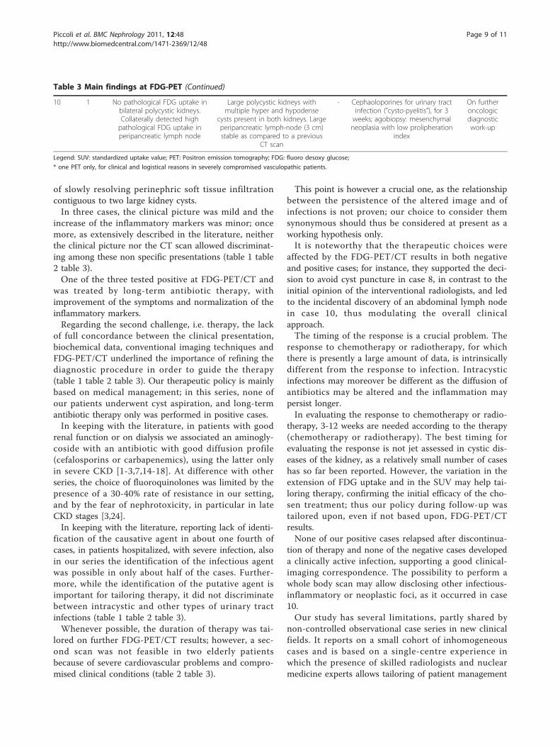
suggestive of infection in 6 patients. The settings were aliver cyst in one case with ADPKD (case 1, Figure 1)and a kidney cyst in 5 cases, 3 affected by ADPKD and2 with multiple cysts (cases 2-6, Figures 2, 3). No meta-bolic activity was recorded in 4 cases (cases 7-10). Incase 10, FDG-PET identified the presence of metabolicactivity in a lymph node (about 3 cm of maximum size),already disclosed by a CT scan one year previously andapparently stable at the CT scan preformed during thestudied episode. Hence, taking into account the highmetabolic activity at the PET scan, the patient under-went an ultrasound-guided agobiopsy, revealing amesenchymal neoplasia with low replicating potential.The patient is presently on oncologic follow-up (table2).
Therapy and follow-upTable 3 summarises the main imaging findings at diag-nosis and during follow-up and the therapeutic choicesperformed integrating the clinical features and the ima-ging data.
Four of the 6 patients testing positive at the first scanunderwent another 8 FDG-PET/CT scans; of note, twocases presented a smouldering picture, in which theFDG-PET/CT scan was the main tool for tailoring theduration of the antibiotic therapy (table 3). In thesecases, the metabolic activity resolved slowly with long-term antibiotic therapy. In two cases, with severely com-promised clinical conditions, only one imaging studywas feasible and long term antibiotic therapy (4-6weeks) was empirically decided on the basis of the firstFDG-PET/CT scan.The therapeutic strategy was based on the clinical pic-
ture and CKD stage. In the patients with GFR > 50 mL/min and in the patient already on dialysis, aminoglyco-sides were used, at least for the first week of therapy, onthe basis of their pharmacological properties, allowingrapid sterilization of blood and urine. In these cases,aminoglycosides were combined with and followed byeither third-generation cefalosporins (ceftriaxone) or bycarbapenemic antibiotics (ertapemen) until resolution ofthe metabolic activity. In the cases with lower GFR,cefalosporins or carbapenemics alone were employed.All patients testing positive at FDG-PET/CT underwentat least one month of intravenous or intramuscular ther-apy (table 3).None of the positive patients with at least 6 months of
follow-up relapsed after the discontinuation of therapy,and no patient testing negative at FDG-PET/CT devel-oped a clinical picture suggestive of intracystic infection(table 3).The therapeutic decision in case 7 (further antibiotic
therapy, in spite of negative FDG-PET/CT) was basedon the presence of a mild residual elevation of CRP (1-1.5 mg/dL, normal values < 0.5 mg/dL), according tothe protocols of the infectivologists, suggesting to dis-continue therapy after one week of normal CRP values.In this case the results of FDG-PET/CT were an argu-ment for avoiding the option of cystic puncture, stronglysuggested by the interventional radiologists.
Concordance analysisAt diagnosis, all but one patient (case 9) displayed amild to severe increase of acute phase reactants. How-ever, the level of CRP, usually chosen as the main mar-ker of infection, was only mildly elevated in 3 cases (2testing positive at FDG-PET/CT scan and 1 testingnegative). The CRP level did not correlate with the pre-sence of fever or the severity of the clinical symptoms,suggesting “background noise” of the presence of multi-ple comorbidities in a relatively old population withCKD (table 1 table 2). Normalization of CRP wasobserved in the positive patients within 3 weeks of anti-biotic therapy; the normalization of CRP preceded the
Piccoli et al. BMC Nephrology 2011, 12:48http://www.biomedcentral.com/1471-2369/12/48
Page 4 of 11
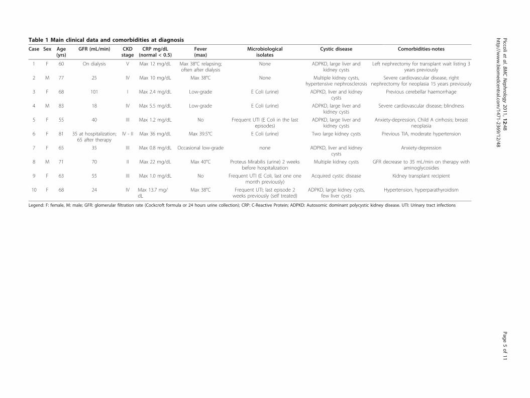
Table 1 Main clinical data and comorbidities at diagnosis
Case Sex Age(yrs)
GFR (mL/min) CKDstage
CRP mg/dL(normal < 0.5)
Fever(max)
Microbiologicalisolates
Cystic disease Comorbidities-notes
1 F 60 On dialysis V Max 12 mg/dL Max 38°C relapsing;often after dialysis
None ADPKD, large liver andkidney cysts
Left nephrectomy for transplant wait listing 3years previously
2 M 77 25 IV Max 10 mg/dL Max 38°C None Multiple kidney cysts,hypertensive nephrosclerosis
Severe cardiovascular disease, rightnephrectomy for neoplasia 15 years previously
3 F 68 101 I Max 2.4 mg/dL Low-grade E Coli (urine) ADPKD, liver and kidneycysts
Previous cerebellar haemorrhage
4 M 83 18 IV Max 5.5 mg/dL Low-grade E Coli (urine) ADPKD, large liver andkidney cysts
Severe cardiovascular disease; blindness
5 F 55 40 III Max 1.2 mg/dL No Frequent UTI (E Coli in the lastepisodes)
ADPKD, large liver andkidney cysts
Anxiety-depression, Child A cirrhosis; breastneoplasia
6 F 81 35 at hospitalization;65 after therapy
IV - II Max 36 mg/dL Max 39.5°C E Coli (urine) Two large kidney cysts Previous TIA, moderate hypertension
7 F 65 35 III Max 0.8 mg/dL Occasional low-grade none ADPKD, liver and kidneycysts
Anxiety-depression
8 M 71 70 II Max 22 mg/dL Max 40°C Proteus Mirabilis (urine) 2 weeksbefore hospitalization
Multiple kidney cysts GFR decrease to 35 mL/min on therapy withaminoglycosides
9 F 63 55 III Max 1.0 mg/dL No Frequent UTI (E Coli, last one onemonth previously)
Acquired cystic disease Kidney transplant recipient
10 F 68 24 IV Max 13.7 mg/dL
Max 38°C Frequent UTI; last episode 2weeks previously (self treated)
ADPKD, large kidney cysts,few liver cysts
Hypertension, hyperparathyroidism
Legend: F: female, M: male; GFR: glomerular filtration rate (Cockcroft formula or 24 hours urine collection); CRP: C-Reactive Protein; ADPKD: Autosomic dominant polycystic kidney disease. UTI: Urinary tract infections
Piccolietal.BM
CNephrology
2011,12:48http://w
ww.biom
edcentral.com/1471-2369/12/48
Page5of
11
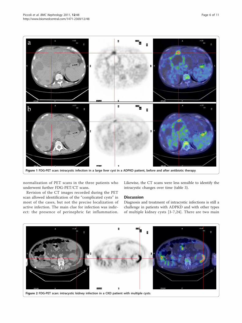
normalization of PET scans in the three patients whounderwent further FDG-PET/CT scans.Revision of the CT images recorded during the PET
scan allowed identification of the “complicated cysts” inmost of the cases, but not the precise localization ofactive infection. The main clue for infection was indir-ect: the presence of perinephric fat inflammation.
Likewise, the CT scans were less sensible to identify theintracystic changes over time (table 3).
DiscussionDiagnosis and treatment of intracystic infections is still achallenge in patients with ADPKD and with other typesof multiple kidney cysts [3-7,24]. There are two main
Figure 1 FDG-PET scan: intracystic infection in a large liver cyst in a ADPKD patient, before and after antibiotic therapy.
Figure 2 FDG-PET scan: intracystic kidney infection in a CKD patient with multiple cysts.
Piccoli et al. BMC Nephrology 2011, 12:48http://www.biomedcentral.com/1471-2369/12/48
Page 6 of 11
points in this challenge: diagnosis of infection and iden-tification of the infected cyst, and definition of type andduration of therapy.The described cases shared the first diagnostic chal-
lenge, i.e. identification of the presence and site of theinfectious foci (table 1; table 2). In contrast with acutepyelonephritis, usually diagnosed in the presence of highfever and severe systemic complaints, the diagnosis ofintracystic infection may not be evident, with non-speci-fic complaints and a smouldering clinical picture[1-7,24]. The wide clinical spectrum of presentations isrepresented in our series, ranging from severe infectionto mild oligosymptomatic disease, also including casesreferred late after several attempts at empirical antibiotictreatment (table 1 table 2 table 3).
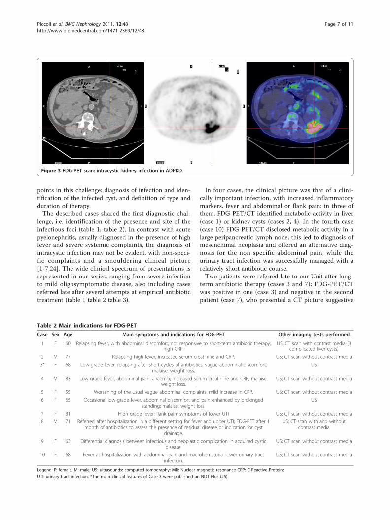
In four cases, the clinical picture was that of a clini-cally important infection, with increased inflammatorymarkers, fever and abdominal or flank pain; in three ofthem, FDG-PET/CT identified metabolic activity in liver(case 1) or kidney cysts (cases 2, 4). In the fourth case(case 10) FDG-PET/CT disclosed metabolic activity in alarge peripancreatic lymph node; this led to diagnosis ofmesenchimal neoplasia and offered an alternative diag-nosis for the non specific abdominal pain, while theurinary tract infection was successfully managed with arelatively short antibiotic course.Two patients were referred late to our Unit after long-
term antibiotic therapy (cases 3 and 7); FDG-PET/CTwas positive in one (case 3) and negative in the secondpatient (case 7), who presented a CT picture suggestive
Figure 3 FDG-PET scan: intracystic kidney infection in ADPKD.
Table 2 Main indications for FDG-PET
Case Sex Age Main symptoms and indications for FDG-PET Other imaging tests performed
1 F 60 Relapsing fever, with abdominal discomfort, not responsive to short-term antibiotic therapy;high CRP.
US; CT scan with contrast media (3complicated liver cysts)
2 M 77 Relapsing high fever, increased serum creatinine and CRP. US; CT scan without contrast media
3* F 68 Low-grade fever, relapsing after short cycles of antibiotics; vague abdominal discomfort,malaise, weight loss.
US
4 M 83 Low-grade fever, abdominal pain; anaemia; increased serum creatinine and CRP, malaise,weight loss.
US; CT scan without contrast media
5 F 55 Worsening of the usual vague abdominal complaints; mild increase in CRP. US; CT scan without contrast media
6 F 65 Occasional low-grade fever, abdominal discomfort and pain enhanced by prolongedstanding; malaise, weight loss.
US
7 F 81 High grade fever; flank pain; symptoms of lower UTI US; CT scan without contrast media
8 M 71 Referred after hospitalization in a different setting for fever and upper UTI; FDG-PET after 1month of antibiotics to assess the presence of residual disease or indication for cyst
drainage.
US; CT scan with and withoutcontrast media
9 F 63 Differential diagnosis between infectious and neoplastic complication in acquired cysticdisease.
US; CT scan without contrast media
10 F 68 Fever at hospitalization with abdominal pain and macrohematuria; lower urinary tractinfection.
US; CT scan without contrast media
Legend: F: female, M: male; US: ultrasounds: computed tomography; MR: Nuclear magnetic resonance CRP: C-Reactive Protein;
UTI: urinary tract infection. *The main clinical features of Case 3 were published on NDT Plus (25).
Piccoli et al. BMC Nephrology 2011, 12:48http://www.biomedcentral.com/1471-2369/12/48
Page 7 of 11
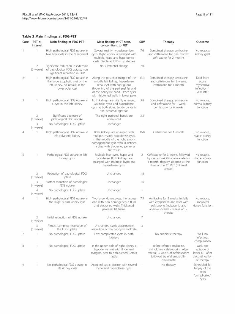
Table 3 Main findings at FDG-PET
Case PET n;interval
Main finding at FDG-PET Main finding at CT scan,concomitant to PET
SUV Therapy Outcome
1 1 High pathological FDG uptake intwo liver cysts in the IV segment
Several mainly hypodense livercysts, Right kidney is enlarged withmultiple, hypo and hyperdensecysts. Stable at follow up studies
7.6 Combined therapy: amikacineand ceftriaxone for one month;
ceftriaxone for 2 months
No relapse;kidney graft
2(6 weeks)
Significant reduction in extensionof pathological FDG uptake; non
significant reduction in SUV
No substantial change 7.0
2* 1 High pathological FDG uptake inthe large exophytic cyst of theleft kidney, no uptake in the
lower polar cyst
Along the posterior margin of themiddle left kidney, hyperdenserenal cyst with contiguous
thickening of the perirenal fat anddense pericystic band. Other cystswith thickened walls in lower pole.
13.3 Combined therapy: amikacineand ceftriaxone for 2 weeks,ceftriaxone for 1 month
Died fromacute
myocardialinfarction 1year later
3 1 High pathological FDG uptake ina cyst in the left kidney
both kidneys are slightly enlarged.Multiple hypo and hyperdense
cysts at both sides. Subtle bands inthe perirenal right fat
3.8 Combined therapy: amikacineand ceftriaxone for 1 week,ceftriaxone for 6 weeks
No relapse,normal kidney
function
2(5 weeks)
Significant decrease ofpathological FDG uptake
The right perirenal bands areattenuated
3.2
3(4 weeks)
No pathological FDG uptake Unchanged -
4 1 High pathological FDG uptake inleft polycystic kidney
Both kidneys are enlarged withmultiple, mainly hypodense cysts,In the middle of the right a non-homogeneous cyst, with ill definedmargins, with thickened perirenal
fat tissue
16.0 Ceftriaxone for 1 month No relapse,stable kidneyfunction
5 1 Pathological FDG uptake in leftkidney cysts
Multiple liver cysts, hyper andhypodense. Both kidneys are
enlarged with multiple, hypo andhyperdense cysts.
2 Ceftriaxone for 3 weeks, followedby oral amoxicillin-clavulanate for1 month; therapy stopped at thetime of the 3rd PET (minimal
uptake)
No relapse,stable kidneyfunction
2(3 weeks)
Reduction of pathological FDGuptake
Unchanged 1.8
3(4 weeks)
Further reduction of pathologicalFDG uptake
Unchanged 1.6
4(4 weeks)
No pathological FDG uptake Unchanged -
6 1 High pathological FDG uptake inthe large (9 cm) kidney cyst
Two large kidney cysts, the largestone with non homogeneous fluidand thickened walls. Thickened
perirenal fat tissue.
7.5 Amikacine for 2 weeks, initiallywith ertapenem, and later withceftriaxone (leukopenia andanemia) overall 9 weeks of i.v.
therapy
No relapse,improved
kidney function
2(3 weeks)
Initial reduction of FDG uptake Unchanged 7
3(5 weeks)
Almost complete resolution ofthe FDG uptake
Unchanged cystic appearance;resolution of the pericystic infiltrate
3
7 1 No pathological FDG uptake Few complicated cysts in bothkidneys
- No antibiotic therapy Well, noinfectious
complication
8 1 No pathological FDG uptake In the upper pole of right kidney ahypodense cyst with ill defined
margins, near to a thickened Gerotafascia
- Before referral: amikacine,chinolones, cefalosporins. Afterreferral: 3 weeks of cefalosporins
followed by oral amoxicillinclavulanate
Well, oneepisode of
lower UTI afterdiscontinuation
of therapy
9 1 No pathological FDG uptake inleft kidney cysts
Acquired cystic disease with severalhypo and hyperdense cysts
- No therapy Scheduled forbiopsy of the
main“complicated”
cysts
Piccoli et al. BMC Nephrology 2011, 12:48http://www.biomedcentral.com/1471-2369/12/48
Page 8 of 11
of slowly resolving perinephric soft tissue infiltrationcontiguous to two large kidney cysts.In three cases, the clinical picture was mild and the
increase of the inflammatory markers was minor; oncemore, as extensively described in the literature, neitherthe clinical picture nor the CT scan allowed discriminat-ing among these non specific presentations (table 1 table2 table 3).One of the three tested positive at FDG-PET/CT and
was treated by long-term antibiotic therapy, withimprovement of the symptoms and normalization of theinflammatory markers.Regarding the second challenge, i.e. therapy, the lack
of full concordance between the clinical presentation,biochemical data, conventional imaging techniques andFDG-PET/CT underlined the importance of refining thediagnostic procedure in order to guide the therapy(table 1 table 2 table 3). Our therapeutic policy is mainlybased on medical management; in this series, none ofour patients underwent cyst aspiration, and long-termantibiotic therapy only was performed in positive cases.In keeping with the literature, in patients with good
renal function or on dialysis we associated an aminogly-coside with an antibiotic with good diffusion profile(cefalosporins or carbapenemics), using the latter onlyin severe CKD [1-3,7,14-18]. At difference with otherseries, the choice of fluoroquinolones was limited by thepresence of a 30-40% rate of resistance in our setting,and by the fear of nephrotoxicity, in particular in lateCKD stages [3,24].In keeping with the literature, reporting lack of identi-
fication of the causative agent in about one fourth ofcases, in patients hospitalized, with severe infection, alsoin our series the identification of the infectious agentwas possible in only about half of the cases. Further-more, while the identification of the putative agent isimportant for tailoring therapy, it did not discriminatebetween intracystic and other types of urinary tractinfections (table 1 table 2 table 3).Whenever possible, the duration of therapy was tai-
lored on further FDG-PET/CT results; however, a sec-ond scan was not feasible in two elderly patientsbecause of severe cardiovascular problems and compro-mised clinical conditions (table 2 table 3).
This point is however a crucial one, as the relationshipbetween the persistence of the altered image and ofinfections is not proven; our choice to consider themsynonymous should thus be considered at present as aworking hypothesis only.It is noteworthy that the therapeutic choices were
affected by the FDG-PET/CT results in both negativeand positive cases; for instance, they supported the deci-sion to avoid cyst puncture in case 8, in contrast to theinitial opinion of the interventional radiologists, and ledto the incidental discovery of an abdominal lymph nodein case 10, thus modulating the overall clinicalapproach.The timing of the response is a crucial problem. The
response to chemotherapy or radiotherapy, for whichthere is presently a large amount of data, is intrinsicallydifferent from the response to infection. Intracysticinfections may moreover be different as the diffusion ofantibiotics may be altered and the inflammation maypersist longer.In evaluating the response to chemotherapy or radio-
therapy, 3-12 weeks are needed according to the therapy(chemotherapy or radiotherapy). The best timing forevaluating the response is not jet assessed in cystic dis-eases of the kidney, as a relatively small number of caseshas so far been reported. However, the variation in theextension of FDG uptake and in the SUV may help tai-loring therapy, confirming the initial efficacy of the cho-sen treatment; thus our policy during follow-up wastailored upon, even if not based upon, FDG-PET/CTresults.None of our positive cases relapsed after discontinua-
tion of therapy and none of the negative cases developeda clinically active infection, supporting a good clinical-imaging correspondence. The possibility to perform awhole body scan may allow disclosing other infectious-inflammatory or neoplastic foci, as it occurred in case10.Our study has several limitations, partly shared by
non-controlled observational case series in new clinicalfields. It reports on a small cohort of inhomogeneouscases and is based on a single-centre experience inwhich the presence of skilled radiologists and nuclearmedicine experts allows tailoring of patient management
Table 3 Main findings at FDG-PET (Continued)
10 1 No pathological FDG uptake inbilateral polycystic kidneys.Collaterally detected highpathological FDG uptake inperipancreatic lymph node
Large polycystic kidneys withmultiple hyper and hypodense
cysts present in both kidneys. Largeperipancreatic lymph-node (3 cm)stable as compared to a previous
CT scan
- Cephaoloporines for urinary tractinfection ("cysto-pyelitis”), for 3weeks; agobiopsy: mesenchymalneoplasia with low prolipheration
index
On furtheroncologicdiagnosticwork-up
Legend: SUV: standardized uptake value; PET: Positron emission tomography; FDG: fluoro desoxy glucose;
* one PET only, for clinical and logistical reasons in severely compromised vasculopathic patients.
Piccoli et al. BMC Nephrology 2011, 12:48http://www.biomedcentral.com/1471-2369/12/48
Page 9 of 11
based on imaging data. As underlined by severalAuthors the diagnosis of intracystic infection is notclear-cut and the criteria are not jet univocal [1-3,7,24].Thus, in a setting where cystic puncture is not routinelyperformed, the diagnosis of intracystic infection cannotdefinitively be proved. However, the concordance withthe follow-up data (no relapse in treated cases; no devel-opment of clinical infection in negative cases) is a strongargument supporting relying on PET results, in a clinicalcontext in which, due also to age and comorbidity, inva-sive procedures are preferentially avoided (table 1).The low availability and high cost of FDG-PET/CT are
probably the main limitations to its wider use. Theselimits are shared also by the two alternative techniquesof scintigraphy with leucocytes labelled with indium orgallium; thus the experience is relatively limited in spiteof a longer history of availability of these techniques[10-12]. The relatively poor spatial discrimination, thehigh radiation activity and the time consuming andcomplex preparation needed for these techniques maysupport a wider use of FDG-PET/CT scan, whose costsare comparable in several settings, including ours [13].However, since less than 30 cases (including ours)
have been reported in the literature, the drawbacks ofthe technique may not yet be clear [24].The strengths of our study are the fact that it’s the
first relatively large prospective study, and the first oneto report on the use of FDG-PET/CT for suspectedinfection not only in ADPKD but also in other cysticdiseases of the kidney.Both the limits and strengths suggest the need of
further studies to define a favourable cost-benefit profileof the procedure and precise indications for itsapplication.
ConclusionsFDG-PET/CT is a very promising tool for the diagnosisand follow-up of infected cysts in ADPKD or in othercystic diseases of the kidney. Further studies are neededto better assess the cost-benefit ratio in order to guidethe systematic use of this technique in the complicationsof ADPKD or other types of cystic kidney diseases.
Acknowledgementsto Dr P Christie for his careful language revision; case 3 was accepted forpublication as Images in Nephrology in Nephrology Dialysis andTransplantation Plus; the publication includes the main clinical features ofthe case and the corresponding images (25). Financial sources: None;conflict of interest: none.
Author details1Department of Clinical and Biological Sciences of the University of Turin,San Luigi Gonzaga Hospital, regione Gonzole 10, Orbassano (TO), 10043 Italy.2Deapartment of Radiology, Ospedale Maria Vittoria, via Cibrario 72, 10100Torino, Italy. 3IRMET SpA, Centro PET, via Onorato Vigliani 89, Torino, 10100Italy.
Authors’ contributionsPG conceived the study, participated in its design and coordination of theclinical management of the patients and drafted the manuscript; CV wasencharged of the clinical management of the patients; DM participated tothe clinical management, and made the bibliographic systematic search; AVcoordinated the imaging studies and participated to the study design; PEwas encharged of the review of the PET analyses; AD participated in thecritical review of the manuscript and in the imaging analysis; PD participatedin the imaging analysis and in the critical review of the images; CGparticipated in the study design, in the manuscript draft and reviewed CTscans. All authors read and approved the final manuscript.The authors also declare that they have made a substantial contribution tothe information or material submitted for publication the manuscript hasbeen read and approved by all authors in this version the manuscript orportions thereof are not under simultaneous consideration by any otherjournal or electronic publication and have not been previously published.
Competing interestsThe authors declare that they have no competing interests.
Received: 25 October 2010 Accepted: 29 September 2011Published: 29 September 2011
References1. Wilson PD: Polycystic kidney disease. New Engl J Med 2004, 350:151-64.2. Torres VE, Harris PC: Autosomal dominant polycystic kidney disease: the
last 3 years. Kidney Int 2009, 76:149-68.3. Sallèe M, Rafat C, Zahar J, Paulmier B, Grunfeld JP, Knebelmann P,
Fakhouri F: Cyst Infections in Patients with Autosomal DominantPolycystic Kidney Disease. Clin J Am Soc Nephrol 2009, 4:1183-1189.
4. Levine E, Cook LT, Grantham JJ: Liver cysts in autosomal dominantpolycystic kidney disease: clinical and computed tomography study. AmJ Roentgenol 1985, 145:229-33.
5. Gibson P, Watson M: Cyst infection in polycystic kidney disease: a clinicalchallenge. Nephrol Dial Transplant 1998, 13:2455-7.
6. Chaveau D, Fakhouri F, Grunfeld JP: Liver involvement in autosomaldominant polycystic kidney disease: therapeutic dilemma. J Am SocNephrol 2000, 11:1767-75.
7. Torres VE, Harris PC, Pirson Y: Autosomal dominant polycystic kidneydisease. Lancet 2007, 369:1287-1301.
8. Levine E, Slusher SL, Grantham JJ, Wetzel LH: Natural history of acquiredrenal cystic disease in dialysis patients: a prospective longitudinal CTstudy. Am J Roentgenol 1991, 156(3):501-6.
9. Ishikawa I, Saito Y, Asaka M, Tomosugi N, Yuri T, Watanabe M, Honda R:Twenty-year follow-up of acquired renal cystic disease. Clin Nephrol 2003,59(3):153-9.
10. Amesur P, Castronuovo JJ, Chandramouly B: Infected cyst localization withgallium SPECT imaging in polycystic kidney disease. Clin Nucl Med 1988,13(1):35-7.
11. Lahiri SA, Halff GA, Speeg KV, Esterl RM Jr: In-111 WBC scan localizesinfected hepatic cysts and confirms their complete resection in adultpolycystic kidney disease. Clin Nucl Med 1998, 23:33-4.
12. Tsang V, Lui S, Hilson A, Moorhead J, Fernando O, Sweny P: Gallium-67scintigraphy in the detection of infected polycystic kidneys in renaltransplant recipients. Nucl Med Commun 1989, 10:167-70.
13. Palestro CJ, Love C, Bhargava KK: Labeled leukocyte imaging: currentstatus and future directions. Q J Nucl Med Mol Imaging 2009, 53:105-23.
14. Kaim AH, Burger C, Ganter CC, Goerres GW, Kamel E, Weishaupt D,Dizendorf E, Schaffner A, von Schulthess GK: PET-CT-guided percutaneouspuncture of an infected cyst in autosomal dominant polycystic kidneydisease: case report. Radiology 2001, 221:818-21.
15. Bleeker-Rovers CP, de Sévaux RG, van Hamersvelt HW, Corstens FH,Oyen WJ: Diagnosis of renal and hepatic cyst infections by 18-F-fluorodeoxyglucose positron emission tomography in autosomaldominant polycystic kidney disease. Am J Kidney Dis 2003, 41:E18-21.
16. Soussan M, Sberro R, Wartski M, Fakhouri F, Pecking AP, Alberini JL:Diagnosis and localization of renal cyst infection by 18F-fluorodeoxyglucose PET/CT in polycystic kidney disease. Ann Nucl Med2008, 22:529-31.
17. DeSouza RM, Prachalias A, Srinivasan P, O’Doherty M, Olsburgh J:Differentiation between infection in kidney and liver cysts in autosomal
Piccoli et al. BMC Nephrology 2011, 12:48http://www.biomedcentral.com/1471-2369/12/48
Page 10 of 11
dominant polycystic kidney disease: use of PET-CT in diagnosis and toguide management. Transplant Proc 2009, 41:1942-5.
18. Jumenez Bonilla JF, Quirce R, Calabia ER, Banzo I, Martinez-Rodriguez I,Carril JM: Hepatorenal polycystic disease and fever: diagnosticcontribution of Gallium Citrate Ga 67 scan and fluorine F18 FDG-PET/CT.Eur Urol 2009, Available online 8 June 2009.
19. Bleeker Rovers CP, Corstens FHM, Van Der Meer JVM, Oyen WJG: Fever ofunknown origin: prospective comparison of diagnostic value of (18)F-FDG PET and (111)In-granulocyte scintigraphy. Eur J Nucl Med MolImaging 2004, 31:1342-1344.
20. Meller J, Altenvoerde G, Munzel U, Jauho A, Behe M, Gratz S, Luig H,Becker W: Fever of unknown origin: prospective comparison of [18F]FDGimaging with a double-head coincidence camera and gallium-67 citrateSPET. Eur J Nucl Med 2000, 27:1617-25.
21. Keidar Z, Gurman-Balbir A, Gaitini D, Israel O: Fever of unknown origin: therole of 18F-FDG PET/CT. J Nucl Med 2008, 49:1980-5.
22. Meller J, Sahlmann CO, Sheel AK: 18F-FDG PET and PET/CT in fever ofunknown origin. J Nucl Med 2007, 48:35-45.
23. Pei Y, Obaji J, Dupuis A, Paterson AD, Magistroni R, Dicks E, Parfrey P,Cramer B, Coto E, Torra R, San Millan SL, Gibson R, Breuning M, Peters D,Ravine D: Unified Criteria for Ultrasonographic diagnosis of ADPKD. J AmSoc Nephrol 2009, 20:205-212.
24. Alam A, Perrone RD: Managing Cyst Infections in ADPKD: An old problemlooking for new answers. Clin J Am Soc Nephrol 2009, 4:1154-1155.
25. Piccoli GB, Arena V, Consiglio V, Depascale A, Deagostini MC: Positronemission tomography: a precious tool in the challenge of the infectedcysts in ADPKD. NDT Plus 2010, 3:492-493.
Pre-publication historyThe pre-publication history for this paper can be accessed here:http://www.biomedcentral.com/1471-2369/12/48/prepub
doi:10.1186/1471-2369-12-48Cite this article as: Piccoli et al.: Positron emission tomography in thediagnostic pathway for intracystic infection in adpkd and “cystic”kidneys. a case series. BMC Nephrology 2011 12:48.
Submit your next manuscript to BioMed Centraland take full advantage of:
• Convenient online submission
• Thorough peer review
• No space constraints or color figure charges
• Immediate publication on acceptance
• Inclusion in PubMed, CAS, Scopus and Google Scholar
• Research which is freely available for redistribution
Submit your manuscript at www.biomedcentral.com/submit
Piccoli et al. BMC Nephrology 2011, 12:48http://www.biomedcentral.com/1471-2369/12/48
Page 11 of 11


![PET/ CT [Positron Emission Tomography]](https://img.pdfslide.net/doc/110x75/56d6bf451a28ab30169592f3/pet-ct-positron-emission-tomography.jpg)


