Embed Size (px)
Citation preview
Positron Emission Tomography (PET) Images �
Each set of PET images below contains four images of a human brain. The four images show cross-sections taken at different levels of the brain.
Set 1 Set 2 a b a b
Set 4 Set 5
a b a b
c d
Set 6
a b
c d c d
Set 3 highest activity a b
c d lowest activity
Cop
yrig
ht ©
200
0 by
BSC
S an
d V
ideo
dis
cove
ry, I
nc. P
erm
issi
on g
rant
ed f
or c
lass
room
use
. Up
dat
ed 2
009.
Master 1.1a
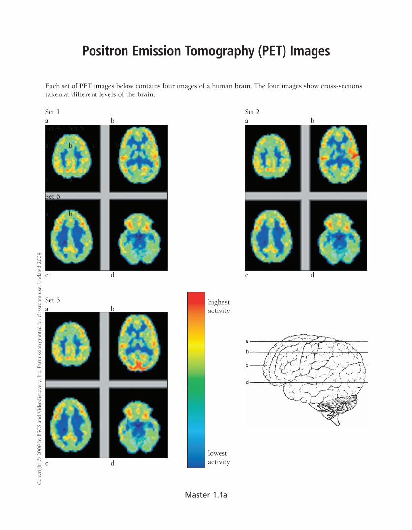
Set 4 Set 5 a b a b
c d c d
Set 6 a b
c d
PET images provided by: �
Sanjiv S. Gambhir, M.D., Ph.D. � UCLA School of Medicine �
Crump Institute for Biological Imaging � Copyright 1998 �
Regents of the University of California �
Cop
yrig
ht ©
200
0 by
BSC
S an
d V
ideo
dis
cove
ry, I
nc. P
erm
issi
on g
rant
ed f
or c
lass
room
use
. Up
dat
ed 2
009.
Master 1.1b
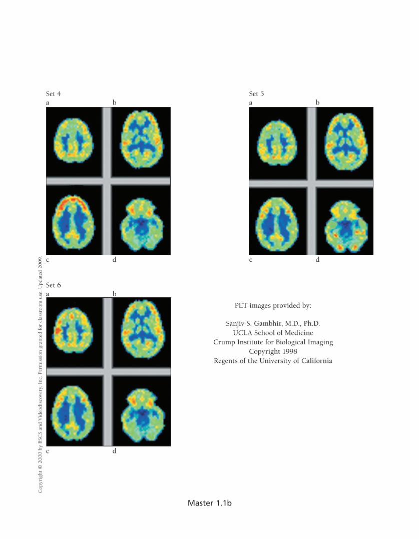
Interpreting PET Images �
Name(s)______________________________________________________________ Date ______________
1. � When you look at the images that make up Set #1 (Master 1.1), how do the four images differ from each other?
2. � Why are four images shown in each set of PET images? Why would scientists need to examine more than one PET image taken of a subject’s brain?
3. � When comparing the images in Set #1 with the images in Sets #2, 3, 4, 5, and 6, how is the activity of the brain in each of these sets different from Set #1’s?
Set Number
Identify the image that shows the greatest change
(a, b, c, or d) Describe the change in brain activity
2
3
4
5
6
Cop
yrig
ht ©
200
0 by
BSC
S an
d V
ideo
dis
cove
ry, I
nc. P
erm
issi
on g
rant
ed f
or c
lass
room
use
. Up
dat
ed 2
009.
Master 1.2a
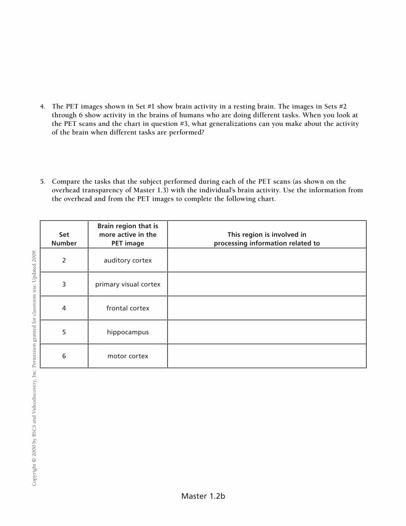
4. The PET images shown in Set #1 show brain activity in a resting brain. The images in Sets #2 through 6 show activity in the brains of humans who are doing different tasks. When you look at the PET scans and the chart in question #3, what generalizations can you make about the activity of the brain when different tasks are performed?
5. Compare the tasks that the subject performed during each of the PET scans (as shown on the overhead transparency of Master 1.3) with the individual’s brain activity. Use the information from the overhead and from the PET images to complete the following chart.
Set Number
Brain region that is more active in the
PET image This region is involved in
processing information related to
2 auditory cortex
3 primary visual cortex
4 frontal cortex
5 hippocampus
6 motor cortex
Cop
yrig
ht ©
200
0 by
BSC
S an
d V
ideo
dis
cove
ry, I
nc. P
erm
issi
on g
rant
ed f
or c
lass
room
use
. Up
dat
ed 2
009.
Master 1.2b
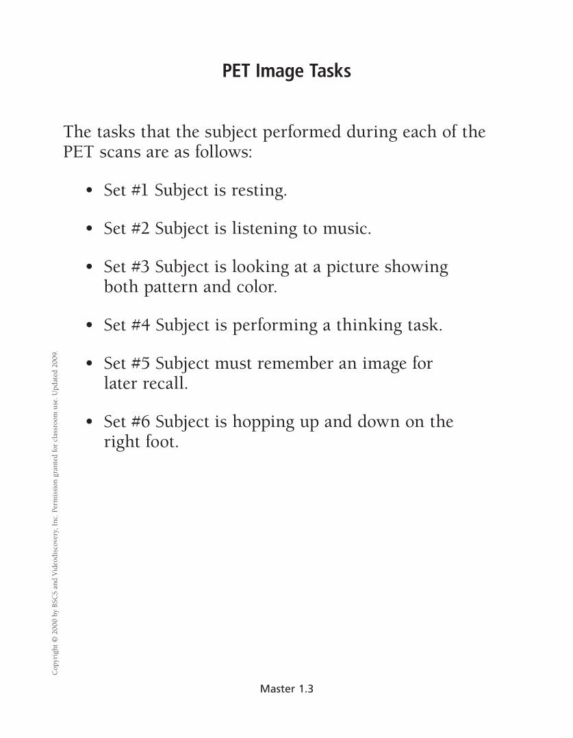
PET Image Tasks �
The tasks that the subject performed during each of the PET scans are as follows:
• Set #1 Subject is resting.
• Set #2 Subject is listening to music.
• Set #3 Subject is looking at a picture showing both pattern and color.
• Set #4 Subject is performing a thinking task.
• Set #5 Subject must remember an image for later recall. �
• Set #6 Subject is hopping up and down on the right foot.
Cop
yrig
ht ©
200
0 by
BSC
S an
d V
ideo
dis
cove
ry, I
nc. P
erm
issi
on g
rant
ed f
or c
lass
room
use
. Up
dat
ed 2
009.
Master 1.3
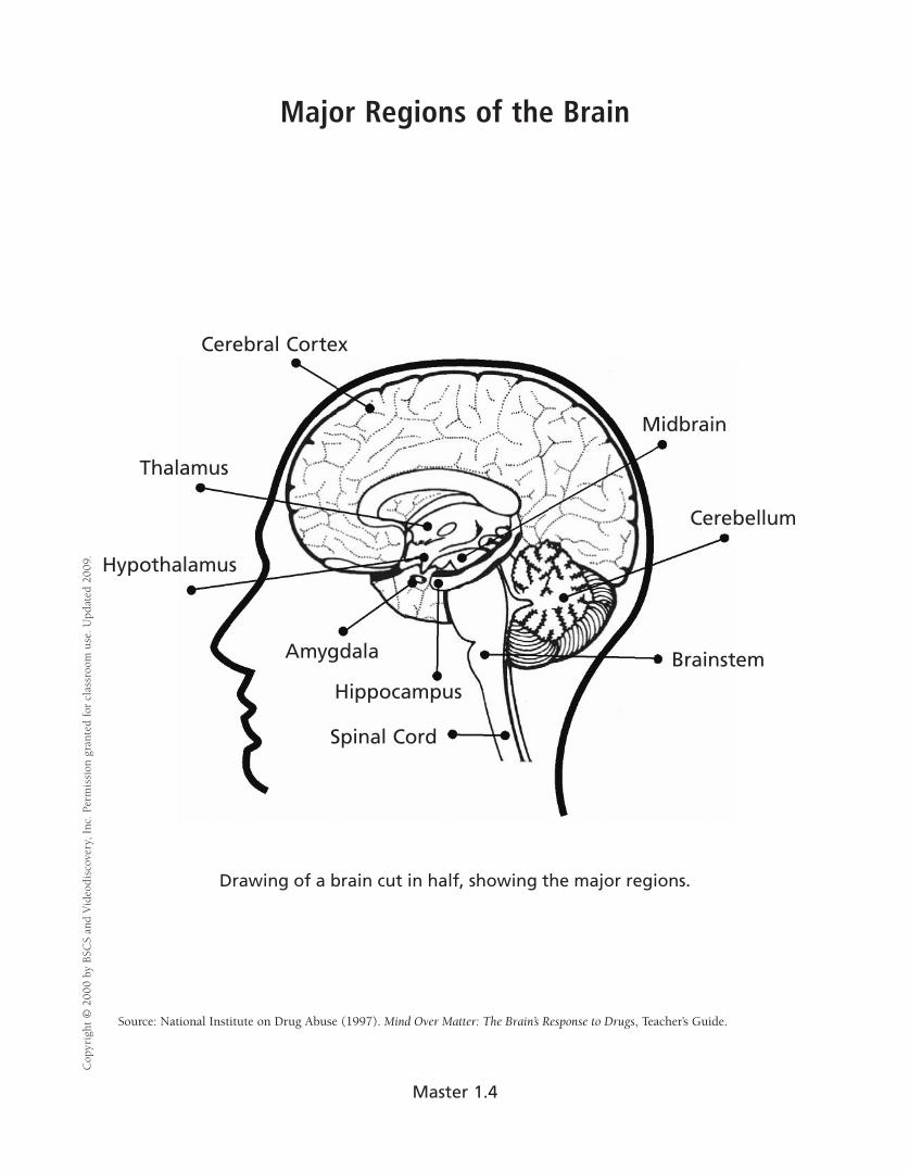
Major Regions of the Brain �
Cerebral Cortex
Midbrain
Thalamus
Cerebellum
Hypothalamus
Amygdala
Hippocampus
Spinal Cord
Brainstem
Drawing of a brain cut in half, showing the major regions.
Source: National Institute on Drug Abuse (1997). Mind Over Matter: The Brain’s Response to Drugs, Teacher’s Guide.
Cop
yrig
ht ©
200
0 by
BSC
S an
d V
ideo
dis
cove
ry, I
nc. P
erm
issi
on g
rant
ed f
or c
lass
room
use
. Up
dat
ed 2
009.
Master 1.4
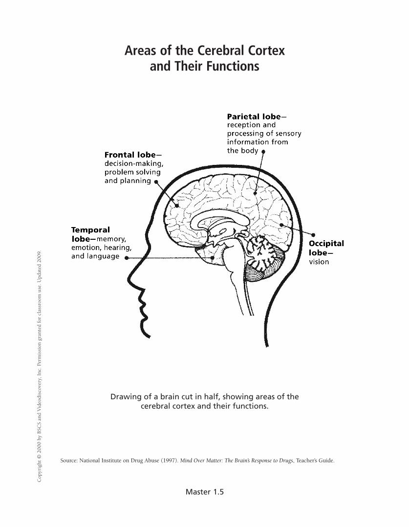
Areas of the Cerebral Cortex and Their Functions �
Drawing of a brain cut in half, showing areas of the cerebral cortex and their functions.
Source: National Institute on Drug Abuse (1997). Mind Over Matter: The Brain’s Response to Drugs, Teacher’s Guide.
Cop
yrig
ht ©
200
0 by
BSC
S an
d V
ideo
dis
cove
ry, I
nc. P
erm
issi
on g
rant
ed f
or c
lass
room
use
. Up
dat
ed 2
009.
Master 1.5
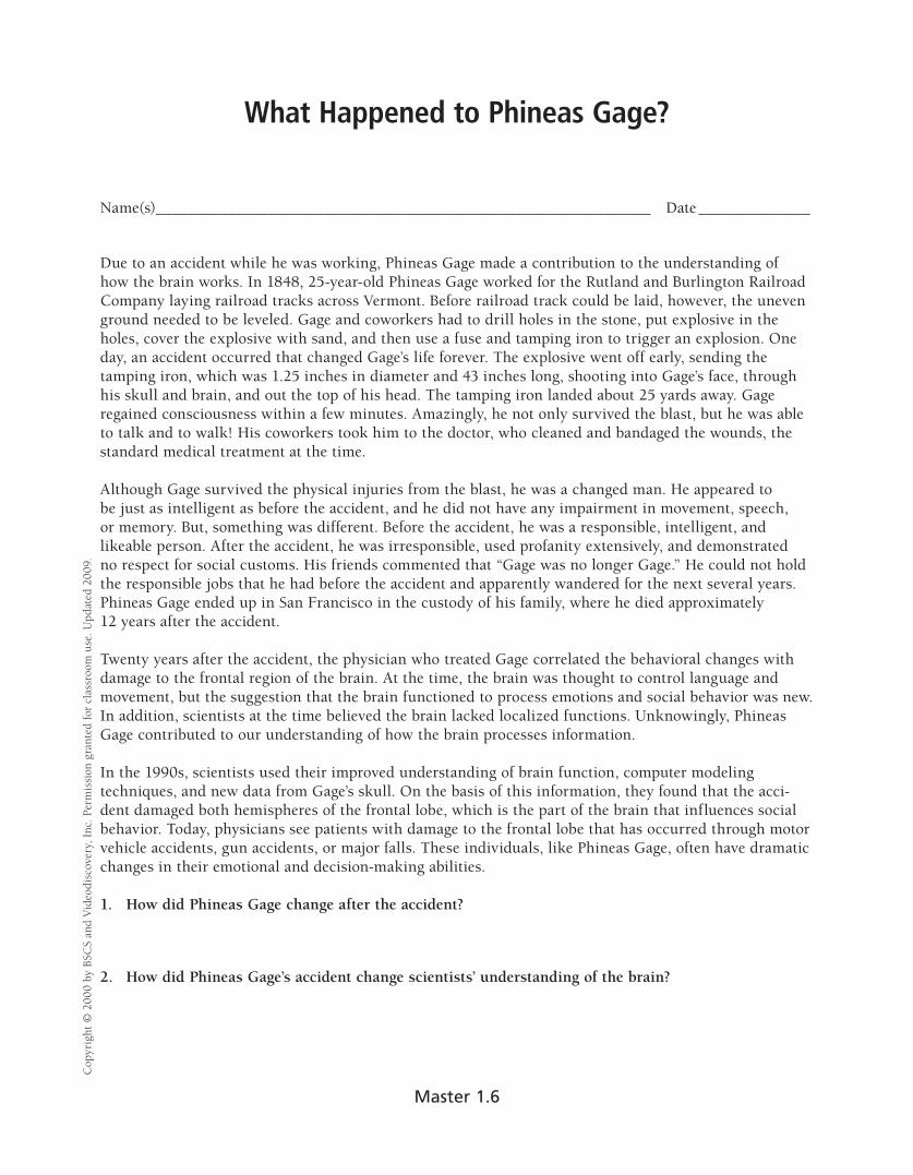
What Happened to Phineas Gage?
Name(s)______________________________________________________________ Date ______________
Due to an accident while he was working, Phineas Gage made a contribution to the understanding of how the brain works. In 1848, 25-year-old Phineas Gage worked for the Rutland and Burlington Railroad Company laying railroad tracks across Vermont. Before railroad track could be laid, however, the uneven ground needed to be leveled. Gage and coworkers had to drill holes in the stone, put explosive in the holes, cover the explosive with sand, and then use a fuse and tamping iron to trigger an explosion. One day, an accident occurred that changed Gage’s life forever. The explosive went off early, sending the tamping iron, which was 1.25 inches in diameter and 43 inches long, shooting into Gage’s face, through his skull and brain, and out the top of his head. The tamping iron landed about 25 yards away. Gage regained consciousness within a few minutes. Amazingly, he not only survived the blast, but he was able to talk and to walk! His coworkers took him to the doctor, who cleaned and bandaged the wounds, the standard medical treatment at the time.
Although Gage survived the physical injuries from the blast, he was a changed man. He appeared to be just as intelligent as before the accident, and he did not have any impairment in movement, speech, or memory. But, something was different. Before the accident, he was a responsible, intelligent, and likeable person. After the accident, he was irresponsible, used profanity extensively, and demonstrated no respect for social customs. His friends commented that “Gage was no longer Gage.” He could not hold the responsible jobs that he had before the accident and apparently wandered for the next several years. Phineas Gage ended up in San Francisco in the custody of his family, where he died approximately 12 years after the accident.
Twenty years after the accident, the physician who treated Gage correlated the behavioral changes with damage to the frontal region of the brain. At the time, the brain was thought to control language and movement, but the suggestion that the brain functioned to process emotions and social behavior was new. In addition, scientists at the time believed the brain lacked localized functions. Unknowingly, Phineas Gage contributed to our understanding of how the brain processes information.
In the 1990s, scientists used their improved understanding of brain function, computer modeling techniques, and new data from Gage’s skull. On the basis of this information, they found that the acci-dent damaged both hemispheres of the frontal lobe, which is the part of the brain that influences social behavior. Today, physicians see patients with damage to the frontal lobe that has occurred through motor vehicle accidents, gun accidents, or major falls. These individuals, like Phineas Gage, often have dramatic changes in their emotional and decision-making abilities.
1. How did Phineas Gage change after the accident?
2. How did Phineas Gage’s accident change scientists’ understanding of the brain?
Cop
yrig
ht ©
200
0 by
BSC
S an
d V
ideo
dis
cove
ry, I
nc. P
erm
issi
on g
rant
ed f
or c
lass
room
use
. Up
dat
ed 2
009.
Master 1.6
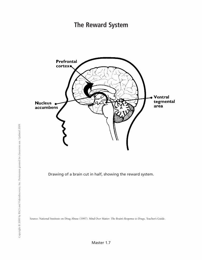
The Reward System �
Drawing of a brain cut in half, showing the reward system.
Source: National Institute on Drug Abuse (1997). Mind Over Matter: The Brain’s Response to Drugs, Teacher’s Guide.
Cop
yrig
ht ©
200
0 by
BSC
S an
d V
ideo
dis
cove
ry, I
nc. P
erm
issi
on g
rant
ed f
or c
lass
room
use
. Up
dat
ed 2
009.
Master 1.7


![PET/ CT [Positron Emission Tomography]](https://img.pdfslide.net/doc/110x75/56d6bf451a28ab30169592f3/pet-ct-positron-emission-tomography.jpg)