Embed Size (px)
Citation preview

Post mortem findings
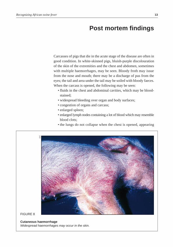
Carcasses of pigs that die in the acute stage of the disease are often ingood condition. In white-skinned pigs, bluish-purple discolourationof the skin of the extremities and the chest and abdomen, sometimeswith multiple haemorrhages, may be seen. Bloody froth may issuefrom the nose and mouth; there may be a discharge of pus from theeyes; the tail and area under the tail may be soiled with bloody faeces.When the carcass is opened, the following may be seen:
• fluids in the chest and abdominal cavities, which may be blood-stained;
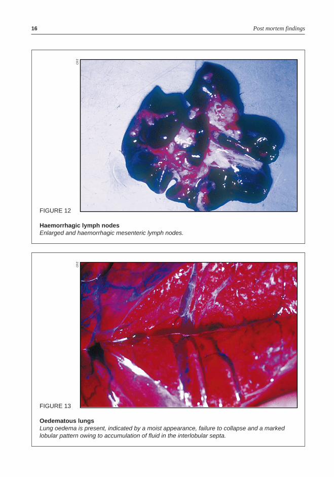
• widespread bleeding over organ and body surfaces;• congestion of organs and carcass;• enlarged spleen;• enlarged lymph nodes containing a lot of blood which may resemble
blood clots;• the lungs do not collapse when the chest is opened, appearing
Recognizing African swine fever 13
FIGURE 8
Cutaneous haemorrhageWidespread haemorrhages may occur in the skin.
OV
I
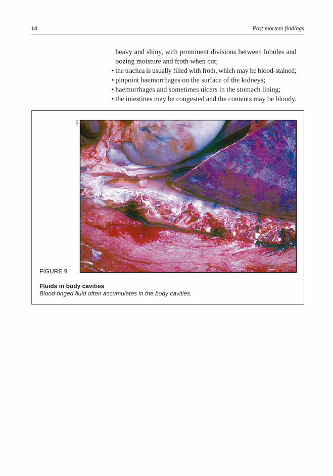
heavy and shiny, with prominent divisions between lobules andoozing moisture and froth when cut;
• the trachea is usually filled with froth, which may be blood-stained;• pinpoint haemorrhages on the surface of the kidneys;• haemorrhages and sometimes ulcers in the stomach lining;• the intestines may be congested and the contents may be bloody.
Post mortem findings14
FIGURE 9
Fluids in body cavitiesBlood-tinged fluid often accumulates in the body cavities.
OV
I
Recognizing African swine fever 15
FIGURE 10
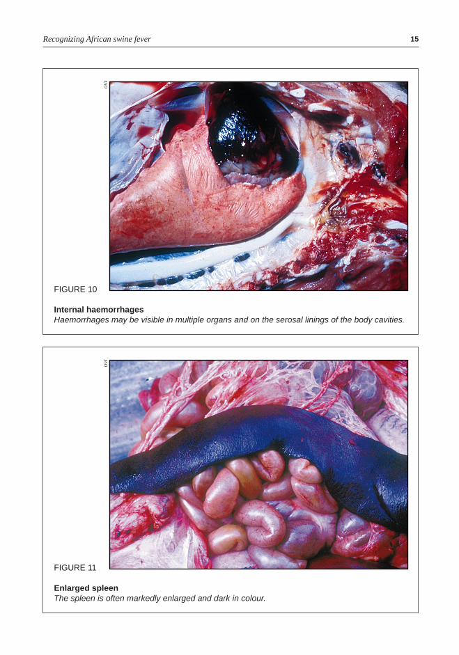
Internal haemorrhagesHaemorrhages may be visible in multiple organs and on the serosal linings of the body cavities.
OV
I
FIGURE 11
Enlarged spleenThe spleen is often markedly enlarged and dark in colour.
OV
I
Post mortem findings16
FIGURE 13
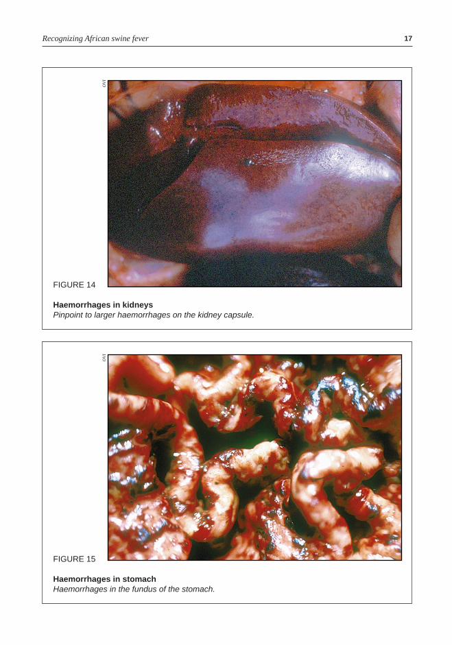
Oedematous lungsLung oedema is present, indicated by a moist appearance, failure to collapse and a markedlobular pattern owing to accumulation of fluid in the interlobular septa.
OV
I
FIGURE 12
Haemorrhagic lymph nodesEnlarged and haemorrhagic mesenteric lymph nodes.
OV
I
Recognizing African swine fever 17
FIGURE 14
Haemorrhages in kidneysPinpoint to larger haemorrhages on the kidney capsule.
OV
I
FIGURE 15
Haemorrhages in stomachHaemorrhages in the fundus of the stomach.
OV
I
Post mortem findings18
FIGURE 16
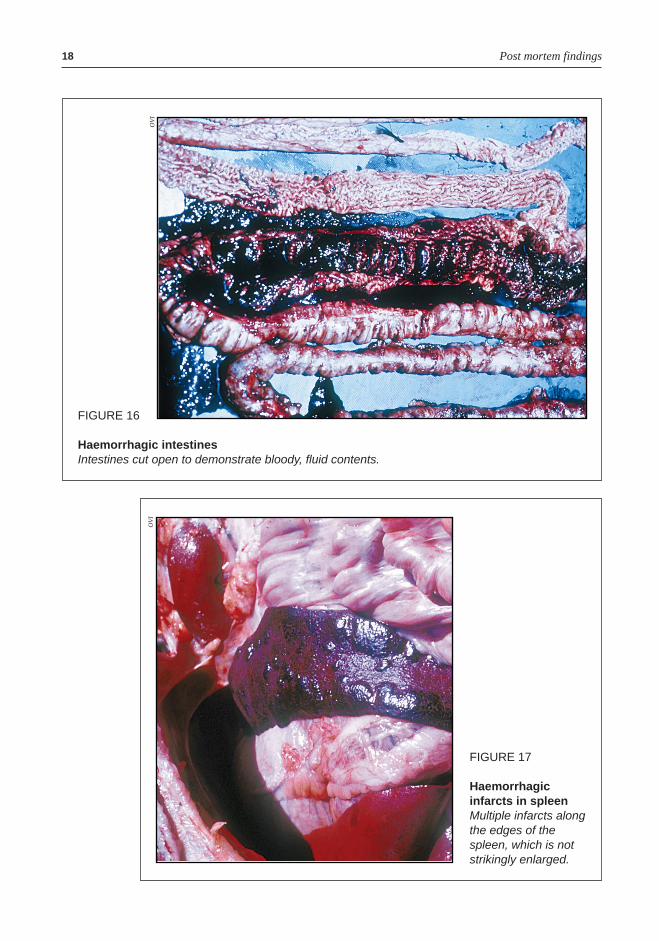
Haemorrhagic intestinesIntestines cut open to demonstrate bloody, fluid contents.
OV
I
FIGURE 17
Haemorrhagicinfarcts in spleenMultiple infarcts alongthe edges of thespleen, which is notstrikingly enlarged.
OV
I
Subacute ASF may be characterized by the following changes:• fluids may be present in body cavities (heart failure);• lymph nodes are enlarged and often haemorrhagic;• fibrin may be present on the surfaces of the lungs and the heart;• lungs may be firm with a mottled appearance, due to pneumonia;• joints may be swollen with accumulated fluid and fibrin.
Chronic ASF is characterized by:• emaciation;• sores and ulcers over bony points;• lymph nodes are enlarged and firm;• a layer of fibrin may be present over the lungs and heart;• swollen joints.
Recognizing African swine fever 19

Differential diagnosis
Few diseases of domestic pigs cause mortality at the rate observed inan acute ASF outbreak in newly infected pig herds. The most importantdifferential diagnosis for ASF is classical swine fever (hog cholera),which is caused by a completely different virus but presents almostidentical clinical signs and post-mortem lesions. The only way todistinguish reliably between classical swine fever and ASF is byidentifying the virus. Post-mortem lesions that have been used todistinguish between the two diseases, such as ulcers in the area wherethe small and large intestines meet and areas of bleeding and tissuedestruction in the spleen, are known as infarcts (Figure 16). These arevariably present and not reliable. It is unwise to attempt vaccinationagainst classical swine fever until the diagnosis is confirmed, as ASFcan easily be spread during a vaccination campaign.
Excessive mortality may be difficult to judge in small pig herds,where four out of five pigs may die from a variety of causes, includingparasitism and malnutrition. When a significant number of pigs in anyherd or group die, it is advisable to find out whether any other pigowners in the area have experienced similar recent losses.Other diseases that my be confused with ASF are as follows:
ErysipelasThis is a bacterial disease and is known as one of the “red fevers”. Pigsof all ages may be affected, and the disease is as likely to affect small-scale and extensive pig farms as commercial, intensive units. Mortalityis usually much lower than in ASF and there are usually some pigs thatwill show the typical diamond-shaped skin lesions. Pigs respond wellto treatment with penicillin. Bacterial isolation will confirm the diagnosis.The microscopic changes differ from those typical of ASF.
Salmonellosis, septicaemic pasteurellosis and otherbacterialsepticaemiasFeatures in common with ASF include fever, loss of appetite, respiratoryor gastro-intestinal disorders and a congested, fevered carcass atslaughter. Pigs of a particular, typically younger, age group are usuallyaffected. Animals treated in time may respond to antimicrobial therapy.Confirmation of the diagnosis is by culture of the bacteria.
Recognizing African swine fever 21
TrypanosomosisTrypanosomosis is caused by blood parasites that are transmitted bytsetse flies. Many deaths among pigs of all ages can occur and the pigsmay die too quickly to develop typical signs of anaemia (lack of blood)or icterus (jaundice). This disease is so severe that pigs are seldomproduced in areas where it occurs. The parasite is easily demonstratedon blood smears stained with Giemsa or Romanoff stains (e.g. diffquick).
When a large number of pigs die suddenly, the possibility of poisoningshould be considered. Few poisons result in the severe bleeding seenin ASF. Coumarin-based rat poisons such as warfarin can causewidespread bleeding but are unlikely to affect more than a few pigsin a herd. Certain fungal poisons found in mouldy feed, such as aflatoxinand stachybotryotoxin, may cause haemorrhage and severe mortality.
Differential diagnosis22