Embed Size (px)
Citation preview
Research ArticlePredictors for Gingival Index in Middle-AgedAsian Indians with Type 2 Diabetes from South India:A Cross-Sectional Observational Study
S. Jai Karthik ,1,2 Shajith Anoop,1 R. Suresh Kumar,3 andM. V. Usha Rani1
1Department of Environmental Sciences, Bharathiar University, Coimbatore, Tamil Nadu, India2RVS Dental College, Coimbatore, Tamil Nadu, India3Suresh Diabetes Centre, Vadavalli, Coimbatore, Tamil Nadu, India
Correspondence should be addressed to S. Jai Karthik; [email protected]
Received 26 June 2017; Revised 2 November 2017; Accepted 19 November 2017; Published 2 January 2018
Academic Editor: Andreas K. Nussler
Copyright © 2018 S. Jai Karthik et al. This is an open access article distributed under the Creative Commons Attribution License,which permits unrestricted use, distribution, and reproduction in any medium, provided the original work is properly cited.
Asian Indians develop type 2 diabetes mellitus (T2DM) much earlier as compared to White Caucasians, due to unique phenotypicand genetic architecture. Periodontitis in T2DM patients is often a neglected clinical feature. This study was conducted to derivepredictor variables for gingival index in middle-aged Asian Indians with T2DM in a semiurban population of Dravidian ethnicityfrom Tamil Nadu, India. T2DM patients (𝑛 = 232, mean age: 50.6 ± 10.4 years) with periodontitis (𝑛 = 123, mean age: 54.3 ± 2.4years) and without periodontitis (𝑛 = 109, mean age: 55.2±3.1 years) were recruited between 2014 and 2016 by purposive samplingmethod. Dental examinations for pocket depth (PD) and clinical attachment level (CAL) were performed and gingival index wascalculated. Fasting venous blood samples were analysed for measures of glycaemia and cholesterol. Significant positive correlation(𝑝 < 0.01) was observed for gingival index with glycosylated haemoglobin (HbA1c), pocket depth, presence of T2DM, and clinicalattachment level. Stepwisemultiple linear regression analysis derived increased pocket depth (𝑝 < 0.01), elevatedHbA1c (𝑝 < 0.01),clinical attachment level (𝑝 < 0.01), and presence of diabetes (𝑝 < 0.01) as significant predictors (𝑟2 value = 0.67) for increasedgingival index inmiddle aged patients with T2DM.These variables significantly (𝑝 < 0.01) predispose middle-aged T2DMpatientsto increased gingival index, thus warranting appropriate intervention.
1. Introduction
The incidence of type 2 diabetes mellitus (T2DM) is evergrowing in Southern Asia and is predicted to increase alongwith urbanization [1]. Among other secondary complications[2, 3] a close link exists between T2DM, periodontitis, andliver diseases [4]. Importantly, periodontal disease is associ-ated with cardiovascular diseases and is predictive of futurecardiac events [5]. A bidirectional relationship exists betweenT2DM and periodontal disease [6] which is attributed to per-sisting hyperglycemia, leading to an exaggerated immuno-inflammatory response to pathogenic microbial challenge inthe gingiva [7]. In T2DM, persisting hyperglycemia causesnon-enzymatic glycation and oxidation of proteins and lipids,leading to the subsequent formation of advanced glycationend products (AGEs) [8]. AGEs with accompanying markers
for increased oxidative stress have been demonstrated inhuman gingiva in T2DM subjects with periodontitis, leadingto rapid and severe periodontal tissue destruction [9].
Importantly, dental health in T2DM patients is indica-tive of glycaemic control and can also predict future riskof retinopathy and neuropathy [10]. With the increasingprevalence of T2DM in Asian Indians and its comorbiditiesincluding periodontitis, it is alarming to note that dentalhealth in T2DM patients, especially the middle aged, is muchneglected.
We hypothesized that glycaemic status in T2DM patientsis closely related to gingival index and aimed to determine theclinical variables that correlatewith gingival index in a sampleof middle aged T2DM subjects with and without chronicperiodontitis from a semiurban population of South India.We aimed to derive simple predictors that can be applied for
Hindawie Scientific World JournalVolume 2018, Article ID 9081572, 7 pageshttps://doi.org/10.1155/2018/9081572
2 The Scientific World Journal
early detection of periodontal risk in T2DM patients agedabove 50 years, such that preventive therapy can be initiated.
2. Methods
This cross-sectional study was conducted in a dental carecentre at Coimbatore, Tamil Nadu. Ethical approval wasobtained from the Institutional Ethics Committee of RVSDental College, Coimbatore, prior to the commencement ofthe study. The objectives were explained to all participatingsubjects and signed and written consent was obtained priorto enrollment in the study.
2.1. Subject Population and Selection. Purposive convenientsamplingmethodwas adopted. Aminimum sample size of 80subjects for each arm was determined with an alpha error of5% at 90% power (95%CI). A total of 232middle-aged (above50 years of age) patients with T2DMwere recruited. Inclusioncriteria were presence of clinically diagnosed T2DM at least12 months prior to the start of the study and at least 4fully erupted teeth. A questionnaire based interview wasadministered to participants to assess awareness of dentalhealth and hygiene.Quality of dental hygienewas determinedas frequency and method of brushing, frequency of toothbrush change, dental flossing, number of dental visits forscaling per year, habitual chewing of tobacco, and pyorrhea.Clinical history of patients was obtained with consent fromthe treating physician and the patient. T2DM subjects withchronic history of smoking, alcoholism, dental abscesses,overt pyorrhea, use of dental braces, and capping or missingteeth were excluded from the study.
2.2. Study Protocol. Periodontal examinations were per-formed using sterile instruments by a single examiner forall study subjects. Pocket depth (PD) measurements wereobtained by using William’s periodontal probe. The PD wasmeasured from the free gingival margin to the base of thepocket. The probe was maintained parallel to the long axis ofthe tooth. Pocket depth and clinical attachment level (CAL)were measured for each tooth at 6 sites, namely, mesiobuccal,midbuccal, distobuccal, mesiolingual, midlingual, and dis-tolingual. Clinical attachment level (CAL) was determinedby measuring the distance from the cement enamel junction(CEJ) to the gingival margin with a William’s periodontalprobe. Gingival index (GI) was determined and recordedat 4 gingival sites per tooth using the following criteria:(0) normal gingiva, (1) mild gingivitis without bleeding onprobing, (2) moderate gingivitis with bleeding on probing,and (3) severe gingivitis with ulceration and spontaneousbleeding. The sum of the scores from the four areas of eachtooth was divided by 4 to derive the gingival index for thattooth. The mean GI score was obtained after calculatingindividual GI [11].
Fasting blood glucose (FBG) after an eight-hourovernight fast, 120min postprandial blood glucose (PPBG),glycosylated hemoglobin (HbA1c), and serum cholesterolwere analysed for all subjects on the same day. FBG andPPBGwere analysed by glucose-oxidase peroxidasemethod. HbA1cwas estimated by high performance liquid chromatography
and expressed as percentage value. Serum lipid profilewas measured by methods mentioned in a previous studyfrom our group [12]. Overweight and obesity were definedin accordance with the cut-offs for Asian Indians [13].Dyslipidemia was defined by the criteria formulated by theNational Cholesterol Education Program, Adult TreatmentPanel III (NCEP–ATP III) [14]. T2DM was diagnosed iffasting plasma glucose was ≥126mg/dL and/or 120minpostprandial plasma glucose was ≥200mg/dL or there wasself-reported medication for diabetes by the participant [15].
3. Data Analysis
Statistical analysis was performed using statistical softwareSTATA (version 14.2, StataCorp, College Station, Texas,USA). Data was checked for normative distribution and anindependent samples t-test was applied for continuous vari-ables. Pearson’s correlation test and stepwise multiple linearregression (SMLR) analysis were used to analyse correlationsand to derive predictor variables, respectively. The 𝑝 value ≤0.05 was considered as statistically significant.
4. Results
The sample size was matched for age and BMI. Of the 232obese, middle aged T2DM subjects recruited in this study,123 subjects with T2DM (53.01%) had no indications ofperiodontitis whereas 109 subjects (46.55%) with T2DM hadchronic periodontitis.Themean values of BMI, FBG, HbA1c,mean pocket depth, and gingival index were comparativelyhigher in T2DM patients with significant differences inclinical attachment level (CAL), probing depth (PPD), andgingival index (Table 1). Genderwise comparisons revealedno significant differences in the clinical profile, clinicalattachment level, gingival index, and pocket depth (Table 2).
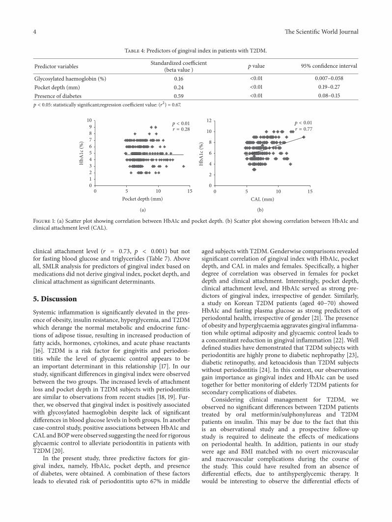
In this study, patients with T2DM and periodontitishad periodontal pockets measuring at least 4mm deep withbleeding on probing and radiographic evidence of boneloss above 50%. T2DM patients without periodontitis hadperiodontal pockets measuring less than 4mm deep withno radiographic evidence of bone loss. Correlation statis-tics revealed significant positive correlation (𝑝 < 0.001)between gingival index, pocket depth, presence of diabetes,and clinical attachment level (Table 3). Significant positivecorrelation was also observed between HbA1c and pocketdepth (Figure 1(a)) and clinical attachment level (Figure 1(b)).Irrespective of age and gender, the predictive variables forgingival index in the study cohort were HbA1c (𝑝 < 0.01),pocket depth (𝑝 < 0.01), and presence of diabetes (𝑝 < 0.01),with a regression coefficient (𝑟2) value of 0.67 (Table 4).
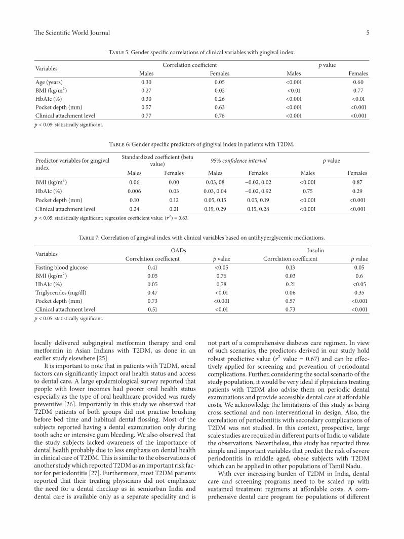
Genderwise correlations revealed significant positive cor-relation of gingival index with age, (𝑝 < 0.001), BMI (𝑝 <0.001), HbA1c (𝑝 < 0.001), pocket depth (𝑝 < 0.001), andclinical attachment level (𝑝 < 0.001) in males. However,though significant correlations were observed for HbA1c,pocket depth (𝑝 < 0.001), and clinical attachment level (𝑝 <0.001), no significant correlation was observed for gingivalindex with age and BMI in females (Table 5).
The Scientific World Journal 3
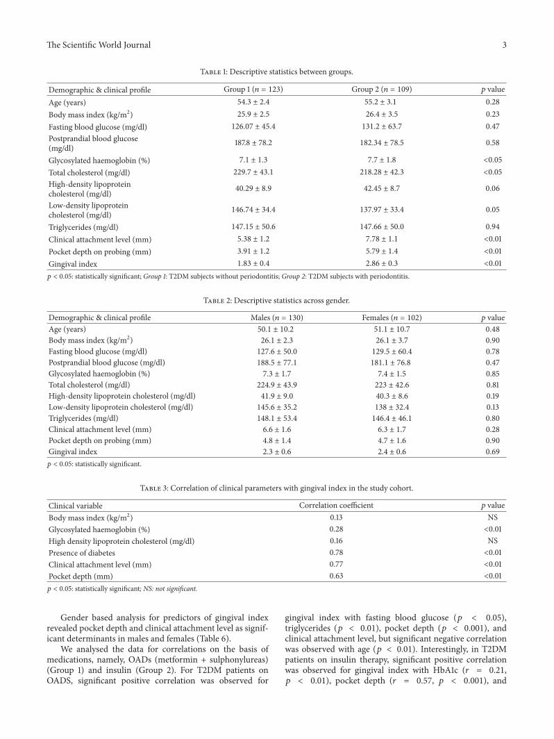
Table 1: Descriptive statistics between groups.
Demographic & clinical profile Group 1 (𝑛 = 123) Group 2 (𝑛 = 109) 𝑝 valueAge (years) 54.3 ± 2.4 55.2 ± 3.1 0.28Body mass index (kg/m2) 25.9 ± 2.5 26.4 ± 3.5 0.23Fasting blood glucose (mg/dl) 126.07 ± 45.4 131.2 ± 63.7 0.47Postprandial blood glucose(mg/dl) 187.8 ± 78.2 182.34 ± 78.5 0.58
Glycosylated haemoglobin (%) 7.1 ± 1.3 7.7 ± 1.8 <0.05Total cholesterol (mg/dl) 229.7 ± 43.1 218.28 ± 42.3 <0.05High-density lipoproteincholesterol (mg/dl) 40.29 ± 8.9 42.45 ± 8.7 0.06
Low-density lipoproteincholesterol (mg/dl) 146.74 ± 34.4 137.97 ± 33.4 0.05
Triglycerides (mg/dl) 147.15 ± 50.6 147.66 ± 50.0 0.94Clinical attachment level (mm) 5.38 ± 1.2 7.78 ± 1.1 <0.01Pocket depth on probing (mm) 3.91 ± 1.2 5.79 ± 1.4 <0.01Gingival index 1.83 ± 0.4 2.86 ± 0.3 <0.01𝑝 < 0.05: statistically significant; Group 1: T2DM subjects without periodontitis; Group 2: T2DM subjects with periodontitis.
Table 2: Descriptive statistics across gender.
Demographic & clinical profile Males (𝑛 = 130) Females (𝑛 = 102) 𝑝 valueAge (years) 50.1 ± 10.2 51.1 ± 10.7 0.48Body mass index (kg/m2) 26.1 ± 2.3 26.1 ± 3.7 0.90Fasting blood glucose (mg/dl) 127.6 ± 50.0 129.5 ± 60.4 0.78Postprandial blood glucose (mg/dl) 188.5 ± 77.1 181.1 ± 76.8 0.47Glycosylated haemoglobin (%) 7.3 ± 1.7 7.4 ± 1.5 0.85Total cholesterol (mg/dl) 224.9 ± 43.9 223 ± 42.6 0.81High-density lipoprotein cholesterol (mg/dl) 41.9 ± 9.0 40.3 ± 8.6 0.19Low-density lipoprotein cholesterol (mg/dl) 145.6 ± 35.2 138 ± 32.4 0.13Triglycerides (mg/dl) 148.1 ± 53.4 146.4 ± 46.1 0.80Clinical attachment level (mm) 6.6 ± 1.6 6.3 ± 1.7 0.28Pocket depth on probing (mm) 4.8 ± 1.4 4.7 ± 1.6 0.90Gingival index 2.3 ± 0.6 2.4 ± 0.6 0.69𝑝 < 0.05: statistically significant.
Table 3: Correlation of clinical parameters with gingival index in the study cohort.
Clinical variable Correlation coefficient 𝑝 valueBody mass index (kg/m2) 0.13 NSGlycosylated haemoglobin (%) 0.28 <0.01High density lipoprotein cholesterol (mg/dl) 0.16 NSPresence of diabetes 0.78 <0.01Clinical attachment level (mm) 0.77 <0.01Pocket depth (mm) 0.63 <0.01𝑝 < 0.05: statistically significant; NS: not significant.
Gender based analysis for predictors of gingival indexrevealed pocket depth and clinical attachment level as signif-icant determinants in males and females (Table 6).
We analysed the data for correlations on the basis ofmedications, namely, OADs (metformin + sulphonylureas)(Group 1) and insulin (Group 2). For T2DM patients onOADS, significant positive correlation was observed for
gingival index with fasting blood glucose (𝑝 < 0.05),triglycerides (𝑝 < 0.01), pocket depth (𝑝 < 0.001), andclinical attachment level, but significant negative correlationwas observed with age (𝑝 < 0.01). Interestingly, in T2DMpatients on insulin therapy, significant positive correlationwas observed for gingival index with HbA1c (𝑟 = 0.21,𝑝 < 0.01), pocket depth (𝑟 = 0.57, 𝑝 < 0.001), and
4 The Scientific World Journal
Table 4: Predictors of gingival index in patients with T2DM.
Predictor variables Standardized coefficient(beta value ) 𝑝 value 95% confidence interval
Glycosylated haemoglobin (%) 0.16 <0.01 0.007–0.058Pocket depth (mm) 0.24 <0.01 0.19–0.27Presence of diabetes 0.59 <0.01 0.08–0.15𝑝 < 0.05: statistically significant;regression coefficient value: (𝑟2) = 0.67.
r = 0.28
0123456789
10
HbA
1c (%
)
5 10 150Pocket depth (mm)
p < 0.01r = 0.77
p < 0.01
5 10 150CAL (mm)
0
2
4
6
8
10
12
HbA
1c (%
)
(a) (b)
Figure 1: (a) Scatter plot showing correlation between HbA1c and pocket depth. (b) Scatter plot showing correlation between HbA1c andclinical attachment level (CAL).
clinical attachment level (𝑟 = 0.73, 𝑝 < 0.001) but notfor fasting blood glucose and triglycerides (Table 7). Aboveall, SMLR analysis for predictors of gingival index based onmedications did not derive gingival index, pocket depth, andclinical attachment as significant determinants.
5. Discussion
Systemic inflammation is significantly elevated in the pres-ence of obesity, insulin resistance, hyperglycemia, and T2DMwhich derange the normal metabolic and endocrine func-tions of adipose tissue, resulting in increased production offatty acids, hormones, cytokines, and acute phase reactants[16]. T2DM is a risk factor for gingivitis and periodon-titis while the level of glycaemic control appears to bean important determinant in this relationship [17]. In ourstudy, significant differences in gingival index were observedbetween the two groups. The increased levels of attachmentloss and pocket depth in T2DM subjects with periodontitisare similar to observations from recent studies [18, 19]. Fur-ther, we observed that gingival index is positively associatedwith glycosylated haemoglobin despite lack of significantdifferences in blood glucose levels in both groups. In anothercase-control study, positive associations between HbA1c andCALandBOPwere observed suggesting the need for rigorousglycaemic control to alleviate periodontitis in patients withT2DM [20].
In the present study, three predictive factors for gin-gival index, namely, HbA1c, pocket depth, and presenceof diabetes, were obtained. A combination of these factorsleads to elevated risk of periodontitis upto 67% in middle
aged subjects with T2DM. Genderwise comparisons revealedsignificant correlation of gingival index with HbA1c, pocketdepth, and CAL in males and females. Specifically, a higherdegree of correlation was observed in females for pocketdepth and clinical attachment. Interestingly, pocket depth,clinical attachment level, and HbA1c served as strong pre-dictors of gingival index, irrespective of gender. Similarly,a study on Korean T2DM patients (aged 40−70) showedHbA1c and fasting plasma glucose as strong predictors ofperiodontal health, irrespective of gender [21]. The presenceof obesity and hyperglycaemia aggravates gingival inflamma-tion while optimal adiposity and glycaemic control leads toa concomitant reduction in gingival inflammation [22]. Welldefined studies have demonstrated that T2DM subjects withperiodontitis are highly prone to diabetic nephropathy [23],diabetic retinopathy, and ketoacidosis than T2DM subjectswithout periodontitis [24]. In this context, our observationsgain importance as gingival index and HbA1c can be usedtogether for better monitoring of elderly T2DM patients forsecondary complications of diabetes.
Considering clinical management for T2DM, weobserved no significant differences between T2DM patientstreated by oral metformin/sulphonylureas and T2DMpatients on insulin. This may be due to the fact that thisis an observational study and a prospective follow-upstudy is required to delineate the effects of medicationson periodontal health. In addition, patients in our studywere age and BMI matched with no overt microvascularand macrovascular complications during the course ofthe study. This could have resulted from an absence ofdifferential effects, due to antihyperglycemic therapy. Itwould be interesting to observe the differential effects of
The Scientific World Journal 5
Table 5: Gender specific correlations of clinical variables with gingival index.
Variables Correlation coefficient 𝑝 valueMales Females Males Females
Age (years) 0.30 0.05 <0.001 0.60BMI (kg/m2) 0.27 0.02 <0.01 0.77HbA1c (%) 0.30 0.26 <0.001 <0.01Pocket depth (mm) 0.57 0.63 <0.001 <0.001Clinical attachment level 0.77 0.76 <0.001 <0.001𝑝 < 0.05: statistically significant.
Table 6: Gender specific predictors of gingival index in patients with T2DM.
Predictor variables for gingivalindex
Standardized coefficient (betavalue) 95% confidence interval 𝑝 value
Males Females Males Females Males FemalesBMI (kg/m2) 0.06 0.00 0.03, 08 −0.02, 0.02 <0.001 0.87HbA1c (%) 0.006 0.03 0.03, 0.04 −0.02, 0.92 0.75 0.29Pocket depth (mm) 0.10 0.12 0.05, 0.15 0.05, 0.19 <0.001 <0.001Clinical attachment level 0.24 0.21 0.19, 0.29 0.15, 0.28 <0.001 <0.001𝑝 < 0.05: statistically significant; regression coefficient value: (𝑟2) = 0.63.
Table 7: Correlation of gingival index with clinical variables based on antihyperglycemic medications.
Variables OADs InsulinCorrelation coefficient 𝑝 value Correlation coefficient 𝑝 value
Fasting blood glucose 0.41 <0.05 0.13 0.05BMI (kg/m2) 0.05 0.76 0.03 0.6HbA1c (%) 0.05 0.78 0.21 <0.05Triglycerides (mg/dl) 0.47 <0.01 0.06 0.35Pocket depth (mm) 0.73 <0.001 0.57 <0.001Clinical attachment level 0.51 <0.01 0.73 <0.001𝑝 < 0.05: statistically significant.
locally delivered subgingival metformin therapy and oralmetformin in Asian Indians with T2DM, as done in anearlier study elsewhere [25].
It is important to note that in patients with T2DM, socialfactors can significantly impact oral health status and accessto dental care. A large epidemiological survey reported thatpeople with lower incomes had poorer oral health statusespecially as the type of oral healthcare provided was rarelypreventive [26]. Importantly in this study we observed thatT2DM patients of both groups did not practise brushingbefore bed time and habitual dental flossing. Most of thesubjects reported having a dental examination only duringtooth ache or intensive gum bleeding. We also observed thatthe study subjects lacked awareness of the importance ofdental health probably due to less emphasis on dental healthin clinical care of T2DM.This is similar to the observations ofanother studywhich reportedT2DMas an important risk fac-tor for periodontitis [27]. Furthermore, most T2DM patientsreported that their treating physicians did not emphasizethe need for a dental checkup as in semiurban India anddental care is available only as a separate speciality and is
not part of a comprehensive diabetes care regimen. In viewof such scenarios, the predictors derived in our study holdrobust predictive value (𝑟2 value = 0.67) and can be effec-tively applied for screening and prevention of periodontalcomplications. Further, considering the social scenario of thestudy population, it would be very ideal if physicians treatingpatients with T2DM also advise them on periodic dentalexaminations and provide accessible dental care at affordablecosts. We acknowledge the limitations of this study as beingcross-sectional and non-interventional in design. Also, thecorrelation of periodontitis with secondary complications ofT2DM was not studied. In this context, prospective, largescale studies are required in different parts of India to validatethe observations. Nevertheless, this study has reported threesimple and important variables that predict the risk of severeperiodontitis in middle aged, obese subjects with T2DMwhich can be applied in other populations of Tamil Nadu.
With ever increasing burden of T2DM in India, dentalcare and screening programs need to be scaled up withsustained treatment regimens at affordable costs. A com-prehensive dental care program for populations of different
6 The Scientific World Journal
socioeconomic strata needs to be envisaged. Interventionalstudies at low cost need to be conducted to ameliorate andmanage dental complications in T2DM.
6. Conclusion
Glycosylated hemoglobin, pocket depth, and clinical attach-ment level can be used in comprehensive screening of T2DMpatients for periodontitis and for initiation of preventivetherapy.
Conflicts of Interest
The authors declare no conflicts of interest regarding thisstudy or the decision to publish it.
Authors’ Contributions
S. Jai Karthik and Shajith Anoop conceptualized anddesigned the study. S. Jai Karthik conducted the study. ShajithAnoop analysed the data, wrote the manuscript, and revisedit. Suresh Kumar recruited T2DM patients for the study.M. V. Usha Rani offered suggestions and participated in thediscussion of the manuscript.
Acknowledgments
Theauthors thank the subjects who participated in this study.
References
[1] A. Cheema, D. Adeloye, S. Sidhu, D. Sridhar, and K. Y. Chan,“Urbanization and prevalence of type 2 diabetes in SouthernAsia: a systematic analysis,” Journal of Global Health, vol. 4, no.1, article 010404, 2014.
[2] A. Kautzky-Willer, J. Harreiter, and G. Pacini, “Sex and genderdifferences in risk, pathophysiology and complications of type 2diabetes mellitus,” Endocrine Reviews, vol. 37, no. 3, pp. 278–316,2016.
[3] Y.-Y. Wu, E. Xiao, and D. T. Graves, “Diabetes mellitus relatedbone metabolism and periodontal disease,” International Jour-nal of Oral Science, vol. 7, no. 2, pp. 63–72, 2015.
[4] P. Han, D. Sun, and J. Yang, “Interaction between periodontitisand liver diseases,”Biomedical Reports, vol. 5, no. 3, pp. 267–276,2016.
[5] T. Dietrich, M. Jimenez, E. A. K. Kaye, P. S. Vokonas, andR. I. Garcia, “Age-dependent associations between chronicperiodontitis/edentulism and risk of coronary heart disease,”Circulation, vol. 117, no. 13, pp. 1668–1674, 2008.
[6] P. M. Preshaw, A. L. Alba, D. Herrera et al., “Periodontitis anddiabetes: a two-way relationship,”Diabetologia, vol. 55, no. 1, pp.21–31, 2012.
[7] J. Hintao, R. Teanpaisan, V. Chongsuvivatwong, C. Ratarasan,and G. Dahlen, “The microbiological profiles of saliva,supragingival and subgingival plaque and dental caries in adultswith and without type 2 diabetes mellitus,” Oral microbiologyand immunology, vol. 22, no. 3, pp. 175–181, 2007.
[8] H. G. Mohamed, S. B. Idris, M. F. Ahmed et al., “Influence oftype 2 diabetes on local production of inflammatory molecules
in adults with and without chronic periodontitis: a cross-sectional study,” BMCOral Health, vol. 15, no. 1, article 86, 2015.
[9] A. Zizzi, G. Tirabassi, S. D. Aspriello, M. Piemontese, C.Rubini, and G. Lucarini, “Gingival advanced glycation end-products in diabetes mellitus-associated chronic periodonti-tis: An immunohistochemical study,” Journal of PeriodontalResearch, vol. 48, no. 3, pp. 293–301, 2013.
[10] P. K. Khanuja, S. C. Narula, R. Rajput, R. K. Sharma, andS. Tewari, “Association of periodontal disease with glycemiccontrol in patients with type 2 diabetes in Indian population,”Frontiers of Medicine, vol. 11, no. 1, pp. 110–119, 2017.
[11] R. T. Demmer, D. R. Jacobs, and M. Desvarieux, “Periodontaldisease and incident type 2 diabetes: Results from the firstnational health and nutrition examination survey and itsepidemiologic follow-up study,” Diabetes Care, vol. 31, no. 7, pp.1373–1379, 2008.
[12] S. Shajithanoop, T. Periyasamy, and M. U. Rani, “DemographicVariations Influence Obesity in a Semi-urban Cohort of TamilNadu, South India,” Life Science Journal, vol. 4, no. 2, pp. 87–95,2017.
[13] A. Misra, P. Chowbey, B. M. Makkar et al., “Consensusstatement for diagnosis of obesity, abdominal obesity and themetabolic syndrome for Asian Indians and recommendationsfor physical activity,medical and surgicalmanagement,” Journalof the Association of Physicians of India, vol. 57, no. 2, pp. 163–170, 2009.
[14] “Third Report of the National Cholesterol Education Program(NCEP) Expert Panel on Detection, Evaluation and treatmentofHigh Blood cholesterol in Adults(Adult Treatment Panel (III)Final report,” Circulation, vol. 106, no. 25, pp. 3143–3421, 2002.
[15] “World Health Organization: Definition, Diagnosis and Classi-fication of Diabetes Mellitus and Its Complications Report ofa WHO Consultation. Part 1: Diagnosis and Classification ofDiabetes Mellitus,” Department of Non communicable DiseaseSurveillance, Geneva, Switzerland, 1999.
[16] R. Ramasamy, S. J. Vannucci, S. S. D. Yan, K. Herold, S. F.Yan, and A.M. Schmidt, “Advanced glycation end products andRAGE: a common thread in aging, diabetes, neurodegenera-tion, and inflammation,” Glycobiology, vol. 15, no. 7, pp. 16R–28R, 2005.
[17] V. S. Patil, V. P. Patil, N. Gokhale, A. Acharya, and P. Kangokar,“Chronic periodontitis in type 2 diabetes mellitus: Oxidativestress as a common factor in periodontal tissue injury,” Journalof Clinical and Diagnostic Research, vol. 10, no. 4, pp. BC12–BC16, 2016.
[18] A. Miyawaki, S. Toyokawa, K. Inoue, Y. Miyoshi, and Y.Kobayashi, “Self-reported periodontitis and incident type 2diabetes among male workers from a 5-year follow-up to MYhealth up study,” PLoS ONE, vol. 11, no. 4, article e0153464, 2016.
[19] I. B. Lamster, B. Cheng, S. Burkett, and E. Lalla, “Periodontalfindings in individuals with newly identified pre-diabetes ordiabetes mellitus,” Journal of Clinical Periodontology, vol. 41, no.11, pp. 1055–1060, 2014.
[20] P. Rajan, M. Nera, A. K. Pavalura, N. Medandrao, and S. C.Kumar, “Comparison of glycosylated hemoglobin (HbA1C) lev-els in patients with chronic periodontitis and healthy controls,”Dental Research Journal (Isfahan), vol. 10, no. 3, pp. 389–393,2013.
[21] E.-K. Kim, S. G. Lee, Y.-H. Choi et al., “Association betweendiabetes-related factors and clinical periodontal parameters intype-2 diabetes mellitus,” BMCOral Health, vol. 13, no. 1, article64, 2013.
The Scientific World Journal 7
[22] R. S. Levine, “Obesity, diabetes and periodontitis - A triangularrelationship?” British Dental Journal, vol. 215, no. 1, pp. 35–39,2013.
[23] W. A. Shultis, E. J. Weil, H. C. Looker et al., “Effect ofperiodontitis on overt nephropathy and end-stage renal diseasein type 2 diabetes,” Diabetes Care, vol. 30, no. 2, pp. 306–311,2007.
[24] T. J. Orchard, K. Y.-Z. Forrest, D. Ellis, and D. J. Becker, “Cumu-lative glycemic exposure and microvascular complications ininsulin-dependent diabetes mellitus: the glycemic thresholdrevisited,” JAMA Internal Medicine, vol. 157, no. 16, pp. 1851–1856, 1997.
[25] A. R. Pradeep, N. S. Rao, S. B. Naik, and M. Kumari, “Efficacyof varying concentrations of subgingivally delivered metforminin the treatment of chronic periodontitis: A randomized con-trolled clinical trial,” Journal of Periodontology, vol. 84, no. 2,pp. 212–220, 2013.
[26] S. Wamala, J. Merlo, and G. Bostrom, “Inequity in access todental care services explains current socioeconomic disparitiesin oral health: The Swedish National Surveys of Public Health2004-2005,” Journal of Epidemiology and Community Health,vol. 60, no. 12, pp. 1027–1033, 2006.
[27] A. Aggarwal and S. R. Panat, “Oral health behavior and HbA1cin Indian adults with type 2 diabetes,” Journal of Oral Science,vol. 54, no. 4, pp. 293–301, 2012.

DentistryInternational Journal of
Hindawiwww.hindawi.com Volume 2018
Environmental and Public Health
Journal of
Hindawiwww.hindawi.com Volume 2018
Hindawi Publishing Corporation http://www.hindawi.com Volume 2013Hindawiwww.hindawi.com
The Scientific World Journal
Volume 2018Hindawiwww.hindawi.com Volume 2018
Public Health Advances in
Hindawiwww.hindawi.com Volume 2018
Case Reports in Medicine
Hindawiwww.hindawi.com Volume 2018
International Journal of
Biomaterials
Scienti�caHindawiwww.hindawi.com Volume 2018
PainResearch and TreatmentHindawiwww.hindawi.com Volume 2018
Preventive MedicineAdvances in
Hindawiwww.hindawi.com Volume 2018
Hindawiwww.hindawi.com Volume 2018
Case Reports in Dentistry
Hindawiwww.hindawi.com Volume 2018
Surgery Research and Practice
Hindawiwww.hindawi.com Volume 2018
BioMed Research International Medicine
Advances in
Hindawiwww.hindawi.com Volume 2018
Hindawiwww.hindawi.com Volume 2018
Anesthesiology Research and Practice
Hindawiwww.hindawi.com Volume 2018
Radiology Research and Practice
Hindawiwww.hindawi.com Volume 2018
Computational and Mathematical Methods in Medicine
EndocrinologyInternational Journal of
Hindawiwww.hindawi.com Volume 2018
Hindawiwww.hindawi.com Volume 2018
OrthopedicsAdvances in
Drug DeliveryJournal of
Hindawiwww.hindawi.com Volume 2018
Submit your manuscripts atwww.hindawi.com