Embed Size (px)
Citation preview

OTOLOGY
Predictors for outcome of paper patch myringoplasty in patientswith chronic tympanic membrane perforations
Shi-Nae Park • Hyo-Min Kim • Kyung-Suk Jin •
Jae-Hoan Maeng • Sang-Won Yeo • So-Young Park
Received: 17 October 2013 / Accepted: 4 December 2013
� Springer-Verlag Berlin Heidelberg 2013
Abstract The purpose of the present study is to evaluate
the outcome of paper patch myringoplasty for chronic
tympanic membrane (TM) perforations and to explore the
predictive factors for a successful closure. A retrospective
study was performed in a tertiary referral center. Data of
the patients who met the inclusion criteria were analyzed:
the treatment outcomes and the potential predictive factors
including age, sex, the affected ear, hearing level, duration
of perforation, causes, location and size of perforations,
relationship between the perforation border and the mal-
leus, status of TM surface, and the number of patch
applications. Complete closure was achieved in 27 of the
total 43 subjects. Among the 11 clinical and TM factors,
only the perforation size remained significant as the pre-
dictor after multivariable logistic regression (p = 0.029,
OR 4.4). The patients with perforation B5 % of the TM
showed higher closure rate (78.3 %) than those with per-
foration [5 % (45.0 %). In conclusion, paper patch
myringoplasty showed overall success rate of 62.8 %. In
patients with perforations smaller than 5 % of the TM, the
closure rate was 78.3 %. The predictor of the treatment
outcome was the perforation size. We can try paper patch
myringoplasty first in patients who had dry chronic per-
forations smaller than 5 % of the TM without middle ear
disease.
Keywords Tympanic membrane perforation � Chronic
otitis media � Paper patching � Myringoplasty
Introduction
Acute traumatic perforations of the tympanic membrane
(TM) have long been treated with paper patching methods,
which showed high closure rates [1–3]. On the other hand,
controversies often exist about how to treat small chronic
TM perforations without middle ear pathology. Some
otologists prefer paper patching, while others prefer sur-
gical myringoplasty using fat or fascia. Chronic TM per-
forations are caused by acute or chronic otitis media,
trauma, ventilation tube removal, iatrogenic complications
and others. Although the TM has an ability to regenerate in
acute perforations, the natural healing process does not
occur in some cases due to repeated infection with pro-
longed otorrhea, large perforation size, atrophic TM,
adjacent myringosclerosis, and defective unknown stimu-
lus factors for the repair. It has been reported that three
principles are required for the office repair of TM perfo-
rations: the edges should be everted and de-epithelialized;
inflammatory response should be created by chemical or
mechanical irritants to promote epithelial proliferation;
materials laid over the perforation provide a scaffold to
support epithelial migration [4].
In the previous clinical studies, paper patch myringo-
plasty for chronic TM perforations has achieved successful
healing in 30 % [5], 55.7 % [6], 66.7 % [7], and 52.2 % [8]
with different methodologies. However, the studies on the
factors that may influence the outcome of this procedure
for chronic perforations have been limited to the perfora-
tion size [6–9], location of the perforations, the time the
perforation has been present, and the age of the patients [6].
The purpose of this study was to evaluate the outcome of
paper patch myringoplasty performed in patients with
chronic TM perforations and to explore the predictive
factors for a successful closure of the perforation.
S.-N. Park � H.-M. Kim � K.-S. Jin � J.-H. Maeng � S.-W. Yeo �S.-Y. Park (&)
Department of Otorhinolaryngology-Head and Neck Surgery,
The Catholic University of Korea College of Medicine,
222 Banpo-daero, Seocho-gu, Seoul 137-701, Korea
e-mail: [email protected]
123
Eur Arch Otorhinolaryngol
DOI 10.1007/s00405-013-2860-y
Materials and methods
Subjects and the potential predictive factors
After obtaining the approval of the institutional review
boards, we retrospectively reviewed the charts of the
patients who underwent paper patch myringoplasty
between July 2002 and December 2012. The candidates for
data analysis were the patients with chronic perforations
that had been present for more than 3 months regardless of
the causes. Paper patch was applied only to the ears
without otorrhea and middle ear/mastoid pathology
checked by otoendoscopy and temporal bone CT scan.
Paper patching itself could be a simple diagnostic test for
the middle ear function. Hearing improved immediately if
the ossicular chain was intact [10]. In addition, only the
patients, who submitted the informed consent after a full
discussion of the possibility of a failure and alternative
surgical options, received this procedure. Audiometric
testing was performed, and pure tone threshold average
(PTA) and air-bone gap were obtained at 0.5, 1, 2, and
3 kHz. Eleven clinical and TM factors were investigated
through the medical records and TM photographs: age, sex,
the affected ear, hearing level, duration of perforation,
causes, location and size of perforations, relationship
between the perforation border and the malleus, status of
TM surface, and the number of patch applications. Perfo-
ration size was measured by the morphometric analysis of
TM photograph using NIH ImageJ software (National
Institutes of Health, USA). The percent of the area of the
perforation with respect to that of total TM—perforation
area index (PAI, %)—was calculated.
Surgical technique
Under the operating microscope, 1 % lidocaine with
1:100,000 epinephrine was injected into the posterosupe-
rior ear canal to anesthetize the TM. The edge of the per-
foration was wounded and freshened by excising the
marginal epithelial layer with a pick and a cup forceps. The
rim was then irritated mechanically with suction tip. Ster-
ilized thin cigarette paper was cut round to a size larger
than the perforated area and coated with ophthalmic anti-
biotic ointment containing oxytetracycline and polymyxin
B. It was placed on the TM using an alligator forceps and
made to overlap the perforated margin enough so there
were no gaps between the TM and the patch. All the pro-
cedures were performed by a single surgeon in the outpa-
tient office. After the procedures, patients were prescribed
oral antibiotics for 1 week, and followed up every week.
When the patch was detached or displaced from the per-
foration at the follow-up visits, a new one was reapplied as
above. A healed TM was confirmed when (1) the complete
closure of perforation was observed under the microscope,
and (2) a normal tympanogram was given for further ver-
ification. Perforations that did not shrink even after three
trials of patching within 3 months were considered a
treatment failure.
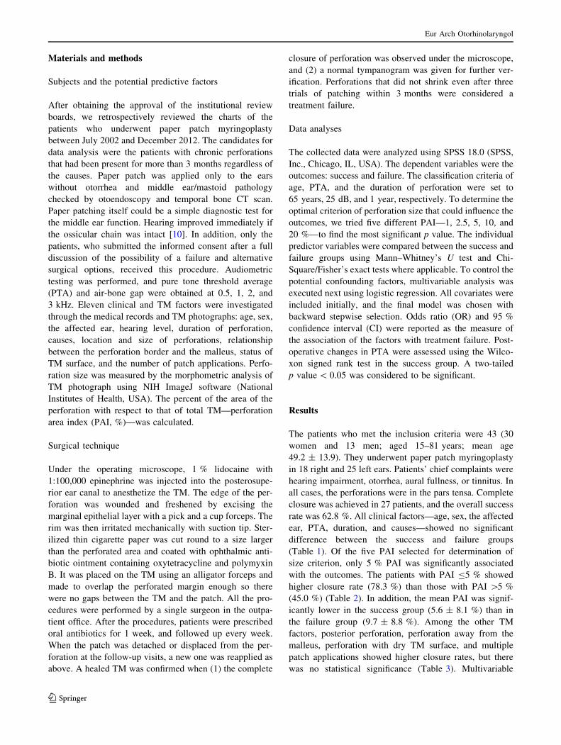
Data analyses
The collected data were analyzed using SPSS 18.0 (SPSS,
Inc., Chicago, IL, USA). The dependent variables were the
outcomes: success and failure. The classification criteria of
age, PTA, and the duration of perforation were set to
65 years, 25 dB, and 1 year, respectively. To determine the
optimal criterion of perforation size that could influence the
outcomes, we tried five different PAI—1, 2.5, 5, 10, and
20 %—to find the most significant p value. The individual
predictor variables were compared between the success and
failure groups using Mann–Whitney’s U test and Chi-
Square/Fisher’s exact tests where applicable. To control the
potential confounding factors, multivariable analysis was
executed next using logistic regression. All covariates were
included initially, and the final model was chosen with
backward stepwise selection. Odds ratio (OR) and 95 %
confidence interval (CI) were reported as the measure of
the association of the factors with treatment failure. Post-
operative changes in PTA were assessed using the Wilco-
xon signed rank test in the success group. A two-tailed
p value \ 0.05 was considered to be significant.
Results
The patients who met the inclusion criteria were 43 (30
women and 13 men; aged 15–81 years; mean age
49.2 ± 13.9). They underwent paper patch myringoplasty
in 18 right and 25 left ears. Patients’ chief complaints were
hearing impairment, otorrhea, aural fullness, or tinnitus. In
all cases, the perforations were in the pars tensa. Complete
closure was achieved in 27 patients, and the overall success
rate was 62.8 %. All clinical factors—age, sex, the affected
ear, PTA, duration, and causes—showed no significant
difference between the success and failure groups
(Table 1). Of the five PAI selected for determination of
size criterion, only 5 % PAI was significantly associated
with the outcomes. The patients with PAI B5 % showed
higher closure rate (78.3 %) than those with PAI [5 %
(45.0 %) (Table 2). In addition, the mean PAI was signif-
icantly lower in the success group (5.6 ± 8.1 %) than in
the failure group (9.7 ± 8.8 %). Among the other TM
factors, posterior perforation, perforation away from the
malleus, perforation with dry TM surface, and multiple
patch applications showed higher closure rates, but there
was no statistical significance (Table 3). Multivariable
Eur Arch Otorhinolaryngol
123

logistic regression also revealed that only the perforation
size based on the criterion of 5 % PAI remained significant
as the predictor of treatment outcome with an OR of 4.4
(p = 0.029, 95 % CI 1.17–16.57).
No patient had otorrhea or other complications during
the follow-up period. In the success group, pre- and post-
operative PTAs were 28.9 ± 25.1 and 24.7 ± 27.9
(p = 0.001), which means that the hearing improved sig-
nificantly after the closure of perforations. The closure
times were 2–8 weeks except five patients. Three of them
were cured by 15 weeks after multiple patch applications,
and in the other two patients, healing was confirmed at 16
and 19 weeks because of the delayed follow-ups.
Discussion
In the present study, the authors intended to report the
success rate of paper patch myringoplasty for chronic TM
perforations in our clinic and to explore the predictors for
Table 1 Clinical factors: success versus failure groups
Success
(n = 27)
Failure
(n = 16)
p value
Age
Mean age (years) 47.5 ± 14.2 51.9 ± 13.2 0.145
\65 years (n = 36) 23 (63.9) 13 (36.1) 1.000
C65 years (n = 7) 4 (57.1) 3 (42.9)
Sex
Male (n = 13) 8 (61.5) 5 (38.5) 1.000
Female (n = 30) 19 (63.3) 11 (36.7)
Affected ear
Right (n = 18) 11 (61.1) 7 (38.9) 0.847
Left (n = 25) 16 (64.0) 9 (36.0)
Hearing
PTA (dB) 28.2 ± 24.9 30.0 ± 12.7 0.152
Air-bone gap (dB) 15.4 ± 11.1 17.0 ± 9.7 0.345
PTA B25 dB (n = 24) 18 (75.0) 6 (25.0) 0.063
PTA [25 dB (n = 19) 9 (47.4) 10 (52.6)
Duration
3 months to 1 year (n = 9) 8 (88.9) 1 (11.1) 0.121
C1 year (n = 34) 19 (55.9) 15 (44.1)
Cause
Chronic otitis media
(n = 30)
19 (63.3) 11 (36.7) 1.000
Trauma or others (n = 13) 8 (61.5) 5 (38.5)
Data presented are mean ± SD or number (%) of patients
Table 2 Patient distributions according to perforation size criteria:
success versus failure groups
Success
(n = 27)
Failure
(n = 16)
p value
PAI = 1 %
B1 % (n = 9) 7 (77.8) 2 (22.2) 0.446
[1 % (n = 34) 20 (58.8) 14 (41.2)
PAI = 2.5 %
B2.5 % (n = 18) 14 (77.8) 4 (22.2) 0.084
[2.5 % (n = 25) 13 (52.0) 12 (48.0)
PAI = 5 %
B5 % (n = 23) 18 (78.3) 5 (21.7) 0.024*
[5 % (n = 20) 9 (45.0) 11 (55.0)
PAI = 10 %
B10 % (n = 34) 24 (70.6) 10 (29.4) 0.058
[10 % (n = 9) 3 (33.3) 6 (66.7)
PAI = 20 %
B20 % (n = 39) 25 (64.1) 14 (35.9) 0.621
[20 % (n = 4) 2 (50.0) 2 (50.0)
Data presented are number (%) of patients. PAI = perforation area
index (the area of perforation/the area of tympanic membrane 9 100)
* p \ 0.05
Table 3 Tympanic membrane factors: success versus failure groups
Success
(n = 27)
Failure
(n = 16)
p value
Location
Anterior (n = 37) 22 (59.5) 15 (40.5) 0.386
Posterior (n = 6) 5 (83.3) 1 (16.7)
Perforation size
Mean PAI (%) 5.6 ± 8.1 9.7 ± 8.8 0.043*
PAI B5 % (n = 23) 18 (78.3) 5 (21.7) 0.024*
PAI [5 % (n = 20) 9 (45.0) 11 (55.0)
Relationship between the perforation border and the malleus
Away from the malleus (n = 34) 23 (67.6) 11 (32.4) 0.257
Touching the malleus (n = 9) 4 (44.4) 5 (55.6)
Surface of TM
Dried-up (n = 19) 14 (73.7) 5 (26.3) 0.189
Slightly moist with mucus
(n = 24)
13 (54.2) 11 (45.8)
Number of patch applications
Single (n = 28) 16 (57.1) 12 (42.9) 0.295
Multiple (n = 15) 11 (73.3) 4 (26.7)
Data presented are mean ± SD or number (%) of patients
PAI perforation area index, TM tympanic membrane
* p \ 0.05
Eur Arch Otorhinolaryngol
123

the outcomes. The use of various artificial membranes and
chemical cautery for the treatment of TM perforations has a
long history. Chemical agents such as silver nitrate, tri-
chloracetic acid, and urea provoke an inflammatory
response to stimulate healing [11]. Goldman reported a
successful closure rate of 64 % using a silver nitrate bead
or a urea ointment patch [12]. CO2 laser has also been used
to trim the perforation margins by Lee et al. [8]. In this
study, we used mechanical method to remove the epithe-
lium and incite an inflammatory healing response at the rim
of the perforation. Rim trimming and irritation are impor-
tant for myringoplasty because more rapidly regenerating
epidermal layer grows inward and migrates over the slowly
regenerating connective tissue layers, interrupting the
repair process [1, 11].
The overall success rate of 62.8 % in the present study is
similar with or slightly higher than those of the previous
reports [5–8]. Furthermore, the closure rate was as high as
78.3 % especially in the patients with perforations smaller
than 5 % of the TM. Perforation size has been known to be
the most important factor for paper patch myringoplasty.
Golz et al. [6] have reported the closure rate of 55.7 % in
perforations \5 mm; Dursun et al. [7], 66.7 % in perfora-
tions \3 mm; Lee et al. [8], 72.9 % in perforations
\4 mm. In rats, the recovery rate was 94.4 % in small
perforations (\30 % of the TM), while 56.2 % in large
perforations [9]. In our study, perforation size was the only
predictor of the outcomes, which agreed with the previous
reports. The 5 % PAI was considered the optimal grouping
criterion of perforation size that could predict the outcome
most correctly among the various size criteria. The odds
ratio of 4.4 (95 % CI 1.17–16.57) means that the likelihood
of treatment failure is 4.4 times more common in patients
who have perforations larger than 5 % of the TM. Mor-
phometric measurement of the area of perforation using
TM photographs and image analysis software may be a
more objective and precise method than manual
measurement of the greatest diameter of perforation using
the surgical instruments such as hook or drill tips. The
schematic drawing of 5 % perforation of the TM is dem-
onstrated in Fig. 1. Although no significant associations
were found between the other factors and the treatment
outcomes, these results may be rather encouraging from a
different perspective: surgeons can be free from the other
risk factors when performing paper patch myringoplasty.
Even if only a small portion of patients with chronic otitis
media benefit from paper patching, it would be worthy of a
try as a first-choice alternative to surgical myringoplasty
when indicated, because paper patch myringoplasty is a
simple, safe, minimally invasive, and cost-effective pro-
cedure performed in an outpatient clinic.
In conclusion, paper patch myringoplasty showed the
favorable success rates: 62.8 % in total and 78.3 % in ears
with perforations smaller than 5 % of the TM. The only
predictor of the treatment outcome was perforation size.
These results suggest that we can try paper patch myrin-
goplasty in selected patients who had dry chronic perfo-
rations smaller than 5 % of the TM without middle ear
disease before considering the other invasive surgical
myringoplasty.
Conflict of interest The authors declare that they have no conflict
of interest.
References
1. Merwin GE, Boies LR Jr (1980) Paper patch repair of blast
rupture of the tympanic membrane. Laryngoscope 90:853–860
2. Camnitz PS, Bost WS (1985) Traumatic perforations of the
tympanic membrane: early closure with paper tape patching.
Otolaryngol Head Neck Surg 93:220–223
3. Saito H, Kazama Y, Yazawa Y (1990) Simple maneuver for
closing traumatic eardrum perforation by micropore strip tape
patching. Am J Otol 11:427–430
4. Kartush JM (2000) Tympanic membrane patcher: a new device to
close tympanic membrane perforations in an office setting. Am J
Otol 21:615–620
5. Spandow O, Hellstrom S, Dahlstrom M, Bohlin L (1995) Com-
parison of the repair of permanent tympanic membrane perfora-
tions by hydrocolloidal dressing and paper patch. J Laryngol Otol
109:1041–1047
6. Golz A, Goldenberg D, Netzer A, Fradis M, Westerman ST,
Westerman LM et al (2003) Paper patching for chronic tympanic
membrane perforations. Otolaryngol Head Neck Surg
128:565–570
7. Dursun E, Dogru S, Gungor A, Cincik H, Poyrazoglu E, Ozdemir
T (2008) Comparison of paper-patch, fat, and perichondrium
myringoplasty in repair of small tympanic membrane perfora-
tions. Otolaryngol Head Neck Surg 138:353–356
8. Lee SH, Jin SM, Lee KC, Kim MG (2008) Paper-patch myrin-
goplasty with CO2 laser for chronic TM perforation. Eur Arch
Otorhinolaryngol 265:1161–1164
9. Imamoglu M, Isik AU, Acuner O, Harova G, Bahadir O (1998)
Fat-plug and paper-patch myringoplasty in rats. J Otolaryngol
27:318–321Fig. 1 Schematic drawing of 5 % perforation of tympanic membrane
Eur Arch Otorhinolaryngol
123

10. Silverstein H, Wycherly BJ, Darley DS, Alameda YA (2012)
Mixed hearing loss in iatrogenic tympanic membrane perfora-
tions over the round window niche and the immediate effect of
paper patch myringoplasty. Audiol Neurootol 17:155–160
11. Dunlap AM, Schuknecht HF (1947) Closure of perforations of the
tympanic membrane. Laryngoscope 57:479–490
12. Goldman NC (2007) Chemical closure of chronic tympanic
membrane perforations. ANZ J Surg 77:850–851
Eur Arch Otorhinolaryngol
123