Embed Size (px)
Citation preview
Preeclampsia / Eclampsia-Diagnosis and Management
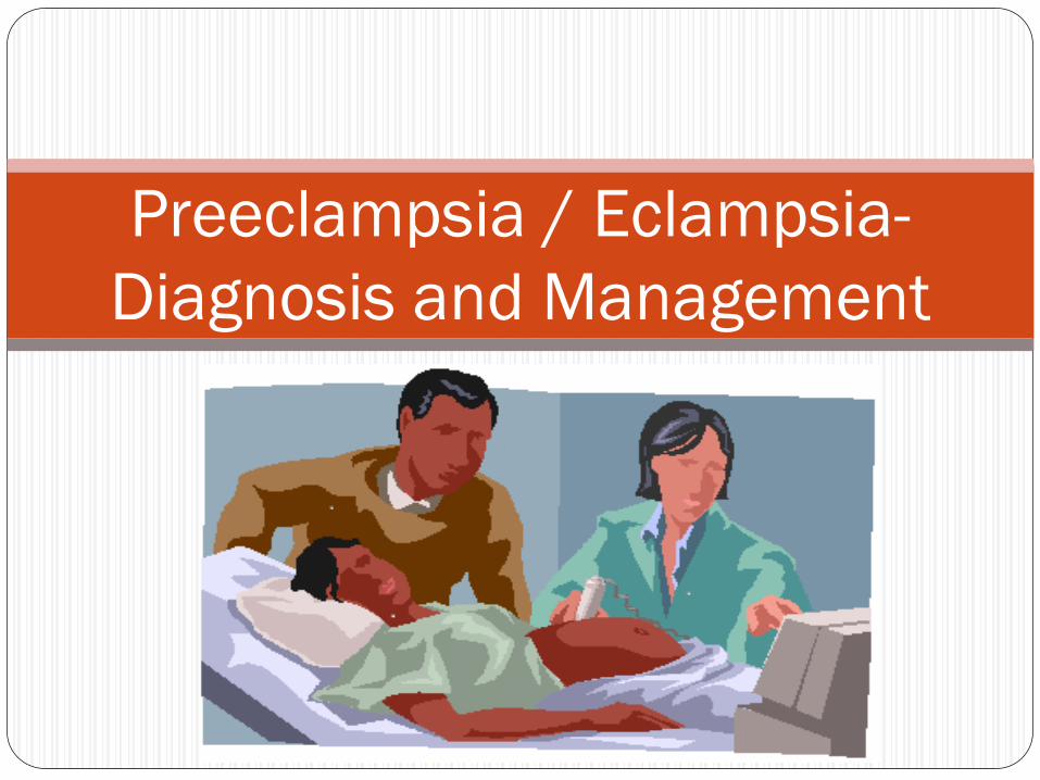
Preeclampsia and Eclampsia Detection and Management During the admission process:
– Review the patient’s record, noting medical history and obstetric history
– Note predisposing factors- Assess the following:
– Baseline BP – Proteinuria – Weight gain – [Sudden excessive wt. gain is sometimes the first sign of impending
preeclampsia. (2# or more per week in the 3rd trimester)] History or current complaint of headache or blurred vision
and/or severe edema of the hands, legs, feet, and face.
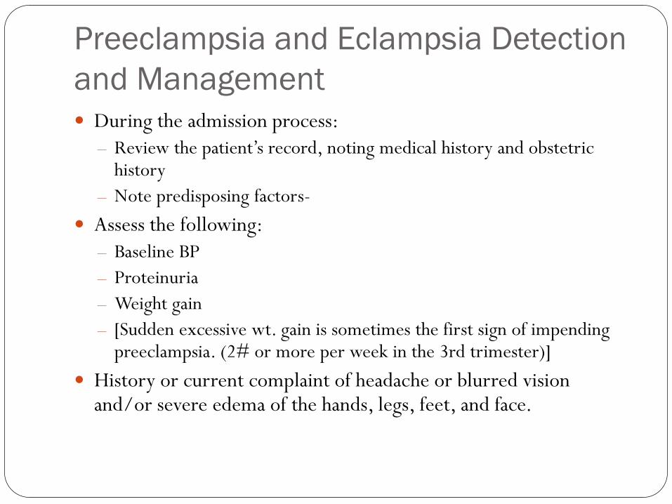
Hypertensive Disorders of Pregnancy Leading cause of maternal morbidity Accounts for 16.1% of maternal deaths in developing
countries Occur in 5%-12% of all pregnancies Encompasses a spectrum of diagnoses
– Chronic hypertension - dx prior to 20 weeks – Preeclampsia - mild or severe – Preeclampsia superimposed on chronic hypertension – Gestational Hypertension - does not meet criteria for
diagnosis of preeclampsia (formerly PIH) – Transient Hypertension
Pathophysiology of Preeclampsia Proteinuria and Pre-eclampsia When the body is not plagued by abnormality, the blood vessels are lined with closely knit cells. These cells and the protein prevent leakage of water into the surrounding tissues. Proteins (made up of 60% albumin) within the cells carry a magnetic charge that is capable of attracting water thus keeping it within the blood vessels. When the blood reaches the kidneys, the pores in the glomerulus are also negatively charged, and prevent the protein from escaping into the surrounding tissues. Pre-eclampsia and eclampsia are characterized by hypertension, proteinuria, and edema. With hypertension, the pores in the glomerulus lose some of their negatively charged capability, the magnetic character of the protein is reduced, and the protein easily escapes into the surrounding tissues (proteinuria). With less protein there is nothing to keep the water (80% of blood is made up of water) within the vessels. In addition, hypertension also causes the blood vessels walls to be damaged; thus allowing the unrestrained water to escape into the surrounding tissues leading to worsening edema and loss of blood volume, an ominous sign of progressive disease.
Preeclampsia Risk Factors Primagravida Multifetal gestation Preeclampsia with a previous pregnancy Chronic hypertension Diabetes Obesity Over age 35 or under age 19 African American
Assessment and Physical Exam on Admission Check BP - Check BP sitting in L arm Check for edema Check deep tendon reflexes Evaluate clean-catch urine Question patient about:
– headaches – blurred vision – loss of consciousness – nausea and vomiting – worsening edema – epigastric pain – general sick feeling
Mild Mild: BP has reached 140/90 to 160/110 mmHg on two different
occasions 4 hours apart. (an increase of the diastolic reading is a more reliable indication of preeclampsia)
Proteinuria is 2+ or between 300 mg and 5 g in a 24 hours urine specimen.
Weight gain / Edema are no longer essential
Mild Preeclampsia Management Confirm Diagnosis-on suspecting diagnosis of preeclampsia
simultaneously treat and further evaluate the mother and fetus
Document fetal well being If > 37 weeks – induction If < 37 weeks
– Inpatient Management – Outpatient Management possible if SBP< 150 or DBP< 100
and normal laboratory findings-recommend daily kick counts and biweekly NST
Severe BP rises to 160/110 mmHg or higher on two different
occasions 4 hours apart. Proteinuria is 5 g or higher in a 24 hours urine specimen. Urine output decreases to less than 500ml in 24 hours. The
kidneys can go into failure with little output. This is a serious situation that can lead to permanent injury.
Note: clinical signs of preeclampsia can appear suddenly. Never underestimate the importance of even mild BP elevations complicating a pregnancy.
Severe preeclampsia symptoms Severe headaches Visual problems Epigastric pain Nausea or vomiting Thrombocytopenia IUGR or Oligohydramnios Elevated serum creatnine Elevated liver functions of unclear etiology Irritability, restlessness, or apprehension Pulmonary edema with respiratory distress Eclampsia Early recognition of worsening preeclampsia when a patient is hospitalized is critical.
Routine Laboratory Analysis Complete Blood Count Platelet count-below 150, 000 thrombocytopenia, below
100, 000 severe coagulapathy, below 50, 000 correct prior to surgery
Liver Function Tests-ALT, AST Renal Function Tests-Creatnine, BUN, Uric Acid-increased
uric acid is an early sign of liver involvement Urinalysis and Microscopy 24 hour urine for Protein and Creatnine Clearance-protein
increases as creatnine clearance decreases Blood Type and antibody screen
Severe Preeclampsia Fetal outcome is poor, often as a result of growth restriction
and/or asphyxia at birth. Incidence of serious complications in the mother is high Signs to watch for:
– Abruptio placentae – HELLP syndrome – Eclampsia – DIC – Acute Renal Failure
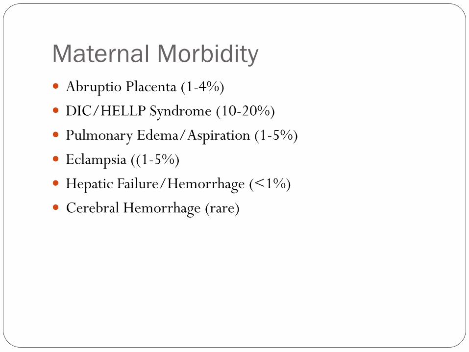
Maternal Morbidity Abruptio Placenta (1-4%) DIC/HELLP Syndrome (10-20%) Pulmonary Edema/Aspiration (1-5%) Eclampsia ((1-5%) Hepatic Failure/Hemorrhage (<1%) Cerebral Hemorrhage (rare)
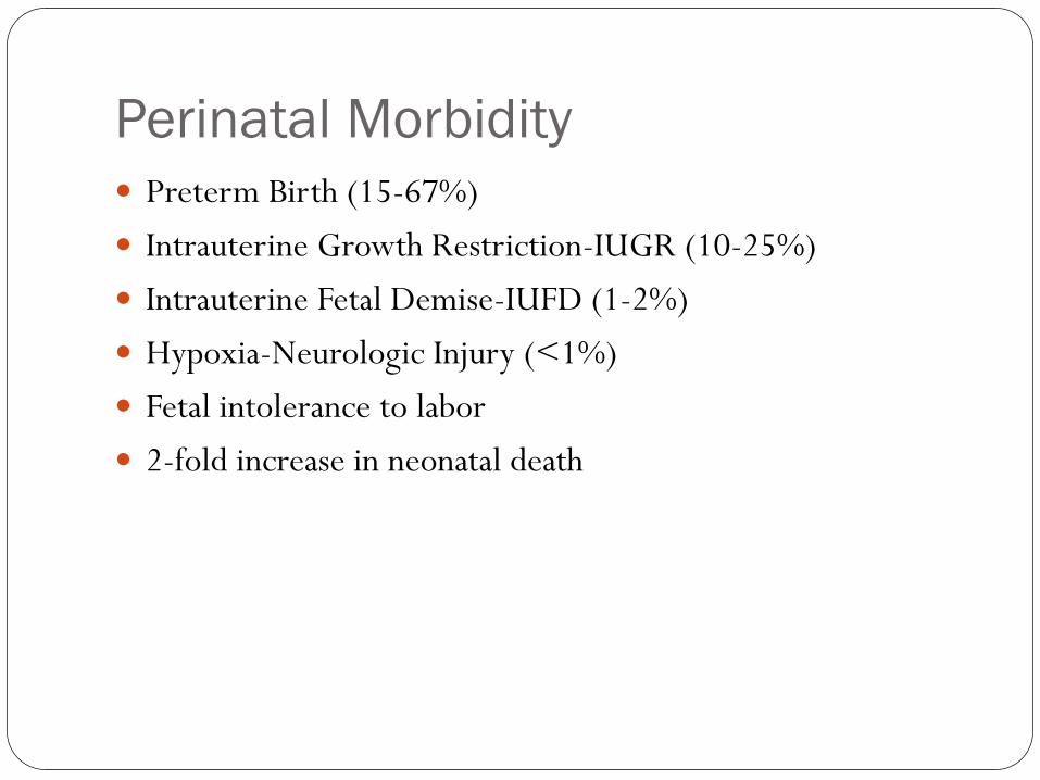
Perinatal Morbidity Preterm Birth (15-67%) Intrauterine Growth Restriction-IUGR (10-25%) Intrauterine Fetal Demise-IUFD (1-2%) Hypoxia-Neurologic Injury (<1%) Fetal intolerance to labor 2-fold increase in neonatal death
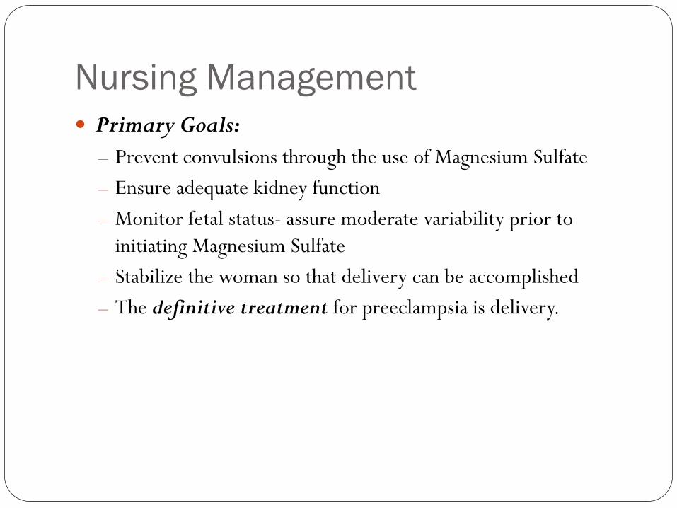
Nursing Management Primary Goals:
– Prevent convulsions through the use of Magnesium Sulfate – Ensure adequate kidney function – Monitor fetal status- assure moderate variability prior to
initiating Magnesium Sulfate – Stabilize the woman so that delivery can be accomplished – The definitive treatment for preeclampsia is delivery.
Nursing Management Intense maternal monitoring MgSO4 administration Antihypertensive medications Depending on the condition of mother and fetus, delivery might be indicated Electronic fetal monitoring 1:1 nurse to patient ratio Assess vital signs based on status to determine worsening of the disease and response to therapy. Obtain BP with consistent methods Record hourly intake and output using a Foley catheter with a urometer. Reduce stimulation from noise and light. Maintain patient on strict bedrest Maintain IV access (D5W or LR at nor more than 150 mL/hr) Lab work as ordered including: type and crossmatch and platelets Test urine for protein Assess deep tendon reflexes Ask patient to tell you if she develops a headache, blurred vision, dizziness, or epigastric pain, or if
she feels uncomfortable or different. Observe the patient for restlessness or apprehension

Magnesium Sulfate Therapy Drug of choice to prevent seizures Loading dose: 4-6 grams Maintenance dose: 2-3 grams/hr Dependent on renal function-if renal function is impaired the kidneys
will not excrete the magnesium Associated with cerebral arterial vasodilation which may relieve cerebral
ischemia Decreases plasma endothelin-1 which protects the vascular endothelial
cells Potentiates beta blockers Increases potency and duration of non depolarizing muscle relaxants-
general anethetic Increases bleeding times Decreases platelet activity-don’t stick as well

Magnesium Sulfate Therapy High alert medication Never a primary line Always use an infusion pump Always document in grams per hour Calcium gluconate immediately available Assess for signs of toxicity Assess DTR’s Assess urine output hourly Assess heart rate and rhytum Auscultate lungs – listen for signs of pulmonary congestion Assess respiratory rate and quality
Antihypertensives Indicated when
– Underlying chronic hypertension – To achieve BP control to prevent cerebral vascular accident
while effecting delivery – Expectant management of severe preeclampsia by BP criteria
alone – Used to prevent cerebral hemorrhage, stroke ( usually BP >
170 )
Hydralazine Direct peripheral vasodilitation Increases cardiac output and heart rate Dosages 5mg IV or 10mg IM
– 5-10mg every 20-30 min – Maximum is 20 IV to 30 IM
Can cause rebound tachycardia and hypotension
Labatelol Selective alpha and non-selective beta antagonist Dosage:
– Initial 20mg IV – If effect sub optimal, then 40mg IV 10 min later – If effect sub optimal, then 80mg IV 10 min later for two
additional doses – Maximum of 300mg
Decreases vascular resistance Works better in stacked doses Can cause fetal bradycardia and neonatal hypoglycemia
Seizure Precautions HAVE EMERGENCY EQUIPMENT READY! Oxygen and suctioning equipment must be immediately
available. Check that the equipment is in operating order. Keep an emergency cart nearby. Administer MgSO4 as ordered. It is currently believed to be
the safest and most effective anticonvulsant drug available.
The patient with severe preeclampsia or eclampsia needs intensive monitoring. She
should never be left alone!
Eclampsia Eclampsia is the generalized seizure or seizues and/ or coma
either during pregnancy or in the postpartum period in the presence of preeclampsia and with no other neurological or medical etiology. The exact cause of the seizures is unknown, although possible mechanisms include cerebral vasospasm and a form of hypertensive encephalopathy.
Eclamptic seiziures are self-limiting and usually last less than 4 min Incidence in the US is 1:3250 births
– Antepartum: 50% of eclamptic seizures occur prior to delivery – Postpartum: early postpartum eclampsia occurs within 48 hours of
delivery. Late postpartum eclampsia occurs between 48hrs and 4 weeks after delivery. Aggressive treatment has shifted the incidence of postpartum eclampsia from early to late- 79% late
2nd most common cause of maternal death 15% of all maternal deaths in the US
Eclampsia Treatment Stabilize the mother and control the seizures
– Call for assistance – Protect the airway – Start medications
Deliver the fetus – During the acute event the mother takes precedence – The fetus will do better when the mother is stabilized – Cesarean section does not need to be performed immediately – Induction of labor is reasonable after the seizure has been
controlled in the term and near term fetus if in a vertex position and fetal distress is resolved