Embed Size (px)
Citation preview
Preparation and Characterization of
Poly Vinyl Alcohol - Gelatin- Carboxy
Methyl Chitosan Polymer Films A THESIS SUBMITTED IN PARTIAL FULFILLMENT
OF THE REQUIREMENT OF THE DEGREE OF
Bachelor of Technology
In
Biotechnology
By
Amit Bothra
Department of Biotechnology and Medical Engineering
National Institute of Technology, Rourkela
2014
i
Department of Biotechnology and Medical Engineering
National Institute of Technology, Rourkela
Certificate
This is to certify that the thesis entitled “Preparation and Characterization of Polymer
Films Based on Poly Vinyl Alcohol - Gelatin- Carboxy Methyl Chitosan” by Amit Bothra
(110BT0542), in partial fulfilment of the requirements for the award of the degree of Bachelor
of Technology in Biotechnology during session 2010-2014 in the Department of
Biotechnology and Medical Engineering, National Institute of Technology Rourkela, is an
authentic work carried out by him under my supervision and guidance. To the best of my
knowledge, the matter embodied in the thesis has not been submitted to any other
University/Institute for the award of any degree or diploma.
Place: NIT Rourkela Dr. Indranil Banerjee
Date: 12th May 2014 Assistant Professor
Biotechnology and Medical Engineering
National Institute of Technology, Rourkela
ii
Acknowledgement
I would really like to take this opportunity to thank my project guide, Dr. Indranil
Banerjee, Department of Biotechnology and Medical Engineering, NIT Rourkela, for his
guidance and support. I am sincerely thankful to Prof. Krishna Pramanik, Head of the
Department, Dept. of Biotechnology and Medical Engineering, NIT, Rourkela, for providing
the necessary facilities for this work. I sincerly thanks to all faculties and to all teaching and
non-teaching staff members of Department of Biotechnology & Medical Engineering, National
Institute of Technology Rourkela, Orissa.
My special thanks to research scholar Mr. AKS Senthilguru. I also owe a debt of
gratitude to Miss Shrutija Pandey, Mr. Tarun Agarwal, Mr. Prerak Gupta and Mr. Goutham
Jillu for all the help and support I got from them.
I would like to express my heartily thanks to my friends Dattaram Sugave, Shambhavi
Singh, Prajna Kabiraj, labmates and others in the department for their help and support. Finally,
I would like to express my heartfelt thanks to my parents for their blessings, support and
constant encouragement and very special thanks to God for showering the blessings on me.
Amit Bothra
110BT0542
iii
Table of Contents
Chapter 1. Introduction 1-5
1.1 Introduction
1.1.1 Polyvinyl alcohol 2
1.1.2 Gelatin 3
1.1.3 Carboxymethyl-chitosan 4
Chapter 2. Objective and Plan of Work 6-7
2.1 Objective 7
2.2 Work Plan 7
Chapter 3. Materials and Methods 8-13
3.1 Materials 9
3.2 Methods
3.2.1 Polymer films preparation 9
3.2.2 Swelling and Biodegradation study 10
3.2.3 Hemocompatibility 11
3.2.4 Cytotoxicity test of polymer films (MTT assay) 12
Chapter 4. Result and Discussion 14-22
4.1 Preparation of polymer films 15
4.2 Swelling and biodegradation
4.2.1 Swelling study 15
4.2.2 Biodegradation study 16
4.3 Hemocompatibility 17
4.4 Cytotoxicity (MTT assay) 19
4.5 Discussion 20
iv
List of Tables
Table 1: Compositions of CMC for polymer films 10
Table 2: Reagents used in preparation of 100ml PBS solution 11
Table 3: Swelling of polymer films with time 16
Table 4: Biodegradation data for polymer film samples 17
Table 5: Absorbance readings of hemocompatibility test for various film samples. 18
Table 6: MTT assay absorbance at 595nm (readings of triplicates) 19
List of Figures
Figure 1: Polymer films with different proportion of CMC 15
Figure 2: Percentage swelling of polymer films with time 16
Figure 3: Degradability index of films with increase in proportion of CMC 17
Figure 4: Hemolysis study of PVA-Gelatin-CMC samples 18
Figure 5: Cell viability index of the polymer samples 19
v
Abstract
Present work delineate the preparation and characterization of polymer films of polyvinyl
alcohol (PVA), gelatin and carboxy methyl chitosan. The films were prepared by esterification
followed by glutaraldehyde crosslinking. Swelling behaviour and biodegradability of the
polymer films were studied at physiological pH. Further, hemocompatibility and
biocompatibility of the polymer film was also evaluated. Result showed that films of different
composition have water-swelling capacity around 80%. Biodegradation study revealed that
35% degradation could be achieved by varying the composition. All the polymeric films were
found hemocompatible and biocompatible. From this preliminary study it can be said that such
films could be used for biomedical application.
KEYWORDS: Polymer films, hemocompatibility, swelling, cytotoxicity, biocompatibility.
1
Chapter 1
Introduction
2
1.1 Introduction:
Now a days, biopolymeric devices are utilized to evaluate, treat, augment or substitute any
tissue, organ or function of the body. In this regard polymeric films shows great versatility.
Polymer films are cross-linked polymeric network. At physiological temperature and pH, these
polymeric materials don’t dissolve in water but swells extensively.
Polymer films are used in artificial corneas, contact lenses, wound dressing, catheters, sutures
and electrode sensors [1]. Cross-linked polymeric films could be obtained by chemical cross-
linking (using epichlorohydrin, glutaraldehyde), radical polymerization or by radiation (e.g.,
gamma radiation, UV radiation). The polymer films characteristics, including the swelling
properties, might be regulated by the amount of reagent used in cross-linking. Naturally
sensitive films can be obtained by the addition of some special monomers. Solubility properties
of water soluble polymers depends upon the presence of functional groups (especially NH2,
OH and COOH) that might be utilized for the preparation of environment sensitive polymer
films [2].
1.1.1 PVA (Polyvinyl Alcohol): PVA was initially manufactured by Hermann and Haehnel in
1924 by hydrolysing polyvinyl acetate in ethanol using potassium hydroxide. Commercial
manufacturing of PVA is done using polyvinyl acetate, normally by sustained process. In the
presence of aqueous sodium hydroxide or anhydrous sodium methylate, the acetate groups are
hydrolysed by ester internally change with methanol. The physical characteristics and its
particular functional utilizations depend on the degree of polymerization and the degree of
hydrolysis [3]. PVA can be divided into two different classes based on hydrolysis: partially
hydrolysed and fully hydrolysed. Generally in the foods, partially hydrolysed PVA is used.
Vinyl acetate monomer is the essential raw material utilized in the preparation of polyvinyl
alcohol. PVA is synthesized using polymerization of vinyl acetate followed by partial
hydrolysis [4]. Whole process of hydrolysis is depend on partial replacement of ester group in
3
vinyl acetate with the hydroxyl group and is finished in the presence of aqueous sodium
hydroxide then slow addition of aqueous saponification reagent. After that precipitated PVA is
washed and dried. At the time when the saponification reaction is stopped properly, the degree
of hydrolysis is determined [5].
PVA is a translucent, tasteless and odourless, cream or white colour granular powder which is
utilized as a moisture barrier film for dry foods with inclusions or foods contain inclusions that
need to be protected from moisture uptake and for food supplement tablets. PVA is slightly
dissolvable in ethanol, soluble in water, but insoluble in other organic solvents. PVA has a
melting point of 179°C to 191°C. It has a molecular weight of between 26,250 and 30,250, and
a degree of hydrolysis of 85.5% to 90%. Ordinarily a 5% solution of PVA exhibits a pH in the
range of 4.5 to 7.0. PVA is unknown to become as a natural product.
Mixtures of PVA with other natural polymers have long been utilized because of film
constructing ability of PVA. Molecular weight and the degree of hydrolysis can affect the
performance properties of PVA. Atomic weight of PVA is ~161 kDa. As polyethylene, PVA
has a planar zigzag structure. PVA grades are promptly dissolvable in water and depend on
components like atomic mass, molecule size distribution, and molecule crystallinity. PVA
exhibits excellent water retention properties because it’s a hydrophilic polymer. For aggregate
disintegration, however, PVA needs water temperatures of about 101˚C with a 30 minutes hold
time [6].
1.1.2 Gelatin: Gelatin is a natural polymer broadly utilized in pharmaceutical, cosmetic,
photographic, and food industries. It is prepared by denaturation and partial hydrolysis of
fibrous collagen. Collagen is the most abundant structural protein of animal skin, bones and
rarely fish scales. And also the major organic factor of skin and bone of vertebrates is collagen.
On the other hand, collagen is consisting a family of proteins with 21 different types of amino
acids.
4
It mainly contains the residues of 3 amino acids—proline, 4-hydroxyproline, and glycine
(arranged every third residue)—in its structure. It consists extended left-handed proline helix
conformations consolidated with 290 to 4100 amino acids. Presence of higher levels of
pyrrolidines in gelatin results in the formation of stronger films or gel. Due to the presence of
the triple helixes, gelatin films have good strength. If the triple-helix content increases, the
strength of the film increases and decreases the swelling property in aqueous. The gelling
properties of the gelatin can be changed by the reaction of chemical cross-links using
glutaraldehyde to cross-link lysine to lysine or transglutaminase to cross-link lysine to
glutamine residues.
Gelatin swells in cold water and is totally dissolve in hot water. In order to release the needed
structure of gelatin in dry state, a temperature of about 60°C is necessary. At the point, when a
gelatin solution is spread in a thin layer over a surface and passes from a sol to a gel, it forms
a film. Gelatin fulfils numerous other functions, such as: gelling ability, stabilising ability,
aerating ability, emulsifying ability, thickening ability, coating, texture improvement,
protecting, preventing syneresis, and gluing. The formation of hard and soft capsules and in
microencapsulation, these properties can be exploited [7].
1.1.3 Carboxymethyl-chitosan: Chitosan is a polysaccharide similar in structure to cellulose.
Chitosan, commonly obtained by partial deacetylation of chitin derived from the exoskeleton
of crustaceans, exhibits various useful physico-chemical. It is a polycationic copolymer
consisting of glucosamine and N-acetyl glucosamine units.
Carboxymethyl-chitosans are a family of water soluble chitosans which forms semipermeable
films and membranes. There are various methods for preparing carboxymethyl-chitosans. The
water dissolving nature of the carboxymethyl-chitosan is greatly dependent on manufacturing
conditions, especially temperature. It seems that higher preparation temperature leads to more
insoluble carboxymethyl-chitosans at neural pH. The nomenclature of carboxymethyl-chitosan
5
depends on which functional group has been altered during preparation. Modifying the amino
or hydroxyl group results in N-carboxymethyl or O-carboxymethyl chitosan, respectively. It is
also possible to modify both the groups to obtain N,O-carboxymethyl chitosan. These
carboxymethyl-chitosans can be further modified to include different functional groups.
Carboxymethyl-chitosan is a tempting biodegradable and biocompatible polymer which is
acquired by the reaction of chitosan with monochloroacetic acid and in basic (alkaline)
medium. Film-forming capability of gelatin and ability to interact with various substances and
dissolving nature in wide range of pH are because of its antimicrobial activity. It can be used
in biomedical and pharmaceutical fields, especially for the controlled drug release. Gelatin is
also used in viscosupplementation and tissue engineering. Comparing with generally used
chitosan, carboxymethyl-chitosan shows multiple potentials in local drug delivery [8].
General application of drug delivery system can be very helpful, both in terms of, preventing
systemic side effects like depression, tachycardia and gastrointestinal complaints and raising
drug concentration directly in the action site. Examples of the benefits of site-specific
employing carboxymethyl-chitosan vehicles include solubility, pH-sensitivity, absorbability,
and bioadhesive ability, controllable biodegradability, nontoxicity of the degradation end
products, ease of administration and sustained release potential. The swelling, drug permeation
and release properties of Carboxymethyl-chitosan can be controlled by the pH changes due to
anionic carboxyl groups and cationic amine groups in matrix [9].

6
Chapter 2
Objective and Plan of work
7
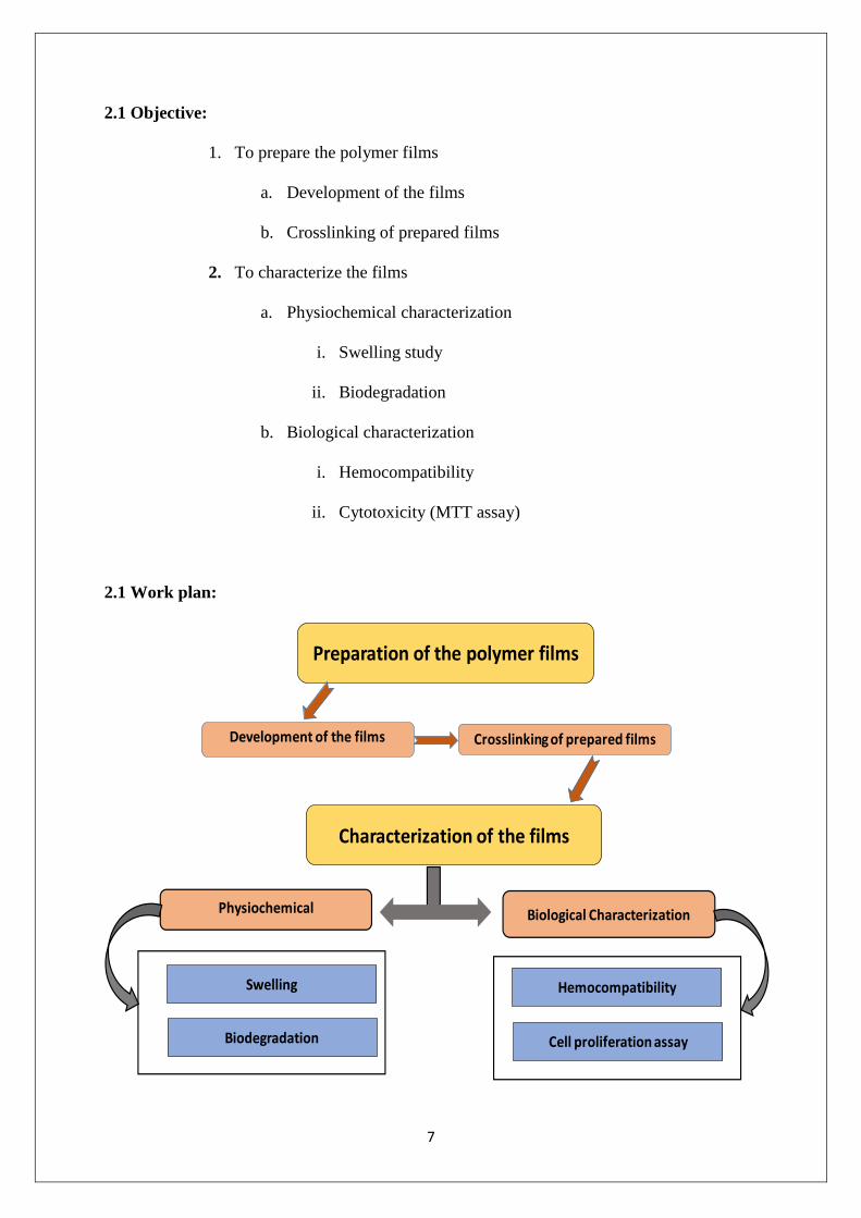
2.1 Objective:
1. To prepare the polymer films
a. Development of the films
b. Crosslinking of prepared films
2. To characterize the films
a. Physiochemical characterization
i. Swelling study
ii. Biodegradation
b. Biological characterization
i. Hemocompatibility
ii. Cytotoxicity (MTT assay)
2.1 Work plan:
Preparation of the polymer films
Characterization of the films
Physiochemical
Swelling
Biodegradation
Biological Characterization
Hemocompatibility
Cell proliferation assay
Development of the films Crosslinking of prepared films
8
Chapter 3
Material and Methods
9
3.1 Materials:
a. Polyvinyl alcohol[Aldrich]
b.Gelatin[LOBAL Chemia]
c. Carboxymethyl chitosan[HIMEDIA]
d.Glycine[HIMEDIA]
e. Glutaraldehyde[RANKEM]
f. Distilled water
g.Ethanol[EMSURE]
3.2 Methods:
3.2.1 Preparation of polymer films:
A) Development of polymer films:
Polymer films were developed using PVA, Gelatin and CMC. 50ml of stock solution
of PVA and gelatin was prepared in distilled water. Proportion of PVA (10%) and
gelatin (2.5%) was kept constant. PVA and gelatin were mixed by stirring at 300rpm,
70˚C for 30 minutes. CMC was weighed for five compositions (Blank, 0.5%, 1.0%,
1.5%, 2.0%). Stock solution was then equally divided into five parts of 10ml each.
CMC was weighed for each sample as per the composition of samples mentioned in
Table 1. CMC was slowly added into the beakers containing PVA, gelatin solution and
stirred at 300rpm, 70˚C. After proper mixing of CMC in the solution, 10 µl of HCl was
added for esterification reaction and stirring was continued at 70˚C, 100rpm for 30
minutes. 2.5 ml of thus prepared solution was poured into small petri dishes. Petri dishes
were labelled with sample code and kept on levelled surface in laminar air flow for 72
10
hours for drying the samples. Polymer films were ready that were taken out using
scalpel (if needed) [10].
Table 1: Compositions of CMC for polymer films
% of CMC in films (Sample code) Weight of CMC(in grams)
Blank (0) 0
0.5 0.05
1.0 0.10
1.5 0.15
2.0 0.20
Table1 for 10 ml of solution carboxymethyl chitosan compositions for polymer films.
B) Crosslinking of polymer films:
Films were washed in distilled water for 5 minutes to remove HCl. 90% ethanol solution
was prepared in 50ml falcon tube (45 ml 100% ethanol + 5 ml water). It was divided
equally into five parts. To each part 25µl of glutaraldehyde (25%) was added. The films
were placed in above mentioned solution for 3 hours for crosslinking. After that films
were kept in laminar air flow for 24 hours to dry. Thus prepared polymer films were
used for physico-chemical characterization of the polymer films [11].
3.2.2 Swelling and biodegradation study:
Swelling and biodegradation study of prepared polymer films was done using PBS
solution. 100ml of PBS solution was prepared [12].
11
Table 2: Composition of PBS (pH 7.4) solution for 100 ml
Polymer films were taken and cut into four pieces. Each piece of all the five samples
were weighed. Each piece of the polymer film was added to the falcon tube containing
5ml PBS. Film pieces were weighed after every 15 minutes for swelling study and the
readings recorded. Readings were taken till 96 hours (Readings were taken after 15, 30,
45, 60, 120, 180, 240 minutes and at 24, 48, 72, 96 hours). After 96 hours, film pieces
were removed and placed in small petri dishes to dry, then weighed and the weight was
recorded. Biodegradation was calculated relative to the dry weight of the samples. The
swelling percentage was calculated as per the below mentioned formula [13].
% Swelling = [(Wt ¬ Ws)/Ws] ×100
Where; Wt = Weight at time during swelling
Ws = Weight before swelling started
Following formula was used to calculated % biodegradation.
% Biodegradation = [(Wt ¬ Ws)/Wt] ×100
Where; Wt = Dry weight after 96hours
Ws = Weight before swelling started
3.2.3 Hemocompatibility:
Hemocompatibility test of films was done using goat blood. Fresh goat blood was
collected in an EDTA containing tube (EDTA was used for less clotting). Goat blood
Na2HPO4 0.238g
KH2PO4 0.019g
NaCl 0.80g
12
was diluted with normal saline in 8:10 ratio. (Normal saline = 9% NaCl in distilled
water). For hemocompatibility study, polymer film pieces were neutralised using 0.1M
glycine for 2 hours at room temperature to neutralize glutaraldehyde. After glycine
treatment the polymer film pieces were dipped in 10ml normal saline at 37˚C for an
hour. After that polymer film pieces were removed and the leachent was added to tubes
containing 0.2ml of diluted goat blood. Samples were then kept at 37˚C for an hour. 0.1
N HCl in diluted goat blood and normal saline were taken as positive and negative
controls respectively. Samples were centrifuged at 4000rpm for 15 minutes. OD value
was measured at 545nm. Positive control was considered as 100% and negative control
as 0% hemolysis. Percentage hemolysis was calculated using following formula [14].
% Hemolysis = [{(OD) test ¬ (OD)-ve} ÷ {(OD) +ve ¬ (OD)-ve}] × 100
Where; (OD) test = OD value of a sample
(OD)-ve = OD value of negative control
(OD)+ve = OD value of positive control
3.2.4 Cytotoxicity test of polymer films (MTT assay):
A) Sample preparation from films:-
Films were neutralized using 0.1M glycine for 2 hours and then washed using PBS
to neutralize remaining glutaraldehyde. For leachent preparation film pieces of
equal weight were dipped into 10ml PBS and kept at 37˚C for an hour. After that
films were removed and leachent was stored at room temperature [15].
B) Seeding of HaCat cells in a 96 well plate for MTT assay:-
A 96 well plate was taken. 1X 10⁴ cells were seeded in each well. 200µl of DMEM
media was added to each well. The cells were maintained in an incubator at 5%
13
CO2, 37˚C. 20µl of different concentration of leachents were added to the wells in
triplicates (1mg/ml, 100µg/ml, and 10µg/ml). Cells without leachents were taken
as control [16].
C) MTT assay of seeded HaCat cells:-
After a day of cell seeding, 20µl of MTT reagent was added to each well. Cells were
incubated for 3 hours with the reagent in an incubator at 37˚C and 5% CO2. After
that the media was removed. 200µl of DMSO was added to each well to dissolve
the formazan crystals. OD value was measured at 595nm [16].
14
Chapter 4
Results and Discussion
15
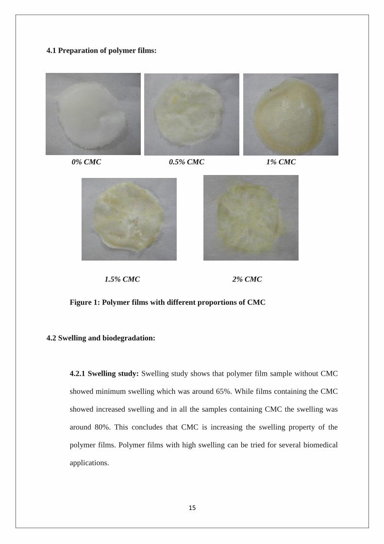
4.1 Preparation of polymer films:
0% CMC 0.5% CMC 1% CMC
1.5% CMC 2% CMC
Figure 1: Polymer films with different proportions of CMC
4.2 Swelling and biodegradation:
4.2.1 Swelling study: Swelling study shows that polymer film sample without CMC
showed minimum swelling which was around 65%. While films containing the CMC
showed increased swelling and in all the samples containing CMC the swelling was
around 80%. This concludes that CMC is increasing the swelling property of the
polymer films. Polymer films with high swelling can be tried for several biomedical
applications.
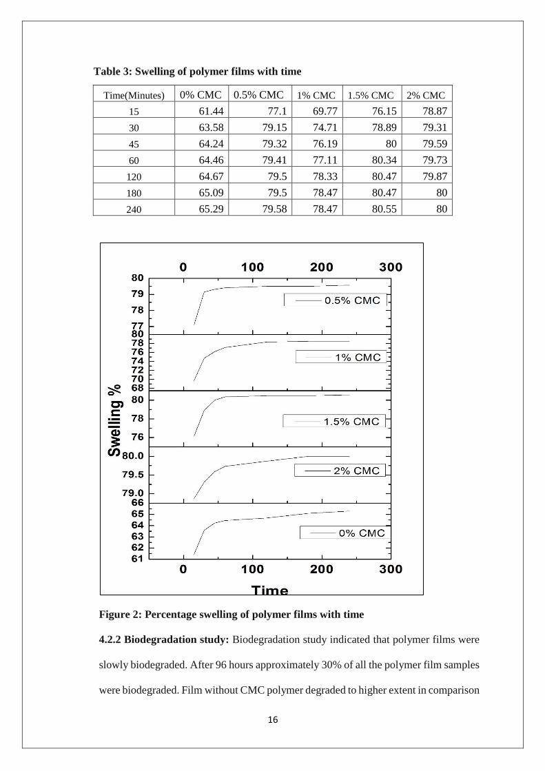
16
Table 3: Swelling of polymer films with time
Figure 2: Percentage swelling of polymer films with time
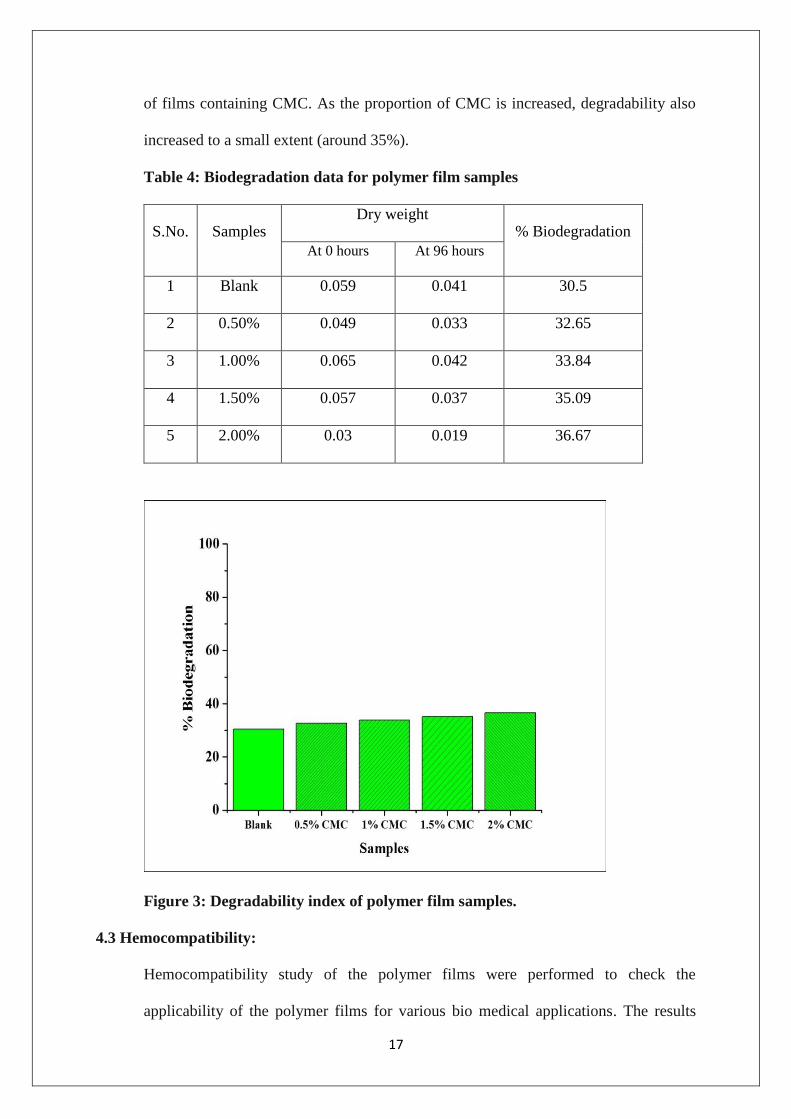
4.2.2 Biodegradation study: Biodegradation study indicated that polymer films were
slowly biodegraded. After 96 hours approximately 30% of all the polymer film samples
were biodegraded. Film without CMC polymer degraded to higher extent in comparison
Time(Minutes) 0% CMC 0.5% CMC 1% CMC 1.5% CMC 2% CMC
15 61.44 77.1 69.77 76.15 78.87
30 63.58 79.15 74.71 78.89 79.31
45 64.24 79.32 76.19 80 79.59
60 64.46 79.41 77.11 80.34 79.73
120 64.67 79.5 78.33 80.47 79.87
180 65.09 79.5 78.47 80.47 80
240 65.29 79.58 78.47 80.55 80
17
of films containing CMC. As the proportion of CMC is increased, degradability also
increased to a small extent (around 35%).
Table 4: Biodegradation data for polymer film samples
S.No. Samples Dry weight
% Biodegradation
At 0 hours At 96 hours
1 Blank 0.059 0.041 30.5
2 0.50% 0.049 0.033 32.65
3 1.00% 0.065 0.042 33.84
4 1.50% 0.057 0.037 35.09
5 2.00% 0.03 0.019 36.67
Figure 3: Degradability index of polymer film samples.
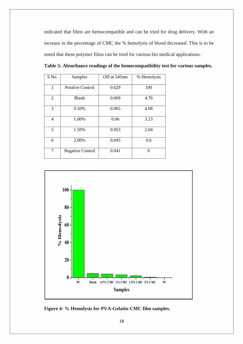
4.3 Hemocompatibility:
Hemocompatibility study of the polymer films were performed to check the
applicability of the polymer films for various bio medical applications. The results
18
indicated that films are hemocompatible and can be tried for drug delivery. With an
increase in the percentage of CMC the % hemolysis of blood decreased. This is to be
noted that these polymer films can be tried for various bio medical applications.
Table 5: Absorbance readings of the hemocompatibility test for various samples.
S.No. Samples OD at 545nm % Hemolysis
1 Positive Control 0.629 100
2 Blank 0.069 4.76
3 0.50% 0.065 4.08
4 1.00% 0.06 3.23
5 1.50% 0.053 2.04
6 2.00% 0.045 0.6
7 Negative Control 0.041 0
Figure 4: % Hemolysis for PVA-Gelatin-CMC film samples.
19
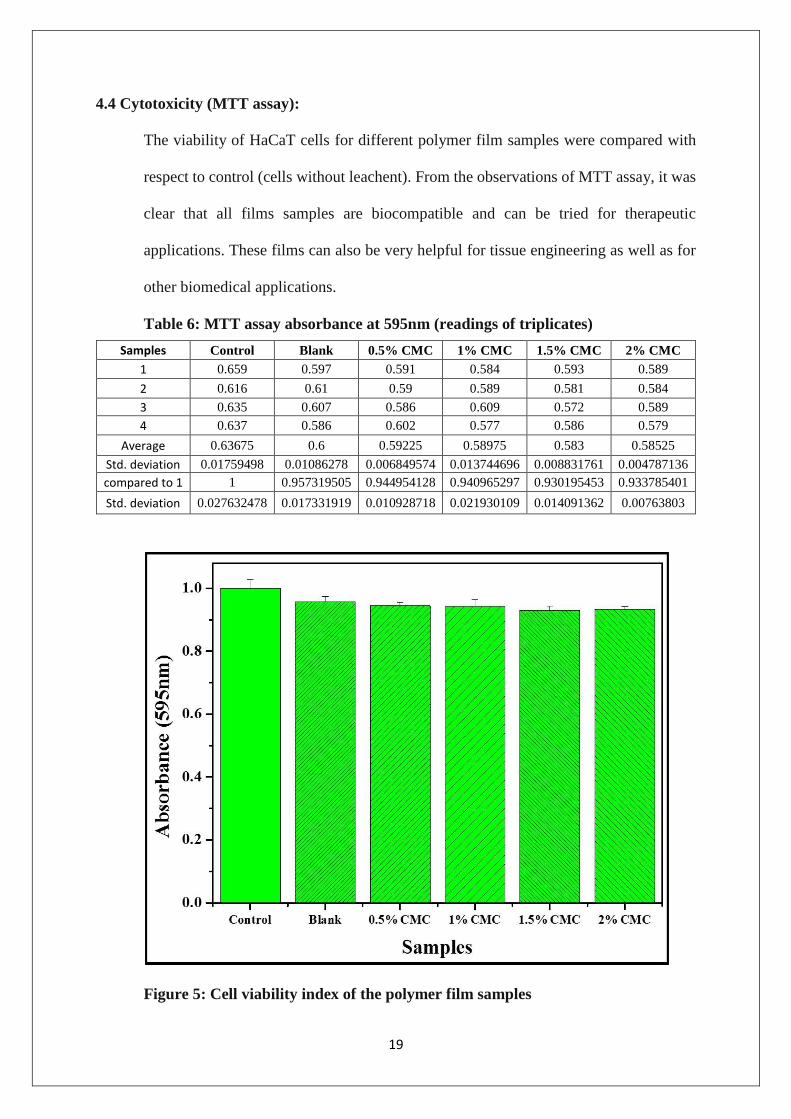
4.4 Cytotoxicity (MTT assay):
The viability of HaCaT cells for different polymer film samples were compared with
respect to control (cells without leachent). From the observations of MTT assay, it was
clear that all films samples are biocompatible and can be tried for therapeutic
applications. These films can also be very helpful for tissue engineering as well as for
other biomedical applications.
Table 6: MTT assay absorbance at 595nm (readings of triplicates)
Figure 5: Cell viability index of the polymer film samples
Samples Control Blank 0.5% CMC 1% CMC 1.5% CMC 2% CMC
1 0.659 0.597 0.591 0.584 0.593 0.589
2 0.616 0.61 0.59 0.589 0.581 0.584
3 0.635 0.607 0.586 0.609 0.572 0.589
4 0.637 0.586 0.602 0.577 0.586 0.579
Average 0.63675 0.6 0.59225 0.58975 0.583 0.58525
Std. deviation 0.01759498 0.01086278 0.006849574 0.013744696 0.008831761 0.004787136
compared to 1 1 0.957319505 0.944954128 0.940965297 0.930195453 0.933785401
Std. deviation 0.027632478 0.017331919 0.010928718 0.021930109 0.014091362 0.00763803
20
4.5 Discussion:
Our study on the polymer films showed that these films can be formed with less
proportion of CMC. Swelling nature of polymer films is constant around 80% and have
good swelling strength. In case of biodegradation, it is increased with increase in
proportion of CMC and is constant around 35%. Hemocompatibility of polymer films
is negligible and can be tried for various tissue engineering applications. Cytotoxicity
(MTT assay) results showed that polymer films biocompatible and can be tried for
several biomedical applications.
21
References

22
1. Zhang, Y.-Q., Applications of natural silk protein sericin in biomaterials.
Biotechnology advances, 2002. 20(2): p. 91-100.
2. Small, P., Some factors affecting the solubility of polymers. Journal of Applied
Chemistry, 1953. 3(2): p. 71-80.
3. Mansfield, S.D., C. Mooney, and J.N. Saddler, Substrate and enzyme characteristics
that limit cellulose hydrolysis. Biotechnology progress, 1999. 15(5): p. 804-816.
4. Peixoto, L.S., et al. Synthesis of Poly (Vinyl Alcohol) and/or Poly (Vinyl Acetate)
Particles with Spherical Morphology and Core‐Shell Structure and its Use in Vascular
Embolization. in Macromolecular Symposia. 2006. Wiley Online Library.
5. Kang, S.T. and J.S. Rhee, Characteristics of immobilized lipase‐catalyzed hydrolysis
of olive oil of high concentration in reverse phase system. Biotechnology and
bioengineering, 1989. 33(11): p. 1469-1476.
6. Miaudet, P., et al., Thermo-electrical properties of PVA–nanotube composite fibers.
Polymer, 2007. 48(14): p. 4068-4074.
7. Gómez-Guillén, M., et al., Structural and physical properties of gelatin extracted from
different marine species: a comparative study. Food Hydrocolloids, 2002. 16(1): p.
25-34.
8. Chen, X.-G. and H.-J. Park, Chemical characteristics of< i> O</i>-carboxymethyl
chitosans related to the preparation conditions. Carbohydrate Polymers, 2003. 53(4):
p. 355-359.
9. Jayakumar, R., et al., Novel carboxymethyl derivatives of chitin and chitosan
materials and their biomedical applications. Progress in Materials Science, 2010.
55(7): p. 675-709.
23
10. Afridi, M., et al., Development of polymer films by the coalescence of polymer
particles in powdered and aqueous polymer-modified mortars. Cement and Concrete
Research, 2003. 33(11): p. 1715-1721.
11. Kaufman, F.B. and E.M. Engler, Solid-state spectroelectrochemistry of crosslinked
donor bound polymer films. Journal of the American Chemical Society, 1979. 101(3):
p. 547-549.
12. Pasparakis, G. and N. Bouropoulos, Swelling studies and in vitro release of verapamil
from calcium alginate and calcium alginate–chitosan beads. International journal of
pharmaceutics, 2006. 323(1): p. 34-42.
13. Pal, K., A. Banthia, and D. Majumdar, Biomedical evaluation of polyvinyl alcohol–
gelatin esterified hydrogel for wound dressing. Journal of Materials Science:
Materials in Medicine, 2007. 18(9): p. 1889-1894.
14. Sutar, P.B., et al., Development of pH sensitive polyacrylamide grafted pectin
hydrogel for controlled drug delivery system. Journal of Materials Science: Materials
in Medicine, 2008. 19(6): p. 2247-2253.
15. Yan, X.L., E. Khor, and L.Y. Lim, Chitosan‐alginate films prepared with chitosans of
different molecular weights. Journal of biomedical materials research, 2001. 58(4): p.
358-365.
16. Gerlier, D. and N. Thomasset, Use of MTT colorimetric assay to measure cell
activation. Journal of immunological methods, 1986. 94(1): p. 57-63.


![Poly(3-hydroxybutyrate)/magnetite Composite Nanofibers ... · PDF fileco-acrylic acid) [24], poly(vinyl chloride) [25] and poly(vinyl alcohol) [26-29]. ... such as stepwise processing](https://img.pdfslide.net/doc/110x75/5a9aa3fe7f8b9a451b8d9cda/poly3-hydroxybutyratemagnetite-composite-nanofibers-acid-24-polyvinyl.jpg)