Embed Size (px)
Citation preview

Revista MVZ Córdoba
ISSN: 0122-0268
Universidad de Córdoba
Colombia
Gómez J, Brigitte; Ramírez R, María; Maldonado E, Juan
Presence of lung metastases in bitches affected by malignant mammary neoplasms in Medellin
(Colombia)
Revista MVZ Córdoba, vol. 17, núm. 2, mayo-agosto, 2012, pp. 2893-2990
Universidad de Córdoba
Montería, Colombia
Available in: http://www.redalyc.org/articulo.oa?id=69323751005
How to cite
Complete issue
More information about this article
Journal's homepage in redalyc.org
Scientific Information System
Network of Scientific Journals from Latin America, the Caribbean, Spain and Portugal
Non-profit academic project, developed under the open access initiative
2983
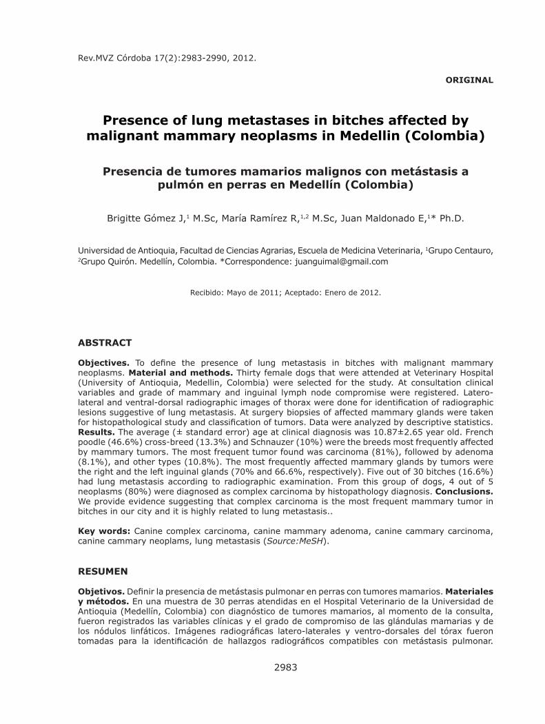
Presence of lung metastases in bitches affected by malignant mammary neoplasms in Medellin (Colombia)
Presencia de tumores mamarios malignos con metástasis a pulmón en perras en Medellín (Colombia)
Brigitte Gómez J,1 M.Sc, María Ramírez R,1,2 M.Sc, Juan Maldonado E,1* Ph.D.
Universidad de Antioquia, Facultad de Ciencias Agrarias, Escuela de Medicina Veterinaria, 1Grupo Centauro, 2Grupo Quirón. Medellín, Colombia. *Correspondence: [email protected]
Recibido: Mayo de 2011; Aceptado: Enero de 2012.
ABSTRACT
Objectives. To define the presence of lung metastasis in bitches with malignant mammary neoplasms. Material and methods. Thirty female dogs that were attended at Veterinary Hospital (University of Antioquia, Medellin, Colombia) were selected for the study. At consultation clinical variables and grade of mammary and inguinal lymph node compromise were registered. Latero-lateral and ventral-dorsal radiographic images of thorax were done for identification of radiographic lesions suggestive of lung metastasis. At surgery biopsies of affected mammary glands were taken for histopathological study and classification of tumors. Data were analyzed by descriptive statistics. Results. The average (± standard error) age at clinical diagnosis was 10.87±2.65 year old. French poodle (46.6%) cross-breed (13.3%) and Schnauzer (10%) were the breeds most frequently affected by mammary tumors. The most frequent tumor found was carcinoma (81%), followed by adenoma (8.1%), and other types (10.8%). The most frequently affected mammary glands by tumors were the right and the left inguinal glands (70% and 66.6%, respectively). Five out of 30 bitches (16.6%) had lung metastasis according to radiographic examination. From this group of dogs, 4 out of 5 neoplasms (80%) were diagnosed as complex carcinoma by histopathology diagnosis. Conclusions. We provide evidence suggesting that complex carcinoma is the most frequent mammary tumor in bitches in our city and it is highly related to lung metastasis..
Key words: Canine complex carcinoma, canine mammary adenoma, canine cammary carcinoma, canine cammary neoplams, lung metastasis (Source:MeSH).
RESUMEN
Objetivos. Definir la presencia de metástasis pulmonar en perras con tumores mamarios. Materiales y métodos. En una muestra de 30 perras atendidas en el Hospital Veterinario de la Universidad de Antioquia (Medellín, Colombia) con diagnóstico de tumores mamarios, al momento de la consulta, fueron registrados las variables clínicas y el grado de compromiso de las glándulas mamarias y de los nódulos linfáticos. Imágenes radiográficas latero-laterales y ventro-dorsales del tórax fueron tomadas para la identificación de hallazgos radiográficos compatibles con metástasis pulmonar.
ORIGINAL
Rev.MVZ Córdoba 17(2):2983-2990, 2012.
2984 REVISTA MVZ CÓRDOBA • Volumen 17(2), Mayo - Agosto 2012
Biopsias mamarias afectadas fueron sometidas a estudio histopatológico y clasificación del tipo de tumor. Los datos fueron analizados mediante estadística descriptiva. Resultados. La edad promedio (± error estándar) al diagnóstico clínico fue 10.87 ± 2.65 años de edad. La raza más frecuentemente afectada fue la French poodle (46.6%) seguida de perros cruzados (13.3%) y Schnauzer (10%). El carcinoma fue el tumor más hallado (81%) seguido del adenoma (8.1%) y otros tipos de tumor (10.8%). Las glándulas mamarias más afectadas fueron las inguinales derecha (70%) e izquierda (66.6%). Cinco de las 30 pacientes (16.6%), presentaron metástasis a pulmón. Entre estas, 4 de 5 (80%) tenían carcinoma complejo. Conclusiones. El carcinoma complejo fue la neoplasia más frecuente y es el tipo más relacionado con metástasis pulmonar.
Palabras clave: Adenoma mamario canino, carcinoma complejo canino, metástasis pulmonar, neoplasia mamaria canina (Fuente:MeSH).
INTRODUCTION
Mammary tumors are one of the most frequently diagnosed neoplasms in bitches, exhibiting a high incidence of malignancy and a high metastasis degree affecting several other tissues (1,2). In addition, they accounts for almost 50% of neoplasm in bitches. The average age at the first clinical evidence is 10 years old, with no predilection for the breed of the bitch (3,4). Canine mammary tumors have a complex pathogenesis, including susceptibility of bitches to their own endogenous sexual steroids (3,5,6), environmental contaminants to which dogs are commonly exposed such as allethrin, cyhalothrin, cypermethrin, deltamethrin and tetramethrin (7), and diet and hormonal components related to obesity (6).
Malignant canine mammary tumors (MCMT) are characterized for presenting an aggressive inflammatory pattern, particularly in the case of carcinoma, it behave as highly aggressive and have a low survival rate. MCMT exhibit unlimited growing capability, and are capable to infiltrate regional lymph drainage and nodes and to cause metastases to other tissues (8). Several studies have reported the incidence of canine mammary tumors: In a report in which 672 Beagle bitches were evaluated, authors found a 71% frequency of mammary tumors, 19% of these were carcinoma and most of the dogs showed a high frequency of metastasis (2). Radiographic evidence of lung metastasis was found in 14 out of 37 bitches having malignant mammary tumors (carcinosarcoma, complex carcinoma, and simple carcinoma) (4). In Colombia, a retrospective study comprising the casuistic of 30 years (1968-1998) performed in the Veterinary Pathology Service at University of Antioquia (n = 232 samples), revealed the presence of carcinoma in 58.6%, benign mammary tumor in 23.7%, mixed malignant tumor in 9.5%, and other type of tumors in 8.2% of the cases (9). However, in this study no data was reported on the incidence and frequency of lung metastases.
Lymph drainage is considered as one of the most important route for dissemination of mammary tumor metastasis in dogs. Canine tumors that induce formation of new lymph vessel have a higher metastasis potential (10). Because lung metastasis are responsible for 25% to 50% of total MCMT metastasis (11), it is important to perform radiographic evaluation of bitches suffering from MCMT on a routine basis, as well as cytologic evaluation by fine needle puncture (12) and evaluation of hepato-renal function. When a non-cavitary interstitial lung pattern at radiography is observed in bitches suffering from MCMT, it would probably reflect the presence of lung metastasis which originated from mammary tumors (4). Accordingly, the clinical prognosis for bitches affected by mammary tumors will depend on the histological pattern of the tumor they suffers, as well as its infiltrating and metastatic capability, characteristics that must be determined by an accurate clinical exam combined with a radiological and histopathological diagnosis. It is also important to consider assessment of the estrogen and progesterone receptor expression (13). This approach could help to define the most precise treatment for the bitch when considering survival probabilities and prognosis of her life quality. Because studies on the frequency of MCMT with lung metastasis have not been reported in Colombia, the main objective of this work was to establish the frequency of lung metastases and the type of histological pattern/classification of MCMT.
MATERIALS AND METHODS
Type of study and sample. A prospective descriptive pilot study was performed in which the records from Veterinary Hospital at University of Antioquia (Medellín, Colombia) were evaluated to define the frequency of malignant canine mammary tumors. Tumor samples from patients
2985Gómez - Presence of lung metastases in bitches affected by malignant
included in the study were obtained only by previous informed consent of the dog owner. Dogs were subjected to surgical procedures according to routinary protocols for ovariohysterectomy (OH) and/or surgical excision of mammary tumors. For the study, data from 30 patients presenting mammary gland neoplasms at consultation were recorded during a 10 month period (June 2008 to April 2009). The group of bitches included in the study were pure or cross-breed females ranging from 7 to 18 years old.
Inclusion criteria. Inclusion criteria of the study were: clinically evident nodes located in mammary glands, with or without compromise of the corresponding inguinal lymph node, and the availability of histopathological exam of a mammary node sample, and a latero-lateral and ventral-dorsal radiography of thorax. The only exclusion criteria were if the bitch was receiving any kind of chemotherapy schedule at the time of clinical diagnosis of mammary gland tumors.
Clinical exam. At clinical examination all the patients were evaluated by palpation to confirm the presence of nodes in mammary glands, if there were left and/or right axillary lymph nodes compromise, left and/or right inguinal superficial lymph node compromise, and/or deep lymph nodes compromise. Similarly, size of the node and its adherence to the surrounding tissues were also evaluated. Then, depending if the type of tumor diagnosed was related to malignancy (14), bitches were subjected to a radiographic evaluation of thorax for estimation of the risk of metastasis. When a radiographic evidence of lung metastases was found, the dog was not subjected to surgical excision of mammary tumors. Otherwise, a post-mortem examination was performed if the dog were died. On the contrary, when the diagnosis was negative for lung metastases, pre-surgical laboratory exams were performed (including Hemogram, alanine aminotransferase, Creatinin, Protrombine, Tromboplastine Partial Time) for defining the clinical status of the dog previous to its programming for surgical excision of the tumors (2).
Surgical and histopathology procedures. Excision of the affected mammary glands was performed only in bitches that met the standard criteria for surgical procedures, according to the surgical protocol for excision of canine mammary neoplasms at “Hospital Veterinario, Universidad de Antioquia”. From each lymph node obtained after surgical excision a piece of 0.5 cm3 was taken, and samples were fixed in 10% formaldehyde. Fixed samples were processed for histopathology and Hematoxilin-Eosin staining performed evaluation under routine procedures at “Laboratorio de
Patología Animal, Universidad de Antioquia”, according to the method by Misdorp et al (15). Samples were double-blind analyzed by a trained histopathologist.
Statistical analysis. Data from quantitative variables of patients included in the study were analyzed by descriptive statistics. Qualitative variables were compared as percentage for each clinical and epidemiological parameter, as well as for histopathological classification of the tumor and radiographic findings. The compromise of each set of lymph node-draining of mammary glands and localization of affected lymph nodes were evaluated as percentages.
RESULTS
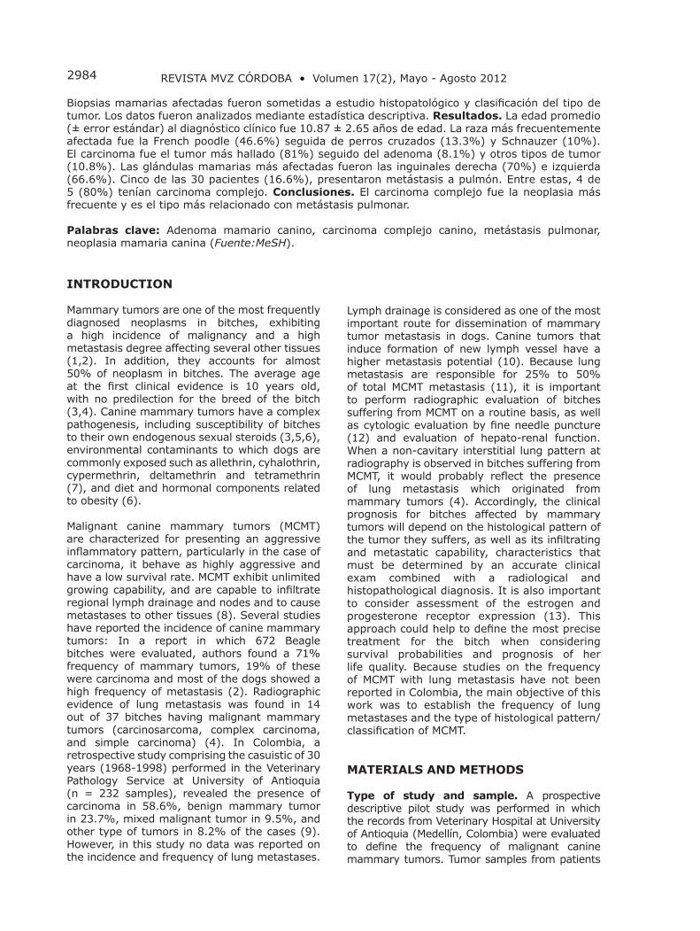
The most relevant clinical and epidemiological data of patients included in the study are presented in table 1. The average age at consultation when the bitch was detected having mammary gland nodes was 10.87±2.65 year (Table 1). Poodle dogs were the genetic predominant group 46.66% (14/30), followed by 13,3% cross-breed (4/30), and 10% Schnauzer (3/30) (Figure 1). At clinical exam 70% of the patients had at least three mammary glands
Breed n % Age (years)
Overall clinical diagnosis
Overall 30 100 10.87 Complex carcinoma.
French poodle 14 46.66 10.57 Complex carcinoma.
Cross-breed 4 13.33 11.5Complex carcinoma, tubular carcinoma, carcinosarcoma.
Schnauzer 3 10 12 Complex carcinoma.
Others 9 29.97 10.66 Complex carcinoma, simple carcinoma.
Table 1. Clinical and epidemiological data of patients included in the study.
Figure 1. Frequency distribution of malignant canine mammary tumors in a sample of bitches in Medellín (Colombia).
2986 REVISTA MVZ CÓRDOBA • Volumen 17(2), Mayo - Agosto 2012
with clinical evidence of tumor, whereas 16.6% and 13.3% had one or two affected mammary glands, respectively.
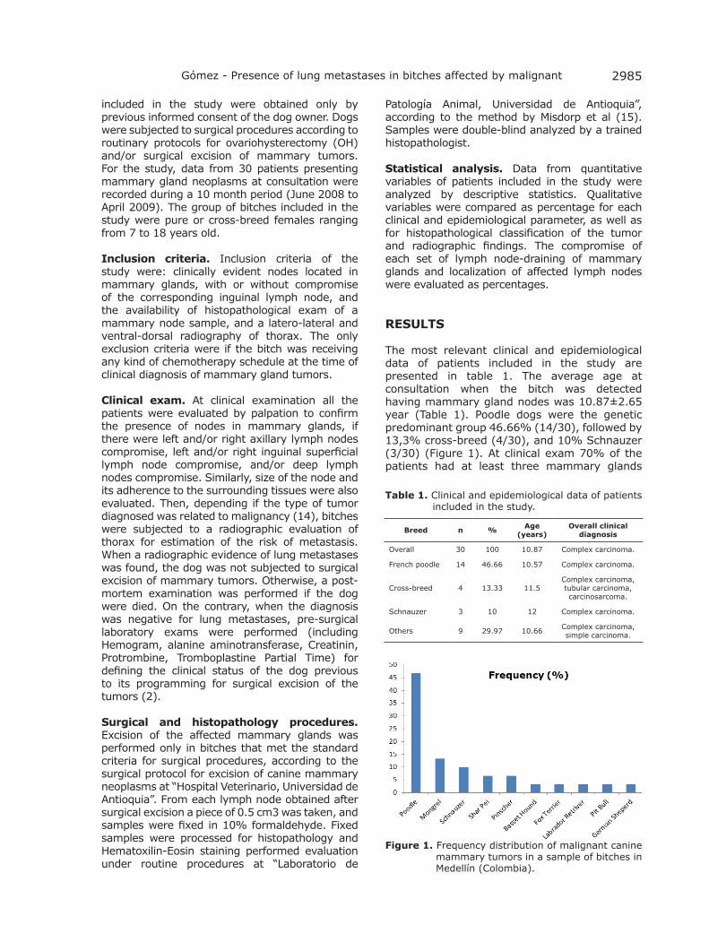
Most of the bitches had compromise of the inguinal mammary gland: 17 out of 30 (56.6%) presented compromise of the right inguinal mammary gland, and 19 out of 30 (63.3%) presented compromise of the left inguinal mammary gland. The remaining affected mammary glands were as follows: 21 out of 30 (70%) the right abdominal 2 mammary gland, and 20 out of 31 (66.6%) the left abdominal 2 mammary gland, 5 out of 30 (16.66%) the right abdominal 1, and 7 out of 30 (23.33%) the left abdominal 1, 10 out of 31 (33.33%) the thoracic right 1, 6 out of 30 (20%) the thoracic left 1, 8 out of 30 (26.66%), the thoracic right 2, and 9 out of 30 (30%) the thoracic left 2 (Figure 2).
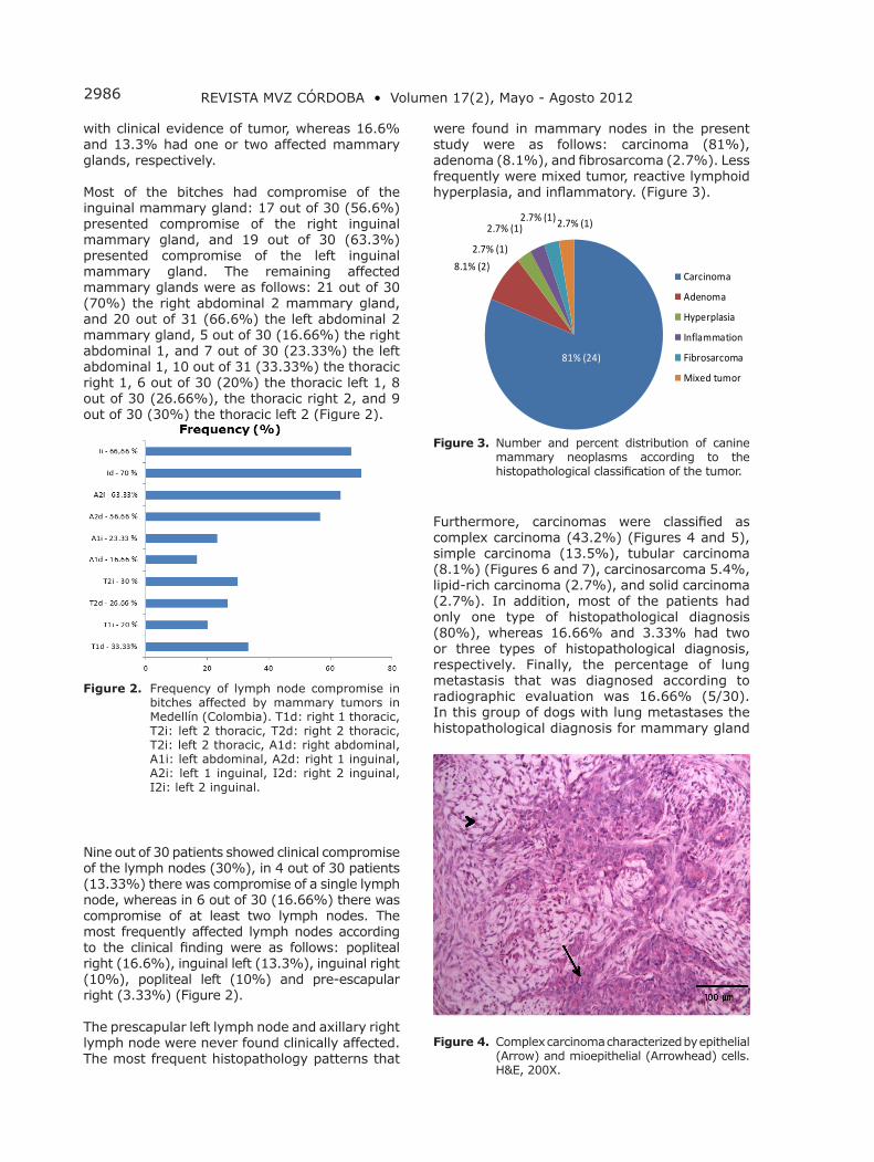
were found in mammary nodes in the present study were as follows: carcinoma (81%), adenoma (8.1%), and fibrosarcoma (2.7%). Less frequently were mixed tumor, reactive lymphoid hyperplasia, and inflammatory. (Figure 3).
Figure 2. Frequency of lymph node compromise in bitches affected by mammary tumors in Medellín (Colombia). T1d: right 1 thoracic, T2i: left 2 thoracic, T2d: right 2 thoracic, T2i: left 2 thoracic, A1d: right abdominal, A1i: left abdominal, A2d: right 1 inguinal, A2i: left 1 inguinal, I2d: right 2 inguinal, I2i: left 2 inguinal.
Nine out of 30 patients showed clinical compromise of the lymph nodes (30%), in 4 out of 30 patients (13.33%) there was compromise of a single lymph node, whereas in 6 out of 30 (16.66%) there was compromise of at least two lymph nodes. The most frequently affected lymph nodes according to the clinical finding were as follows: popliteal right (16.6%), inguinal left (13.3%), inguinal right (10%), popliteal left (10%) and pre-escapular right (3.33%) (Figure 2).
The prescapular left lymph node and axillary right lymph node were never found clinically affected. The most frequent histopathology patterns that
81% (24)
8.1% (2)2.7% (1)
2.7% (1)2.7% (1) 2.7% (1)
Carcinoma
Adenoma
Hyperplasia
Inflammation
Fibrosarcoma
Mixed tumor
Figure 3. Number and percent distribution of canine mammary neoplasms according to the histopathological classification of the tumor.
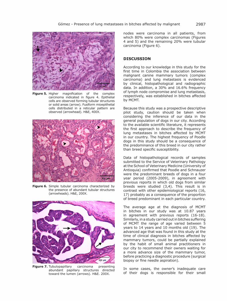
Figure 4. Complex carcinoma characterized by epithelial (Arrow) and mioepithelial (Arrowhead) cells. H&E, 200X.
Furthermore, carcinomas were classified as complex carcinoma (43.2%) (Figures 4 and 5), simple carcinoma (13.5%), tubular carcinoma (8.1%) (Figures 6 and 7), carcinosarcoma 5.4%, lipid-rich carcinoma (2.7%), and solid carcinoma (2.7%). In addition, most of the patients had only one type of histopathological diagnosis (80%), whereas 16.66% and 3.33% had two or three types of histopathological diagnosis, respectively. Finally, the percentage of lung metastasis that was diagnosed according to radiographic evaluation was 16.66% (5/30). In this group of dogs with lung metastases the histopathological diagnosis for mammary gland
2987Gómez - Presence of lung metastases in bitches affected by malignant
nodes were carcinoma in all patients, from which 80% were complex carcinomas (Figures 4 and 5) and the remaining 20% were tubular carcinoma (Figure 6).
DISCUSSION
According to our knowledge in this study for the first time in Colombia the association between malignant canine mammary tumors (complex carcinoma) and lung metastasis is evidenced by clinical, histopathological and radiographic data. In addition, a 30% and 16.6% frequency of lymph node compromise and lung metastasis, respectively, was established in bitches affected by MCMT.
Because this study was a prospective descriptive pilot study, caution should be taken when considering the inference of our data in the general population of dogs in our city. According to the available scientific literature, it represents the first approach to describe the frequency of lung metastases in bitches affected by MCMT in our country. The highest frequency of Poodle dogs in this study should be a consequence of the predominance of this breed in our city rather than breed specific susceptibility.
Data of histopathological records of samples submitted to the Service of Veterinary Pathology at the School of Veterinary Medicine (University of Antioquia) confirmed that Poodle and Schnauzer were the predominant breeds of dogs in a four year period (2005-2009), in agreement with previous reports in which old dogs from similar breeds were studied (3,4). This result is in contrast with other epidemiological reports (16, 17) probably as a consequence of the proportion of breed predominant in each particular country.
The average age at the diagnosis of MCMT in bitches in our study was at 10.87 years in agreement with previous reports (16-18). Similarly, in a study carried out in bitches suffering of MCMT the range of age varied between 5 years to 14 years and 10 months old (19). The advanced age that was found in this study at the time of clinical diagnosis in bitches affected by mammary tumors, could be partially explained by the habit of small animal practitioners in our city to recommend their owners waiting for a more advance size of the mammary tumor, before practicing a diagnostic procedure (surgical biopsy or fine needle aspiration).
In some cases, the owner’s inadequate care of their dogs is responsible for their small
Figure 6. Simple tubular carcinoma characterized by the presence of abundant tubular structures (arrowheads). H&E, 200X.
Figure 7. Tubulopapillary carcinoma presenting abundant papillary structures directed toward the lumen (arrows). H&E. 200X.
Figure 5. Higher magnification of the complex carcinoma indicated in figure 4. Epithelial cells are observed forming tubular structures or solid areas (arrow). Fusiform mioepithelial cells distributed in a reticular pattern are observed (arrowhead). H&E, 400X.
2988 REVISTA MVZ CÓRDOBA • Volumen 17(2), Mayo - Agosto 2012
mammary gland nodes to develop toward an advanced clinical condition of mammary tumors before consulting the veterinarian, which causes misdiagnosis at early stages of the condition in the affected dog. In other cases and due to the owner’s negligence, the affected mammary glands develop toward a more advanced stage of the tumor, including variable grades of clinical signs such as inflamed/ulcerated mammary glands, even with a critical general compromise of the patient. Representative cases of this situation have been reported elsewhere (for example, see figure 1 in the report by Andrade et al, 2010) (7) and figure 6 in the report by Otini et al, 2010 (19). Because in dogs there is a normal course of mammary tumor progression from a benign pattern toward malignancy (18), an early diagnosis and treatment of the condition would probably avoid the affected bitch to develop a malignant form of the neoplasm, and would prevent a potential risk of metastases.
The most frequently affected glands in bitches in our study were the inguinal mammary glands, which is in agreement with previous reports (10,20). The growing capability of inguinal mammary glands is greater than that of the other glands, a fact that could be explained by its greater vascular supply and its greater lymph drainage. This particular physiological situation could support its major compromise in neoplasm development and metastasis (20). In the original reports by Patsikas and Dessiris (21) the authors suggested the existence of lymphatic vessel connections between the lymph nodes draining both sides of the mammary gland, in which most of the connections are established with the inguinal lymph nodes.
The finding of a greater compromise of inguinal and popliteal lymph nodes was as expected, because these lymph nodes drains the inguinal mammary gland, which was found the most affected gland in this study, in agreement with previous reports in dogs (10,20,22) and cats (23), which suggest there are an intricate communication between the lymph nodes draining the mammary gland. Together with the high frequency of compromise of the inguinal mammary gland in our study, these results explain, at least partially, the most frequent compromise of the inguinal lymph nodes in bitches affected by MCMT. As we shall see, this would be a rational explanation for the incidence of lung metastasis in bitches.
In our study most of the mammary glands affected by neoplasm (83.74%) exhibited histopathological findings of malignancy, mainly carcinoma (81%), and in a lesser percentage
fibrosarcoma (2.7%), whereas only 8.1% of the samples showed benign characteristics (adenomas). Our data are in agreement with a previous study by Lockett (2005) and with retrospective data from our Laboratory, in which the frequency of malignant mammary tumors was 68.1%, from which 58.6% of tumors were diagnosed as carcinoma (9). Similarly, other authors found a predominant diagnosis of coriocarcinoma in bitches affected by mammary tumors (16,18). On the contrary, our data are not in agreement with previous reports in which predominance of benign (60% to 40%) versus malignant (30%-40%) mammary tumors was reported, where malignant tumors included complex carcinoma, simple carcinoma (tubulopapilar, solid, cribiforme and anaplasic, and solid carcinoma), and sarcoma (fibrosarcoma, osteosarcoma, and carcinosarcoma) (24,25).
When lung metastases were confirmed on the basis of radiographic findings, it was found in 5 out of 30 (16.6%) bitches in our study. This data support the concept that MCMT, mainly carcinoma, behaves as a highly invasive and metastatic tumor, because of its high growing capability and its infiltration potential (8). The radiographic diagnosis of lung metastases is characterized by a structured interstitial pattern, non-cavitary, characterized by the presence of multiple nodules of approximately 5-6 mm diameter (11), whereas nodules having a lesser diameter should be grouped together for being visible at radiographic examination.
The finding of 16.66% frequency of lung metastases is lower than a previous study where the frequency of metastases reported was 25% to 50% (4). In the work by Clement et al, 2010, authors provided evidence that computed tomography is a more accurate method than radiography for the diagnosis of lung metastases (17). For instance, our results on the frequency of lung metastases based on radiographic diagnosis should be considered cautiously if used for comparative purposes. It is important to recall that the postsurgical recurrence rate of bitches affected by MCMT is up to 58% (26) as well as the relationship between prognostic factors (27) and type of tumor and survival rate (28,29).
Regarding sample size in our study (n=30 cases in 10 months period of study), the results of a previous retrospective study performed to evaluate the frequency of canine mammary tumors in Colombia, in a sample of 173 tumors in a 25 year sample period, represented a mean frequency of 7 cases per year (30). In this study authors found that 18.73% of the bitches were included in the range of age of 120 months,
2989Gómez - Presence of lung metastases in bitches affected by malignant
comparable with the average age of bitches in our study (Table 1). The predominant breed found by these authors was Cocker spaniel (23.8%) —this finding would probably reflect the predominance of this breed during the time period of sampling (1975-2000). The second predominant breed they found was poodle (30), in agreement with our results (Figure 1).
In the study by Torres and Botero (2008) malignancy was reported in 41% of the cases. Mixt Malignant Mammary tumor (recently classified as carcinosarcoma) was found in 12 out of 32 (37.5%) cases and simple tubular carcinoma was found in 4 out of 32 (12.5%) cases, in the group of age greater than 120 months (see table 4 in reference 30) corresponding to the range of age of bitches in our study. Data from this report are partially similar with our data in which complex carcinoma (43.2%), simple carcinoma (13.5%), and tubular carcinoma (8.1%) were the predominant type of tumors. Data from the present pilot study do not exclude the possibility that dogs were affected by
other primary tumors or had presented other metastatic sites. For that reason, further studies in bitches affected by MCMT should include the histopathological evaluation of regional lymph nodes, and abdominal ultrasound evaluation for defining other metastatic sites different to lung. Similarly, it would support a more precise surgical procedure used for resection of affected mammary glands.
Finally, our data also corroborates the importance for establishing a radiographic evaluation of female dogs affected by mammary gland tumors as a routine practice, due to its valuable prognostic value for diagnosis of metastases (24). If the veterinarian uses a precise protocol on a routinely basis, a great advance on earlier detection of MCMT and its metastases should be gained, with implications for the clinical evolution of the affected patient. Furthermore, the results of this study supports the concept that a finding of a clinically normal lymph node draining an affected mammary gland, do not mean they are not histopathologically affected, and suggest that sampling the affected lymph node should be always considered.
1. Morris P, Dobson J. Oncología en pequeños animales. Buenos Aires: Intermédica; 2002.
2. Welch T. Small animal surgery. 3rd ed. New York: Mosby; 2007.
3. Hermo G, García M, Torres P. Gobello C. Tumores de mama en la perra. Cienc Vet 2005; 7:1-25.
4. Lockett M, Merlo W, Rosciana A, Maccio O, Guaimás L. Evaluación radiológica en caninos para detección de metástasis de tumores mamarios malignos en tórax y abdomen. Comunicaciones científicas y tecnológicas [en línea] 2005 [fecha de acceso 15 de febrero de 2010]. URL disponible en: http://www.unne.edu.ar/Web/cyt/com2005/4-Veterinaria/V-025.pdf
5. Munson L, Moresco A. Comparative pathology of mammary gland cancers in domestic and wild animals. Breast Dis 2007; 28:7-21.
REFERENCES
6. German AJ. The growing problem of obesity in dogs and cats. J Nutr 2006; 136(7 Suppl):1940S-1946S.
7. Andrade FH, Figueiroa FC, Bersano PR, Bissacot DZ, Rocha NS. Malignant mammary tumor in female dogs: environmental contaminants. Diagn Pathol 2010; 5:45.
8. Flores P, Cattaneo U. Tumores mamarios en caninos domésticos, epidemiología, criterios de diagnóstico y enfoque terapéutico. Monografías de medicina veterinaria [en línea] 2001 [fecha de acceso 15 de febrero de 2010]. URL disponible en: http://www.monografiasveterinaria.uchile.cl/CDA/mon_vet_seccion/0,1419,SCID%253D8149%2526ISID%253D416,00.html)
9. Ferreira G. Patología Veterinaria. Medellín: Universidad de Antioquia; 2003.
10. Patsikas M, Karayannopoulou M, Kaldrymidoy E, Papazoglou L, Papadopoulou P. The lymph drainage of the neoplastic mammary glands in the bitch: A lymphographic study. Anat Histol Embryol 2006; 35:228-234.

2990 REVISTA MVZ CÓRDOBA • Volumen 17(2), Mayo - Agosto 2012
11. Thrall D. Manual de diagnóstico radiológico Veterinario. 3ra ed. Madrid: Elsevier; 2003.
12. Hellmén E, Lindgren A. The accuracy of cytology in diagnosis and DNA analysis of canine mammary tumours. J Comp Pathol 1989; 101(4):443-450.
13. Chang CC, Tsai MH, Liao JW, Chan JP, Wong ML, Chang SC. Evaluation of hormone receptor expression for use in predicting survival of female dogs with malignant mammary gland tumours. J Am Vet Med Assoc. 2009; 235(4):391-396.
14. Withrow S, Vail D. Small animal clinical oncology. 4th ed. Philadelphia: Saunders; 2007.
15. Misdorp W, Else R, Hellmen E and Lipscomb T. Histological classification of mammary tumors of the dog and cat. Estados Unidos: Armed Forces Institute of Pathology, 1999.
16. Brønden LB, Nielsen SS, Toft N, Kristensen AT. Data from the Danish veterinary cancer registry on the occurrence and distribution of neoplasms in dogs in Denmark. Vet Rec 2010; 166:586-590.
17. Clemente M, Pérez-Alenza MD, Peña L. Metastasis of canine inflammatory versus non-inflammatory mammary tumours. J Comp Pathol 2010; 143:157-163.
18. Sorenmo KU, Kristiansen VM, Cofone MA, Shofer FS, Breen AM, Langeland M, Mongil CM, Grondahl AM, Teige J, Goldschmidt MH. Canine mammary gland tumours; a histological continuum from benign to malignant; clinical and histopathological evidence. Vet Comp Oncol 2009; 7:162-172.
19. Otoni CC, Rahal SC, Vulcano LC, Ribeiro SM, Hette K, Giordano T, Doiche DP, Amorim RL. Survey radiography and computerized tomography imaging of the thorax in female dogs with mammary tumors. Acta Vet Scand 2010; 52:20.
20. Pereira C, Rahal S, De Carvalho Balieiro J, Ribeiro A. Lymphatic drainage on healthy and neoplasic mammary glands in female dogs: can it really altered. Anat Histol Embryol 2003; 32: 282-290.
21. Patsikas MN, Karayannopoulou M, Kaldrymidoy E, Papazoglou LG, Papadopoulou PL, Tzegas SI, Tziris NE, Kaitzis DG, Dimitriadis AS, Dessiris A. The lymph drainage of the mammary glands in the Bitch: a lymphographic study. Anat Histol Embryol 2006; 35(4):228-234.
22. Pereira CT, Navarro-Marques LF, Williams J, De Martin WB, Bombonato PP. 99mTc-labeled dextran for mammary lymphoscintigraphy in dogs. Vet Radiol Ultrasound 2008; 49:487-491.
23. Papadopoulou PL, Patsikas MN, Charitanti A, Kazakos GM, Papazoglou LG, Karayannopoulou M, Chrisogonidis I, Tziris N, Dimitriadis A. The lymph drainage pattern of the mammary glands in the cat: a lymphographic and computerized tomography lymphographic study. Anat Histol Embryol 2009; 38:292-299.
24. Meuten D. Tumors in domestic animals. 4th ed. New York: Blackwell; 2002.
25. Dobson J, Lascelles D. BSAVA manual of canine and feline oncology. 2nd ed. London: Blackwell publishing; 2003.
26. Stratmann N, Failing K, Richter A, Wehrend A. Mammary tumor recurrence in bitches after regional mastectomy. Vet Surg 2008; 37:82-86.
27. Marconato L, Romanelli G, Stefanello D, Giacoboni C, Bonfanti U, Bettini G, Finotello R, Verganti S, Valenti P, Ciaramella L, Zini E. Prognostic factors for dogs with mammary inflammatory carcinoma: 43 cases (2003-2008). J Am Vet Med Assoc 2009; 235:967-972.
28. Queiroga FL, Pires I, Lobo L, Lopes CS. The role of Cox-2 expression in the prognosis of dogs with malignant mammary tumours. Res Vet Sci 2010; 88(3):441-445.
29. Ness M. Survival time of dogs with
inflammatory mammary cancer. Vet Rec 2009; 165:272.
30. Torres Vidales G, Botero Espinosa L. Estudio histopatológico retrospectivo de neoplasias de glándula mamaria en caninos (1975-2000). Revista Orinoquia 2008; 12(1): 80-88.


![CASE REPORT Open Access Metastatic colorectal carcinoma ...the female reproductive system most commonly affected by metastases [3]. Ovarian metastases occur in 3 to 8% of women with](https://img.pdfslide.net/doc/110x75/60df4bb305bcd923ec2815ba/case-report-open-access-metastatic-colorectal-carcinoma-the-female-reproductive.jpg)


![ZOGZAG 34 [PRETTY AND THE BITCHES]](https://img.pdfslide.net/doc/110x75/579053301a28ab900c8b549b/zogzag-34-pretty-and-the-bitches.jpg)