Embed Size (px)
Citation preview
#DMD-AR-2020-000340
1
Primary human hepatocyte spheroids as an in vitro tool for
investigating drug compounds with low hepatic clearance
Julia Riede, Birgit M. Wollmann, Espen Molden, Magnus Ingelman-Sundberg
Section of Pharmacogenetics, Department of Physiology and Pharmacology, Karolinska
Institutet, SE-171 77 Stockholm, Sweden (JR, MIS)
Center for Psychopharmacology, Diakonhjemmet Hospital, N-0319 Oslo, Norway (BMW,
EM)
Present address: Julia Riede, PK Sciences, Novartis Institutes for BioMedical Research, CH-
4056 Basel, Switzerland
This article has not been copyedited and formatted. The final version may differ from this version.DMD Fast Forward. Published on June 1, 2021 as DOI: 10.1124/dmd.120.000340
at ASPE
T Journals on February 15, 2022
dmd.aspetjournals.org
Dow
nloaded from
#DMD-AR-2020-000340
2
Running Title
Clearance predictions using human hepatocyte spheroids
Corresponding author
Magnus Ingelman-Sundberg
Section of Pharmacogenetics
Department of Physiology and Pharmacology
Biomedicum 5B
Karolinska Institutet
Solnavägen 9
SE-171 77 Stockholm
Sweden
Email: [email protected]
Number of text pages: 18
Number of tables: 1
Number of figures: 4
Number of references: 51
Number of words in abstract: 221
Number of words in introduction: 568
Number of words in discussion: 1680
This article has not been copyedited and formatted. The final version may differ from this version.DMD Fast Forward. Published on June 1, 2021 as DOI: 10.1124/dmd.120.000340
at ASPE
T Journals on February 15, 2022
dmd.aspetjournals.org
Dow
nloaded from
#DMD-AR-2020-000340
3
List of abbreviations
AAFE absolute average fold error
AFE average fold error
ADME absorption, distribution, metabolism, excretion
ATP adenosine triphosphate
CLh hepatic organ clearance
CLint intrinsic clearance
CYP cytochrome P450
Eh hepatic extraction ratio
IVIVE in vitro-in vivo extrapolation
MPCC micro-patterned co-culture
UGT uridine 5'-diphospho‐glucuronosyltransferase
UPLC-MS/MS ultra performance liquid chromatography-tandem mass spectrometry
This article has not been copyedited and formatted. The final version may differ from this version.DMD Fast Forward. Published on June 1, 2021 as DOI: 10.1124/dmd.120.000340
at ASPE
T Journals on February 15, 2022
dmd.aspetjournals.org
Dow
nloaded from
#DMD-AR-2020-000340
4
Abstract
Characterizing the pharmacokinetic properties of drug candidates represents an essential
task during drug development. In the past, liver microsomes and primary suspended
hepatocytes have been extensively used for this purpose, but their relatively short stability
limits the applicability of such in vitro systems for drug compounds with low metabolic
turnover. In the present study, we used 3D primary human hepatocyte spheroids to predict
the hepatic clearance of seven drugs with low to intermediate clearance in humans. Our
results indicate that hepatocyte spheroids maintain their in vivo like phenotype during
prolonged incubations allowing to monitor the depletion of parent drug for seven days. In
contrast, attempts to increase the relative metabolic capacity by pooling hepatocyte
spheroids resulted in an immediate fusion of the spheroids followed by hepatocellular de-
differentiation processes, demonstrating limited applicability of the pooling approach for
quantitative pharmacokinetic studies. The hepatic clearance values obtained from
incubations with individual spheroids were in close correlation with the clinical reference data
with six out of seven drug compounds being predicted within a three-fold deviation and
average fold and absolute average fold errors of 0.57 and 1.74, respectively. In conclusion,
the hepatocyte spheroid model enables accurate hepatic clearance predictions for slowly
metabolized drug compounds and represents a valuable tool for determining the
pharmacokinetic properties of new drug candidates as well as for mechanistic
pharmacokinetic studies.
This article has not been copyedited and formatted. The final version may differ from this version.DMD Fast Forward. Published on June 1, 2021 as DOI: 10.1124/dmd.120.000340
at ASPE
T Journals on February 15, 2022
dmd.aspetjournals.org
Dow
nloaded from
#DMD-AR-2020-000340
5
Significance Statement
Traditional in vitro systems often fail to predict the hepatic clearance of slowly metabolized
drug compounds. The current study demonstrates the ability of primary human hepatocyte
spheroids to provide accurate projections on the hepatic clearance of drug compounds with
low and intermediate clearance.
This article has not been copyedited and formatted. The final version may differ from this version.DMD Fast Forward. Published on June 1, 2021 as DOI: 10.1124/dmd.120.000340
at ASPE
T Journals on February 15, 2022
dmd.aspetjournals.org
Dow
nloaded from
#DMD-AR-2020-000340
6
Introduction
Appropriate absorption, distribution, metabolism, and excretion (ADME) properties are crucial
for selecting drug candidates and avoiding late-stage failures of drug development programs.
Therefore, new chemical entities routinely undergo in vitro high-throughput ADME screenings
at early drug discovery stages in order to identify and eliminate compounds with undesirable
pharmacokinetic properties such as short half-life and poor bioavailability. During lead
optimization, medicinal chemists further improve the metabolic stability and other
pharmacokinetic properties of lead compounds (Obach et al., 1997; Kratochwil et al., 2017).
Consequently, a large number of compounds entering preclinical development displays low
or no turnover in metabolic stability assays typically conducted using liver microsomes in the
presence of nicotinamide adenine dinucleotide phosphate (Di and Obach, 2015).
Although prolonged metabolic stability is a highly desirable pharmacokinetic attribute
allowing for a once daily dosing regimen, the characterization of such low clearance
compounds is extremely challenging for ADME scientists during the later preclinical
development, since common in vitro systems as used in screening assays are no longer
applicable. The preclinical testing is further challenged by the fact that not only slowly
metabolized compounds but also compounds eliminated by non-oxidative metabolism (e.g.
phase II conjugation reactions by uridine 5'-diphospho‐glucuronosyltransferases (UGTs)) and
pathways beyond hepatic metabolism (e.g. transporter-mediated permeability or extrahepatic
metabolism) are preferably selected by the microsomal screening approach (Argikar et al.,
2016). For instance, UGT-mediated metabolism can be covered in microsomal incubations
by adding corresponding co-factors, whereas microsomal fractions lack cytosolic enzymes
and are likewise not applicable for compounds with low (rate-limiting) active and/or passive
permeability (Kusuhara and Sugiyama, 2009; Camenisch, 2016). The use of primary
hepatocytes can partly meet these challenges as they express the entirety of hepatic drug-
metabolizing enzymes and transporters, yet primary hepatocytes rapidly de-differentiate in
suspension or 2D monolayer culture and the activity of enzymes and transporters begins to
This article has not been copyedited and formatted. The final version may differ from this version.DMD Fast Forward. Published on June 1, 2021 as DOI: 10.1124/dmd.120.000340
at ASPE
T Journals on February 15, 2022
dmd.aspetjournals.org
Dow
nloaded from
#DMD-AR-2020-000340
7
decrease within a few hours (Di and Obach, 2015). For drug compounds with low metabolic
clearance in vitro, this period is too short to achieve sufficient depletion of parent drug
compound that would allow for reliable intrinsic clearance (CLint) estimations (Chan et al.,
2013; Kratochwil et al., 2017).
Recently, 3D spheroid cultures of primary human hepatocytes have been established
allowing the culture of phenotypically stable hepatocytes for several weeks (Lin and Chang,
2008; Messner et al., 2013; Bell et al., 2016). Extensive characterization studies by our
laboratory revealed that primary human hepatocyte spheroids maintain their morphology,
viability, hepatocellular function, and the activity of drug-metabolizing enzymes over time,
closely resembling hepatocytes in vivo (Bell et al., 2016; Bell et al., 2017; Vorrink et al.,
2017). Moreover, we have successfully applied hepatocyte spheroids for hepatotoxicity
screenings and mechanistic investigations as well as for the modeling of liver diseases
(Hendriks et al., 2016; Kozyra et al., 2018; Vorrink et al., 2018; Hendriks et al., 2019; Hurrell
et al., 2020).
In the present study, we established primary human hepatocyte spheroids as a novel in vitro-
in vivo extrapolation (IVIVE) tool for predicting the hepatic clearance of low and intermediate
turnover drugs. Following the functional validation of hepatocyte spheroids, the CLint of seven
drugs with low and intermediate clearance, which are metabolized by different cytochrome
P450 (CYP) enzymes, was determined in hepatocyte spheroids from three different donors
and compared to clinical reference data. Furthermore, the effect of pooling individual
spheroids in order to enhance the metabolic capacity was investigated with regard to cell
morphology, hepatocellular function, and expression of hepatic drug-metabolizing enzymes
and transporters.
This article has not been copyedited and formatted. The final version may differ from this version.DMD Fast Forward. Published on June 1, 2021 as DOI: 10.1124/dmd.120.000340
at ASPE
T Journals on February 15, 2022
dmd.aspetjournals.org
Dow
nloaded from
#DMD-AR-2020-000340
8
Materials and Methods
Cell culture. Cryopreserved primary human hepatocytes were obtained from
BioreclamationIVT (USA) or KalyCell (France). Donor information of hepatocyte lots are
summarized in Supplementary Table 1. Hepatocytes were thawed according to the
manufacturer’s instruction and hepatocyte spheroids were generated as described previously
(Bell et al., 2016). In brief, hepatocytes from individual donors were seeded in culture media
(William’s E medium (Life Technologies, Thermo Fisher Scientific) supplemented with 2 mM
L-glutamine, 100 units/mL penicillin, 100 μg/mL streptomycin, 100 nM dexamethasone,
5.5 μg/mL transferrin, 6.7 ng/mL sodium selenite, and 10% fetal bovine serum) in 96-well
ultra-low attachment plates (Corning, USA) at a density of 1’500 viable cells per well. After
cell aggregation on Day 5, the media was replaced by serum-free culture media and drug
exposures were initiated on Day 7.
Media-loss assay. The in vitro intrinsic clearance (CLint) of seven drugs with low (apixaban
[CYP3A4], carbamazepine [CYP3A4], clozapine [CYP1A2], olanzapine [CYP1A2]) and
intermediate (nortriptyline [CYP2D6], risperidone [CYP2D6], venlafaxine
[CYP2D6/CYP2C19]) clearance was estimated during up to seven days. Hepatocyte
spheroids were incubated in 100 μL serum-free culture media containing either 1 μM
apixaban, 1 μM carbamazepine, 1 μM clozapine, 0.1 μM nortriptyline, 1 μM olanzapine,
0.01 μM risperidone, or 1 μM venlafaxine at 37°C and 5% CO2 in a humidified incubator.
Nominal drug concentrations were at least ten-fold below the reported Michaelis-Menten
constant (Km) (Ring et al., 1996; Linnet and Olesen, 1997; Fogelman et al., 1999;
Venkatakrishnan et al., 1999; Yasui-Furukori et al., 2001; Cazali et al., 2003; Wang et al.,
2010) to avoid saturation of drug-metabolizing enzymes (Obach, 2001). The DMSO
concentration was adjusted to 0.2 % in all incubations. Nortriptyline, risperidone, and
venlafaxine were exclusively investigated in Donor C, which was identified as a CYP2D6
extensive metabolizer (Supplemental Table 1). At designated time points (10 min, 4 h, 24 h,
48 h, 72 h, 96 h, 120 h, and 168 h), 40 μL media from three individual wells was pooled and
This article has not been copyedited and formatted. The final version may differ from this version.DMD Fast Forward. Published on June 1, 2021 as DOI: 10.1124/dmd.120.000340
at ASPE
T Journals on February 15, 2022
dmd.aspetjournals.org
Dow
nloaded from
#DMD-AR-2020-000340
9
quenched with 240 μL acetonitrile containing internal standard (either 0.5 μM apixaban-d3
(Toronto Research Chemicals, Canada), 0.5 μM carbamazepine-d8 (Toronto Research
Chemicals, Canada), 0.5 μM [13C,2H3]-clozapine (Alsachim, France), 0.05 μM nortriptyline-d3
hydrochloride (Toronto Research Chemicals, Canada), 0.5 μM olanzapine-d3 (CDN isotopes,
Canada), 0.005 μM risperidone-d4, (Toronto Research Chemicals, Canada), or 0.5 μM D,L-
venlafaxine-d6 (Toronto Research Chemicals, Canada)). Following one freeze-thaw cycle,
samples were centrifuged at a speed of 14’000 xg for 15 min at 4°C and drug concentrations
were quantified in the supernatant by ultra performance liquid chromatography-tandem mass
spectrometry (UPLC-MS/MS).
UPLC-MS/MS analysis. Test drugs were analyzed using sample preparation procedures
and UPLC-MS/MS instrumentation validated and certified for therapeutic drug monitoring
purposes. Samples were mixed with equal volumes of cold acetonitrile/methanol (90/10)
containing internal standard and were mixed for 5 sec, frozen for 10 min at -20°C, and
centrifuged at a speed of 3’100 xg for 10 min at 2°C. The samples were injected onto a BEH
Shield RP18 column (1.7 µm, 1.0x100 mm; Waters, USA) and eluted with a mobile phase
gradient of acetonitrile-ammonium acetate buffer (10 mM, pH 4.8) at a flow rate of
0.2 mL/min on a Acquity UPLC system (Waters, USA). Determination of the analyte
concentrations was performed using a Quattro Premier mass spectrometer (Waters, USA)
operated in positive electrospray ionization multiple reaction monitoring mode using selected
transitions for each of the compounds. Calibration standards and quality control samples
were prepared in phosphate buffered saline from working solutions containing the respective
compounds, and the UPLC gradient was adapted depending on the test drug. The
performance of the analytical assays were within the general requirements defined by the US
Food and Drug Administration with intra-day and inter-day inaccuracies and imprecisions of
<15 % for all analytes.
Processing of in vitro clearance data. The in vitro CLint [μL/min/106 cells] was determined
from the linear phase of parent drug depletion according to eq. 1 (Obach et al., 1997):
This article has not been copyedited and formatted. The final version may differ from this version.DMD Fast Forward. Published on June 1, 2021 as DOI: 10.1124/dmd.120.000340
at ASPE
T Journals on February 15, 2022
dmd.aspetjournals.org
Dow
nloaded from
#DMD-AR-2020-000340
10
𝐶𝐿𝑖𝑛𝑡 =−𝑘𝑒
0.015 106𝑐𝑒𝑙𝑙𝑠
𝑚𝐿 × 120
106𝑐𝑒𝑙𝑙𝑠
𝑔 𝑙𝑖𝑣𝑒𝑟× 25.7
𝑔 𝑙𝑖𝑣𝑒𝑟
𝑘𝑔 𝑏𝑜𝑑𝑦 𝑤𝑒𝑖𝑔ℎ𝑡 (1)
where ke [h] is the elimination rate constant representing the slope of the linear regression
from the ln-transformed percentage of remaining parent drug versus incubation time. Scaling
factors for hepatocellularity and liver weight were taken from Smith et al. (2008) and Davies
and Morris (1993), respectively. Incubational binding was taken into account by determining
parent drug concentrations in media at early time points (i.e 10 min after application of the
dosing solution), representing unbound initial drug concentrations in serum-free media.
The hepatic organ clearance (CLh) was predicted using the “well-stirred” liver model
according to eq. 2 (Pang and Rowland, 1977):
𝐶𝐿ℎ =𝑄ℎ×𝑓𝑢𝑏×𝐶𝐿𝑖𝑛𝑡
𝑄ℎ+𝑓𝑢𝑏×𝐶𝐿𝑖𝑛𝑡 (2)
where Qh is the hepatic blood flow (20.7 mL/min/kg) and fub is the unbound fraction in blood
(Supplementary Table 2).Clinical pharmacokinetic parameters used to derive the clinical
reference CLh data as well as corresponding literature references are provided in
Supplementary Table 2. Clinical CLh values were derived from the total oral plasma
clearance taking into account the oral bioavailability and the fractional contribution of renal
drug clearance, assuming only hepatic and renal drug elimination and a human body weight
of 70 kg as described in the Supplementary Information. The classification of low and
intermediate clearance was derived based on the corresponding hepatic extraction ratio
(Eh = CLh/Qh) in humans, where low (CLh < 6.2 mL/min/kg) and intermediate hepatic
clearance (6.2 < CLh < 14.5 mL/min/kg) refers to Eh < 0.3 and 0.3 < Eh < 0.7, respectively
(Benet and Zia-Amirhosseini, 1995).
The statistical significance of parent drug depletion over time was determined using an F-test
(GraphPad Prism 9.0.1, Suite, US). Slopes of the concentration-time profiles were
considered to be significantly non-zero if a P value of <0.05 was obtained. The accuracy of
This article has not been copyedited and formatted. The final version may differ from this version.DMD Fast Forward. Published on June 1, 2021 as DOI: 10.1124/dmd.120.000340
at ASPE
T Journals on February 15, 2022
dmd.aspetjournals.org
Dow
nloaded from
#DMD-AR-2020-000340
11
CLh predictions was assessed by calculating the average fold error (AFE) and absolute
average fold error (AAFE) according to eq. 3 and eq. 4, respectively:
𝑨𝑭𝑬 = 𝟏𝟎∑ 𝒍𝒐𝒈
𝒑𝒓𝒆𝒅𝒐𝒃𝒔
𝑵 (3)
𝑨𝑨𝑭𝑬 = 𝟏𝟎∑|𝒍𝒐𝒈
𝒑𝒓𝒆𝒅𝒐𝒃𝒔
|
𝑵 (4)
where pred and obs are the predicted and observed CLh values, respectively and N is the
number of data points.
Cell viability. The viability of hepatocyte spheroids was assessed by determining the cellular
adenosine triphosphate (ATP) content using the CellTiter-Glo Luminescent Cell Viability
Assay (Promega, USA) according to the manufacturer’s instructions. Luminescence per
spheroid was measured on a MicroBeta LumiJET 2460 Microplate Counter (Perkin Elmer,
USA).
Albumin secretion. The secretion of albumin by hepatocyte spheroids was determined by
quantifying the albumin concentration in the cell culture medium using the Human Albumin
ELISA Kit (Bethyl laboratories, USA) according to the manufacturer’s instructions.
Gene expression profiling. Total RNA was isolated using the Qiazol Lysis Reagent
(Qiagen, USA). RNA was reverse-transcribed into cDNA using SuperScript III reverse
transcriptase and analyzed by reverse transcription polymerase chain reaction (RT-qPCR)
using a TaqMan Universal mix and Taqman probes (Supplementary Table 3) on a 7500 Fast
Real-Time PCR system. Data analysis was performed using the 2-ΔΔCt method and TATA-
binding protein as housekeeping gene.
CYP3A4 protein expression. Total protein was extracted from hepatocyte spheroids using
RIPA Lysis and Extraction buffer (Thermo Fisher Scientific, USA) supplemented with
cOmplete protease inhibitor (Roche, Switzerland). The protein content was determined using
a Bradford assay and 25 µg of protein was separated using a 10% Mini-PROTEAN® TGX™
This article has not been copyedited and formatted. The final version may differ from this version.DMD Fast Forward. Published on June 1, 2021 as DOI: 10.1124/dmd.120.000340
at ASPE
T Journals on February 15, 2022
dmd.aspetjournals.org
Dow
nloaded from
#DMD-AR-2020-000340
12
Precast Protein Gel (Bio-Rad Laboratories, USA) and transferred onto an AmershamProtran
membrane (GE Healthcare Life Sciences, UK). The membrane was incubated with anti-
CYP3A4 (1:1’000, Cypex, UK) and anti-vinculin (1:10’000, Abcam, UK) followed by goat anti-
rabbit IgG/HRP (1:10’000, DAKO, Denmark) and was visualized using Super Signal West
Femto Chemiluminescent Substrate (Thermo Fisher Scientific, USA).
This article has not been copyedited and formatted. The final version may differ from this version.DMD Fast Forward. Published on June 1, 2021 as DOI: 10.1124/dmd.120.000340
at ASPE
T Journals on February 15, 2022
dmd.aspetjournals.org
Dow
nloaded from
#DMD-AR-2020-000340
13
Results
Viability and functionality of hepatocyte spheroids. Hepatocyte spheroids have
previously been shown to maintain their phenotype and the activity of drug-metabolizing
enzymes over several weeks (Bell et al., 2016), enabling chronic drug exposures for
hepatotoxicity screening as well as mechanistic investigations (Vorrink et al., 2018; Hendriks
et al., 2019). Chronic hepatotoxicity testing in a metabolically active in vitro system requires
regular supply with new parent drug compound to substitute for the loss of metabolized drug
(i.e. drug exposures every –two to three days). Yet, clearance experiments do not allow
regular media changes in order to quantitatively monitor the loss of parent drug or the
formation of metabolite(s) over time. In particular, investigations of drugs with low clearance
require extended incubations for several days exceeding the common period between media
changes. In order to validate persistent cell viability and functionality in the absence of
regular nutrient supply, the morphology of hepatocyte spheroids as well as the secretion of
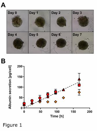
albumin as a marker of hepatocellular function were monitored over seven days. Bright-field
images of hepatocyte spheroids indicated no changes in cellular morphology and spheroid
size over the investigated period (Fig. 1A). Moreover, the albumin concentrations in the
culture media steadily increased over time suggesting stable albumin production and
hepatocellular function of spheroids from all donors (Fig. 1B). The average rates of albumin
secretion were 19.9 ±3.4, 23.3 ±7.5, and 12.0 ±5.0 pg/hepatocyte/day for Donor A, B, and C,
respectively, whereby the albumin secretion rate in vivo is reported to be
17.8 pg/hepatocyte/day (Jang et al., 2015).
Loss of hepatocellular phenotype in pooled hepatocyte spheroids. In order to enhance
the metabolic capacity of hepatocyte spheroids to reduce the assay duration, we
hypothesized that pooling of spheroids should increase the depletion of parent drug as well
as metabolite formation. While the amount of enzyme is adjustable in traditional in vitro
systems like liver microsomes or hepatocytes (Obach, 2001; Nordell et al., 2013), the
presented spheroid system was developed using only 1’500 - 2’000 hepatocytes per
This article has not been copyedited and formatted. The final version may differ from this version.DMD Fast Forward. Published on June 1, 2021 as DOI: 10.1124/dmd.120.000340
at ASPE
T Journals on February 15, 2022
dmd.aspetjournals.org
Dow
nloaded from
#DMD-AR-2020-000340
14
spheroids to prevent the risk for intra-spheroid hypoxia, which is expected in larger spheroids
(Lin and Chang, 2008). Thus, we attempted to increase the cell number per incubation by a
factor of 10 by pooling hepatocyte spheroids (5 spheroids/50 μL media as opposed to
1 spheroid/100 μL media) following completed spheroid formation on Day 7 post-seeding and
investigated potential changes in the morphology and hepatocellular function over time
(Fig. 2).
As indicated by bright-field images, the hepatocyte spheroids formed loose aggregates within
24 hours after pooling. After 48 hours, the spheroids entirely fused forming solid and
compact aggregates (Fig. 2A). In line with the changes in spheroid morphology, the albumin
secretion per cell decreased about two-fold in pooled spheroids as compared to individual
spheroids (Fig. 2B) with estimated average albumin secretion rates in pooled spheroids from
Donor A, B, and C being 12.1 ±0.6, 11.7 ±2.2, and 7.3 ±1.6 pg/hepatocyte/day, respectively.
In contrast, the cellular ATP content indicated stable cell viability of pooled spheroids from
Donors A and B during seven days, while the cellular ATP content in spheroids from Donor C
decreased by about 40% within seven days after pooling (Fig. 2C). Furthermore, the mRNA
expression of major hepatic CYP enzymes and uptake transporters was analyzed during
seven days after pooling (Fig. 2D), indicating a rapid decrease in CYP3A4, CYP2C8,
CYP2C9, and CYP1A2 mRNA expression. In contrast, the CYP2C19, CYP2D6, and organic
cation transporter (OCT) 1 mRNA expression decreased less than two-fold and no change in
the mRNA expression of organic anion transporting polypeptide (OATP) 1B1 was detected
during seven days. Similarly, the protein expression of CYP3A4, which was analyzed as a
representative CYP enzyme, immediately decreased after pooling the spheroids with hardly
no CYP3A4 protein detectable by Western Blot after five and seven days (Fig. 2E). In
contrast, the CYP3A4 protein expression did not decrease in spheroids from the same donor,
which were cultured in individual wells.
In vitro CLint incubations and prediction of CLh. In order to examine the feasibility of CLh
predictions, seven test drugs with reported low to intermediate hepatic clearance were
This article has not been copyedited and formatted. The final version may differ from this version.DMD Fast Forward. Published on June 1, 2021 as DOI: 10.1124/dmd.120.000340
at ASPE
T Journals on February 15, 2022
dmd.aspetjournals.org
Dow
nloaded from
#DMD-AR-2020-000340
15
incubated with individual hepatocytes spheroids and the disappearance of parent drug was
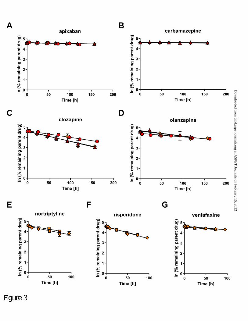
monitored over time (Fig. 3). The low clearance drugs apixaban, carbamazepine, clozapine,
and olanzapine (reported CLh in humans ranging from 0.110 to 3.55 mL/min/kg, Table 1)
were incubated with spheroids from Donor A, B, and C during seven days without medium
change to allow for sufficient removal of parent drug from the incubations. Significant
depletion of three out of four low clearance drugs was observed in hepatocyte spheroids
from all three donors and concentration-time profiles are shown in Fig. 3 (panel A-D).
Exception was carbamazepine for which no significant depletion of parent drug was
measured in hepatocyte spheroids from any of the tested donors. Although not significant, a
small decline in parent drug concentrations over time was detected in incubations with
hepatocyte spheroids from Donor A and C and the obtained in vitro data are shown in
Table 1, yet carbamazepine was excluded for the subsequent comparison to clinical
reference data and further statistical tests. In contrast, no depletion of carbamazepine was
observed in Donor B (data not shown), whose concentration-time profile was not considered
for the predicted CLh value presented in Table 1.
In addition, the intermediate clearance drugs nortriptyline, risperidone, and venlafaxine
(reported CLh in humans ranging from 6.68 to 8.79 mL/min/kg, Table 1) were investigated in
hepatocyte spheroids from Donor C. All three drug compounds are predominately eliminated
by the highly polymorphic CYP2D6 enzyme (Fogelman et al., 1999; Venkatakrishnan et al.,
1999; Yasui-Furukori et al., 2001). In order to facilitate the comparison to clinical CLh
reference data, incubations were conducted in Donor C only, which was identified to inhere
the CYP2D6 extensive metabolizer (“wild-type”) genotype (Supplementary Table 1). Similar
to previous incubations, parent drug depletion was observed for all three test compounds
over time (Fig. 3, panel E-G), yet, the in vitro half-life was generally markedly lower than for
the investigated low clearance drugs (Table 1). Accordingly, incubations with the
intermediate clearance drugs could be terminated already after three to four days.
This article has not been copyedited and formatted. The final version may differ from this version.DMD Fast Forward. Published on June 1, 2021 as DOI: 10.1124/dmd.120.000340
at ASPE
T Journals on February 15, 2022
dmd.aspetjournals.org
Dow
nloaded from
#DMD-AR-2020-000340
16
Next, we performed CLh predictions based on the in vitro CLint data and compared the
obtained values with clinical reference data (Table 1). The predicted CLh values ranged from
0.434 to 1.64 mL/min/kg and from 3.00 to 5.59 mL/min/kg for drugs with low and intermediate
clearance, respectively. The direct comparison with clinical reference data indicated a close
correlation between predicted and observed values with an AFE of 0.57, an AAFE of 1.74
and six out of seven drug compounds predicted within a three-fold deviation when excluding
carbamazepine (Fig. 4).
To investigate potential changes in the metabolic activity following the pooling of hepatocyte
spheroids, we monitored the depletion of the low clearance drugs apixaban, carbamazepine,
clozapine, and olanzapine in pooled spheroids from Donor A, B, and C during seven days.
The drug compounds were cleared from the media to a higher extent in all incubations with
pooled spheroids as compared to individual spheroid incubations (substrate depletion-time
profiles are shown in Supplemental Fig. 1). Yet, after taking the hepatocyte number into
account, the CLint data derived from pooled spheroids were lower, notably underestimating
the hepatic clearance of the majority of the investigated compounds. The fold deviation
between predicted and observed CLh data ranged from 0.1 to 0.6 and the AAFE increased to
4.95 for the four test drugs (Table 1).
This article has not been copyedited and formatted. The final version may differ from this version.DMD Fast Forward. Published on June 1, 2021 as DOI: 10.1124/dmd.120.000340
at ASPE
T Journals on February 15, 2022
dmd.aspetjournals.org
Dow
nloaded from
#DMD-AR-2020-000340
17
Discussion
Anticipating the clearance of slowly metabolized drugs is particularly challenging as common
in vitro systems feature short-term stability and do not allow to reliably determine the
clearance of such compounds (Di and Obach, 2015). Here, we established primary human
hepatocyte spheroids as a new IVIVE tool for predicting the hepatic clearance of compounds
with low and intermediate metabolic turnover. Hepatocyte spheroids demonstrated
phenotypical stability during seven days allowing for extended incubations in order to
quantify the disappearance of parent drug from the culture media. Using this experimental
setup, we were able to determine the in vitro CLint of six low and intermediate clearance
drugs from individual spheroids and successfully predicted the hepatic clearance of these
compounds within a three-fold deviation from the clinical reference data (AAFE of 1.74).
The demands for in vitro ADME test systems undergo fundamental changes as current drug
discovery programs generate compounds with uncommon physicochemical properties from
unprecedented chemical space (Tu et al., 2013). Non-oxidative metabolism and transporter-
mediated pathways like sinusoidal uptake or renal and biliary secretion are often involved in
the elimination of such compounds. These processes can largely be covered in profiling
assays by using more holistic in vitro systems such as primary hepatocytes or cytosolic
fractions (Sahi et al., 2010). Furthermore, static hepatic clearance models such as the
Extended Clearance Model have been developed, allowing the integration of individual
hepatic in vitro process clearances in order to predict the hepatic drug clearance in humans
(Kusuhara and Sugiyama, 2009; Camenisch et al., 2015). Nevertheless, the clearance of
compounds with high metabolic stability cannot be estimated in short-term in vitro assays
requiring time and cost-intense allometric scaling approaches (Hutzler et al., 2015). In the
present study, we found that hepatocyte spheroids can help to overcome these challenges
due to their long-term stability (Fig. 1), allowing for sufficient depletion of parent drug even for
compounds with low hepatic clearance such as apixaban (clinical CLh = 0.6 mL/min/kg).
Exception was the test drug carbamazepine (clinical CLh = 0.11 mL/min/kg) for which no
This article has not been copyedited and formatted. The final version may differ from this version.DMD Fast Forward. Published on June 1, 2021 as DOI: 10.1124/dmd.120.000340
at ASPE
T Journals on February 15, 2022
dmd.aspetjournals.org
Dow
nloaded from
#DMD-AR-2020-000340
18
significant loss could be detected during seven days of incubation. Although not statistically
significant, minor depletion of carbamazepine over time was observed, which likely resulted
from metabolic elimination rather than cellular accumulation of carbamazepine, considering
the small number of cells in relation to the media volume. For instance, even in the situation
of a high cell-to-media drug concentration ratio (Kp) of 100, cellular accumulation would
account for the disappearance of only 0.33% of carbamazepine from the media (based on a
cell volume of 2.2 μL/106 hepatocytes (Riede et al., 2017)). Yet, parent drug depletion due to
chemical instability can generally not be excluded. In order to account for non-specific
compound loss, additional incubations in the absence of hepatocyte spheroids could be
performed allowing to correct for potential chemical instability of the test compounds.
We previously demonstrated that hepatocytes in spheroid culture maintain the expression of
important hepatic genes involved in ADME including phase I and II drug-metabolizing
enzymes as well as uptake and efflux transporters on the transcriptomic and proteomic level,
mimicking in vivo liver tissue (Bell et al., 2016; Bell et al., 2017). Although the present study
did not include drugs being primarily eliminated by non-CYP enzymes or (rate-limiting)
transporter-mediated permeability, hepatocyte spheroids are expected to provide equally
accurate hepatic clearance projections for such compounds, making them an attractive tool
for pharmaceutical companies. Yet, the applicability of hepatocyte spheroids for assessing
the metabolic stability of a vast amount of compounds in a drug discovery setting is limited
until robotic platforms suitable for automated formation and handling of spheroids are
routinely available to decrease the assay costs (Lucendo-Villarin et al., 2020). Furthermore,
hepatocyte spheroids preserve the hepatocyte donor’s phenotype and genotype (Bell et al.,
2016), which is of great advantage for studying specific mutations (i.e. polymorphic drug-
metabolizing enzymes) or liver diseases (Prill et al., 2019; Hurrell et al., 2020). Accordingly,
hepatocyte spheroids from single donors do not reflect the diversity in the population, which
would require the simultaneous screening of drug compounds with several spheroid batches
while microsomal or hepatocyte assays are typically conducted with pooled material from up
This article has not been copyedited and formatted. The final version may differ from this version.DMD Fast Forward. Published on June 1, 2021 as DOI: 10.1124/dmd.120.000340
at ASPE
T Journals on February 15, 2022
dmd.aspetjournals.org
Dow
nloaded from
#DMD-AR-2020-000340
19
to 50 and 200 donors, respectively (Izumi et al., 2017). Although hepatocyte spheroids can
be produced from multiple donor hepatocyte lots (Kermanizadeh et al., 2019), not every
platable hepatocyte lot is suitable for producing spheroids and the use of multiple donor
hepatocyte lots would require careful monitoring to ensure equal incorporation and survival of
hepatocytes from all donors in spheroids. Due to these limitations, the application of
hepatocyte spheroids in high-throughput screening assays in drug discovery remains
challenging, nevertheless, they can be considered as a second tier approach for drug
compounds with low turnover when standard in vitro systems have failed as well as for
characterizing compounds with complex enzyme-transporter interplay and other tailor-made
mechanistic studies.
The use of commercial long-term in vitro liver systems for pharmacokinetic studies
continuously increases and most often micro-patterned co-cultures (MPCC) of primary
human hepatocytes with stromal cells are applied. MPCC systems likewise feature long-term
stability and have been successfully used for IVIVE of low turnover compounds (Chan et al.,
2013; Hultman et al., 2016; Umehara et al., 2020). These studies yielded overall a
comparable accuracy with 70 - 92% of compounds being predicted within a three-fold
deviation whereby our spheroid model predicted 86% of compounds within a three-fold
deviation. Nevertheless, it has to be noted that the selection of compounds differed between
studies. In particular Umehara et al. (2020) included a diverse set of uptake transporter and
non-CYP substrates, while our compound set consisted of CYP substrates. A face-to-face
comparison of a harmonized set of compounds and the same hepatocyte lots would be
required to directly compare the performance of hepatocyte spheroid and MPCC models.
Similarly, another recent study by Kanebratt et al. (2021) applied hepatocyte spheroids to
predict the clearance of compounds with low turnover. Despite introducing an additional
empirical scaling factor to account for systemic under-prediction, their study achieved less
accurate predictions as indicated by AFE and AAFE values of 0.53 and 2.7, respectively,
whereas our study obtained AFE and AAFE values of 0.57 and 1.74, respectively, without
This article has not been copyedited and formatted. The final version may differ from this version.DMD Fast Forward. Published on June 1, 2021 as DOI: 10.1124/dmd.120.000340
at ASPE
T Journals on February 15, 2022
dmd.aspetjournals.org
Dow
nloaded from
#DMD-AR-2020-000340
20
applying empirical scaling factors. Furthermore, the study by Kanebratt et al. (2021) indicated
a trend towards more pronounced deviation between predicted and observed clearance data
for drug compounds with higher clearance, which was not evident in our study. Interestingly,
the authors pooled three spheroids per incubation in order to increase the metabolic capacity
and observed comparable CLint data using pooled and individual hepatocyte spheroids. Our
attempts to improve the cell to media volume ratio provided markedly lower CLint values in
pooled spheroids (Table 1). Follow-up investigations on the effect of pooling spheroids
indicated merging of the individual spheroids within 24 - 48 hours, which was accompanied
by reduced albumin secretion as well as reduced mRNA expression of major hepatic drug-
metabolizing enzymes and uptake transporters as well as reduced CYP3A4 protein
expression (Fig. 2). The fusion of spheroids likely introduced major rearrangements in the
cellular structures resulting in de-differentiation and entire loss of the hepatocellular
phenotype. We did not investigate whether the hepatocellular phenotype reverses over time
(like during normal spheroid formation), however, hepatocytes in the center of the fused
spheroid structure are likely not sufficiently supplied with oxygen and nutrients due to the
larger spheroid diameter (Lin and Chang, 2008) and will be susceptible to early cell death.
Consequently, we recommend to avoid pooling of spheroids but to perform extended
incubations with individual spheroids to allow for sufficient parent drug depletion. As an
alternative, plates with integrated microwells could be considered (Wassmer et al., 2020).
The good performance of our model might result from the ability of hepatocyte spheroids to
continuously secrete albumin at in vivo rate (Fig. 1). The presence of albumin (or other
protein) in the medium is assumed to support the cellular uptake, a process known as
protein-mediated uptake. The underlying mechanism is not fully understood and current
hypotheses were reviewed in detail elsewhere (Bowman and Benet, 2018; Bteich et al.,
2019). In disagreement with the free-drug hypothesis (Pang and Rowland, 1977), different
studies observed that cellular uptake rates decreased less than expected in the presence of
plasma protein when considering the unbound drug concentration and this effect was
This article has not been copyedited and formatted. The final version may differ from this version.DMD Fast Forward. Published on June 1, 2021 as DOI: 10.1124/dmd.120.000340
at ASPE
T Journals on February 15, 2022
dmd.aspetjournals.org
Dow
nloaded from
#DMD-AR-2020-000340
21
increased for compounds with higher plasma protein binding (Miyauchi et al., 2018; Poulin
and Haddad, 2018; Bowman et al., 2019). Furthermore, under-prediction of drug clearance
by in vitro approaches was reported to be proportional to the extent of plasma protein binding
(Liang et al., 2020). In line with these observations, a recent study applying MPCC reported
superior clearance predictions using hepatocyte donors with high albumin secretion rate (Da-
Silva et al., 2018), and likely both MPCC and spheroids models profit from the integrated
albumin secretion. On the other hand, the increasing concentrations of albumin over time
complicate the estimation of intrinsic clearance as the unbound fraction of drug in the media
changes over time. In the present study, initial unbound drug concentrations in the media
were determined from supernatant sampled from the assay plate shortly upon applying the
dosing solution, assuming complete absence of protein in the media. With regard to the very
low albumin concentrations in the media observed after seven days and the rather small
degree of plasma protein binding of the investigated compounds (fraction unbound in plasma
≥0.05, Supplementary Table 2), no effects of albumin on the fraction unbound in the media
and obtained CLint estimates are expected for our dataset. In order to account for the time-
dependent increase of albumin for drugs with extensive plasma protein binding, measured
drug concentrations in the media could be corrected with the fraction unbound for the
respective albumin concentration (estimated using plasma protein binding data).
In conclusion, we demonstrated that primary human hepatocyte spheroids remain
phenotypically stable for up to seven days without medium change allowing for extended
incubations to accurately predict the hepatic clearance of drug compounds with low to
intermediate hepatic clearance. Hepatocyte spheroids represent a valuable IVIVE tool and
are expected to significantly facilitate the characterization of slowly metabolized compounds
to improve IVIVE during pharmaceutical development.
This article has not been copyedited and formatted. The final version may differ from this version.DMD Fast Forward. Published on June 1, 2021 as DOI: 10.1124/dmd.120.000340
at ASPE
T Journals on February 15, 2022
dmd.aspetjournals.org
Dow
nloaded from
#DMD-AR-2020-000340
22
Acknowledgements
The authors would like to thank Vlasia Kastrinou-Lampou for experimental support and Dr.
Birk Poller for his critical review of this manuscript.
Authorship Contributions
Participated in research design: Riede, Wollmann, Molden, Ingelman-Sundberg
Conducted experiments: Riede, Wollmann
Performed data analysis: Riede, Wollmann
Wrote or contributed to the writing of the manuscript: Riede, Wollmann, Molden, Ingelman-
Sundberg
This article has not been copyedited and formatted. The final version may differ from this version.DMD Fast Forward. Published on June 1, 2021 as DOI: 10.1124/dmd.120.000340
at ASPE
T Journals on February 15, 2022
dmd.aspetjournals.org
Dow
nloaded from
#DMD-AR-2020-000340
23
References
Argikar UA, Potter PM, Hutzler JM, and Marathe PH (2016) Challenges and Opportunities
with Non-CYP Enzymes Aldehyde Oxidase, Carboxylesterase, and UDP-
Glucuronosyltransferase: Focus on Reaction Phenotyping and Prediction of Human
Clearance. AAPS J 18:1391-1405.
Bell CC, Hendriks DF, Moro SM, Ellis E, Walsh J, Renblom A, Fredriksson Puigvert L,
Dankers AC, Jacobs F, Snoeys J, Sison-Young RL, Jenkins RE, Nordling A, Mkrtchian S,
Park BK, Kitteringham NR, Goldring CE, Lauschke VM, and Ingelman-Sundberg M (2016)
Characterization of primary human hepatocyte spheroids as a model system for drug-
induced liver injury, liver function and disease. Sci Rep 6:25187.
Bell CC, Lauschke VM, Vorrink SU, Palmgren H, Duffin R, Andersson TB, and Ingelman-
Sundberg M (2017) Transcriptional, Functional, and Mechanistic Comparisons of Stem
Cell-Derived Hepatocytes, HepaRG Cells, and Three-Dimensional Human Hepatocyte
Spheroids as Predictive In Vitro Systems for Drug-Induced Liver Injury. Drug metabolism
and disposition: the biological fate of chemicals 45:419-429.
Benet LZ and Zia-Amirhosseini P (1995) Basic principles of pharmacokinetics. Toxicologic
pathology 23:115-123.
Bowman CM and Benet LZ (2018) An examination of protein binding and protein-facilitated
uptake relating to in vitro-in vivo extrapolation. European journal of pharmaceutical sciences
: official journal of the European Federation for Pharmaceutical Sciences 123:502-514.
Bowman CM, Okochi H, and Benet LZ (2019) The Presence of a Transporter-Induced
Protein Binding Shift: A New Explanation for Protein-Facilitated Uptake and Improvement
for In Vitro-In Vivo Extrapolation. Drug metabolism and disposition: the biological fate of
chemicals 47:358-363.
Bteich M, Poulin P, and Haddad S (2019) The potential protein-mediated hepatic uptake:
discussion on the molecular interactions between albumin and the hepatocyte cell surface
This article has not been copyedited and formatted. The final version may differ from this version.DMD Fast Forward. Published on June 1, 2021 as DOI: 10.1124/dmd.120.000340
at ASPE
T Journals on February 15, 2022
dmd.aspetjournals.org
Dow
nloaded from
#DMD-AR-2020-000340
24
and their implications for the in vitro-to-in vivo extrapolations of hepatic clearance of drugs.
Expert opinion on drug metabolism & toxicology 15:633-658.
Camenisch G, Riede J, Kunze A, Huwyler J, Poller B, and Umehara K (2015) The extended
clearance model and its use for the interpretation of hepatobiliary elimination data. ADMET
& DMPK 3:1-14.
Camenisch GP (2016) Drug Disposition Classification Systems in Discovery and
Development: A Comparative Review of the BDDCS, ECCS and ECCCS Concepts.
Pharmaceutical research 33:2583-2593.
Cazali N, Tran A, Treluyer JM, Rey E, d'Athis P, Vincent J, and Pons G (2003) Inhibitory
effect of stiripentol on carbamazepine and saquinavir metabolism in human. Br J Clin
Pharmacol 56:526-536.
Chan TS, Yu H, Moore A, Khetani SR, and Tweedie D (2013) Meeting the challenge of
predicting hepatic clearance of compounds slowly metabolized by cytochrome P450 using a
novel hepatocyte model, HepatoPac. Drug metabolism and disposition: the biological fate of
chemicals 41:2024-2032.
Da-Silva F, Boulenc X, Vermet H, Compigne P, Gerbal-Chaloin S, Daujat-Chavanieu M,
Klieber S, and Poulin P (2018) Improving Prediction of Metabolic Clearance Using
Quantitative Extrapolation of Results Obtained From Human Hepatic Micropatterned
Cocultures Model and by Considering the Impact of Albumin Binding. Journal of
pharmaceutical sciences 107:1957-1972.
Davies B and Morris T (1993) Physiological parameters in laboratory animals and humans.
Pharmaceutical research 10:1093-1095.
Di L and Obach RS (2015) Addressing the challenges of low clearance in drug research.
AAPS J 17:352-357.
Fogelman SM, Schmider J, Venkatakrishnan K, von Moltke LL, Harmatz JS, Shader RI, and
Greenblatt DJ (1999) O- and N-demethylation of venlafaxine in vitro by human liver
microsomes and by microsomes from cDNA-transfected cells: effect of metabolic inhibitors
and SSRI antidepressants. Neuropsychopharmacology 20:480-490.
This article has not been copyedited and formatted. The final version may differ from this version.DMD Fast Forward. Published on June 1, 2021 as DOI: 10.1124/dmd.120.000340
at ASPE
T Journals on February 15, 2022
dmd.aspetjournals.org
Dow
nloaded from
#DMD-AR-2020-000340
25
Hendriks DF, Fredriksson Puigvert L, Messner S, Mortiz W, and Ingelman-Sundberg M
(2016) Hepatic 3D spheroid models for the detection and study of compounds with
cholestatic liability. Sci Rep 6:35434.
Hendriks DFG, Hurrell T, Riede J, van der Horst M, Tuovinen S, and Ingelman-Sundberg M
(2019) Mechanisms of chronic fialuridine hepatotoxicity as revealed in primary human
hepatocyte spheroids. Toxicological sciences : an official journal of the Society of
Toxicology.
Hultman I, Vedin C, Abrahamsson A, Winiwarter S, and Darnell M (2016) Use of HmuREL
Human Coculture System for Prediction of Intrinsic Clearance and Metabolite Formation for
Slowly Metabolized Compounds. Mol Pharm 13:2796-2807.
Hurrell T, Kastrinou-Lampou V, Fardellas A, Hendriks DFG, Nordling A, Johansson I, Baze
A, Parmentier C, Richert L, and Ingelman-Sundberg M (2020) Human Liver Spheroids as a
Model to Study Aetiology and Treatment of Hepatic Fibrosis. Cells 9.
Hutzler JM, Ring BJ, and Anderson SR (2015) Low-Turnover Drug Molecules: A Current
Challenge for Drug Metabolism Scientists. Drug metabolism and disposition: the biological
fate of chemicals 43:1917-1928.
Izumi S, Nozaki Y, Komori T, Takenaka O, Maeda K, Kusuhara H, and Sugiyama Y (2017)
Comparison of the Predictability of Human Hepatic Clearance for Organic Anion
Transporting Polypeptide Substrate Drugs Between Different In Vitro-In Vivo Extrapolation
Approaches. Journal of pharmaceutical sciences 106:2678-2687.
Jang M, Neuzil P, Volk T, Manz A, and Kleber A (2015) On-chip three-dimensional cell
culture in phaseguides improves hepatocyte functions in vitro. Biomicrofluidics 9:034113.
Kanebratt KP, Janefeldt A, Vilen L, Vildhede A, Samuelsson K, Milton L, Bjorkbom A,
Persson M, Leandersson C, Andersson TB, and Hilgendorf C (2021) Primary Human
Hepatocyte Spheroid Model as a 3D In Vitro Platform for Metabolism Studies. Journal of
pharmaceutical sciences 110:422-431.
This article has not been copyedited and formatted. The final version may differ from this version.DMD Fast Forward. Published on June 1, 2021 as DOI: 10.1124/dmd.120.000340
at ASPE
T Journals on February 15, 2022
dmd.aspetjournals.org
Dow
nloaded from
#DMD-AR-2020-000340
26
Kermanizadeh A, Brown DM, Moritz W, and Stone V (2019) The importance of inter-
individual Kupffer cell variability in the governance of hepatic toxicity in a 3D primary human
liver microtissue model. Sci Rep 9:7295.
Kozyra M, Johansson I, Nordling A, Ullah S, Lauschke VM, and Ingelman-Sundberg M
(2018) Human hepatic 3D spheroids as a model for steatosis and insulin resistance. Sci
Rep 8:14297.
Kratochwil NA, Meille C, Fowler S, Klammers F, Ekiciler A, Molitor B, Simon S, Walter I,
McGinnis C, Walther J, Leonard B, Triyatni M, Javanbakht H, Funk C, Schuler F, Lave T,
and Parrott NJ (2017) Metabolic Profiling of Human Long-Term Liver Models and Hepatic
Clearance Predictions from In Vitro Data Using Nonlinear Mixed-Effects Modeling. AAPS J
19:534-550.
Kusuhara H and Sugiyama Y (2009) In vitro-in vivo extrapolation of transporter-mediated
clearance in the liver and kidney. Drug Metab Pharmacokinet 24:37-52.
Liang X, Park Y, DeForest N, Hao J, Zhao X, Niu C, Wang K, Smith B, and Lai Y (2020) In
Vitro Hepatic Uptake in Human and Monkey Hepatocytes in the Presence and Absence of
Serum Protein and Its In Vitro to In Vivo Extrapolation. Drug metabolism and disposition:
the biological fate of chemicals 48:1283-1292.
Lin RZ and Chang HY (2008) Recent advances in three-dimensional multicellular spheroid
culture for biomedical research. Biotechnol J 3:1172-1184.
Linnet K and Olesen OV (1997) Metabolism of clozapine by cDNA-expressed human
cytochrome P450 enzymes. Drug metabolism and disposition: the biological fate of
chemicals 25:1379-1382.
Lucendo-Villarin B, Meseguer-Ripolles J, Drew J, Fischer L, Ma WSE, Flint O, Simpson K,
Machesky L, Mountford J, and Hay D (2020) Development of a cost effective automated
platform to produce human liver spheroids for basic and applied research. Biofabrication.
Messner S, Agarkova I, Moritz W, and Kelm JM (2013) Multi-cell type human liver
microtissues for hepatotoxicity testing. Arch Toxicol 87:209-213.
This article has not been copyedited and formatted. The final version may differ from this version.DMD Fast Forward. Published on June 1, 2021 as DOI: 10.1124/dmd.120.000340
at ASPE
T Journals on February 15, 2022
dmd.aspetjournals.org
Dow
nloaded from
#DMD-AR-2020-000340
27
Miyauchi S, Masuda M, Kim SJ, Tanaka Y, Lee KR, Iwakado S, Nemoto M, Sasaki S,
Shimono K, Tanaka Y, and Sugiyama Y (2018) The Phenomenon of Albumin-Mediated
Hepatic Uptake of Organic Anion Transport Polypeptide Substrates: Prediction of the In
Vivo Uptake Clearance from the In Vitro Uptake by Isolated Hepatocytes Using a
Facilitated-Dissociation Model. Drug metabolism and disposition: the biological fate of
chemicals 46:259-267.
Nordell P, Svanberg P, Bird J, and Grime K (2013) Predicting metabolic clearance for drugs
that are actively transported into hepatocytes: incubational binding as a consequence of in
vitro hepatocyte concentration is a key factor. Drug metabolism and disposition: the
biological fate of chemicals 41:836-843.
Obach RS (2001) The prediction of human clearance from hepatic microsomal metabolism
data. Current opinion in drug discovery & development 4:36-44.
Obach RS, Baxter JG, Liston TE, Silber BM, Jones BC, MacIntyre F, Rance DJ, and Wastall
P (1997) The prediction of human pharmacokinetic parameters from preclinical and in vitro
metabolism data. J Pharmacol Exp Ther 283:46-58.
Pang KS and Rowland M (1977) Hepatic clearance of drugs. I. Theoretical considerations of
a "well-stirred" model and a "parallel tube" model. Influence of hepatic blood flow, plasma
and blood cell binding, and the hepatocellular enzymatic activity on hepatic drug clearance.
Journal of pharmacokinetics and biopharmaceutics 5:625-653.
Poulin P and Haddad S (2018) Extrapolation of the Hepatic Clearance of Drugs in the
Absence of Albumin In Vitro to That in the Presence of Albumin In Vivo: Comparative
Assessement of 2 Extrapolation Models Based on the Albumin-Mediated Hepatic Uptake
Theory and Limitations and Mechanistic Insights. Journal of pharmaceutical sciences
107:1791-1797.
Prill S, Caddeo A, Baselli G, Jamialahmadi O, Dongiovanni P, Rametta R, Kanebratt KP,
Pujia A, Pingitore P, Mancina RM, Linden D, Whatling C, Janefeldt A, Kozyra M, Ingelman-
Sundberg M, Valenti L, Andersson TB, and Romeo S (2019) The TM6SF2 E167K genetic
This article has not been copyedited and formatted. The final version may differ from this version.DMD Fast Forward. Published on June 1, 2021 as DOI: 10.1124/dmd.120.000340
at ASPE
T Journals on February 15, 2022
dmd.aspetjournals.org
Dow
nloaded from
#DMD-AR-2020-000340
28
variant induces lipid biosynthesis and reduces apolipoprotein B secretion in human hepatic
3D spheroids. Sci Rep 9:11585.
Riede J, Camenisch G, Huwyler J, and Poller B (2017) Current In Vitro Methods to
Determine Hepatic Kpuu: A Comparison of Their Usefulness and Limitations. Journal of
pharmaceutical sciences 106:2805-2814.
Ring BJ, Catlow J, Lindsay TJ, Gillespie T, Roskos LK, Cerimele BJ, Swanson SP, Hamman
MA, and Wrighton SA (1996) Identification of the human cytochromes P450 responsible for
the in vitro formation of the major oxidative metabolites of the antipsychotic agent
olanzapine. J Pharmacol Exp Ther 276:658-666.
Sahi J, Grepper S, and Smith C (2010) Hepatocytes as a tool in drug metabolism, transport
and safety evaluations in drug discovery. Curr Drug Discov Technol 7:188-198.
Smith R, Jones RD, Ballard PG, and Griffiths HH (2008) Determination of microsome and
hepatocyte scaling factors for in vitro/in vivo extrapolation in the rat and dog. Xenobiotica;
the fate of foreign compounds in biological systems 38:1386-1398.
Tu M, Mathiowetz AM, Pfefferkorn JA, Cameron KO, Dow RL, Litchfield J, Di L, Feng B, and
Liras S (2013) Medicinal chemistry design principles for liver targeting through OATP
transporters. Curr Top Med Chem 13:857-866.
Umehara K, Cantrill C, Wittwer MB, Di Lenarda E, Klammers F, Ekiciler A, Parrott N, Fowler
S, and Ullah M (2020) Application of the Extended Clearance Classification System (ECCS)
in Drug Discovery and Development: Selection of Appropriate In Vitro Tools and Clearance
Prediction. Drug metabolism and disposition: the biological fate of chemicals 48:849-860.
Venkatakrishnan K, von Moltke LL, and Greenblatt DJ (1999) Nortriptyline E-10-
hydroxylation in vitro is mediated by human CYP2D6 (high affinity) and CYP3A4 (low
affinity): implications for interactions with enzyme-inducing drugs. Journal of clinical
pharmacology 39:567-577.
Vorrink SU, Ullah S, Schmidt S, Nandania J, Velagapudi V, Beck O, Ingelman-Sundberg M,
and Lauschke VM (2017) Endogenous and xenobiotic metabolic stability of primary human
hepatocytes in long-term 3D spheroid cultures revealed by a combination of targeted and
This article has not been copyedited and formatted. The final version may differ from this version.DMD Fast Forward. Published on June 1, 2021 as DOI: 10.1124/dmd.120.000340
at ASPE
T Journals on February 15, 2022
dmd.aspetjournals.org
Dow
nloaded from
#DMD-AR-2020-000340
29
untargeted metabolomics. FASEB journal : official publication of the Federation of American
Societies for Experimental Biology 31:2696-2708.
Vorrink SU, Zhou Y, Ingelman-Sundberg M, and Lauschke VM (2018) Prediction of Drug-
Induced Hepatotoxicity Using Long-Term Stable Primary Hepatic 3D Spheroid Cultures in
Chemically Defined Conditions. Toxicological sciences : an official journal of the Society of
Toxicology 163:655-665.
Wang L, Zhang D, Raghavan N, Yao M, Ma L, Frost CE, Maxwell BD, Chen SY, He K,
Goosen TC, Humphreys WG, and Grossman SJ (2010) In vitro assessment of metabolic
drug-drug interaction potential of apixaban through cytochrome P450 phenotyping,
inhibition, and induction studies. Drug metabolism and disposition: the biological fate of
chemicals 38:448-458.
Wassmer CH, Bellofatto K, Perez L, Lavallard V, Cottet-Dumoulin D, Ljubicic S, Parnaud G,
Bosco D, Berishvili E, and Lebreton F (2020) Engineering of Primary Pancreatic Islet Cell
Spheroids for Three-dimensional Culture or Transplantation: A Methodological Comparative
Study. Cell Transplant 29:963689720937292.
Yasui-Furukori N, Hidestrand M, Spina E, Facciola G, Scordo MG, and Tybring G (2001)
Different enantioselective 9-hydroxylation of risperidone by the two human CYP2D6 and
CYP3A4 enzymes. Drug metabolism and disposition: the biological fate of chemicals
29:1263-1268.
Financial support
This work was funded by the Swedish Cancer Society [grant agreement 17 0599], the
European Research Council (ERC)‐Advanced Grant (AdG) project HEPASPHER [grant
agreement 742020], and the European Union’s Horizon 2020 research and innovation
program [grant agreement 668353/U-PGx]. None of the authors has any conflict of interest to
this study to declare.
This article has not been copyedited and formatted. The final version may differ from this version.DMD Fast Forward. Published on June 1, 2021 as DOI: 10.1124/dmd.120.000340
at ASPE
T Journals on February 15, 2022
dmd.aspetjournals.org
Dow
nloaded from
#DMD-AR-2020-000340
30
Figure Legends
FIGURE 1. Hepatocyte spheroids remain viable and functional during extended
incubations. A, Bright-field images of hepatocyte spheroids from Donor A over the time
course of seven days. B, Media concentration of albumin secreted by hepatocyte spheroids
over seven days. Data points represent mean values of quadruplicates ±standard deviation.
The dashed line represents the albumin secretion rate in vivo (17.8 pg/hepatocyte/day (Jang
et al., 2015)). Δ, Donor A; Ο, Donor B; ◇, Donor C.
FIGURE 2. Loss of hepatocellular phenotype in pooled hepatocyte spheroids. A, Bright-
field images of pooled hepatocyte spheroids from Donor B over the time course of seven
days. B, Albumin secretion of pooled hepatocyte spheroids over seven days. Data points
represent mean values of quadruplicates ±standard deviation. The dashed line represents
the albumin secretion rate in vivo (17.8 pg/hepatocyte/day (Jang et al., 2015)). C, Cellular
viability (total cellular ATP) of pooled hepatocyte spheroids over seven days. Data points
represent mean values of quadruplicates ±standard deviation. Δ, Donor A; Ο, Donor B; ◇,
Donor C. D, Fold change in mRNA expression (relative to Day 0) of major hepatic CYP
enzymes and uptake transporters in pooled hepatocyte spheroids during seven days. Data
points represent mean values of the measured fold change in mRNA expression in
hepatocyte spheroids from Donor A, B, C, and D. E, Western Blot of CYP3A4 and vinculin
(loading control) in individual and pooled hepatocyte spheroids from Donor D during 7 days.
FIGURE 3. Substrate depletion-time profiles in hepatocyte spheroids. A-D, Depletion of
compounds with low hepatic clearance in individual hepatocyte spheroids from Donor A, B,
and C. E-G, Depletion of compounds with intermediate hepatic clearance in individual
hepatocyte spheroids from Donor C (data obtained in two independent experiments). Parent
drug concentrations were quantified in cell culture media and CLint was estimated from the
linear phase of parent drug depletion as described in Materials & Methods. Data points
This article has not been copyedited and formatted. The final version may differ from this version.DMD Fast Forward. Published on June 1, 2021 as DOI: 10.1124/dmd.120.000340
at ASPE
T Journals on February 15, 2022
dmd.aspetjournals.org
Dow
nloaded from
#DMD-AR-2020-000340
31
represent mean values of duplicates ±standard deviation. Δ, Donor A; Ο, Donor B; ◇ and □,
Donor C.
FIGURE 4. Comparison of predicted and observed CLh data. Data points represent mean
CLh values ±standard deviation obtained using hepatocyte spheroids from Donor A, B, and C
(apixaban, clozapine, olanzapine) or mean CLh values of duplicates ±standard deviation
obtained using hepatocyte spheroids from Donor C in two independent experiments
(nortriptyline, risperidone, venlafaxine). The solid line is the line of unity and dotted lines
represent three-fold deviations. api, apixaban; clo, clozapine; nor, nortriptyline; ola,
olanzapine; ris, risperidone; ven, venlafaxine.
This article has not been copyedited and formatted. The final version may differ from this version.DMD Fast Forward. Published on June 1, 2021 as DOI: 10.1124/dmd.120.000340
at ASPE
T Journals on February 15, 2022
dmd.aspetjournals.org
Dow
nloaded from
#DMD-AR-2020-000340
32
Tables
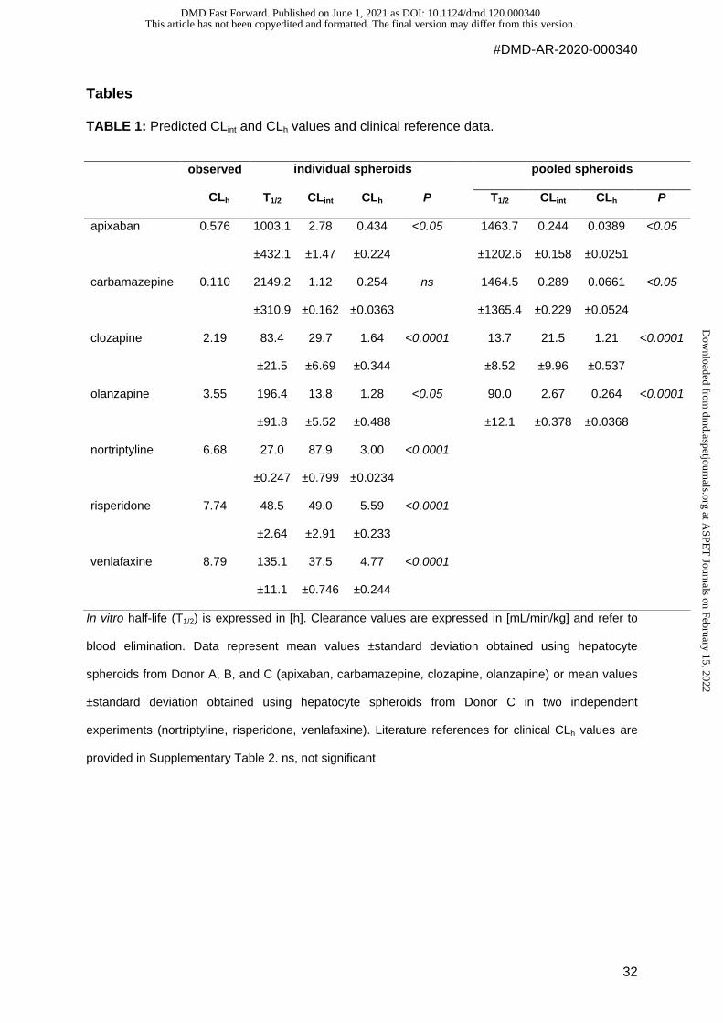
TABLE 1: Predicted CLint and CLh values and clinical reference data.
In vitro half-life (T1/2) is expressed in [h]. Clearance values are expressed in [mL/min/kg] and refer to
blood elimination. Data represent mean values ±standard deviation obtained using hepatocyte
spheroids from Donor A, B, and C (apixaban, carbamazepine, clozapine, olanzapine) or mean values
±standard deviation obtained using hepatocyte spheroids from Donor C in two independent
experiments (nortriptyline, risperidone, venlafaxine). Literature references for clinical CLh values are
provided in Supplementary Table 2. ns, not significant
observed
CLh
individual spheroids pooled spheroids
T1/2 CLint CLh P T1/2 CLint CLh P
apixaban 0.576 1003.1
±432.1
2.78
±1.47
0.434
±0.224
<0.05 1463.7
±1202.6
0.244
±0.158
0.0389
±0.0251
<0.05
carbamazepine 0.110 2149.2
±310.9
1.12
±0.162
0.254
±0.0363
ns 1464.5
±1365.4
0.289
±0.229
0.0661
±0.0524
<0.05
clozapine 2.19 83.4
±21.5
29.7
±6.69
1.64
±0.344
<0.0001 13.7
±8.52
21.5
±9.96
1.21
±0.537
<0.0001
olanzapine 3.55 196.4
±91.8
13.8
±5.52
1.28
±0.488
<0.05 90.0
±12.1
2.67
±0.378
0.264
±0.0368
<0.0001
nortriptyline 6.68 27.0
±0.247
87.9
±0.799
3.00
±0.0234
<0.0001
risperidone 7.74 48.5
±2.64
49.0
±2.91
5.59
±0.233
<0.0001
venlafaxine 8.79 135.1
±11.1
37.5
±0.746
4.77
±0.244
<0.0001
This article has not been copyedited and formatted. The final version may differ from this version.DMD Fast Forward. Published on June 1, 2021 as DOI: 10.1124/dmd.120.000340
at ASPE
T Journals on February 15, 2022
dmd.aspetjournals.org
Dow
nloaded from
Background
This article has not been copyedited and formatted. The final version may differ from this version.DMD Fast Forward. Published on June 1, 2021 as DOI: 10.1124/dmd.120.000340
at ASPE
T Journals on February 15, 2022
dmd.aspetjournals.org
Dow
nloaded from

-
This article has not been copyedited and formatted. The final version may differ from this version.DMD Fast Forward. Published on June 1, 2021 as DOI: 10.1124/dmd.120.000340
at ASPE
T Journals on February 15, 2022
dmd.aspetjournals.org
Dow
nloaded from
This article has not been copyedited and formatted. The final version may differ from this version.DMD Fast Forward. Published on June 1, 2021 as DOI: 10.1124/dmd.120.000340
at ASPE
T Journals on February 15, 2022
dmd.aspetjournals.org
Dow
nloaded from

This article has not been copyedited and formatted. The final version may differ from this version.DMD Fast Forward. Published on June 1, 2021 as DOI: 10.1124/dmd.120.000340
at ASPE
T Journals on February 15, 2022
dmd.aspetjournals.org
Dow
nloaded from


![Research Paper in vitro model using spheroids-laden nanofibrous … · 2021. 1. 22. · study, an endothelial spheroid was guided to sprout along the fibrin supporting structure [10]](https://img.pdfslide.net/doc/110x75/60faffc1807d4143cf588331/research-paper-in-vitro-model-using-spheroids-laden-nanofibrous-2021-1-22-study.jpg)