Embed Size (px)
Citation preview
EXPERIMENTAL AND THERAPEUTIC MEDICINE 16: 2287-2294, 2018
Abstract. The aim of the current study was to evaluate cell viability and osteogenic differentiation potential in cell spher-oids composed of varying ratios of gingiva-derived and bone marrow stem cells cultured in concave microwells. Cell spher-oids were established from bone marrow and gingiva-derived stem cells in ratios of 6:0 (Group 1), 2:1 (Group 2), 3:3 (Group 3), 1:2 (Group 4), and 0:6 (Group 5). On days 3 and 5, the viability of the cell spheroids was qualitatively analyzed using a calcein acetoxymethyl ester working solution and an ethidium homodimer-1 live/dead assay. On days 1, 3, 5 and 7, a quantitative cell viability analysis was performed using a Cell Counting Kit-8. Alkaline phosphatase activity assays were performed using a commercially available kit on day 7 to assess osteogenic differentiation. In addition, reverse transcription-quantitative polymerase chain reaction and western blot analysis were performed to evaluate runt-related transcription factor 2 (Runx2) and osteocalcin expression. The ratio of gingiva-derived to bone marrow stem cells did not affect the stem cell spheroid morphology. No significant changes in cell viability were noted among the different groups following incubation for 7 days. A consistent alkaline phos-phatase activity was measured in co-cultured gingiva-derived and bone marrow stem cell spheroids of varying composi-tions. Runx2 and osteocalcin expression was increased when co-cultured compared with pure gingiva-derived or bone marrow stem cells. In conclusion, stem cell spheroids estab-lished by co-culturing maintained morphology, viability and a high osteogenic differentiation potential during the experi-mental period of 7 days. These spheroids containing human gingiva-derived and bone marrow stem cells may enhance the osteogenic differentiation potential. The use of multicell
spheroids may be a simple and effective strategy for improving stem cell therapy.
Introduction
Mesenchymal stem cells are stromal cells that can differen-tiate into bone, fat and cartilage cells (1). These stem cells can be found in various tissues, including bone marrow and fat (2). A marrow-derived mesenchymal stem cell sheet with platelet-rich plasma can promote bone regeneration (3). Bone marrow is an attractive source for stem cells, but accessing stem cells from bone marrow can lead to pain and complica-tions, including bleeding and infection (4). In the dental field, stem cells can be obtained from a variety of tissues, including periodontal ligaments and dental pulp (5,6). Gingiva has been demonstrated to serve as a feasible source for stem cells and obtaining gingival-derived stem cells solely requires local anesthesia, which is associated with less pain compared with bone marrow extractions (7).
Extensive research has been conducted on the use of stem cells for recovery and regeneration of teeth and surrounding tissues (8-12). The use of a three-dimensional cell culture has important effects on mimicking the native environment on intercellular interactions that regulate self-renewal and differentiation of stem cells (13). Two-dimensional cultures may provide a stiffer environment compared with the natural environment and this may lead to changes in cell function (14). Stem cell spheroids obtained from three-dimensional cultures have demonstrated increased differentiation abilities (15,16). Cell spheroids have the ability to reflect the paracrine effects of producing growth factors and metabolites (17).
The current study was performed using concave microwells to evaluate viability and osteogenic differentiation potential of cell spheroids established using varying ratios of gingiva-derived and bone marrow stem cells. The efficacy of cell stem spheroids was investigated by comparing viability and osteogenic differ-entiation ability of three-dimensional cell cultures composed of gingiva-derived and bone marrow stem cells compared with spheroids containing cells of a single origin.
Materials and methods
Formation of cell spheroids using human gingiva‑derived and bone marrow stem cells. Silicone elastomer-based concave
Osteogenic potential of cell spheroids composed of varying ratios of gingiva‑derived and bone marrow stem cells using concave microwells
JAE-YONG TAE, HYUNA LEE, HYUNJIN LEE, YOUNGKYUNG KO and JUN-BEOM PARK
Department of Periodontics, College of Medicine, The Catholic University of Korea, Seoul 06591, Republic of Korea
Received March 21, 2018; Accepted June 8, 2018
DOI: 10.3892/etm.2018.6462
Correspondence to: Professor Jun-Beom Park, Department of Periodontics, College of Medicine, The Catholic University of Korea, 222 Banpo-Daero, Seocho-Gu, Seoul 06591, Republic of KoreaE-mail: [email protected]
Key words: bone marrow, cell differentiation, cellular spheroids, gingival, osteogenesis, stem cells
TAE et al: OSTEOGENIC DIFFERENTIATION OF STEM CELL SPHEROIDS2288
microwells (H389600; StemFIT 3D; MicroFIT, Seongnam, Korea) with 600 µm diameter were used to prepare the stem cell spheroids. A total of 1x10⁶ cells at varying ratios were loaded into each well and cultured in osteogenic medium containing α-minimal essential medium (α-MEM; Gibco; Thermo Fisher Scientific, Inc., Waltham, MA, USA) comprising 200 mM L-glutamine (Sigma-Aldrich; Merck KGaA, Darmstadt, Germany), 10 mM ascorbic acid 2-phosphate (Sigma-Aldrich; Merck KGaA), 100 µg/ml streptomycin (Sigma-Aldrich; Merck KGaA), 15% fetal bovine serum (Gibco; Thermo Fisher Scientific, Inc.), 100 U/ml penicillin, 2 mg/ml glycerophos-phate disodium salt hydrate and 38 µg/ml dexamethasone at 37˚C in a humidified incubator with 5% CO2 and 95% O2 to evaluate cellular responses. The Catholic Institute of Cell Therapy (Seoul, Korea) provided bone marrow-derived mesenchymal stem cells (Catholic MASTER Cells). Isolation and propagation of bone marrow-derived mesenchymal stem cells were performed following previously reported method (18). In short, human bone marrow aspirates were obtained from the iliac crest of healthy male donors in their 40s in March 2012 after approval by the Institutional Review Board of Seoul St. Mary's Hospital was obtained. Bone marrow aspirate from each consented donor was collected and sent to the good manufacturing practice-compliant facility of Catholic Institute of Cell Therapy for the isolation, expansion and quality control. Human gingiva-derived stem cells were obtained as reported previously (19). Samples from a 91-year old female participant from Seoul St Mary's Hospital recruited in July 2013 were used in the current study. The gingival tissue was harvested during surgical procedure visiting Department of Periodontics, Seoul St Mary's Hospital. The participant was physically healthy without severe medical or psychological disease and written consent was obtained. The study was approved by the Institutional Review Board at Seoul St Mary's Hospital's College of Medicine and the Catholic University of Korea (KC17SESI0290; Seoul, Korea). All experiments were performed according to the Declaration of Helsinki.
Human bone marrow-derived mesenchymal and gingiva-derived stem cells were loaded at densities of 1.2x10⁶ cells/microwell and in ratios of 6:0 (Group 1), 2:1 (Group 2), 3:3 (Group 3), 1:2 (Group 4) and 0:6 (Group 5). Cells of each stock were added to the well and mixed in the well in case of Groups 2-4. The formation of spheroids and morphological changes were observed under an inverted microscope (Leica DM IRM; Leica Microsystems GmbH, Wetzlar, Germany).
Cell viability. Stem-cell spheroids were cultured in osteogenic medium at 37˚C for 3‑5 days.
A commercially available live/dead assay (LIVE/DEAD Cell Viability assays; Molecular Probes; Thermo Fisher Scientific, Inc.) for qualitative analysis of the stem cell spher-oids was performed, following the manufacturer's instructions on days 3 and 5 of culturing. Spheroids were washed twice with growth medium and suspended in 1 ml α-MEM, containing 2 µl calcein acetoxymethyl ester working solution (50 mM) and 4 µl ethidium homodimer-1 (2 mM) and cultured for 30 min at room temperature. In intact cells intracellular esterases produce green fluorescence by reacting with the nonfluores-cent, cell-permeant calcein acetoxymethyl ester (20). The
ethidium homodimer can enter damaged cells and produce red fluorescence (21). Following staining, the spheroids were observed under a fluorescence microscope (magnification, x100; Axiovert 200; Zeiss GmbH, Jena, Germany).
On days 1, 3, 5 and 7 of culturing, a Cell Counting Kit-8 (CCK-8; Dojindo Molecular Technologies, Inc., Kumamoto, Japan) was used for the quantitative analysis of cell viability according to manufacturer's instructions. Initially, 2-(2-methoxy-4-nitrophenyl)-3-(4-nitrophenyl)-5-(2,4-disulfophenyl)-2H tetrazolium, monosodium salt was added and stem cell spheroids were cultured for 45 min at 37˚C. The absorbance at 450 nm was measured using a microplate reader (BioTek Instruments, Inc., Winooski, VT, USA).
Alkaline phosphatase activity assays. Cell spheroids were obtained on day 7, following growth in culture plates with osteogenic media at 37˚C. A commercially available kit (Alkaline Phosphatase Activity Colorimetric Assay kit; K412-500; BioVision, Inc., Milpitas, CA, USA) was used to evaluate alkaline phosphatase activity according to manufac-turer's instructions. Cells (1.0x10⁴) were suspended with assay buffer, sonicated and centrifuged at 13,000 x g for 3 min at 4˚C to remove insoluble material. The supernatant was mixed with a p‑nitrophenylphosphate substrate and incubated at 25˚C for 40 min. The absorbance of the resultant p-nitrophenol was measured spectrophotometrically at 405 nm and the activity was calculated based on the absorbance.
Total RNA extraction and reverse transcription‑quantitative polymerase chain reaction (RT‑qPCR). Samples were harvested on day 10 (12,000 x g; 5 min; 4˚C). A GeneJET RNA Purification kit (Thermo Fisher Scientific, Inc.) was used to isolate the total RNA according to the manufacturer's instructions. A total of 1 ng RNA was used as template for RT using the SuperiorScript II Reverse Transcriptase (Invitrogen; Thermo Fisher Scientific, Inc.). Purity was determined spectrophotometrically (ND-2000; Thermo Fisher Scientific, Inc.) on day 10, with the ratios of absorbance measured at 260 and 280 nm.
qPCR was performed to detect mRNA expression using SYBR-Green Real-Time PCR Master mixes (Enzynomics Co., Ltd., Daejeon, Republic of Korea) according to the manufac-turer's protocols. Primers were designed using GenBank (22). Primer sequences were as follows: Runt-related transcription factor 2 (Runx-2), forward 5'-AAT GAT GGT GTT GAC GCT GA-3' and reverse 5'-TTG ATA CGT GTG GGA TGT GG-3'; osteocalcin, forward 5'-AGG GAG GTG TGT GAG CTC AA-3' and reverse 5'-CCG TAG AAG CGC CGA TAG G-3'; and β-actin, forward 5'-TGG CAC CCA GCA CAA TGA A-3' and reverse 5'-CTA AGT CAT AGT CCG CCT AGA AGC A-3' following the following the manufacture's instructions. mRNA levels were normalized to β-actin and expressed as a fold change calculated using the 2-ΔΔCq method (23).
Western blot analysis. On day 7, samples were washed twice with ice-cold PBS and solubilized in a radioimmunopre-cipitation lysis buffer (Thermo Fisher Scientific, Inc.) with protease inhibitors (PPI1015; Quartett Immunodiagnostika, Biotechnologie & Kosmetik Vertriebs GmbH, Berlin Germany) for 30 min at 4˚C. Lysates were then centrifuged at 13,000 x g for 10 min at 4˚C. SDS‑PAGE (7% Mini‑PROTEAN® TGX™
EXPERIMENTAL AND THERAPEUTIC MEDICINE 16: 2287-2294, 2018 2289
Precast Gels; Bio-Rad Laboratories, Inc., Hercules, CA, USA) was used to separate the samples (15 µg). Protein concentra-tion was determined as previously reported (24). Samples were transferred to polyvinylidene difluoride membranes (IB24002, Immun-Blot®; Bio-Rad, Laboratories, Inc.) using a transfer apparatus (iBlot® 2 Transfer Stacks, Bio-Rad Laboratories, Inc.), prior to blocking with 5% skim milk at room tempera-ture for 1 h, incubation with corresponding antibodies (primary antibodies, 2 h at 4˚C; secondary antibodies, 2 h at room temperature) and enhanced chemiluminescent detection (Chemiluminescent HRP Substrate; Immobilon™ Western; Merck KGaA). Primary antibodies against Runx2 (ab76956; Abcam, Cambridge, MA, USA), osteocalcin (ab13418; Abcam) and GAPDH (ab9485; Abcam) at 1:300 dilution were used in the analysis. Horseradish peroxidase-conjugated secondary antibodies, goat anti-mouse IgG F(ab')2 (ADI-SAB-100-J; Enzo Life Sciences, Inc., Farmingdale, NY, USA) and goat anti-rabbit IgG (ADI-SAB-300-J; Enzo Life Sciences, Inc.) were incubated at 1:10,000 dilution for 2 h at room tempera-ture. The quantitative analysis of the protein expression for Runx2, osteocalcin and GAPDH was conducted using image processing and analysis software (ImageJ v1.8.0_112; National Institutes of Health, Bethesda, MD, USA).
Statistical analysis. Data are represented as the mean ± stan-dard deviation (n=3). Using commercially available software (SPSS 12; SPSS Inc., Chicago, IL, USA), tests of normality,
one-way analysis of variance with a post hoc Tukey's tests and Kruskal-Wallis tests were performed to determine the differ-ences among the groups. P<0.05 was considered to indicate a statistically significant difference.
Results
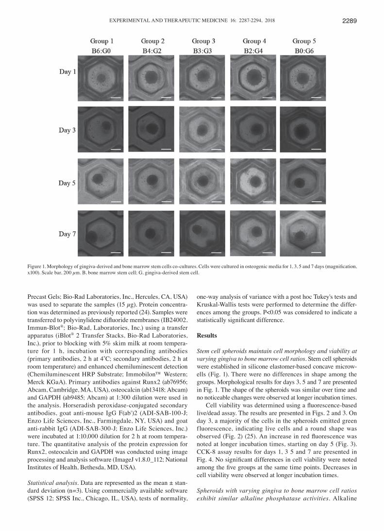
Stem cell spheroids maintain cell morphology and viability at varying gingiva to bone marrow cell ratios. Stem cell spheroids were established in silicone elastomer-based concave microw-ells (Fig. 1). There were no differences in shape among the groups. Morphological results for days 3, 5 and 7 are presented in Fig. 1. The shape of the spheroids was similar over time and no noticeable changes were observed at longer incubation times.
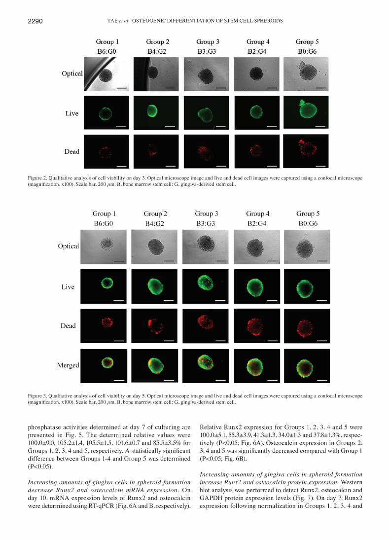
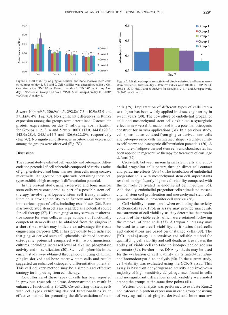
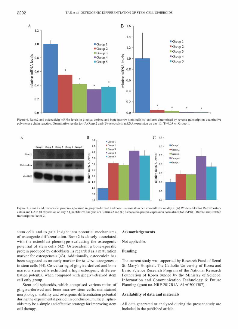
Cell viability was determined using a fluorescence‑based live/dead assay. The results are presented in Figs. 2 and 3. On day 3, a majority of the cells in the spheroids emitted green fluorescence, indicating live cells and a round shape was observed (Fig. 2) (25). An increase in red fluorescence was noted at longer incubation times, starting on day 5 (Fig. 3). CCK-8 assay results for days 1, 3 5 and 7 are presented in Fig. 4. No significant differences in cell viability were noted among the five groups at the same time points. Decreases in cell viability were observed at longer incubation times.
Spheroids with varying gingiva to bone marrow cell ratios exhibit similar alkaline phosphatase activities. Alkaline
Figure 1. Morphology of gingiva‑derived and bone marrow stem cells co‑cultures. Cells were cultured in osteogenic media for 1, 3, 5 and 7 days (magnification, x100). Scale bar, 200 µm. B, bone marrow stem cell; G, gingiva-derived stem cell.
TAE et al: OSTEOGENIC DIFFERENTIATION OF STEM CELL SPHEROIDS2290
phosphatase activities determined at day 7 of culturing are presented in Fig. 5. The determined relative values were 100.0±9.0, 105.2±1.4, 105.5±1.5, 101.6±0.7 and 85.5±3.5% for Groups 1, 2, 3, 4 and 5, respectively. A statistically significant difference between Groups 1-4 and Group 5 was determined (P<0.05).
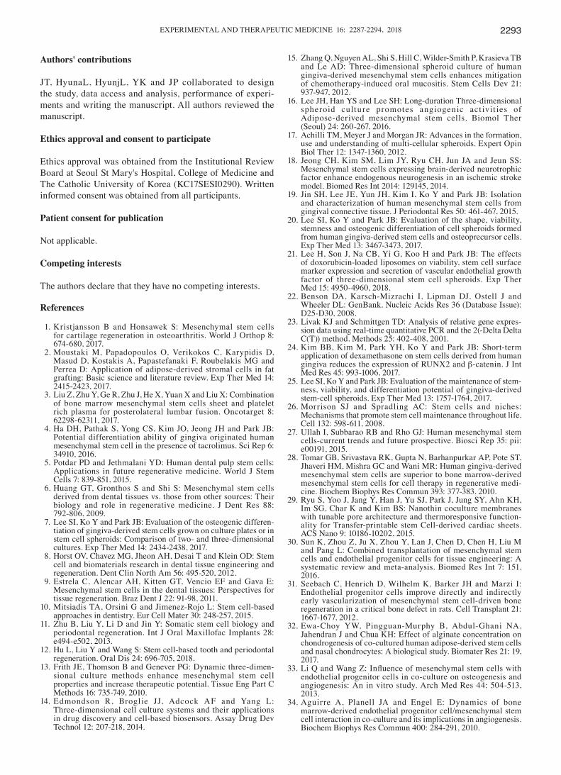
Increasing amounts of gingiva cells in spheroid formation decrease Runx2 and osteocalcin mRNA expression. On day 10, mRNA expression levels of Runx2 and osteocalcin were determined using RT-qPCR (Fig. 6A and B, respectively).
Relative Runx2 expression for Groups 1, 2, 3, 4 and 5 were 100.0±5.1, 55.3±3.9, 41.3±1.3, 34.0±1.3 and 37.8±1.3%, respec-tively (P<0.05; Fig. 6A). Osteocalcin expression in Groups 2, 3, 4 and 5 was significantly decreased compared with Group 1 (P<0.05; Fig. 6B).
Increasing amounts of gingiva cells in spheroid formation increase Runx2 and osteocalcin protein expression. Western blot analysis was performed to detect Runx2, osteocalcin and GAPDH protein expression levels (Fig. 7). On day 7, Runx2 expression following normalization in Groups 1, 2, 3, 4 and
Figure 2. Qualitative analysis of cell viability on day 3. Optical microscope image and live and dead cell images were captured using a confocal microscope (magnification, x100). Scale bar, 200 µm. B, bone marrow stem cell; G, gingiva‑derived stem cell.
Figure 3. Qualitative analysis of cell viability on day 5. Optical microscope image and live and dead cell images were captured using a confocal microscope (magnification, x100). Scale bar, 200 µm. B, bone marrow stem cell; G, gingiva‑derived stem cell.
EXPERIMENTAL AND THERAPEUTIC MEDICINE 16: 2287-2294, 2018 2291
5 were 100.0±9.5, 306.9±14.5, 292.8±17.3, 410.9±32.9 and 371.1±43.4% (Fig. 7B). No significant differences in Runx2 expression among the groups were determined. Osteocalcin protein expressions on day 7 following normalization for Groups 1, 2, 3, 4 and 5 were 100.0±17.0, 144.6±20.3, 142.9±28.4, 245.1±44.7 and 186.6±22.8%, respectively (Fig. 7C). No significant differences in osteocalcin expression among the groups were observed (Fig. 7C).
Discussion
The current study evaluated cell viability and osteogenic differ-entiation potential of cell spheroids composed of various ratios of gingiva-derived and bone marrow stem cells using concave microwells. It suggested that spheroids containing these cell types exhibit a high osteogenic differentiation potential.
In the present study, gingiva-derived and bone marrow stem cells were considered as part of a possible stem cell therapy involving allogeneic stem cell transplantation. Stem cells have the ability to self-renew and differentiate into various types of cells, including osteoblasts (26). Bone marrow-derived stem cells are regarded as a potential source for cell therapy (27). Human gingiva may serve as an alterna-tive source for stem cells, as large numbers of functionally competent stem cells can be obtained from the gingiva in a short time, which may indicate an advantage for tissue engineering purposes (28). It has previously been indicated that gingiva-derived stem cell spheroids exhibited increased osteogenic potential compared with two-dimensional cultures, including increased level of alkaline phosphatase activity and mineralization (20). Stem cell spheroids in the current study were obtained through co-culturing of human gingiva-derived and bone marrow stem cells and results suggested an enhanced osteogenic differentiation potential. This cell delivery method may be a simple and effective strategy for improving stem cell therapy.
Co-culturing of these types of cells has been reported in previous research and was demonstrated to result in enhanced functionality (14,20). Co-culturing of stem cells with cell types exhibiting desired functionalities is an effective method for promoting the differentiation of stem
cells (29). Implantation of different types of cells into a test object has been widely applied in tissue engineering in recent years (30). The co-culture of endothelial progenitor cells and mesenchymal stem cells exhibited a synergistic effect in new-vessel formation and it is a potential osteogenic construct for in vivo applications (31). In a previous study, cell spheroids co-cultured from gingiva-derived stem cells and osteoprecursor cells maintained shape, viability, ability to self-renew and osteogenic differentiation potentials (20). A co-culture of adipose-derived stem cells and chondrocytes has been applied in regenerative therapy for treatment of cartilage defects (32).
Cross-talk between mesenchymal stem cells and endo-thelial progenitor cells occurs through direct cell contact and paracrine effects (33,34). The incubation of endothelial progenitor cells with mesenchymal stem cell supernatants resulted in significantly higher cell viability compared with the controls cultivated in endothelial cell medium (35). Additionally, endothelial progenitor cells stimulated mesen-chymal stem cell proliferation and mesenchymal stem cells promoted endothelial progenitor cell survival (36).
Cell viability is considered when evaluating the toxicity of chemicals (20). Protein assays may provide inaccurate measurement of cell viability, as they determine the protein content of the viable cells, which were retained following the removal of dead cells (37). A trypan blue assay may be used to assess cell viability, as it stains dead cells and calculations are based on unstained cells (38). The [51Cr-uptake] assay is a sensitive and reliable method for quantifying cell viability and cell death, as it evaluates the ability of viable cells to take up isotope-labeled sodium chromate (39). Furthermore, DNA synthesis may be used for the evaluation of cell viability via tritiated-thymidine and bromodeoxyuridine analysis (40). In the current study, cell viability was evaluated using the CCK-8 assay. This assay is based on dehydrogenase activity and involves a majority of high-sensitivity dehydrogenases found in cells and no significant differences in cell viability were noted among the groups at the same time points (41).
Western blot analysis was performed to evaluate Runx2 and osteocalcin protein expression in each group consisting of varying ratios of gingiva-derived and bone marrow
Figure 4. Cell viability of gingiva-derived and bone marrow stem cells co-cultures on day 1, 3, 5 and 7. Cell viability was determined using a Cell Counting Kit-8. *P<0.05 vs. Group 1 on day 1; **P<0.05 vs. Group 2 on day 1; #P<0.05 vs. Group 3 on day 1; ##P<0.05 vs. Group 4 on day 1; †P<0.05 vs. Group 5 on day 1.
Figure 5. Alkaline phosphatase activity of gingiva-derived and bone marrow stem cells co-cultures on day 7. Relative values were 100.0±9.0, 105.2±1.4, 105.5±1.5, 101.6±0.7 and 85.5±3.5% for Groups 1, 2, 3, 4 and 5, respectively. *P<0.05 vs. Group 1.
TAE et al: OSTEOGENIC DIFFERENTIATION OF STEM CELL SPHEROIDS2292
stem cells and to gain insight into potential mechanisms of osteogenic differentiation. Runx2 is closely associated with the osteoblast phenotype evaluating the osteogenic potential of stem cells (42). Osteocalcin, a bone‑specific protein produced by osteoblasts, is regarded as a maturation marker for osteogenesis (43). Additionally, osteocalcin has been suggested as an early marker for in vitro osteogenesis in stem cells (44). Co-culturing of gingiva-derived and bone marrow stem cells exhibited a high osteogenic differen-tiation potential when compared with gingiva-derived stem cell only group.
Stem-cell spheroids, which comprised various ratios of gingiva-derived and bone marrow stem cells, maintained morphology, viability and osteogenic differentiation potential during the experimental period. In conclusion, multicell spher-oids may be a simple and effective strategy for improving stem cell therapy.
Acknowledgements
Not applicable.
Funding
The current study was supported by Research Fund of Seoul St. Mary's Hospital, The Catholic University of Korea and Basic Science Research Program of the National Research Foundation of Korea funded by the Ministry of Science, Information and Communication Technology & Future Planning (grant no. NRF-2017R1A1A1A05001307).
Availability of data and materials
All data generated or analyzed during the present study are included in the published article.
Figure 7. Runx2 and osteocalcin protein expression in gingiva-derived and bone marrow stem cells co-cultures on day 7. (A) Western blot for Runx2, osteo-calcin and GAPDH expression on day 7. Quantitative analysis of (B) Runx2 and (C) osteocalcin protein expression normalized to GAPDH. Runx2, runt-related transcription factor 2.
Figure 6. Runx2 and osteocalcin mRNA levels in gingiva-derived and bone marrow stem cells co-cultures determined by reverse transcription-quantitative polymerase chain reaction. Quantitative results for (A) Runx2 and (B) osteocalcin mRNA expression on day 10. *P<0.05 vs. Group 1.
EXPERIMENTAL AND THERAPEUTIC MEDICINE 16: 2287-2294, 2018 2293
Authors' contributions
JT, HyunaL, HyunjL, YK and JP collaborated to design the study, data access and analysis, performance of experi-ments and writing the manuscript. All authors reviewed the manuscript.
Ethics approval and consent to participate
Ethics approval was obtained from the Institutional Review Board at Seoul St Mary's Hospital, College of Medicine and The Catholic University of Korea (KC17SESI0290). Written informed consent was obtained from all participants.
Patient consent for publication
Not applicable.
Competing interests
The authors declare that they have no competing interests.
References
1. Kristjansson B and Honsawek S: Mesenchymal stem cells for cartilage regeneration in osteoarthritis. World J Orthop 8: 674-680, 2017.
2. Moustaki M, Papadopoulos O, Verikokos C, Karypidis D, Masud D, Kostakis A, Papastefanaki F, Roubelakis MG and Perrea D: Application of adipose-derived stromal cells in fat grafting: Basic science and literature review. Exp Ther Med 14: 2415-2423, 2017.
3. Liu Z, Zhu Y, Ge R, Zhu J, He X, Yuan X and Liu X: Combination of bone marrow mesenchymal stem cells sheet and platelet rich plasma for posterolateral lumbar fusion. Oncotarget 8: 62298-62311, 2017.
4. Ha DH, Pathak S, Yong CS, Kim JO, Jeong JH and Park JB: Potential differentiation ability of gingiva originated human mesenchymal stem cell in the presence of tacrolimus. Sci Rep 6: 34910, 2016.
5. Potdar PD and Jethmalani YD: Human dental pulp stem cells: Applications in future regenerative medicine. World J Stem Cells 7: 839-851, 2015.
6. Huang GT, Gronthos S and Shi S: Mesenchymal stem cells derived from dental tissues vs. those from other sources: Their biology and role in regenerative medicine. J Dent Res 88: 792-806, 2009.
7. Lee SI, Ko Y and Park JB: Evaluation of the osteogenic differen-tiation of gingiva-derived stem cells grown on culture plates or in stem cell spheroids: Comparison of two- and three-dimensional cultures. Exp Ther Med 14: 2434-2438, 2017.
8. Horst OV, Chavez MG, Jheon AH, Desai T and Klein OD: Stem cell and biomaterials research in dental tissue engineering and regeneration. Dent Clin North Am 56: 495-520, 2012.
9. Estrela C, Alencar AH, Kitten GT, Vencio EF and Gava E: Mesenchymal stem cells in the dental tissues: Perspectives for tissue regeneration. Braz Dent J 22: 91-98, 2011.
10. Mitsiadis TA, Orsini G and Jimenez-Rojo L: Stem cell-based approaches in dentistry. Eur Cell Mater 30: 248-257, 2015.
11. Zhu B, Liu Y, Li D and Jin Y: Somatic stem cell biology and periodontal regeneration. Int J Oral Maxillofac Implants 28: e494-e502, 2013.
12. Hu L, Liu Y and Wang S: Stem cell-based tooth and periodontal regeneration. Oral Dis 24: 696-705, 2018.
13. Frith JE, Thomson B and Genever PG: Dynamic three-dimen-sional culture methods enhance mesenchymal stem cell properties and increase therapeutic potential. Tissue Eng Part C Methods 16: 735-749, 2010.
14. Edmondson R, Broglie JJ, Adcock AF and Yang L: Three-dimensional cell culture systems and their applications in drug discovery and cell-based biosensors. Assay Drug Dev Technol 12: 207-218, 2014.
15. Zhang Q, Nguyen AL, Shi S, Hill C, Wilder-Smith P, Krasieva TB and Le AD: Three-dimensional spheroid culture of human gingiva-derived mesenchymal stem cells enhances mitigation of chemotherapy-induced oral mucositis. Stem Cells Dev 21: 937-947, 2012.
16. Lee JH, Han YS and Lee SH: Long-duration Three-dimensional spheroid culture promotes angiogenic act ivit ies of Adipose-derived mesenchymal stem cells. Biomol Ther (Seoul) 24: 260-267, 2016.
17. Achilli TM, Meyer J and Morgan JR: Advances in the formation, use and understanding of multi-cellular spheroids. Expert Opin Biol Ther 12: 1347-1360, 2012.
18. Jeong CH, Kim SM, Lim JY, Ryu CH, Jun JA and Jeun SS: Mesenchymal stem cells expressing brain-derived neurotrophic factor enhance endogenous neurogenesis in an ischemic stroke model. Biomed Res Int 2014: 129145, 2014.
19. Jin SH, Lee JE, Yun JH, Kim I, Ko Y and Park JB: Isolation and characterization of human mesenchymal stem cells from gingival connective tissue. J Periodontal Res 50: 461-467, 2015.
20. Lee SI, Ko Y and Park JB: Evaluation of the shape, viability, stemness and osteogenic differentiation of cell spheroids formed from human gingiva-derived stem cells and osteoprecursor cells. Exp Ther Med 13: 3467-3473, 2017.
21. Lee H, Son J, Na CB, Yi G, Koo H and Park JB: The effects of doxorubicin-loaded liposomes on viability, stem cell surface marker expression and secretion of vascular endothelial growth factor of three-dimensional stem cell spheroids. Exp Ther Med 15: 4950-4960, 2018.
22. Benson DA, Karsch-Mizrachi I, Lipman DJ, Ostell J and Wheeler DL: GenBank. Nucleic Acids Res 36 (Database Issue): D25-D30, 2008.
23. Livak KJ and Schmittgen TD: Analysis of relative gene expres-sion data using real-time quantitative PCR and the 2(-Delta Delta C(T)) method. Methods 25: 402-408, 2001.
24. Kim BB, Kim M, Park YH, Ko Y and Park JB: Short-term application of dexamethasone on stem cells derived from human gingiva reduces the expression of RUNX2 and β-catenin. J Int Med Res 45: 993-1006, 2017.
25. Lee SI, Ko Y and Park JB: Evaluation of the maintenance of stem-ness, viability, and differentiation potential of gingiva-derived stem-cell spheroids. Exp Ther Med 13: 1757-1764, 2017.
26. Morrison SJ and Spradling AC: Stem cells and niches: Mechanisms that promote stem cell maintenance throughout life. Cell 132: 598-611, 2008.
27. Ullah I, Subbarao RB and Rho GJ: Human mesenchymal stem cells-current trends and future prospective. Biosci Rep 35: pii: e00191, 2015.
28. Tomar GB, Srivastava RK, Gupta N, Barhanpurkar AP, Pote ST, Jhaveri HM, Mishra GC and Wani MR: Human gingiva-derived mesenchymal stem cells are superior to bone marrow-derived mesenchymal stem cells for cell therapy in regenerative medi-cine. Biochem Biophys Res Commun 393: 377-383, 2010.
29. Ryu S, Yoo J, Jang Y, Han J, Yu SJ, Park J, Jung SY, Ahn KH, Im SG, Char K and Kim BS: Nanothin coculture membranes with tunable pore architecture and thermoresponsive function-ality for Transfer-printable stem Cell-derived cardiac sheets. ACS Nano 9: 10186-10202, 2015.
30. Sun K, Zhou Z, Ju X, Zhou Y, Lan J, Chen D, Chen H, Liu M and Pang L: Combined transplantation of mesenchymal stem cells and endothelial progenitor cells for tissue engineering: A systematic review and meta-analysis. Biomed Res Int 7: 151, 2016.
31. Seebach C, Henrich D, Wilhelm K, Barker JH and Marzi I: Endothelial progenitor cells improve directly and indirectly early vascularization of mesenchymal stem cell-driven bone regeneration in a critical bone defect in rats. Cell Transplant 21: 1667-1677, 2012.
32. Ewa-Choy YW, Pingguan-Murphy B, Abdul-Ghani NA, Jahendran J and Chua KH: Effect of alginate concentration on chondrogenesis of co-cultured human adipose-derived stem cells and nasal chondrocytes: A biological study. Biomater Res 21: 19, 2017.
33. Li Q and Wang Z: Influence of mesenchymal stem cells with endothelial progenitor cells in co-culture on osteogenesis and angiogenesis: An in vitro study. Arch Med Res 44: 504-513, 2013.
34. Aguirre A, Planell JA and Engel E: Dynamics of bone marrow-derived endothelial progenitor cell/mesenchymal stem cell interaction in co-culture and its implications in angiogenesis. Biochem Biophys Res Commun 400: 284-291, 2010.
TAE et al: OSTEOGENIC DIFFERENTIATION OF STEM CELL SPHEROIDS2294
35. Raida M, Heymann AC, Gunther C and Niederwieser D: Role of bone morphogenetic protein 2 in the crosstalk between endo-thelial progenitor cells and mesenchymal stem cells. Int J Mol Med 18: 735-739, 2006.
36. Steiner D, Köhn K, Beier JP, Stürzl M, Horch RE and Arkudas A: Cocultivation of mesenchymal stem cells and endothelial progenitor cells reveals antiapoptotic and proangiogenic effects. Cells Tissues Organs 204: 218-227, 2017.
37. Park JB: Effects of 17-α ethynyl estradiol on proliferation, differ-entiation & mineralization of osteoprecursor cells. Indian J Med Res 136: 466-470, 2012.
38. Lee SI, Yeo SI, Kim BB, Ko Y and Park JB: Formation of size-controllable spheroids using gingiva-derived stem cells and concave microwells: Morphology and viability tests. Biomed Rep 4: 97-101, 2016.
39. Neville ME: 51Cr-uptake assay. A sensitive and reliable method to quantitate cell viability and cell death. J Immunol Methods 99: 77-82, 1987.
40. Madhavan H: Simple laboratory methods to measure cell proliferation using DNA synthesis property. J Stem Cells Regen Med 3: 12-14, 2007.
41. Lee H and Park JB: Evaluation of the effects of dimethylsul-phoxide on morphology, cellular viability, mRNA, and protein expression of stem cells culture in growth media. Biomed Rep 7: 291-296, 2017.
42. Bruderer M, Richards RG, Alini M and Stoddart MJ: Role and regulation of RUNX2 in osteogenesis. Eur Cell Mater 28: 269-286, 2014.
43. Stein GS, Lian JB and Owen TA: Relationship of cell growth to the regulation of tissue‑specific gene expression during osteo-blast differentiation. FASEB J 4: 3111-3123, 1990.
44. Nakamura A, Dohi Y, Akahane M, Ohgushi H, Nakajima H, Funaoka H and Takakura Y: Osteocalcin secretion as an early marker of in vitro osteogenic differentiation of rat mesenchymal stem cells. Tissue Eng Part C Methods 15: 169-180, 2009.
This work is licensed under a Creative Commons Attribution-NonCommercial-NoDerivatives 4.0 International (CC BY-NC-ND 4.0) License.