Embed Size (px)
DESCRIPTION
Chapter 1 The thirteenth edition of the phenomenally successful Principles of Anatomy and Physiology continues to set the standard for the discipline. The authors maintained a superb balance between structure and function and continue to emphasize the correlations between normal physiology and pathophysiology, normal anatomy and pathology, and homeostasis and homeostatic imbalances.
Citation preview
Copyright © John Wiley & Sons, Inc. All rights reserved.
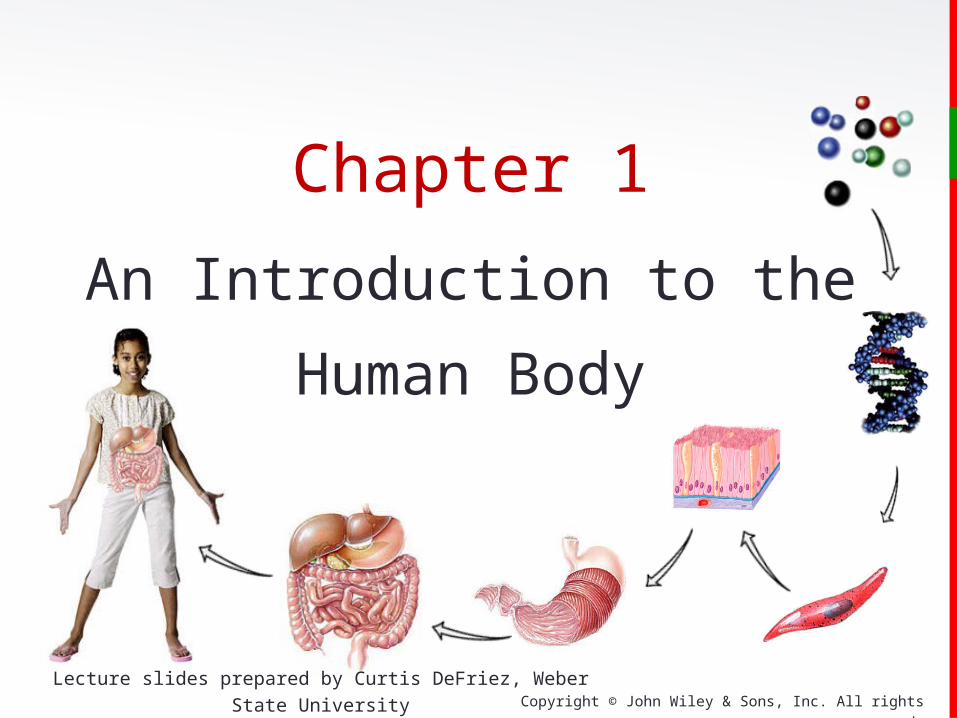
Chapter 1
An Introduction to the
Human Body
Lecture slides prepared by Curtis DeFriez, Weber State University
Copyright © John Wiley & Sons, Inc. All rights reserved.
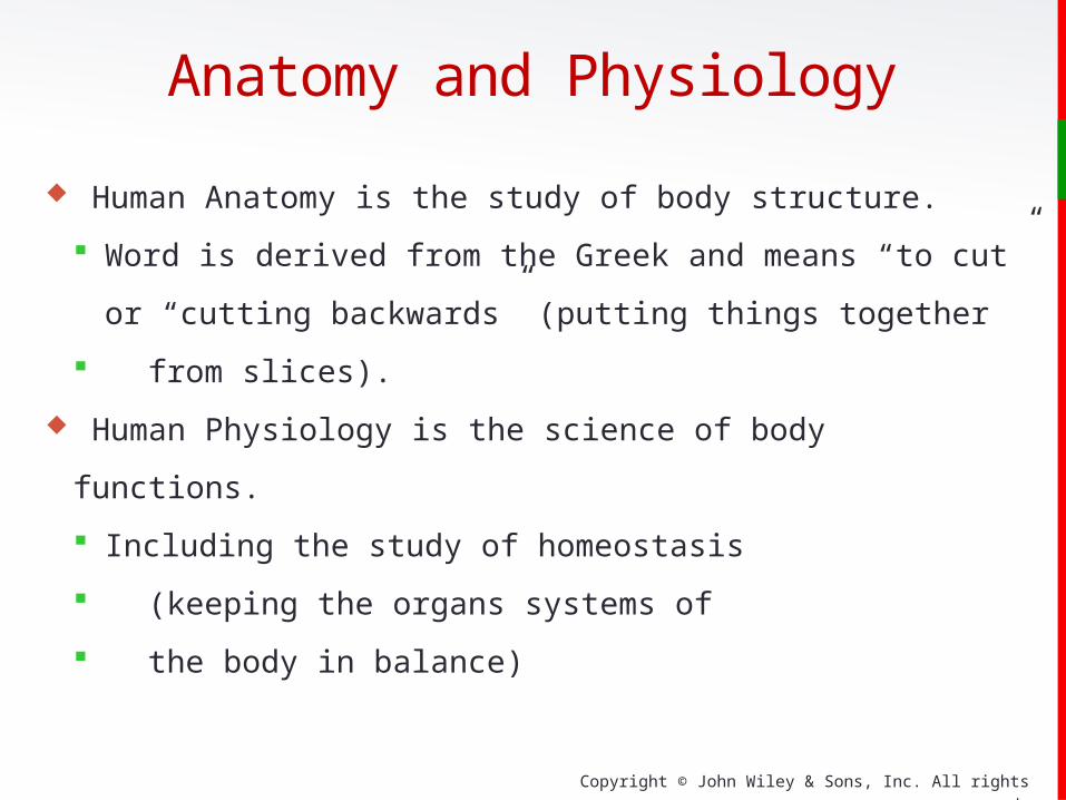
Anatomy and Physiology
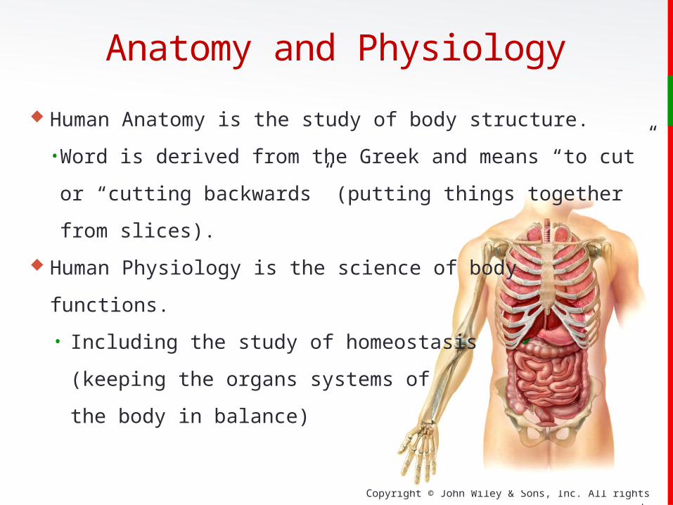
Human Anatomy is the study of body structure.
Word is derived from the Greek and means “to cut” or
“cutting backwards” (putting things together
from slices).
Human Physiology is the science of body functions.
Including the study of homeostasis
(keeping the organs systems of
the body in balance)
Copyright © John Wiley & Sons, Inc. All rights reserved.
Anatomy and Physiology
Human Anatomy is the study of body structure.
• Word is derived from the Greek and means “to cut” or “cutting
backwards” (putting things together
from slices).
Human Physiology is the science of body
functions.
• Including the study of homeostasis
(keeping the organs systems of
the body in balance)
Copyright © John Wiley & Sons, Inc. All rights reserved.
Anatomy and PhysiologyStructure and function of the body are closely related:
Structure mirrors function
Bones of the skull
are heavy and secure to
protect brain function.
The thin air sacs of the
lungs permit movement
of gases from the lungs to the blood.

Copyright © John Wiley & Sons, Inc. All rights reserved.
This structure is the liver, which has the function of filtering blood and producing bile. Can you see how the function is determined by the structure, and vice versa?
Anatomy and Physiology
Structure mirrors function

Copyright © John Wiley & Sons, Inc. All rights reserved.
Subdivisions of AnatomySurface Anatomy is the study of form and markings of the
body surface, often explored through visualization or
palpation (without any “cutting”).
Gross Anatomy is the study of anatomical structures visible
to unaided eye. After making the appropriate surface
marking in the prior picture, the gross dissection proceeds
through “cutting.”
Copyright © John Wiley & Sons, Inc. All rights reserved.
Subdivisions of AnatomyGross Anatomy can be studied by two general
approaches:
Systemic approach (Systemic Anatomy):
• Study all of the blood vessels, or all of the muscles, or
all of the bones… at once.
Regional approach (Regional Anatomy)
• All anatomical structures of a specific region (e.g. the
thorax, or the Head and Neck) are all studied together.
Copyright © John Wiley & Sons, Inc. All rights reserved.
Subdivisions of Anatomy
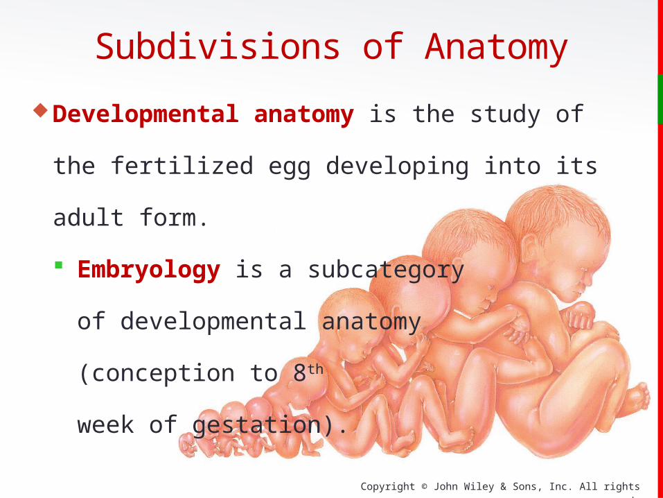
Developmental anatomy is the study of the fertilized egg
developing into its adult form.
Embryology is a subcategory
of developmental anatomy
(conception to 8th
week of gestation).

Copyright © John Wiley & Sons, Inc. All rights reserved.
Subdivisions of Anatomy
Histology is the study of tissues.
Cytology, like histology, uses a
microscope, but restricts the study to
individual cellular structures .
This micrograph is typical of
an histological and cytological
examination under light
microscopy

Copyright © John Wiley & Sons, Inc. All rights reserved.
Subdivisions of Anatomy Pathology is the study of anatomical changes due to disease .
Pathologists use gross inspection, as well as cytologic,
histologic, and laboratory
examinations to discover the
source of the disease.
This is a section of a human colon
opened by a pathologist to reveal polyps that would become
cancerous in a few years (premalignant).

Copyright © John Wiley & Sons, Inc. All rights reserved.
An autopsy is a postmortem (after death) examination of the
body and internal organs performed by a pathologist.
An autopsy is usually done to :
Determine the cause of death
Identify diseases not detected during life
Determine the extent of injuries and contribution to death
Identify hereditary conditions
Clinical Connection

Copyright © John Wiley & Sons, Inc. All rights reserved.
Levels of Organization
In this course, we will
study Anatomy and
Physiology by starting with
the most basic level of
organization (atoms) and
“working our way up”.

Copyright © John Wiley & Sons, Inc. All rights reserved.
The chemical level of organization is discussed in Chapter 2:
Atoms
Inorganic Molecules (inorganic chemistry)
Organic Molecules (organic chemistry)
Levels of Organization
Copyright © John Wiley & Sons, Inc. All rights reserved.
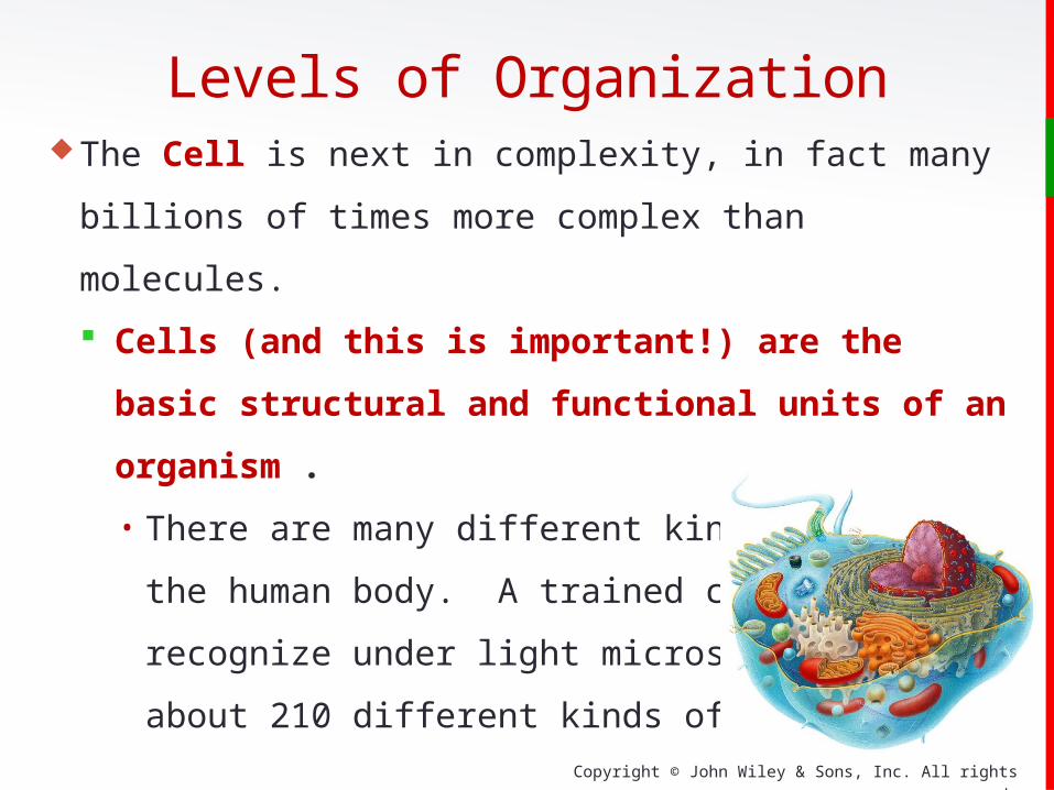
Levels of OrganizationThe Cell is next in complexity, in fact many billions of times
more complex than molecules.
Cells (and this is important!) are the basic structural
and functional units of an organism .
• There are many different kinds of cells in the human
body. A trained cytologist can
recognize under light microscopy
about 210 different kinds of cells.

Copyright © John Wiley & Sons, Inc. All rights reserved.
Levels of OrganizationTissues are groups of cells that work together to perform a
similar function.
While there are many different types of cells, they all work to
form 4 basic types of tissues:
Epithelium
Connective Tissue
Muscle
Nerves
Copyright © John Wiley & Sons, Inc. All rights reserved.
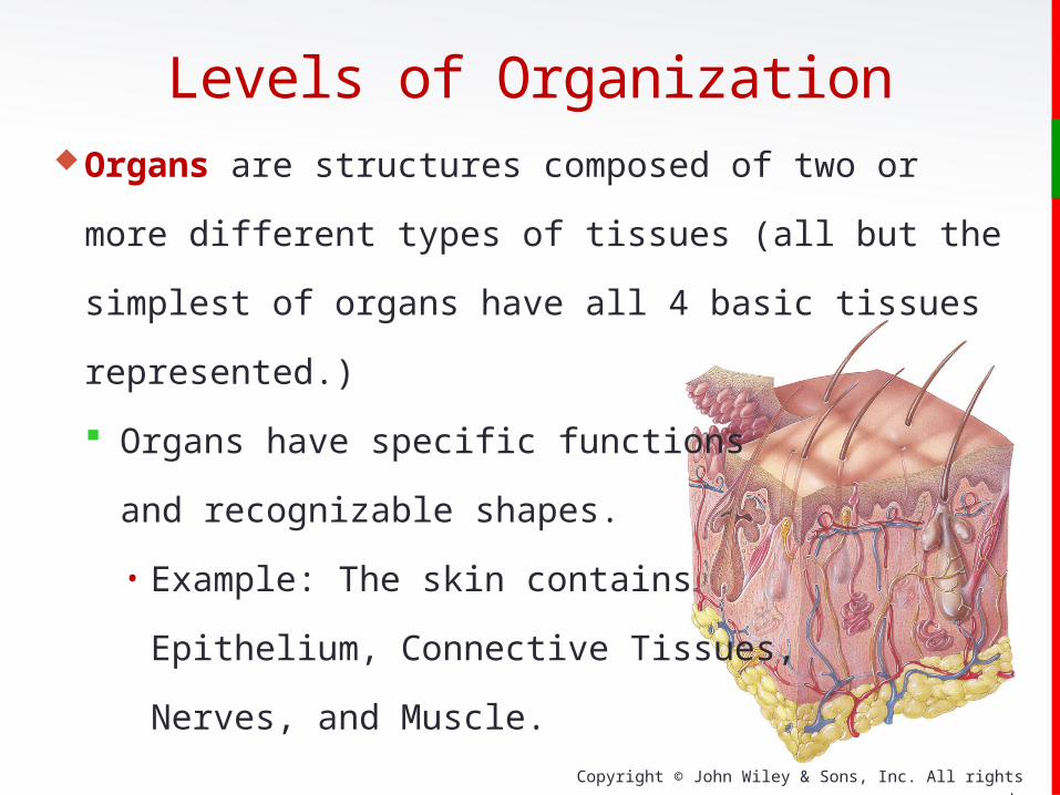
Organs are structures composed of two or more different
types of tissues (all but the simplest of organs have all 4
basic tissues represented.)
Organs have specific functions
and recognizable shapes.
• Example: The skin contains
Epithelium, Connective Tissues,
Nerves, and Muscle.
Levels of Organization
Copyright © John Wiley & Sons, Inc. All rights reserved.
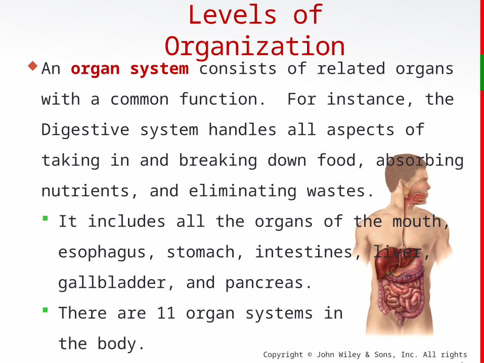
An organ system consists of related organs with a common
function. For instance, the Digestive system handles all
aspects of taking in and breaking down food, absorbing
nutrients, and eliminating wastes.
It includes all the organs of the mouth,
esophagus, stomach, intestines, liver,
gallbladder, and pancreas.
There are 11 organ systems in
the body.
Levels of Organization

Copyright © John Wiley & Sons, Inc. All rights reserved.
An organism consists of a collection of organ systems.
Six important life processes:
• Metabolism
• Responsiveness
• Movement
• Growth
• Differentiation
• Reproduction
In health, all parts of the body must be functioning together
in a process called homeostasis.
Levels of Organization
Copyright © John Wiley & Sons, Inc. All rights reserved.
Essential Life ProcessesMetabolism is the sum of all the catabolic (breaking
down) and anabolic (building up) chemical processes that
occur in the body.
Responsiveness is the body’s ability to detect and respond
to changes which might represent an opportunity… or a
threat!
Decrease in body temperature
Responding to sound
Nerve (electrical signals) and muscle cells (contracting)
Copyright © John Wiley & Sons, Inc. All rights reserved.
Movement is any motion, including movement of tiny
subcellular structures, or movement inside cells or organs.
Leg muscles move the body from one place to another.
Growth involves an increase in body size due to an
increase in existing cells, number of cells, or both.
In bone growth, materials between cells increase.
Essential Life Processes

Copyright © John Wiley & Sons, Inc. All rights reserved.
Differentiation is the development of a cell from an
unspecialized to specialized state. Cells have specialized
structures and functions that differ from precursor cells.
Stem cells give rise to cells that undergo differentiation.
Reproduction is the formation of new cells (growth, repair,
or replacement) or the production of a new individual.
Essential Life Processes
Copyright © John Wiley & Sons, Inc. All rights reserved.
Homeostasis
A condition of equilibrium (balance) in the body’s internal
environment. It is a dynamic condition meant to keep body
functions in the narrow range compatible with maintaining
life.
Blood glucose levels range between 70 and 110 mg of
glucose/dL of blood.
Copyright © John Wiley & Sons, Inc. All rights reserved.
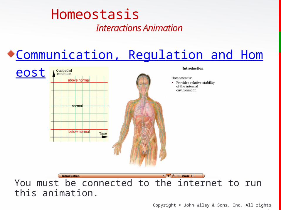
Homeostasis Interactions Animation
Communication, Regulation and Homeostasis
You must be connected to the internet to run this animation.
Copyright © John Wiley & Sons, Inc. All rights reserved.
HomeostasisBody fluids are defined as dilute, watery solutions
containing dissolved chemicals inside or outside of the cell.
Maintaining the volume and composition of body fluids is
important.
Intracellular Fluid (ICF) is the fluid within cells
Extracellular Fluid (ECF) is the fluid outside cells
• Interstitial fluid is ECF between cells and tissues

Copyright © John Wiley & Sons, Inc. All rights reserved.
Homeostasis
Some important body fluids:
Blood Plasma is the ECF within blood vessels.
Lymph is the ECF within lymphatic vessels.
Cerebrospinal fluid (CSF) is the ECF in the brain and
spinal cord.
Synovial fluid is the ECF in joints.
Aqueous humor is the ECF in eyes.

Copyright © John Wiley & Sons, Inc. All rights reserved.
Cellular function depends on the regulation of the
composition of the interstitial fluid.
Composition of interstitial fluid changes as substances
move between plasma and the interstitial fluid.
Movement back and forth across capillary walls provides
nutrients (glucose, oxygen, ions) to tissue cells and
removes waste (carbon dioxide).
Homeostasis
Copyright © John Wiley & Sons, Inc. All rights reserved.
Control of homeostasis is constantly being challenged by:
Physical insults such as intense heat or lack of oxygen
Changes in the internal environment such as a drop in
blood glucose due to lack of food
Physiological stress such as demands of work or school
Disruptions are mild if balance is quickly restored.
Intense disruptions are often prolonged and result in disease
(poisoning or severe infections) or death.
Homeostasis

Copyright © John Wiley & Sons, Inc. All rights reserved.
Cycle of events:
Body is monitored and re-monitored.
Each monitored variable is termed
a controlled condition.Three basic components:
Receptor
Control center
Effector
Feedback System
Copyright © John Wiley & Sons, Inc. All rights reserved.
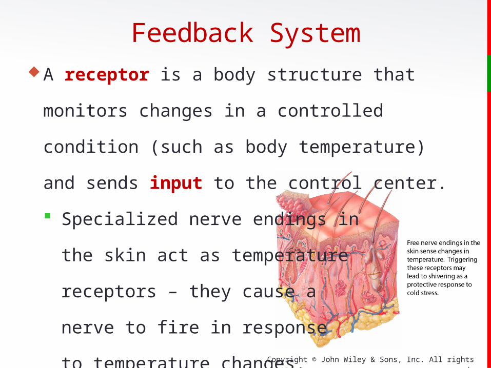
A receptor is a body structure that monitors changes in a
controlled condition (such as body temperature) and sends
input to the control center.
Specialized nerve endings in
the skin act as temperature
receptors – they cause a
nerve to fire in response
to temperature changes.
Feedback System
Copyright © John Wiley & Sons, Inc. All rights reserved.
The control center sets the range of values to be
maintained – usually this is done by the brain.
Evaluates input received from receptors and generates
output command
Output involves nerve impulses, hormones, or other
chemical agents.
• Brain acts as a control center receiving nerve
impulses from skin temperature receptors.
Feedback System

Copyright © John Wiley & Sons, Inc. All rights reserved.
The effector receives output from the control center and
produces a response or effect that changes the controlled
condition.
Nearly every organ or tissue can serve as an effector.• Body temperature drops.• The brain sends an impulse to the skeletal muscles to
contract .• Shivering occurs to generate heat.
Feedback System
Copyright © John Wiley & Sons, Inc. All rights reserved.
Negative Feedback systems:
Reverses a change in a controlled condition
• Regulation of blood pressure (force exerted by blood as
it presses again the walls of the blood vessels)
Positive Feedback systems:
Strengthens or reinforces a change in one of the body’s
controlled conditions
• Normal child birth
Feedback System
Copyright © John Wiley & Sons, Inc. All rights reserved.
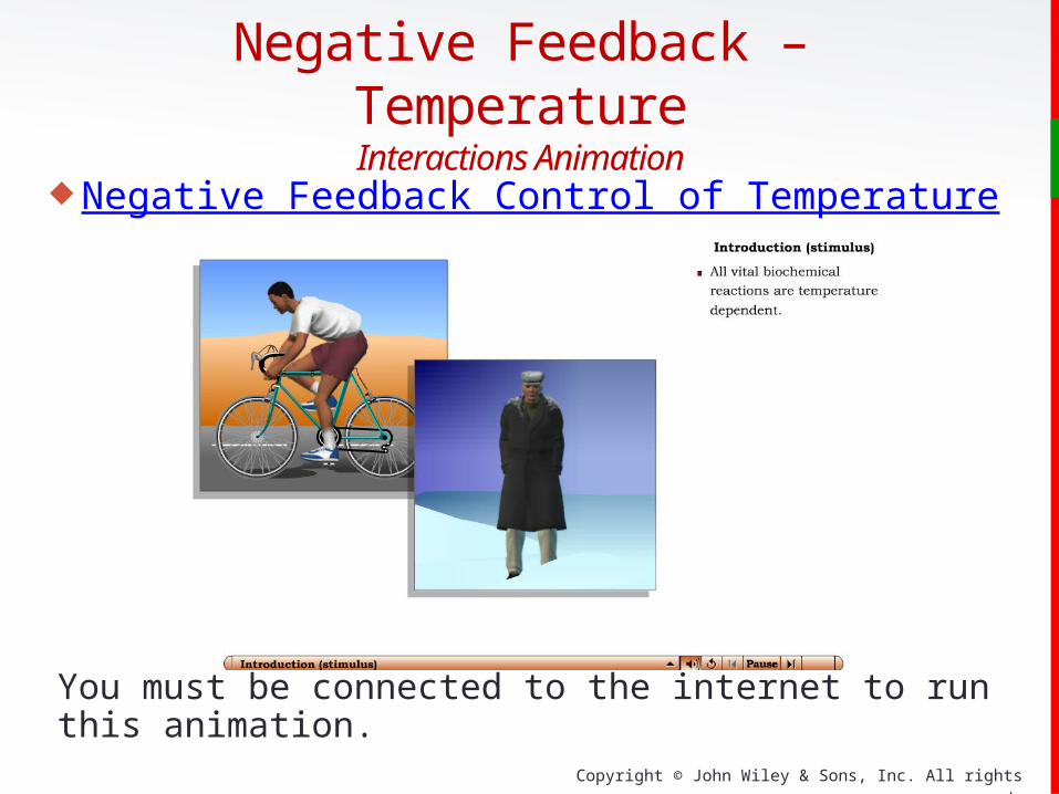
Negative Feedback – Temperature Interactions Animation
Negative Feedback Control of Temperature
You must be connected to the internet to run this animation.

Copyright © John Wiley & Sons, Inc. All rights reserved.
Blood Pressure regulation is a negative feedback system.
External or internal stimulus increases BP.
Baroreceptors (pressure sensitive
receptors) detect higher BP and send a
nerve impulse to the brain (interpretation).
Responses sent via nerve impulses
to the heart and blood vessels cause the
BP to drop (homeostasis is restored.)
Feedback System
Copyright © John Wiley & Sons, Inc. All rights reserved.
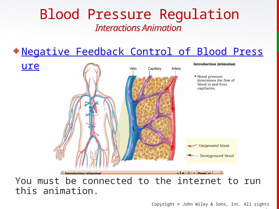
Blood Pressure RegulationInteractions Animation
Negative Feedback Control of Blood Pressure
You must be connected to the internet to run this animation.

Copyright © John Wiley & Sons, Inc. All rights reserved.
Childbirth is an example of a positive
feedback system:
Uterine contractions cause vagina to open.
Stretch-sensitive receptors in cervix send
impulses to brain.
Oxytocin is released into the blood.
Contractions enhanced and baby pushes
farther down the uterus.
Cycle continues to the birth of the
baby (no stretching).
Feedback System

Copyright © John Wiley & Sons, Inc. All rights reserved.
Positive Feedback – LaborInteractions Animation
Positive Feedback Control of Labor
You must be connected to the internet to run this animation.

Copyright © John Wiley & Sons, Inc. All rights reserved.
Clinical ConnectionDiagnosis of Disease is done by assessing:
Signs and symptoms
Medical history
• Collecting information about event
• Present illnesses and past medical problems
Physical examination:
• Orderly evaluation of the body and its function
• Noninvasive techniques and other vital signs (pulse)

Copyright © John Wiley & Sons, Inc. All rights reserved.
Organ Systems of the Body
Integumentary System (Chapter 5)
consists of the skin and related
structures (hair, nails, and glands).
Protects body, regulates
temperature, and eliminates
wastes through sweat and other
secretions

Copyright © John Wiley & Sons, Inc. All rights reserved.
Skeletal System (Chapters 6-9) consists of the bones and
joints.
Provides protection and support
Houses cells that will
become red blood cells,
white blood cells, and
platelets
Organ Systems of the Body

Copyright © John Wiley & Sons, Inc. All rights reserved.
Muscular System (Chapters 10-11) consists of the
named skeletal muscles, as well as
smooth muscle and cardiac muscle.
Participates with the skeletal
system to facilitate movement
and maintain posture
Generates the heat necessary
for warm-blooded organisms to
maintain a constant body temp.
Organ Systems of the Body

Copyright © John Wiley & Sons, Inc. All rights reserved.
Nervous System (Chapters 12-17) consists of the brain,
spinal cord, nerves, and sensory
organs).
Senses and responds to body
conditions through
nerve impulses
Organ Systems of the Body

Copyright © John Wiley & Sons, Inc. All rights reserved.
Endocrine System (Chapter 18) consists of hormone-
producing cells and glands
scattered throughout the
body.
Regulates the body
through chemical
mechanisms (by releasing
hormones into the blood)
Organ Systems of the Body

Copyright © John Wiley & Sons, Inc. All rights reserved.
Cardiovascular (Chapters 19-21) consists of the heart,
blood, and blood vessels.
Carries blood and nutrients to
specific locations
Regulates body temperature,
and water balance
Organ Systems of the Body
Copyright © John Wiley & Sons, Inc. All rights reserved.
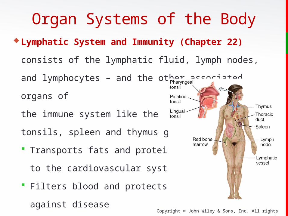
Lymphatic System and Immunity (Chapter 22) consists of
the lymphatic fluid, lymph nodes, and lymphocytes – and the
other associated organs of
the immune system like the
tonsils, spleen and thymus gland.
Transports fats and proteins
to the cardiovascular system
Filters blood and protects
against disease
Organ Systems of the Body

Copyright © John Wiley & Sons, Inc. All rights reserved.
Respiratory System (Chapter 23) consists of the upper
airways, the trachea and major
bronchi, and the lungs.
Extracts O2 and
eliminates CO2
In conjunction with the
kidneys, regulates
acid/base balance
Organ Systems of the Body

Copyright © John Wiley & Sons, Inc. All rights reserved.
Organ Systems of the BodyDigestive System (Chapter 24) consists of the esophagus,
stomach and intestines, and
the accessory digestive
glands like the salivary
glands, liver, and gallbladder.
Accomplishes the physical
and chemical breakdown
of food and elimination of waste
Copyright © John Wiley & Sons, Inc. All rights reserved.
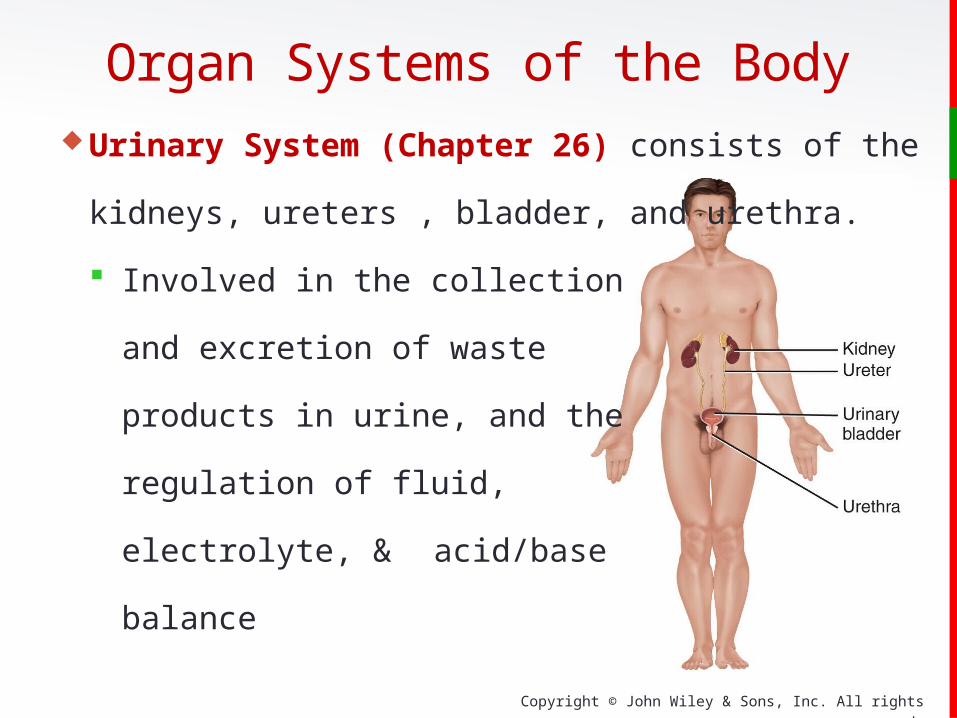
Urinary System (Chapter 26) consists of the kidneys,
ureters , bladder, and urethra.
Involved in the collection
and excretion of waste
products in urine, and the
regulation of fluid,
electrolyte, & acid/base
balance
Organ Systems of the Body
Copyright © John Wiley & Sons, Inc. All rights reserved.
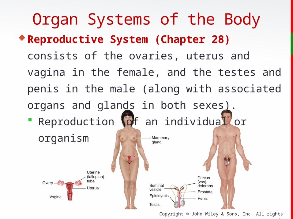
Reproductive System (Chapter 28) consists of the
ovaries, uterus and vagina in the female, and the testes and
penis in the male (along with associated organs and glands
in both sexes).
Reproduction of an individual or organism
Organ Systems of the Body

Copyright © John Wiley & Sons, Inc. All rights reserved.
The systems of the body may appear to be separate and
distinct, but the maintenance of most body functions
requires the integration of many systems working together.
For example, regulation of body temperature involves
the muscular, cardiovascular, nervous, and integumentary
systems all working together to produce and distribute
body heat appropriately.
Organ Systems of the Body
Copyright © John Wiley & Sons, Inc. All rights reserved.
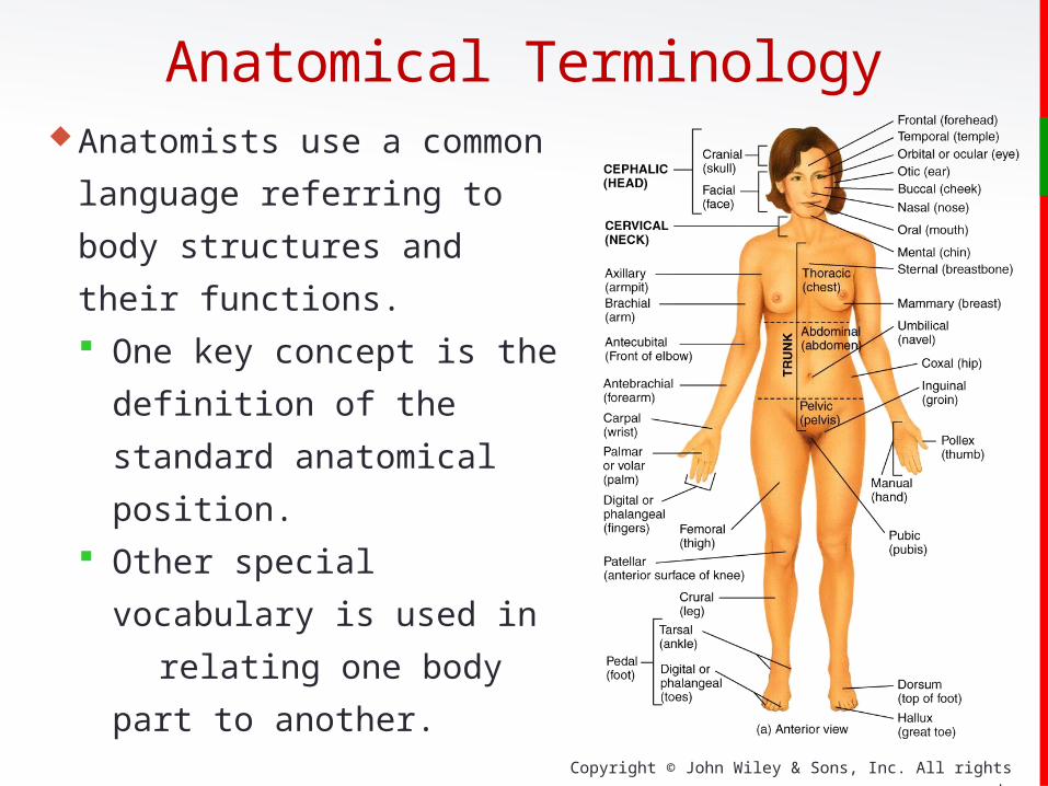
Anatomists use a common
language referring to body
structures and their functions.
One key concept is the
definition of the standard
anatomical position.
Other special vocabulary is
used in relating one
body part to another.
Anatomical Terminology

Copyright © John Wiley & Sons, Inc. All rights reserved.
Anatomical Position
In the anatomical position, the subject stands
erect facing the observer with the head
level, the eyes facing forward, feet flat on
the floor directed forward, and the arms
at their sides, palms forward.
All anatomical descriptions are in
reference to this position.
Anatomical Terminology
Copyright © John Wiley & Sons, Inc. All rights reserved.
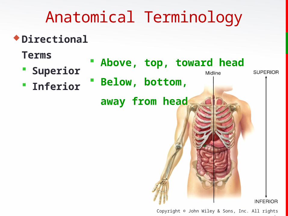
Directional Terms
Superior
Inferior Above, top, toward
head
Below, bottom,
away from head
Anatomical Terminology
Copyright © John Wiley & Sons, Inc. All rights reserved.
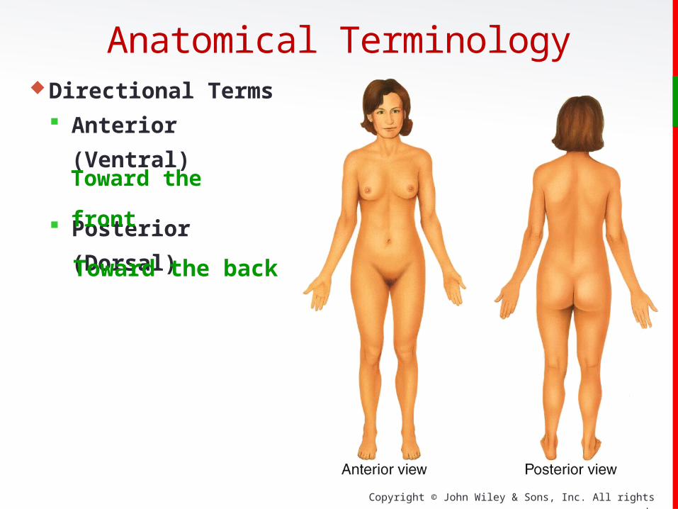
Directional Terms
Anterior (Ventral)
Posterior (Dorsal)
Anatomical Terminology
Toward the front
Toward the back
Copyright © John Wiley & Sons, Inc. All rights reserved.
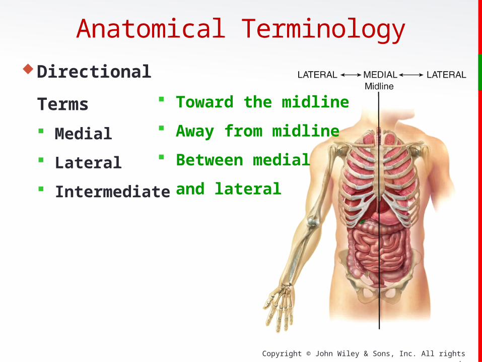
Toward the midline
Away from midline
Between medial
and lateral
Anatomical TerminologyDirectional Terms
Medial
Lateral
Intermediate

Copyright © John Wiley & Sons, Inc. All rights reserved.
Nearest to the
origination
Farther from
origination
Anatomical Terminology
Directional
Terms
Proximal
Distal
Copyright © John Wiley & Sons, Inc. All rights reserved.
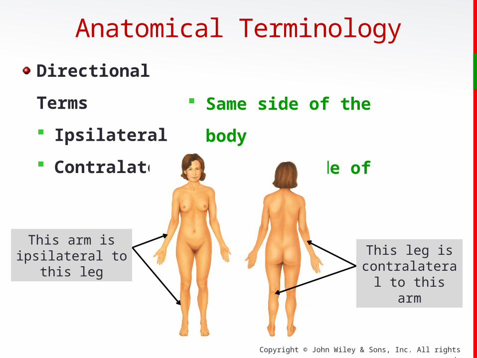
Same side of the body
Opposite side of the body
Anatomical Terminology
Directional Terms
Ipsilateral
Contralateral
This arm is ipsilateral to this leg This leg is
contralateral to this arm

Copyright © John Wiley & Sons, Inc. All rights reserved.
Directional Terms
Superficial
Deep Towards the surface
Towards the core of the body
Anatomical Terminology
DeepSuperficial Superficial
Superficial
Superficial
Copyright © John Wiley & Sons, Inc. All rights reserved.
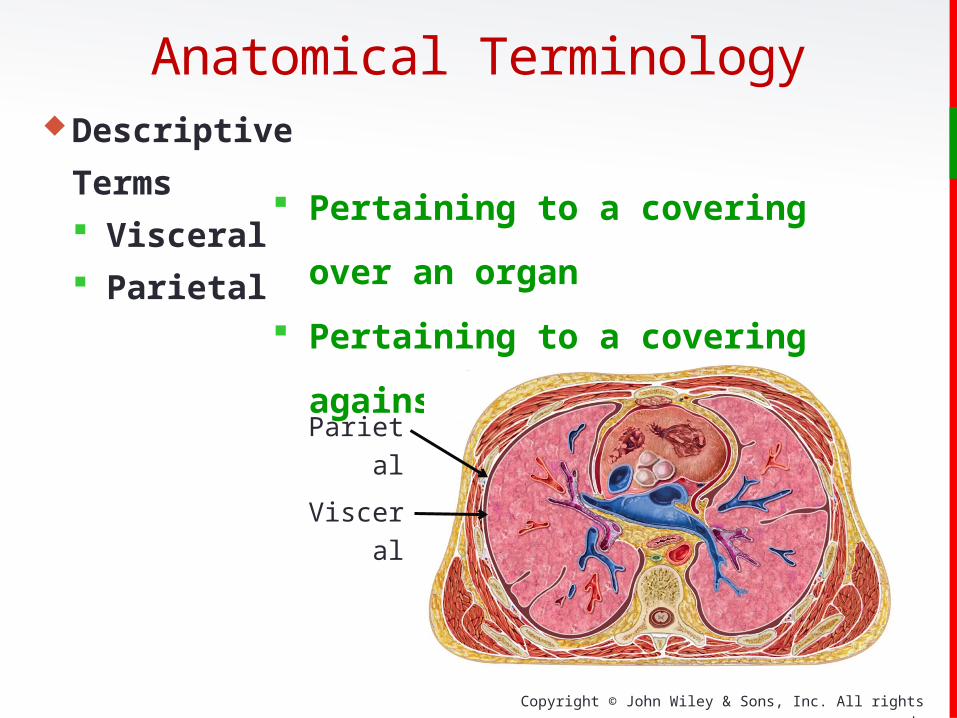
Descriptive Terms
Visceral
Parietal Pertaining to a covering over an organ
Pertaining to a covering against a
cavity wall
Anatomical Terminology
Visceral
Parietal
Copyright © John Wiley & Sons, Inc. All rights reserved.
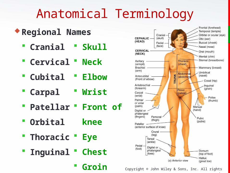
Regional Names
Cranial
Cervical
Cubital
Carpal
Patellar
Orbital
Thoracic
Inguinal
Skull
Neck
Elbow
Wrist
Front of knee
Eye
Chest
Groin
Anatomical Terminology

Copyright © John Wiley & Sons, Inc. All rights reserved.
Regional Names
Metacarpal
Plantar
Buccal
Axillary
Femoral
Gluteal
Tarsal
Digital
or Phalangeal
• Hand/palm
• Sole of foot
• Cheek
• Armpit
• Thigh
• Buttock
• Ankle
• Toes
or Fingers
Anatomical Terminology
Copyright © John Wiley & Sons, Inc. All rights reserved.
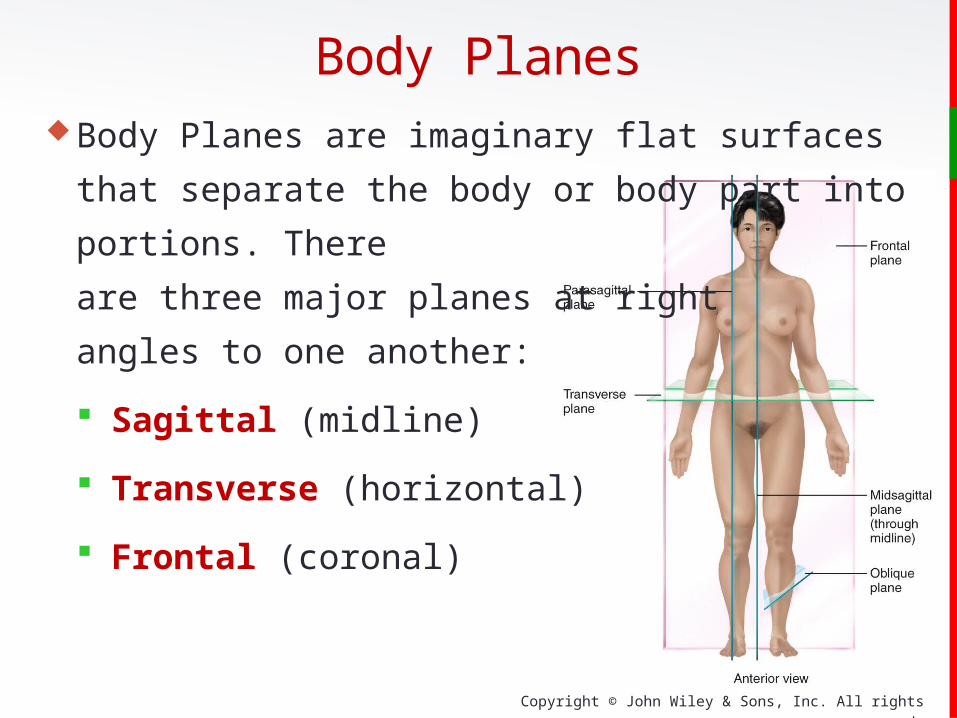
Body Planes are imaginary flat surfaces that separate the
body or body part into portions. There
are three major planes at right
angles to one another:
Sagittal (midline)
Transverse (horizontal)
Frontal (coronal)
Body Planes
Copyright © John Wiley & Sons, Inc. All rights reserved.
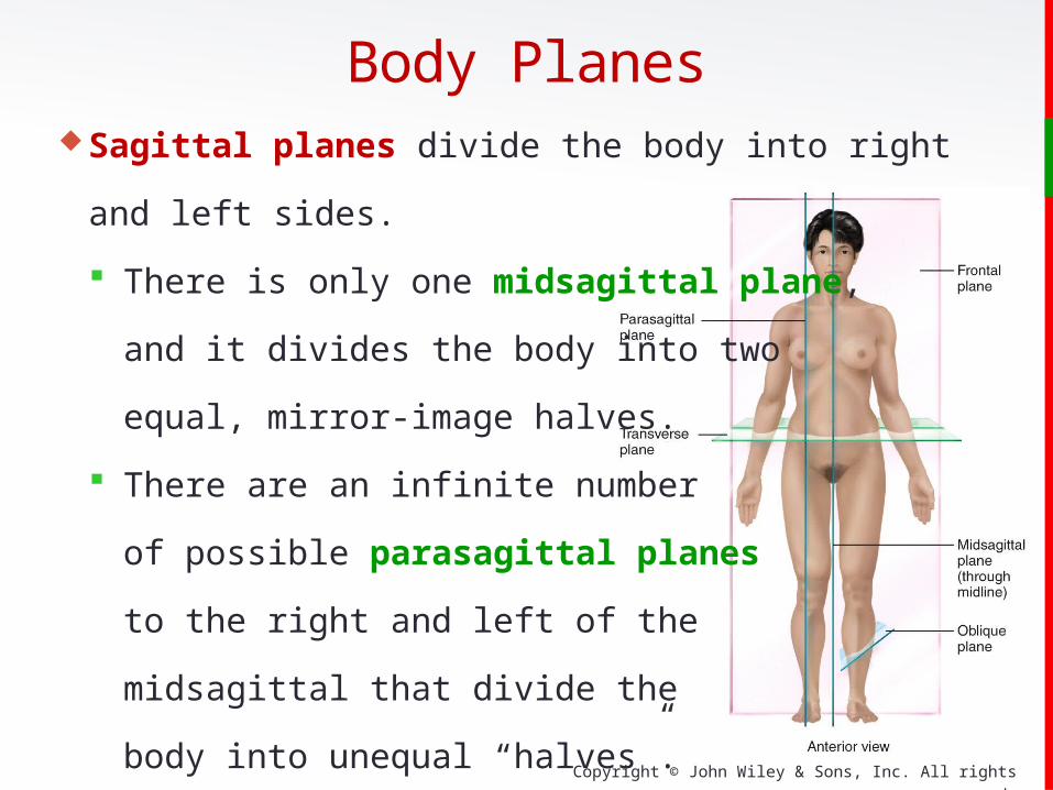
Sagittal planes divide the body into right and left sides.
There is only one midsagittal plane,
and it divides the body into two
equal, mirror-image halves.
There are an infinite number
of possible parasagittal planes
to the right and left of the
midsagittal that divide the
body into unequal “halves”.
Body Planes

Copyright © John Wiley & Sons, Inc. All rights reserved.
Body PlanesFrontal or coronal planes divide the body (or an organ)
into anterior (front) and posterior
(back) portions.
Transverse planes (also called
cross-sectional or horizontal
planes) divide the body into
superior (upper) and inferior
(lower) portions.
Copyright © John Wiley & Sons, Inc. All rights reserved.
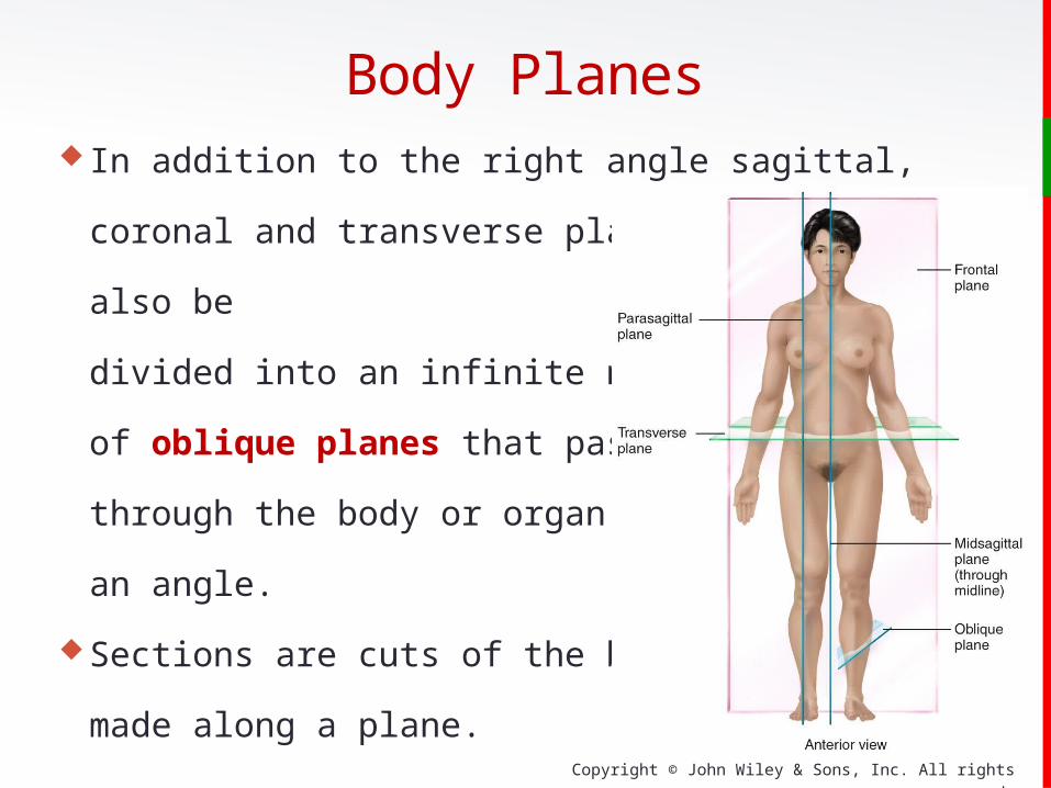
In addition to the right angle sagittal, coronal and transverse
planes, the body can also be
divided into an infinite number
of oblique planes that pass
through the body or organ at
an angle.
Sections are cuts of the body
made along a plane.
Body Planes

Copyright © John Wiley & Sons, Inc. All rights reserved.
Body Planes
A midsagittal section of the human brain
A frontal (or coronal) brain section
A transverse (or horizontal) brain section
Copyright © John Wiley & Sons, Inc. All rights reserved.
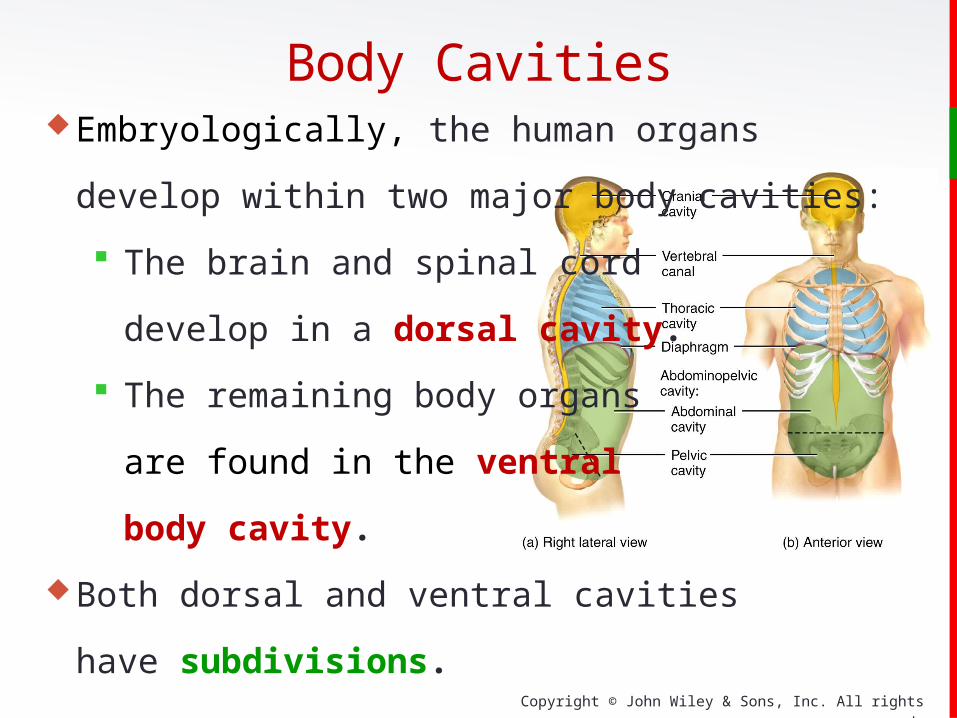
Body CavitiesEmbryologically, the human organs develop
within two major body cavities:
The brain and spinal cord
develop in a dorsal cavity.
The remaining body organs
are found in the ventral
body cavity.
Both dorsal and ventral cavities
have subdivisions.

Copyright © John Wiley & Sons, Inc. All rights reserved.
Body Cavities
Copyright © John Wiley & Sons, Inc. All rights reserved.
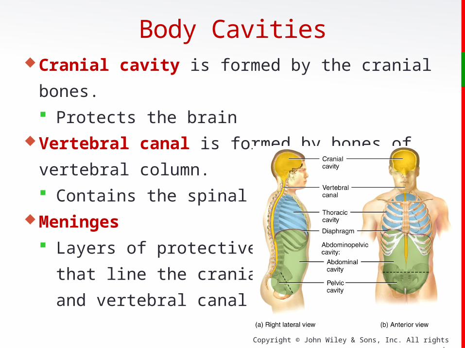
Cranial cavity is formed by the cranial bones.
Protects the brainVertebral canal is formed by bones of vertebral column.
Contains the spinal cord Meninges
Layers of protective tissue
that line the cranial cavity
and vertebral canal
Body Cavities
Copyright © John Wiley & Sons, Inc. All rights reserved.
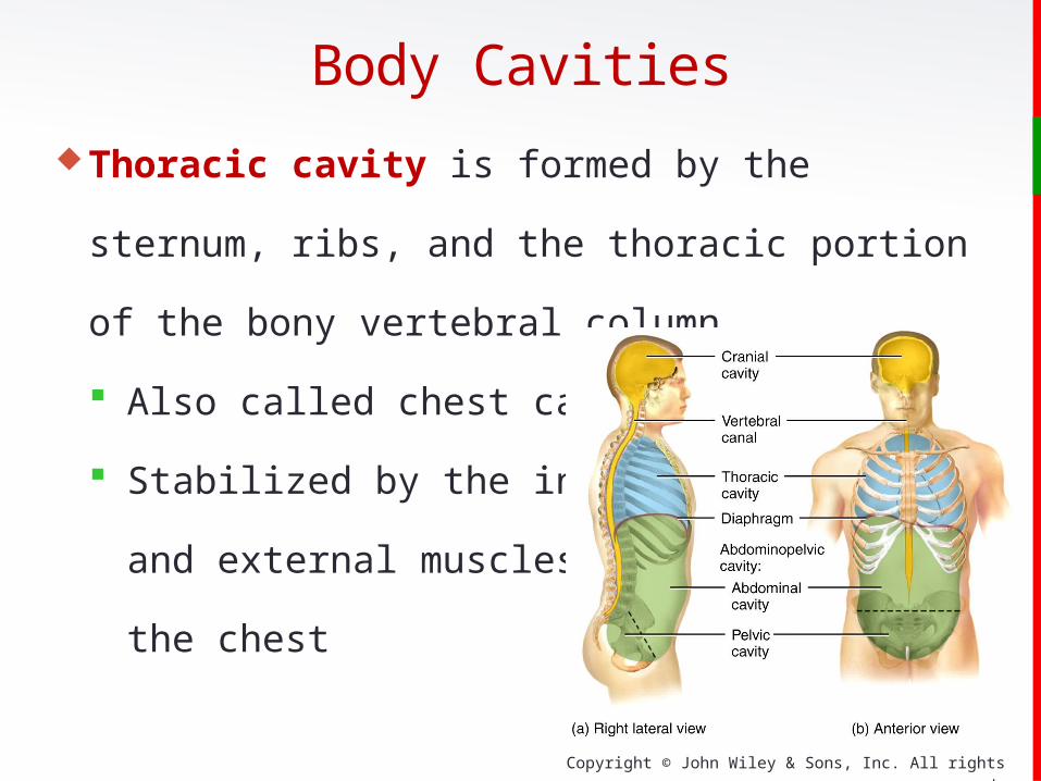
Thoracic cavity is formed by the sternum, ribs, and the
thoracic portion of the bony vertebral column.
Also called chest cavity
Stabilized by the internal
and external muscles of
the chest
Body Cavities
Copyright © John Wiley & Sons, Inc. All rights reserved.
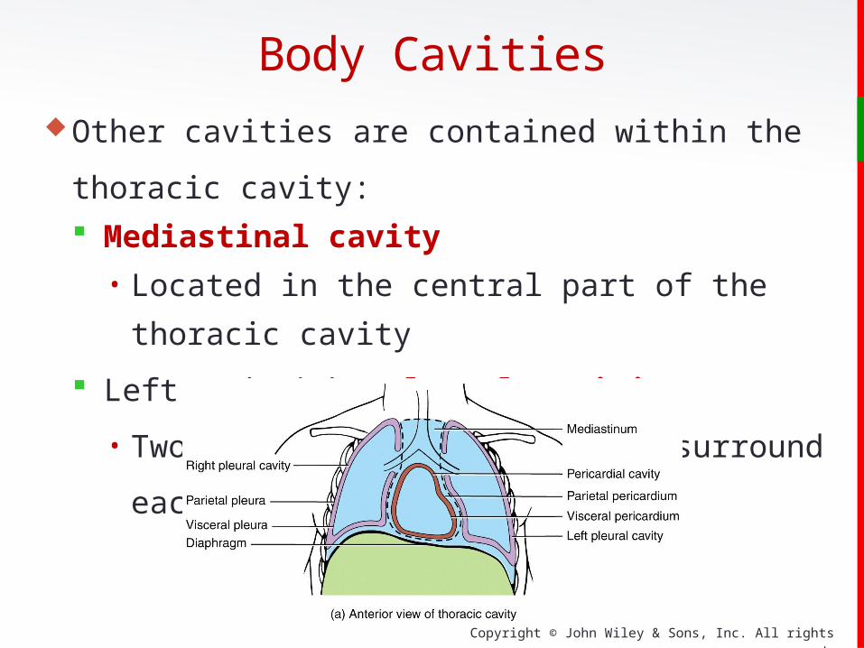
Other cavities are contained within the thoracic cavity:
Mediastinal cavity • Located in the central part of the thoracic cavity
Left and Right Pleural cavities
• Two fluid-filled spaces that surround each lung
Body Cavities
Copyright © John Wiley & Sons, Inc. All rights reserved.
Pericardial cavity is itself located within the middle part of
the mediastinal cavity in the thoracic cavity (like a set of
Russian nesting dolls of decreasing size—one placed inside
the other).
Fluid-filled space that
surrounds the heart
Body Cavities
Insert new photo

Copyright © John Wiley & Sons, Inc. All rights reserved.
The pericardial cavity is shown here nestled in
the middle mediastinum:
Body Cavities

Copyright © John Wiley & Sons, Inc. All rights reserved.
Abdominopelvic Cavity extends from the diaphragm to the
groin and is encircled by the abdominal wall and bones and
muscles of the pelvis.
Divided into two portions:• Abdominal cavity contains the stomach, spleen, liver,
gallbladder, small and large intestines.• Pelvic cavity contains the urinary bladder, internal
organs of reproductive system, and portions of the large
intestine.
Body Cavities
Copyright © John Wiley & Sons, Inc. All rights reserved.
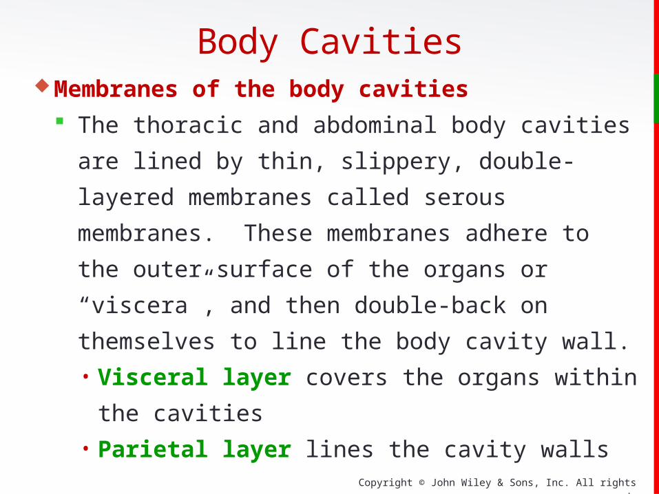
Membranes of the body cavities
The thoracic and abdominal body cavities are lined by
thin, slippery, double-layered membranes called serous
membranes. These membranes adhere to the outer surface
of the organs or “viscera”, and then double-back on
themselves to line the body cavity wall.• Visceral layer covers the organs within the cavities• Parietal layer lines the cavity walls
Body Cavities
Copyright © John Wiley & Sons, Inc. All rights reserved.
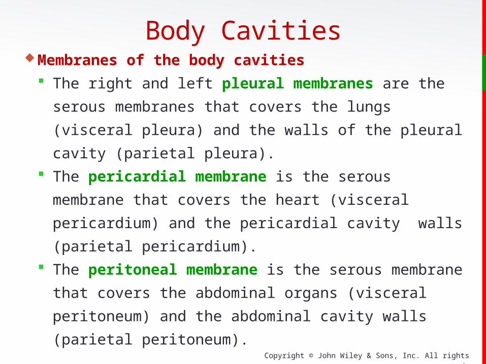
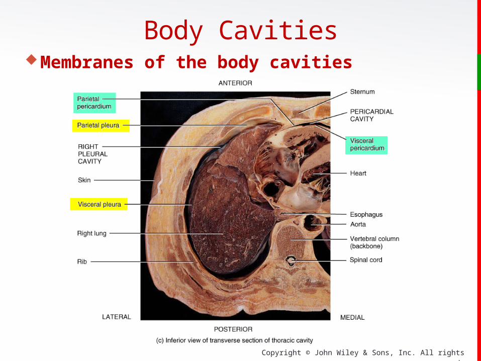
Membranes of the body cavities
The right and left pleural membranes are the serous
membranes that covers the lungs (visceral pleura) and the
walls of the pleural cavity (parietal pleura).
The pericardial membrane is the serous membrane that
covers the heart (visceral pericardium) and the pericardial
cavity walls (parietal pericardium).
The peritoneal membrane is the serous membrane that
covers the abdominal organs (visceral peritoneum) and
the abdominal cavity walls (parietal peritoneum).
Body Cavities
Copyright © John Wiley & Sons, Inc. All rights reserved.
Membranes of the body cavities
Body Cavities

Copyright © John Wiley & Sons, Inc. All rights reserved.
Other body cavities
Oral (mouth) cavity contains the tongue and teeth.
Nasal cavity is part of the upper airways (Chapter 23).
Orbital cavities contain the eyeballs and various nerves
and blood vessels.
Middle ear cavities contain the small bones of the middle
ear.
Synovial cavities are found in freely moveable joints like
the large joints of the shoulder and hip.
Body Cavities
Copyright © John Wiley & Sons, Inc. All rights reserved.
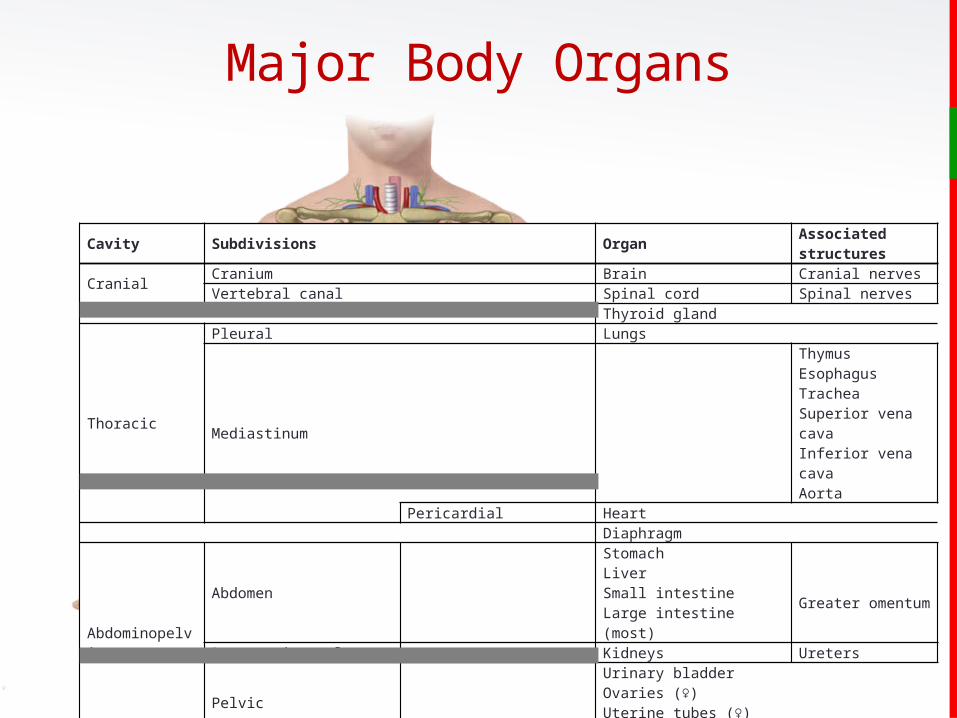
Cavity Subdivisions OrganAssociated structures
CranialCranium Brain Cranial nervesVertebral canal Spinal cord Spinal nerves
Thyroid gland
Thoracic
Pleural Lungs
Mediastinum
ThymusEsophagusTracheaSuperior vena cavaInferior vena cavaAorta
Pericardial HeartDiaphragm
Abdominopelvic
Abdomen
StomachLiverSmall intestineLarge intestine (most)
Greater omentum
Retroperitoneal Kidneys Ureters
Pelvic
Urinary bladderOvaries (♀) Uterine tubes (♀)Uterus (♀)
Testes (♂)
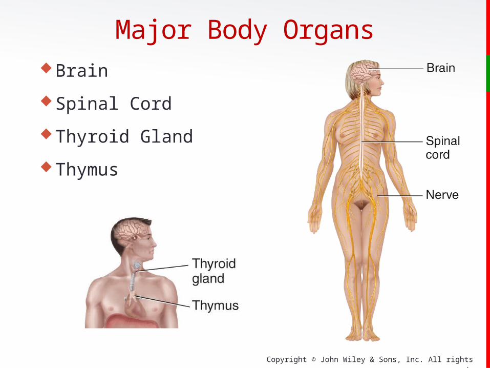
Major Body Organs
Copyright © John Wiley & Sons, Inc. All rights reserved.
Major Body OrgansBrain
Spinal Cord
Thyroid Gland
Thymus
Copyright © John Wiley & Sons, Inc. All rights reserved.
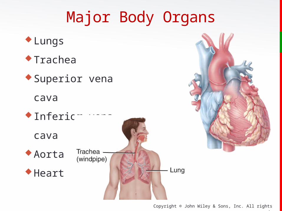
Major Body OrgansLungs
Trachea
Superior vena cava
Inferior vena cava
Aorta
Heart
Copyright © John Wiley & Sons, Inc. All rights reserved.
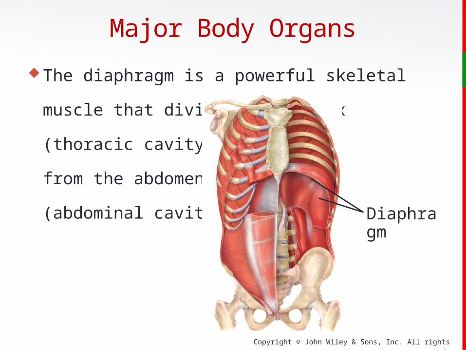
Major Body Organs
The diaphragm is a powerful skeletal muscle that divides
the thorax
(thoracic cavity)
from the abdomen
(abdominal cavity).
Diaphragm

Copyright © John Wiley & Sons, Inc. All rights reserved.
Major Body Organs
Trachea
Esophagus
Stomach
Liver
Small Intestine
Large Intestine
Copyright © John Wiley & Sons, Inc. All rights reserved.
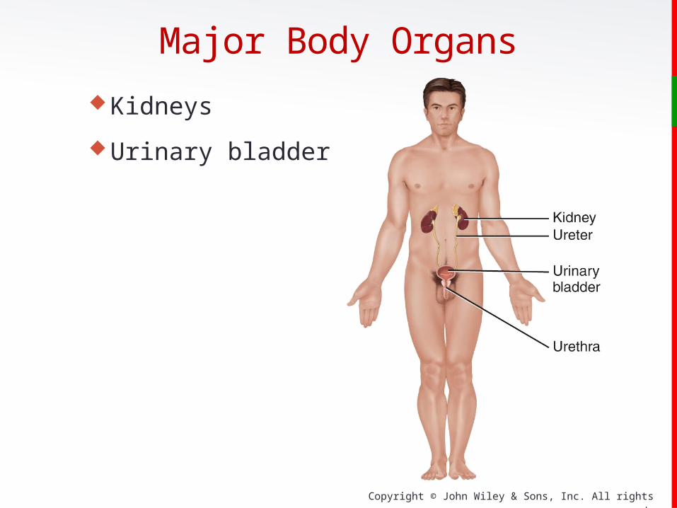
Major Body Organs
Kidneys
Urinary bladder

Copyright © John Wiley & Sons, Inc. All rights reserved.
Major Body Organs
Ovaries
Uterine tubes
Uterus
Testes

Copyright © John Wiley & Sons, Inc. All rights reserved.
Abdominopelvic Quadrants & Regions
Identification of quadrants and regions in the
abdominopelvic cavity helps clinicians describe the
location of the many abdominal and pelvic organs.
There are 4 abdominopelvic quadrants and 9
regions.
The dividing lines between these are centered
on the umbilicus (“belly button”).

Copyright © John Wiley & Sons, Inc. All rights reserved.
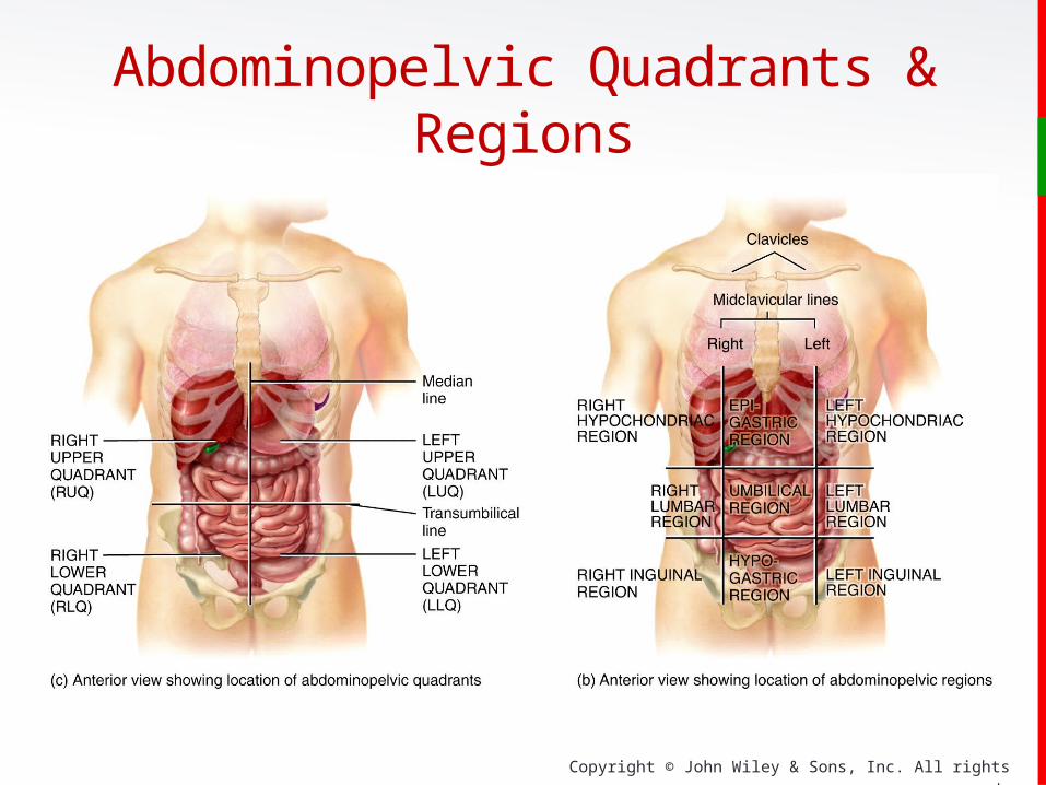
Vertical and horizontal lines pass through the umbilicus
Right upper quadrant (RUQ)• liver
Left upper quadrant (LUQ)
• spleen and left kidney
Right lower quadrant (RLQ)
• appendix
Left lower quadrants (LLQ)
• left ovary ( )
Abdominopelvic Quadrants & Regions
Copyright © John Wiley & Sons, Inc. All rights reserved.
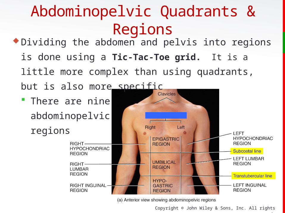
Dividing the abdomen and pelvis into regions is done using
a Tic-Tac-Toe grid. It is a little more complex than using
quadrants,
but is also more specific
There are nine
abdominopelvic
regions
Abdominopelvic Quadrants & Regions
Copyright © John Wiley & Sons, Inc. All rights reserved.
Abdominopelvic Quadrants & Regions
Copyright © John Wiley & Sons, Inc. All rights reserved.
Medical ImagingTechniques and procedures used to create images of the
human body
Allow visualization of structures inside the body
Diagnosis of anatomical and physiological disorders
Conventional radiography (X-rays) have been in use
since the late 1940’s
Copyright © John Wiley & Sons, Inc. All rights reserved.
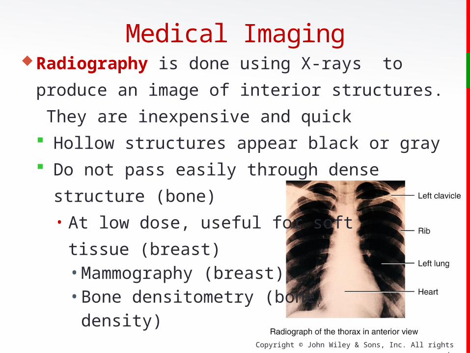
Radiography is done using X-rays to produce an image of
interior structures. They are inexpensive and quick
Hollow structures appear black or gray
Do not pass easily through dense structure (bone)• At low dose, useful for soft
tissue (breast)• Mammography (breast)• Bone densitometry (bone
density)
Medical Imaging
Copyright © John Wiley & Sons, Inc. All rights reserved.
Magnetic Resonance Imaging (MRI) is done using an
extremely powerful magnetic field. It is a safe procedure
but cannot be used on patients containing metal.
Protons in body fluid align with field
Used for differentiating normal and abnormal tissues
(tumors, brain abnormalities, blood flow)
2D and 3D color images can be viewed on a video
monitor.
Medical Imaging

Copyright © John Wiley & Sons, Inc. All rights reserved.
Medical ImagingComputed Tomography or CT-Scans are done using a
computer to organize x-rays to form a 3D image. It is used
to visualize soft tissue in more detail than conventional
radiography.
Tissue intensities show
varying degrees of gray.
Whole-body CT scans
expose the body to a high
dose of x-rays.

Copyright © John Wiley & Sons, Inc. All rights reserved.
Here are 3 cross sectional images of a
head from the Visible Human Project.
They are done using the three
modalities discussed above.
From top to bottom:
Photograph of frozen, sawed head
CT scan of the same level/plane
MRI scan of the same level/plane
http
://vh
p.m
ed
.um
ich.e
du
/
Objective 10
Medical Imaging

Copyright © John Wiley & Sons, Inc. All rights reserved.
Medical ImagingUltrasound Scanning (sonography) is done using high
frequency sound waves. It is noninvasive and painless.
Because of its safety profile,
it is commonly used to
monitor the progress of
fetal development during
pregnancy.

Copyright © John Wiley & Sons, Inc. All rights reserved.
Radionuclide Scanning is done by giving a radioactive
substance (radionuclide) intravenously.
Gamma rays emitted by tissues that take up the
radionuclide are detected by a camera and displayed on a
video monitor. The color intensity represents the amount of
uptake.
Single-photo-emission
computerized tomography
(SPECT) is a specialized
form of this technique.
Medical Imaging

Copyright © John Wiley & Sons, Inc. All rights reserved.
Positron Emission Tomography (PET scan) is done by
injecting a substance emitting positively charged particles
into the body. The collision between positrons and negatively
charged electron in
body tissues produce gamma rays
used to form a computer assisted
image.
Used to study physiology of
body structures (metabolism)
Medical Imaging
Copyright © John Wiley & Sons, Inc. All rights reserved.
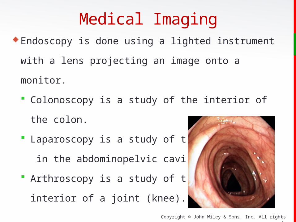
Endoscopy is done using a lighted instrument with a lens
projecting an image onto a monitor.
Colonoscopy is a study of the interior of the colon.
Laparoscopy is a study of the organs
in the abdominopelvic cavity.
Arthroscopy is a study of the
interior of a joint (knee).
Medical Imaging
Copyright © John Wiley & Sons, Inc. All rights reserved.
Clinical ConnectionNoninvasive Diagnostic Techniques are used to inspect
different aspects of the body:
Is often done to access structure and function and to search
for the presence of disease.
• Palpation is gently touching body surfaces with hands.
• Auscultation is listening to body sounds (stethoscope).
• Percussion is tapping on the body surface with
fingertips and listening to echoes.

Copyright © John Wiley & Sons, Inc. All rights reserved.
End of Chapter 1
Copyright 2012 John Wiley & Sons, Inc.
All rights reserved. Reproduction or translation of this work beyond that
permitted in section 117 of the 1976 United States Copyright Act without
express permission of the copyright owner is unlawful. Request for
further information should be addressed to the Permission Department,
John Wiley & Sons, Inc. The purchaser may make back-up copies for
his/her own use only and not for distribution or resale. The Publishers
assumes no responsibility for errors, omissions, or damages caused by the
use of these programs or from the use of the information herein.