Embed Size (px)
Citation preview
Proc 8th Int Coral Reef Sym 1:607-612. 1997
MICROBIAL PESTS CORAL DISEASE IN THE WESTERN ATLANTIC
D.L. Santavy' and E.C. Peters ,
U.S. Environmental Protection ~gency, Gulf Ecology Division, 1 Sabine Dr., Gulf Breeze, Florida 32561, U.S.A., Tetra Tech. , Inc. , 10306 Eaton Place, Suite 340, Fairfax, Virginia 22030 U.S.A.
ABSTRACT
Diseases of scleractinian corals have increased signifi-cantly over the last decade, affecting greater numbers ofspecies around the world. Gross signa of coral disease areoften observed as tissue loss on the skeleton, makingdifferential diagnosis difficult. Using histopathologicaland ultrastructural techniques, coupled with microbiolog-ical analyses, the importance of microorganisms as patho-gens in coral diseases is becoming more apparent. Thispaper addresses the ecology of pathogens on reefs,specifically bacteria and cyanobacteria that producedisease in scleractinian and alcyonarian corals. We reviewthe nature of disease and the influence of adverseenvironmental conditions. An update is presented onresearch concerning the bacteria associated with black- andwhite-band diseases; observations are presented concerningother coral diseases in the western Atlantic that appearto be caused by bacteria. We conclude with suggestions forimproving the recognition of coral diseases that includeapproaches for conducting research to identify bacterialpathogens and the role of environmental factors in thedevelopment of coral disease.
INTRODUCTION
Unprecedented decreases of cover by stony corals, alter-ing both total abundance and species richness, have beendocumented during the last decade ~Brown 1987; Ginsberg1993; Hughes 1994; Sebens 1994) .Increases have beenreported in the incidence of physiological disorders,bleaching (loss of obligate symbiotic algae and/or algalpigments), and other diseases that cause loss of tissue.Accounts of declining reef quality from geographically-distant regions include the Florida Keys (Porter and Meier1992, Zorpette 1995), Fiji (Littler and Littler 1996),Hawaii (Hunter and Evans 1995), the Red Sea (Antonius1988), and the Great Barrier Reef (Dinsdale 1994;Glazebrook and Streiner 1994) .These disorders have beenobserved primarily among the scleractinian corals, butother organisms in the reef ecosystem have been affected(Lessios et al. 1983; Hallock et al. 1993; Peters 1993) .
Although diseases of corals have been reported from aroundthe world, they have been documente~ most widely in thetropical western Atlantic (Peters 1993) .Black-band andwhite-band diseases have been reported most frequently inthis region, since the first observations were made in theearly 1970s (Antonius 1977; Gladf~lter et al. 1977) .Subsequently, other maladies have been observed, includingyellow-blotch disease on stony corals and red-band diseaseon octocorals. Most "band" diseases have been associatedwith microorganisms that appear to have a role in thedevelopment of lesions and the loss qf coral tissue. Mostcoral diseases observed on reefs have not been thoroughlydescribed. Their etiologies, including identification ofthe causative agent, pathogenesis, hosts affected, andinteractions with anthropogenic stressors, are unknown.Knowledge of coral disease mechanisms, processes, andmanifestation, and the role of microbial "pests" indevelopment of these diseases might help to explain thestriking increase in reef destruction.
Disease is any impairment of an organism's vital functionsor systems, including interruption, cessation, prolifer-ation, or other malfunction. Each Organism has optimizedbehavioral responses, physiological status, and biochemicalconditions that allow the constant regulation of itsinternal environment. The regulation of these responsesallows an organism to maintain its health during exposureto stressors, including pathogens. As the intensity of astressor or number of stressors ~o which the host isexposed increases, energy usage increases to counteract theimpact, at the expense of growth and reproduction. Whenthe organism can no longer maint~in critical physio-logical and biochemical functions or reverse the con-dition, it succumbs to disease. The organism's competenceto resist disease can be altered by environmentalconditions, physiological status, nutrition, developmentalstage, and genetic factors. Although the causal agent ofa disease can be biotic or abiot~c, both are closelyrelated in disease manifestation; thus, most diseases arenot caused by a single factor, and determining the primarycause can be difficult.
Biotic diseases are produced by parasites or pathogens.Potential infectious agents of corals include bacteria,viruses, protozoans, fungi, and macroparasites such as,helminths and arthropods. To date, no coral diseases areknown to be caused by viruses. Abiotic diseases arestructural and functional impairments to the organism thatresult from exposure to extreme physical and chemicalconditions. Physical stress can be induced by changes insalinity, temperature, light intensity, radiation,sedimentation, oxygen, and water flow, as well as by directinjury to coral tissues. Abiotic diseases are noninfect-ious and cannot be spread by contact with an affectedindividual. Abiotic stressors can lead to greater suscept-ibility to disease by parasites and pathogens, and mademore severe by the invasion of secondary pathogens.Secondary or opportunistic pathogens cause disease in ahost whose defense mechanisms are compromised and whonormally would not be infected by these microorganisms.
It is increasingly evident that abiotic factors mightinfluence the distribution and impact of biotic diseases incorals. Anthropogenic inputs enrich nutrients, increasesedimentation, and add pesticides and other toxicchemicals, enteric bacteria and viruses to coastal waters(Sindermann 1995) .Additionally, agricultural and
industrial contaminants are released into watersheds anddischarged into the sea (McIvor et al. 1994) .Thedischarge of these materials in the vicinity of reefsrepresent inputs of biotic and abiotic stressors to whichcorals might be exposed with the potential to overburdenthe reef ecosystem. Increased landscape modification,sewage outfalls, and injection well sites used forwastewater combine to facilitate the dissolution ofcarbonate sediments on offshore reefs, which might allowpercolation of elevated nutrient loads in these areas(Tomascik and Sander 1987; Shinn 1993; Porter et al. 1994;Szmant and Forrester 1996) .Whether these areas ofenriched water correspond to areas of increased diseaseactivity on corals has not been thoroughly investigated.It is recognized, however, that enriched nutrients,increased water temperatures, and exposure to high levelsof UV radiation or toxicants cause mortality of coralsunder experimental conditions (Dustan and Halas 1987; Glynnet al. 1989; Glynn and D'Croz 1990; williams and Bunkley-Williams 1990) .
The first line of defense of the coral against biotic andabiotic materials is the production of mucus. Mucusenables coral to shed sediments, invading microorganisms,and other irritants from its surface. Many bacteriacolonize the surface of coral and feed on the mucus in theabsence of disease, representing a complex balance betweena host and its commensal bacterial flora under normalconditions (Ducklow and Mitchell 1979; Pascal and Vacelet1981; Paul et al. 1986; Vacelet and Thomassin 1991) .Thecommunity of commensal bacteria associated with mucusvaries depending on the species of coral and physiologicalstatus of the host (Ducklow and Mitchell 1979; Pascal andVacelet 1981; Coffroth 1990; Santavy et al. 1992; Ritchieand Smith 1995a, 1995b) .When mucus production increasesafter exposure to irritants, bacterial numbers alsoincrease (Mitchell and Chet 1975; Rublee et al. 1980;Segel and Ducklow 1982) .With a physical or environmentalchange, the equilibrium between the coral and its commensalbacteria in the mucus can be disturbed, causing substantivechanges in the species composition of these bacteria(Santavy, 1995) .For example, the ubiquitous marine
bacterium, Vibrio alginolyticus out competed and dominatedthe natural bacterial communities in the mucus of stressedcorals relative to bacterial communities found in the mucusof nonstressed corals (Ducklow and Mitchell, 1979) .Acutecoral mortality has been observed after excessive mucusproduction, leading to subsequent bacterial growth andtissue invasion following exposure of corals to stressors(Mitchell and Chet 1975; Knap et al. 1985) .
The effects of temperature, dissolved organic carbon, andother factors, on the growth of bacteria in the mucus havethe potential to increase their numbers and pathogenicityon reef corals. New species of bacteria dominating themucus might stress the host, and induce greater suscept-ibility to diseases. Exposure of corals to oil inducedtissue swelling, copious mucus production, bleaching, and
608 Santavy and Peters
tissue loss resulting from bacterial infections andundescribed coral tissue loss (Loya and Rinkevich 1980;Segel and Ducklow 1982; Jackson et al. 1989) .Field andlaboratory studies have provided evidence that somebleaching in one species of coral is induced by a bacteriumclassified as a Vibrio (Kushmaro et al. 1996) .Somebacteria can infect and degrade coral cells, causing moredamage than exposure to the stressor alone (Mitchell andChet 1975; Hodgson 1990) .Hodgson (1990) speculated thatphysiological resistance to bacterial infection might be"the most important determinant of sedimentation toleranceof reef corals". Comprehensive studies to test thesepotential synergistic causes and effects in diseasemanifestation between biotic pathogens and abioticstressors could significantly advance our understanding ofcause and effect relationships.
BACTERIAL DISEASES OF WESTERN ATLANTIC CORALS
Significant coral mortality has been reported around theworld from black-band disease, white-band disease, and red-band disease, which have been associated with cyanobacteriaand bacterial infections. Microbiological and moleculartechniques are being applied to increase our understandingof the role of microbial "pests" in the development ofthese diseases. Additional observations on westernAtlantic reefs suggest that microorganisms are importantcontributors to other types of coral tissue loss.
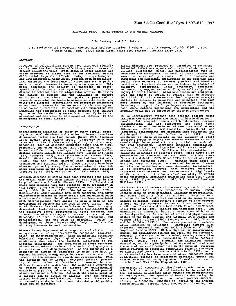
Black-Band DiseaseBlack-band disease (BBD) was the first disease reported toaffect scleractinian corals. It was discovered in the1970s on reefs off Belize, Bermuda, and the Florida Keys(Antonius 1973; Garrett and Ducklow 1975) and found
throughout the western Atlantic and Caribbean basin in the1980s (Peters 1993). Subsequently, cases of BBD have beenreported off the Phillippines (Antonius 1985), in the RedSea (Antonius 1988), off Fiji (Littler and Littler 1996),and on the Great Barrier Reef (Dinsdale 1994; Glazebrookand Streiner 1994; Miller 1996) .The diseased tissueinterface appears as a black band, a few mm to a few cm inwidth, with a clean, tissue-depleted skeleton next toapparently healthy tissue (Fig. 1) .BBD affects primarilyfaviid corals in the Western Atlantic and is resisted bymost other Scleractinia including the acroporids (Rutzleret al. 1983) .Recently, infections on acroporids on theGreat Barrier Reef were reported (Miller 1996) .Thisdisease has also been documented on milleporid hydrocorals,gorgonians, and other families of Scleractinia (Rutzler etal. 1983; Antonius 1985,1988; Feingold 1988; Glazebrook andStreiner 1994; Miller 1996) .The disease proceeds at arapid rate, with the band destroying several cm'tissue/day(Rutzler et al. 1983) .
It is unknown how the disease is originally established,although corals with physical injury or sloughing tissueappear most susceptible (Antonius 1985) .BBD is usuallyfound only on scattered coral colonies on reefs (Garrettand Ducklow 1975; Edmunds 1991), but extensive outbreaksalso have been reported in the Caribbean (Peters 1993) .Aclumped distribution has been found in the Florida Keys,suggesting the disease is infectious (Kuta and Richardson1994) .Several factors appear to promote BBD infections;they include increased temperatures, nutrient loading,sedimentation, and water current patterns (Antonius 1985;Peters 1993; Littler and Littler 1996; Bruckner et al. inpress) .
The black band is composed of a consortium ofmicroorganisms including the cyanobacteria Phormidiumcorallyticum and Spirulina sp., sulfate-reducing bacteria,the sulfur-oxidizing bacterium Beggiatoa, ciliates, andheterotrophic bacteria (Rutzler and Santavy 1983) .Thedominant organism in BBD is the cyanobacterium P.corallyticum (Rutzler and Santavy 1983) which forms aminiature microbial algal mat. Examinations of the matusing oxygen and sulfide microelectrodes have demonstratedvery steep oxygen and sulfide gradients. The gradientscorrespond to the vertical migration of the cyanobacterialfilaments in the band following a diurnal cycle related tooxygen production by photosynthesis (Carlton and Richardson1995) .The presence of H2S in the anoxic zone probablyinduces necrosis of the coral tissue. The bacteria consumeorganic compounds released as the cells lyse, leavingbehind bare skeleton. The compounds released from thenecrotic tissue could enable heterotrophic growth of P.corallyticum and the microbial community (Taylor 1983) .
Previously, P. corallyticum had been found associated onlywith the coral disease, never reported in the free-livingform. It has a very distinct trichome morphology associatedwith the disease and in culture, allowing easy distinctionfrom other filamentous cyanobacteria. Direct sequencecomparisons of the 165 rRNA have verified P. corallyticumto be a distinct species (Santavy et al. 1995b) .Species-specific oligonucleotide probes were used to examinediseased corals and environmental samples from adjacentareas in the Bahamas, Puerto Rico, and the Florida Keys.A very large black algal mat was found in the Bahamasoverlaying the sand; closer examination confirmed thecharacteristic trichome morphology of P. corallyticum.Oligonucleotide probing using a fluorescence in situhybridization technique confirmed the cyanobacterialfilaments to be P. corallyticum (Santavy et al. 1996) .Further studies will increase our knowledge aboutdistribution of this cyanobacterium in the noninfectiousstate. Eventually, other samples from distant locationscan be probed to determine whether P. corallyticum isassociated with BBD worldwide or whether othercyanobacterial species can colonize compromised corals andproduce BBD.
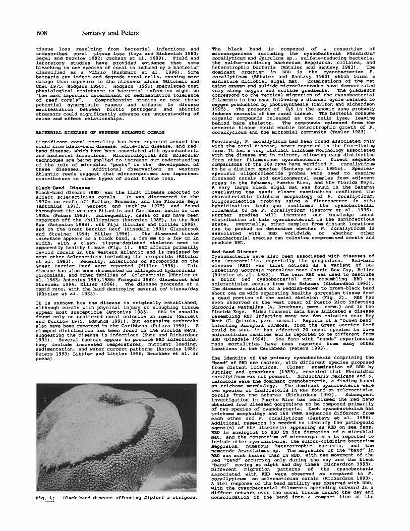
Red-Band DiseaseCyanobacteria have also been associated with diseases ofthe Octocorallia, especially the gorgonians. Red-banddisease (RBD) was first noticed as a variant of BBDinfecting Gorgonia ventalina near Carrie Bow Cay, Belize(Rutzler et al. 1983) .The term RBD was used to describe
a brick red cyanobacterial mat resembling BBD onscleractinian corals from the Bahamas (Richardson 1993) .The disease consists of a reddish-brown to brown-black bandabout one cm wide separating healthy gorgonian tissue froma dead portion of the axial skeleton (Fig. 2) .RBD hasbeen observed on the west coast of Puerto Rico infectingGorgonia ventalina (A. Bruckner, pers. corom.) and in theFlorida Keys. Video transect data have indicated a diseaseresembling RBD infecting many sea fan colonies near KeyWest (C. Quirolo, pers. corom.) .Reports of a "brown band"infecting Acropora formosa, from the Great Barrier Reefcould be RBD, it has affected 20 coral species in fivescleractinian families and is reported to be different fromBBD (Dinsdale 1994) .Sea fans with "bands" experiencingmass mortalities have been reported from many otherlocations in the Caribbean (Peters 1993) .
The identity of the primary cyanobacteria comprising the"band" of RBD are unclear, with different species proposedfrom distant locations. Closer examination of RBD byRutzler and coworkers (1983), revealed that Phormidiumcorallyticum was not present. Schizothrix mexicana and S.calcicola were the dominant cyanobacteria, a finding basedon trichome morphology. The dominant cyanobacteria weretwo species of Oscillatoria in RBD found on scleractiniancorals from the Bahamas (Richardson 1993) .Subsequentinvestigation in Puerto Rico has confirmed the red bandobtained from diseased gorgonians to be composed primarilyof two species of cyanobacteria. Each cyanobacterium hastrichome morphology and 165 rRNA sequences different fromeach other and P. corallyticum (Santavy et al. 1996) .Additional research is needed to identify the pathogenicagent(s) of the disease(s) appearing as RBD on sea fans.RBD is analogous to BBD in its formation of a microbialmat, and the consortium of microorganisms is reported toinclude other cyanobacteria, the sulfur-oxidizing bacteriumBeggiatoa, numerous heterotrophic bacteria, and thenematode Araeolaimus sp. The migration of the "band" inBBD was much faster than in RBD, with the movement of thered "band" occurring only during the day and the black"band" moving at night and day times (Richardson 1993) .Different migration patterns of the cyanobacteriaassociated with RBD were observed as compared to P.corallyticum on scleractinian corals (Richardson 1993) .A diel response of the band motility was observed with RBD,with the cyanobacterial filaments spreading outward in adiffuse network over the coral tissue during the day andconsolidation of the band into a compact line at theBlack-band disease affecting Diplori a strigosa.~
609Coral Diseases in Western Atlantic
interface between the live tissue and denuded coralskeleton at night (Richardson 1993) , RED probably createsa microbial microenvironment with oxygen and sulfurdynamics analogous to those in BBD (Carlton and Richardson1995) .
to 4-5 mm/day, much greater than the growth of regeneratingareas, which is approximately 0.3mm/day, and the growth ofrecruits at approximately 0.1-0.2 mmlday (Davis et al.1986) .
The etiology of WBD is uncertain, with ambiguous reportsconfusing our present understanding. Aggregates ofbacteria were reported in acroporid colonies affected withWBD from St. Croix, Bonaire, and the Netherland Antilles(Peters et al. 1983) .Bacterial aggregates were found alsoin apparently healthy colonies at St. Croix, but withinfive years up to 95 percent of the Acorpora palmatacolonies had died (Peters 1984). Bacterial aggregates havebeen found in acroporids without signs of WBD from otherwestern Atlantic locations (Peters, unpub. observ.), butthe role of the bacteria in the development of the diseaseis still unclear. Although the number of bacterialaggregates in grossly diseased and healthy coloniesoverlapped, the counts of bacterial aggregates per area oftissue were significantly higher ~n the diseased coloniesfrom St. Croix (Peters 1984) .Diseased colonies from theIndo-Pacific region and the Red Sea have not been examinedhistologically; therefore, it is unclear whether thebacterial aggregates are present.
~ Gorgonia ventalina affected with red-band disease,note that the axial skeleton remains devoid of purpletissue.
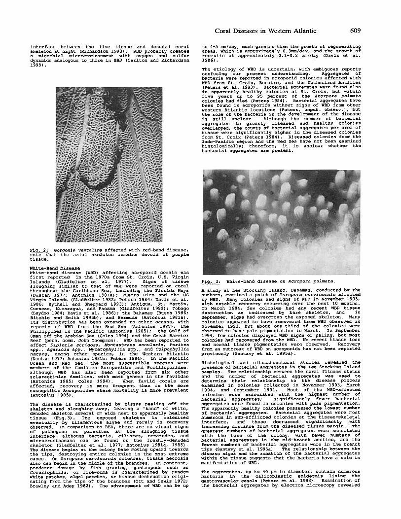
White-Band DiseaseWhite-band disease (WBD) affecting acroporid corals wasfirst reported in the 1970s from St.Croix, U.S. VirginIslands (Gladfelter et al. 1977) .Signs of tissuesloughing similar to that of WBD were reported on coralthroughout the Caribbean Sea, including the Florida Keys(Dustan 1977; Antonius 1981a); puerto Rico and the US
Virgin Islands (Gladfelter 1982; Peters 1984; Davis et al.1986; Bythell and Sheppard 1993); Antigua, St. Martin,Curacao, Nicaragua, and Panama (Gladfelter 1982); Tobago(Laydoo 1984; Davis et al. 1986); the Bahamas (Busch 1986;Ritchie and Smith 1995b); and Bermuda (Antonius 1981a) .Its distribution has been extended to other oceans, withreports of WBD from the Red Sea (Antonius 1988); thePhilippines in the Pacific (Antonius 1985); the Gulf ofoman off the Arabian Sea (Coles 1994) and the Great BarrierReef (pers. comm. John Thompson) .WBD has been reported toaffect Diploria strigosa, Montastraea annularis, Poritesspp., Agaricia spp., Mycetophyllia spp., and Colpophyllianatans, among other species, in the Western Atlantic(Dustan 1977; Antonius 1981b; Peters 1984). In the Pacific
Ocean and Red Sea, the most susceptible species weremembers of the families Acroporidae and Pocilloporidae,although WBD has also been reported from six otherscleractinian families, with most genera in the Faviidae(Antonius 1985; Coles 1994) .When faviid corals areaffected, recovery is more frequent than in the moresusceptible Acroporidae where mortality is more prevalent(Antonius 1985).
The disease, is characterized by tissue peeling off theskeleton and sloughing away, leaving a "band" of white,denuded skeleton several cm wide next to apparently healthytissue (Fig.3) .The exposed skeleton is colonizedeventually by filamentous algae and rarely is recoveryobserved. In comparison to BBD, there are no visual signsof pathogens or parasites at the sloughing tissueinterface, although bacteria, ciliates, nematodes, andmicrocrustaceans can be found on the freshly-denudedskeleton (Gladfelter et al. 1977; Antonius 1981a, 1985) .The disease begins at the colony base moving upward towardsthe tips, destroying entire colonies in the most extremecases. On Acropora cervicornis colonies, tissue necrosisalso can begin in the middle of the branches. In contrast,predator damage by fish grazing, gastropods such asCorallophilia, or fireworms is characterized by randomwhite patches, algal patches, or tissue destruction origi-nating from the tips of the branches (Ott and Lewis 1972;Brawley and Adey 1982) .The advancement of WBD can be up
~: White-band disease on Acropora palmata.
A study at Lee Stocking Island, Bahamas, conducted by theauthors, examined a patch of Acropora cervicornis affectedby WBD. Many colonies had signs of WBD in November 1993,with notable recovery occurring over the next 10 months.In March 1994, few colonies had any recent WBD tissuedestruction as indicated by bare skeleton, and inSeptember, algae had overgrown the exposed skeleton. Manycolonies appeared to have recovered from WBD observed inNovember 1993, but about one-third of the colonies wereobserved to have pale pigmentation in March. In September1994, few colonies displayed WBD signs or paling, but mostcolonies had recovered from the WBD. No recent tissue lossand normal tissue pigmentation were observed. Recoveryfrom an outbreak of WBD on acroporids has not been reportedpreviously (Santavy et al. 1995a) .
Histological and ultrastructural studies revealed thepresence of bacterial aggregates in the Lee Stocking Islandsamples. The relationship between the coral fitness statusand the number of bacterial aggregates was used todetermine their relationship to the disease processexamined in colonies collected in November 1993, March1994, and September 1994. Most of the WBD-affectedcolonies were associated with the highest number ofbacterial aggregates; significantly fewer bacterialaggregates were found in colonies with pale pigmentation.The apparently healthy colonies possessed the lowest numberof bacterial aggregates. Bacterial aggregates were mostnumerous in WBD-afflicted colonies at the tissue-skeletalinterface, and these decreased significantly withincreasing distance from the diseased tissue margin. Thegreatest numbers of bacterial aggregates were associatedwith the base of the colony, with fewer numbers ofbacterial aggregates in the mid-branch section, and thefewest numbers of bacterial aggregates were in the branchtips (Santavy et al. 1995a) .The relationship between thedisease signs and the zonation of the bacterial aggregateswithin the tissue suggests that the bacteria have a role inmanifestation of WBD.
The aggregates, up to 40 ~ in diameter, contain numerousbacteria in the calicoblastic epidermis lining thegastrovascular canals (Peters et al. 1983). Examination ofthe bacterial aggregates by electron microscopy revealed
610 Santavy and Peters
crystalline-like material in the gastric cavity.Examination of the tissue with electron microscopy andadditional planned studies will also help to determine ifbacteria playa major role in this deposition and itsrelationship to the disease state.
FACTORS TO BE CONSIDERED IN DISEASE STUDIES
Our understanding of how changes in environmental condit-ions can influence coral diseases is limited, and thecausal agents currently are undescribed for most coraldiseases. Many times similar gross signs of coralabnormalities are classified as the same disease by thosemaking observations in the field. After histopathologicalevaluation of these diseased corals, it is apparent thatthere are different pathologies associated with the samegross signs. Therefore, many different histologicalconditions are being classified into few describeddiseases. Consequently, many of the abnormalities are notadequately characterized and cannot be distinguished byuntrained observers. Closer observation of coral diseasesindicate that a single disease state might actually becaused by multiple biotic or abiotic factors: the unhealthycondition of a coral can be manifested in a limited numberof ways. Subtle variations in gross appearance and speciesafflicted by particular disease signs are indicative ofdifferent diseases with different etiologies (e.g.,WBD vs.stress-related necrosis vs. white plague or temperature-related bleaching vs. UV-related bleaching vs. micropara-sites) (Brown 1990: Peters 1993) .
that each bacterial aggregate was separated from theadjacent coral cells by a bilayered membrane of uncertainorigin (Santavy et al. 1995a) .The bacterium containedwithin the aggregates appeared as bundles of filaments,possessed a typical Gram-negative wall, and appeared to beeither unicellular in the state of division ormulticellular as evidenced by the septal formation betweenelongated cells (Santavy et al. 1995a) .
An enigma that remains to be resolved is the inconsistencyof bacterial aggregates associated with Acroporacervicornis and A. palmata with gross signs of WED.Bacterial aggregates were not found in all corals withdegenerating tissue from other locations examined by theauthors. Bacterial aggregates were found in tissues of A.cervicornis affected with WED from Rainbow Gardens Reef inthe vicinity of Lee Stocking Island, Bahamas. But WED-affected A. cervicornis did not contain any bacterialaggregates at the proximal reef location, Norman's PondReef, and at South Carysfort Reef in the Florida Keys(Kozlowski 1996). Furthermore, histopathological analysisof A. palmata from Puerto Rico with putative WED did notcontain these bacterial aggregates, but did contain otherpotentially pathogenic organisms and tissue deterioration.The histopathology of the corals from Puerto Rico appearedvery different from that observed in A. cervicornis fromRainbow Gardens (R. Bruckner and D.L. Santavy, unpublisheddata) .The absence of any obvious infectious agent orbacterial aggregates in these corals with similar grosscharacteristics of WED, suggests that either visible signsof this disease do not represent the same disease, or thatWED is caused primarily by other factors.
Colonies could possess similar external or gross diseasesigns but might be afflicted with different pathogens orstressed by abiotic factors. Perhaps this explanation isapplicable to WED, a term that generally has been appliedto all species of corals in which tissue is sloughing fromthe skeleton. "White plague" was transmitted by inoculatingmaterial from the affected tissue interface onto thesurface of another coral that had been intentionallyinjured (Dustan 1977). Only material obtained fromdiseased Mycetophyllia ferox, M. lamarkiana, andColpophyllia natans was able ~o produce infection inhealthy corals of the same species. Bacteria were observedin the inoculant; however, no microbiological studies wereperformed to isolate and characterize potential bacterialpathogens. White plague obtained from the previous speciesdid not affect M. annularis, Porites astreoides, orStephanocoenia michelini. It was hypothesized that theappearance of similar signs of disease in the latterspecies might be due to another agent, or those specieswere resistant to the inoculum (Dustan 1977). In contrast,xenografts of affected and healthy coral species did nottransfer WED to healthy colonies (Antonius 1981a, b) .Thus, the term "stress-related necrosis" was proposed todescribe the condition in which degenerative changes incell structure are observed in the absence of obviouspathogens, after histopathological observations (Peters1984) .Ritchie and Smith (1995a,b) have examined bacteriafrom the coral surface mucus layer in another WED-likedisease affecting A. cervicornis from San Salvador,Bahamas .
During the summer of 1995, a new disease emerged in theFlorida Keys. It began affecting only Dichocoeniastokesii, but by the end of the season, it was reportedto affect over a dozen different coral species (Zorpette1995). Superficially, signs of tissue loss were similar toWED described from other areas, but the rate of coraltissue loss was greater. Rapid tissue necrosis wasobserved on massive, branching, and encrusting corals, withsloughing tissue progressing at a rate of over a cm perday, much faster than any other reports of WED, whitedeath, or white plague. The disease destroyed smallcolonies in a matter of weeks. The extremely rapid rate oftissue loss and ability to cause mortalities in manyspecies of corals make this condition of particularconcern, not only for the Keys, but also for the rest ofthe tropical western Atlantic. The etiology of the diseaseis under investigation.
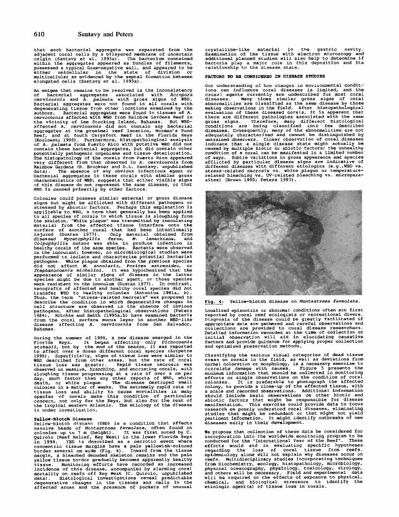
Yellov-Blotch DiseaseYellow-blotch disease (YBD) is a condition that affectsmassive heads of Montastraea faveolata, often found oncolonies up to 7 m (height) .It was first noted by C.Quirolo (Reef Relief, Key West) in the lower Florida Keysin 1994. YBD is described as a necrotic event whereconcentric tissue margins have a pale yellowish-coloredborder several cm wide (Fig. 4). Inward from the tissuemargin, a bleached denuded skeleton remains and the paleyellow tissue border gradually becomes apparently healthytissue. Monitoring efforts have recorded an increasedincidence of this disease, accompanied by alarming coralmortality on reefs off Key West (C. Quirolo, unpublisheddata) .Histological investigations reveal predictabledegenerative changes in the tissues and cells in theaffected areas and the presence of pockets of unusual
~: Yellow-b~otch disease on Montastraea faveolata.
Localized epizootics or abnormal conditions often are firstreported by coral reef ecologists or recreational divers.Diagnosis of the disease could be greatly facilitated ifappropriate data are gathered and careful observations andcollections are provided to coral disease researchers.Detailed information recorded at the time of collection orinitial observation will aid in elucidating causativefactors and provide guidance for applying proper collectionand optimized preservation methods.
Classifying the various visual categories of dead tissueareas on corals in the field, as well as deviations fromnormal coloration or morphology, is a necessary exercise tocorrelate damage with causes. Figure 5 presents theminimum information that should be collected in monitoringefforts or single observations on the condition of coralcolonies. It is preferable to photograph the affectedcolony, to provide a close-up of the affected tissue, witha scale and recorded observations. Additional informationshould include basic observations on other biotic andabiotic factors that might be responsible for diseasemanifestation. This exercise could provide data to focusresearch on poorly understood coral diseases, eliminatingstudies that might be redundant or that might not yieldpertinent information. It might identify outbreaks of newdiseases early in their development.
We propose that collection of these data be considered forincorporation into the worldwide monitoring program to beconducted for the "International Year of the Reef". Theseefforts would aid in evaluating specific hypothesesregarding the loss of coral tissue from reefs.Epidemiology alone will not explain why diseases occur onreefs. Multidisciplinary studies incorporating techniquesfrom biochemistry, ecology, histopathology, microbiology,physical oceanography, physiology, toxicology, virology,and others will be necessary. Field and experimental datawill be required on the effects of exposure to physical,chemical, and biological stressors to identify theetiologic agent(s) of tissue loss in corals.
612 Santavy and Peters
Glazebrook, JS and Streiner, HM (1994) Pathologyassociated with tumors and black-band disease in coralsfrom Agincourt Reef. Joint Conf on Sci, Mgt andSustainability of Mar Hab GBR
Glynn, PW, Szmant, AM, Corcoran, EF and Cofer-Shabica, SV(1989) Condition of coral reef cnidarians from thenorthern Florida reef tract: pesticides, heavymetals, and histopathological examination. Mar Poll Bull20:568-576
Glynn, PW, and D'Croz, L. (1990) Experimental evidencefor high temperature stress as the cause of El Hino-coincident coral mortality. Coral reefs 8:181-191
Hallock, P, Muller-Karger, FE and Halas, JC (1993) Coralreef decline: Anthropogenic nutrients and thedegradation of Western Atlantic and Caribbean coralreefs. Hat Geog Res Expl 9:358-378
Hodgson, G (1990) Tetracycline reduces sedimentationdamage to corals. Mar Biol 104:493-496
Hughes, TP (1994) Catastrophes, phase shifts, and large-scale degradation of a Caribbean coral reef. Science265:1547-1551
Hunter, CL and Evans, CE (1995) Coral reefs in KaneoheBay, Hawaii: Two centuries of western influence and twodecades of data. Bull Mar Sci 57:501-515
Jackson, JBC, Cubit, JD, Keller, BD, Batista, V, Burns,Caffey, K, Caldwell, RL, Garrity, SD, Getter, CD,Gonzales, C, Guzman, HN, ~aufmann, KW, Knap, AH,Levings, SC, Marshall, MJ, Steger, R, Thompson, RC andWeil, E (1989) Ecological effects of a major oil spillon Panamanian coastal marine communities. Science243:37-44
Knap, AH, Wyers, SC, Dodge, RE, Sleeter, TD, Frith, HR,Smith, SR and Cook, CB (1985) The effects ofchemically and physically dispersed oil on the braincoral Diploria strigosa (Dana)-a summary review. Proc1985 Oil Spill Conf:547-551
Kozlowski, J (1996) Investigations of the occurrence anddistribution of bacterial aggregates in Acroporacervicornis from reefs in the Bahamas and the FloridaKeys. Nat Res. Princess Anne, Univ MD 100 pp
Kushmaro, A, Loya, Y, Fine, M and Rosenberg, E (1996)Bacterial infection and coral bleaching. Nature380:396
Kuta, KG and Richardson, LL (1994) Distribution andfrequepcy patterns of black-band disease in the NorthernFlorida Keys. Bull Mar Sci 54:1078
Laydoo, R (1984) Inference of a "white-band" epidemicin the elkhorn coral, Acropora palmata, populations inTobago, WI. Proc, Assoc Isl Mar Labs Carib 18:12
Lessios, HA, Glynn, PW and Robertson, DR (1983) Massmortalities of coral reef organisms. Science 182:715
Littler, MM and Littler, DS (1996) Black-band diseasein the South Pacific. Coral Reefs 15:20
Loya, Y and Rinkevich, B (1980) Effects of oilpollution on coral reef communities. Mar Ecol Progr Ser3:167-180
McIvor, CC, Ley, JA and Bjork, RD (1994) Changes infreshwater inflow from the Everglades to Florida Bayincluding effects on biota and biotic processes: Areview. Delray Beach, FL, St. Lucie Press
Miller, I (1996) Black-band disease on the Great BarrierReef. Coral Reefs 15:67
Mitchell, Rand Chet, I (1975) Bacterial attack ofcorals in polluted seawater. Microbial Ecol 2:227-233
Ott, B and Lewis, JB (1972) The importance of thegastropod Coralliophila abbreviata (Lamark) and thepolychaete Hermodice carunculata (Pallas) as coral reefpredators. Can Zool 50:1651-1656
Pascal, Hand Vacelet, E (1981) Bacterial utilizationof mucus on the coral reefs of Aqaba (Red Sea). Proc 4thInt Coral Reef Symp 1:669-677
Paul, JH, DeFlaun, MF and Jeffrey, WH (1986) Elevatedlevels of microbial activity in the coral surfacemicrolayer. Mar Ecol Progr Ser 33:29-40
Peters, EC (1984) A survey of cellular reactions toenvironmental stress and disease in Caribbeanscleractinian corals. Hel Meer 37:113-137
Peters, EC (1993) Diseases of other invertebrate phyla:Pori.fera, Cnidaria, Ctenophora, Annelida, Echinodermata.In: J.A. Couch and J.W. Fournie (ed) Pathobiology ofmarine and estuarine organisms CRC Press. Boca Raton,FL:388-444
Peters, EC, Oprandy, JJ and Yevich, PP (1983) possiblecausal agent of "white band disease" in CaribbeanAcroporid Corals. J Invert Path 41:394-396
Porter, JW and Meier, OW (1992) Quantification of lossand change in Floridian Reef Coral Populations. Am Zool32:625-640
Porter, JW, Meier, OW, Tougas, JI and Lewis, SK (1994)Modification of the South Florida hydroscape and itseffect on coral reef survival in the Florida Keys. BullEcol Soc Am Abst Sup 75:184
Richardson, LL (1993) Red band disease: Anewcyanobacterial infestation of corals. Am Acad Und Sciloth Ann Sci Div Symp:153-160
Ritchie, KB and Smith, GW (1995a) Carbon-sourceutilization patterns of coral-associated marineheterotrophs. J Mar Biotech 3:105-107
Ritchie, KB and Smith, GW (1995b) Preferential carbonutilization by surface bacterial communities from watermass, normal, and white-band diseased Acroporacervicornis. J Mar Biotech 4:345-352
Rublee, PA, Lasker, HR, Gottfried, M and Roman, MR(1980) production and bacterial colonization of mucusfrom the soft coral Briareum asbestinum. Bull Mar Sci30:888-893
Rlitzler, K and Santavy, DL (1983) The black-banddisease of Atlantic reef corals I. Description of thecyanophyte pathogen. PSZNI Mar Ecol 4:301-319
Rlitzler, K, Santavy, DL and Antonius, A (1983) Theblack band disease of Atlantic reef corals III.Distribution, ecology, and development. PSZNI Mar Ecol4:301-319
Santavy, DL (1995) The diversity of microorganismsassociated with marine invertebrates and their roles inthe maintainenance of ecosystems. In: MicrobialDiversity and Ecosystem Function. Ed. by D Allsopp, RRColwell, and DL Hawksworth. CAB International,Wallingford, UK, pp 211-230
Santavy, DL, Jeffrey, WH, Snyder, RA, Campbell, J,Malouin, P and Cole, L (1992) Microbial communitydynamics in the mucus of healthy and stressed corals.Bull Mar Sci 54:1077-1078
Santavy, DL, Peters, EC, Kozlowski, J and Wilkinson, SS(1995a) Characterization of the bacterium suspected in
the incidence of white-band disease. 95th Gen Mtg ASM,Abst N-l:332
Santavy, DL, Schmidt, TM and Wilkinson, SS (1995b)Phylogeny of Phormidium corallyticum (Rlitzler & Santavy,1983) using 16S rRNA. Proc 7th All Invest Mtg for EPA'sEnv Rel of Biotec Prod Risk Assess Prog
Santavy, DL, Schmidt, T, Wilkinson, SS, Buckley, DH andBruckner, AW (1996) The phylogency and alliance ofcyanobacteria affiliated with two band diseases incorals from the Western Atlantic. 96th Gen Mtg ASM:325
Sebens, KP (1994) Biodiversity of coral reefs: what arewe losing and why. Am Zool 34:115-133
Segel, LA and Ducklow, HW (1982) A theoreticalinvestigation into the influence of sublethal stresseson coral-bacterial ecosystem dynamics. Bull Mar Sci32:919-935
Shinn, EA (1993) Geology and human activity in theFlorida Keys. Washington DC, USGS/US Dept Interior
Sindermann, CJ (1995) Ocean pollution effects on livingresources and humans. Boca Raton, FL, CRC Press
Szmant, AM and Forrester, A (1996) Water column andsediment nitrogen and phosphorus distribution patternsin the Florida Keys, USA. Coral Reefs 15:21-42
Taylor, DL (1983) The black-band disease of Atlanticreef corals. III. Isolation, cultivation and growth ofPhormidium corallyticum. PSZNI Mar Ecol 4:321-328
Tomascik, T and Sander, F (1987) Effects ofeutrophication on reef-building corals. II. Structureof scleractinian coral communities on £ringing reefs,Barbados, West Indies. Mar Biol 94:53-75
Vacelet, E and Thomassin, BA (1991) Microbialutilization of coral mucus in long term in situincubation over a coral reef. Hydrobio1211:19-32
Williams, EH and Bunkley-Williams, L (1990) The world-wide coral reef bleaching cycle and related sources ofcoral mortality. Atoll Res Bull 335:1-71
Zorpette, G (1995) More coral trouble. Sci Amer 273:36-37