Embed Size (px)
Citation preview
1
2
Proceeding Book
PERTEMUAN ILMIAH REGIONAL PERHIMPUNAN DOKTER SPESIALIS PATOLOGI INDONESIA. JOGLOSEMAR-PURWOKERTO
Gastrointestinal Pathology and Recent Updates On Neuroendocrine Tumors: From Morphology To Clinical Management in Personalized Medicine Era
Royal Ambarukmo Hotel Yogyakarta, 5 April 2018
2018
3
Proceeding Book
PERTEMUAN ILMIAH REGIONAL PERHIMPUNAN DOKTER SPESIALIS PATOLOGI INDONESIA. JOGLOSEMAR-PURWOKERTO
Gastrointestinal Pathology and Recent Updates On Neuroendocrine Tumors: From Morphology To Clinical Management in Personalized Medicine Era
Royal Ambarukmo Hotel Yogyakarta, 5 April 2018
Editor
Dr. dr. Irianiwati, Sp.PA(K)
dr. Didik Setyo Heriyanto, Sp.PA, Ph.D
dr. Paranita Ferronika, Sp.PA
dr. Intannuary Paringga
dr. Fitria
dr. Deflen Jumatul Sastri
dr. Muchamad Ridotu Solichin
4
DAFTAR ISI
Halaman Judul ..................................................................................................................................... 1 Daftar isi ............................................................................................................................................................ 3 Kata pengantar ................................................................................................................................................ 6 Program................................................................................................................................................ 7 Abstrak Pembicara Pertenuan Ilmiah Regional 2018 Yogyakarta P1. ETHICS IN ANATOMIC PATHOLOGY SERVICE (Indra Wijaya) .............................................. 9 P2. CLINICAL ASPECT OF NEUROENDOCRINE TUMORS OF THE GASTROINTESTINAL
TRACT (Neneng Ratnasari) ....................................................................................................... 10 P3. CHALLENGE OF SURGICAL ASPECT IN NEUROENDOCRINE TUMORS OF THE
LOWER GASTROINTESTINAL TRACT (Yuda Handaya) ......................................................... 11 P4. RECENT UPDATE ON GASTROINTESTINAL TRACT NEUROENDOCRINE
NEOPLASM (Ahmad Ghozali) .................................................................................................... 12 P5. CASE SERIES OF NEUROENDOCRINE TUMORS OF THE GASTROINTESTINAL
TRACT (Susanna Hutajulu) ........................................................................................................ 13 P6. ALL-RAS AND BRAF MUTATION IN METASTASIS COLORECTAL CARCINOMA:
WHAT THE CLINICIANS NEED AND THE PATHOLOGISTS PERFORM (Didik Setyo Heriyanto) ................................................................................................................................... 14
P7. DIAGNOSTIC PITFALLS IN GASTROINTESTINAL TUMORS (Antony Chan) ......................... 15 P8. WORKSHOP ON GASTROENTEROPANCREATIC NEUROENDOCRINE TUMORS
(Antony Chan) ............................................................................................................................. 16 Abstrak Peserta Pertenuan Ilmiah Regional 2018 Yogyakarta R1. ASSOCIATION BETWEEN AGE AND SEX WITH HISTOLOGICAL TYPE OF
CARCINOMA THYROID IN MARGONO SOEKARJO HOSPITAL PURWOKERTO ................. 17 R2. BASAL CELL CARCINOMA IN YOGYAKRTA, INDONESIA: A CLINICAL AND
PATHOLOGICAL STUDY ........................................................................................................... 18 R3. CASE SERIES: INCIDENT NEUROENDOCRINE TUMOUR IN Dr.KARIADI GENERAL
HOSPITAL .................................................................................................................................. 19 R4. CLINICOPATHOLOGICAL PROFILE OF WILMS TUMOR IN DR. SARDDJITO
GENERAL HOSPITAL, YOGYAKARTA, INDONESIA ............................................................... 20 R5. CORRELATION BETWEEN GENDER, AGE AND TOPOGRAPHY WITH
HISTOLOGICAL TYPE OF COLORECTAL CANCER (A cross sectional study in Margono Soekarjo Hospital Purwokerto) .................................................................................... 21
R6. DIAGNOSTIC VALUE OF FINE NEEDLE ASPIRATION BIOPSY IN BREAST LESIONS AT dr. SOERADJI TIRTONEGORO HOSPITAL, KLATEN ........................................................ 22
R7. HISTOPATHOLOGICAL PATTERNS OF GALL BLADDER DISEASES WITH SPECIAL REFERENCE TO INCIDENTAL CASES IN BOYOLALI PANDAN ARANG GENERAL HOSPITAL .................................................................................................................................. 23
R8. MOLECULAR TYPES ON VARIOUS CHARACTERISTICS OF BREAST CARSINOMA PATIENTS AT GENERAL HOSPITAL CENTER OF DR. KARIADI IN 2015-2017 .................... 24
C1. APPENDICEAL NEUROENDOCRIN TUMOR GRADE I : A CASE REPORT ........................... 25 C2. BENIGN CHONDROID SYRINGOMA OF THE ORBIT: A RARE CAUSE OF
EXOPHTHALMOS ...................................................................................................................... 26 C3. CASE REPORT A RARE CASE OF MEDULLARY THYROID CARCINOMA ........................... 27 C4. CASE REPORT A RARE CASE OF NEUROENDOKRINE TUMOR IN DISTAL
PANKREAS ................................................................................................................................ 28 C5. CHILDHOOD SIGNET RING CELL CARCINOMA OF THE COLON : A RARE CASE
REPORT ..................................................................................................................................... 29 C6. COLON ADENOCARCINOMA, CRIBRIFORM COMEDO-TYPE .............................................. 30 C7. CELLULAR CONGENITAL MESOBLASTIC NEPHROMA: A RARE CASE OF
MESENCHYMAL RENAL TUMOR ............................................................................................. 31
C8. GASTRIC NEUROENDOCRINE TUMOR: A RARE CASE REPORT ....................................... 32 C9. INFLAMMATORY MYOFIBROBLASTIC TUMOR COLON: A RARE CASE REPORT ............. 33 C10. LACRIMAL GLAND ADENOID CYSTIC CARCINOMA .............................................................. 34 C11. LIPOBLASTOMA: A RARE CASE REPORT .............................................................................. 35
5
C12. METAPLASTIC BREAST CARCINOMA WITH CARCINOSARCOMA DIFFERENTIATION: A RARE CASE REPORT WITH REVIEW OF LITERATURE .................. 36
C13. MALIGNANT HEMANGIOPERICYTOMA (HPC) ON REGION ANTEBRACHII DEXTRA ........ 37 C14. METASTASIS OF HEPATOCELLULAR CARCINOMA IN MANDIBLE, A VERY RARE
CASE REPORT .......................................................................................................................... 38 C15. METASTATIC SIGNET RING CELL CARCINOMA WITH NEGATIVE RAS AND RAF
PROFILE IN 10-YEAR-OLD BOY: POSSIBILITY OF ANTI-EGFR USE ................................... 39 C16. NEUROENDOCRINE CARCINOMA ARISING IN GLUTEUS ON FOUR YEAR OLD BOY ...... 40 C17. POSSIBLE PROGRESSION OF OLIGODENDROGLIOMA TO ANAPLASTIC
OLIGODENDROGLIOMA: A CASE REPORT ........................................................................... 41 C18. PRIMARY MALIGNANT MELANOMA OF THE BREAST .......................................................... 42 C19. PRIMARY CUTANEOUS BLASTOMYCOSIS: HISTOPATHOLOGY AND
HISTOCHEMISTRY APPROACH .............................................................................................. 43 C20. RECIDIVE OF PAPILLARY THYROID CARCINOMA WITH BRAF STATUS MUTATION
IN JUVENILE BOY ...................................................................................................................... 44 C21. RECTAL MIXED ADENONEUROENDOCRINE CARCINOMA: A RARE CASE REPORT ....... 45 C22. RECTOSIGMOID ENDOMETRIOSIS CLINICALLY MIMICKING COLORECTAL
MALIGNANT TUMOUR: A RARE CASE REPORT .................................................................... 46 C23. ROLE & PITFALL OF IMMUNOHISTOCHEMISTRY IN DIAGNOSING SMALL ROUND
BLUE CELL TUMOR: LESSON LEARNED FROM DIFFICULT CASE OF EXTRASKELETAL EWING SARCOMA PREVIOUSLY DIAGNOSED AS T CELL LYMPHOMA ............................................................................................................................... 47
C24. SMALL CELL NEUROENDOCRINE CARCINOMA OF LARYNX A CASE REPORT ............... 48 C25. SPINDE TYPE NEUROENDOCRINE OF MULTIPLE METASTATIC RECTAL
CARCINOMA: A RARE CASE REPORT.................................................................................... 49 C26. THE ROLE OF RAPID ON-SITE EVALUATION (ROSE) WITH
CYTOHISTOPATHOLOGICAL CORRELATION IN DIAGNOSIS OF CHOLANGIOCARCINOMA: AN EFFORT TO IMPROVE DIAGNOSIS ACCURACY AND ADEQUACY ................................................................................................................................ 50
C27. THE USE OF FINE NEEDLE ASPIRATION CYTOLOGY SPECIMEN FOR DETERMINING PROGNOSIS IN THE CASE OF HEPATOBLASTOMA: AN APPLICATION OF microRNA PROFILING (miR-122, miR-195, miR-210, miR-214, and miR-221) AS A BIOMARKER ..................................................................................................... 51
C28. UROTHELIAL CARCINOMA WITH GLANDULAR DIFFERENTIATION: A HISTOPATHOLOGICAL CHALLANGE OF UNUSUAL VARIANT ............................................. 52
C29. VARICELLA MIMICKING BULLOUS PEMPHIGOID IN IMMUNOCOMPROMISED PATIENT ..................................................................................................................................... 53
C30. METASTASIS DETECTION OF RECTAL NEUROENDOCRINE CARCINOMA IN THE BONE .......................................................................................................................................... 54
Abstrak pembicara Seminar Nasional Ikatan Teknisi Patologi Anatomi (ITPAI) I1. ASPEK HUKUM DAN ETIKA PROFESI AHLI TEKNOLOGI LABORATORIUM MEDIK
DALAM TATA LAKSANA PEMERIKSAAN SAMPEL (Yanuar Amin) ........................................ 57 I2. PERAN DAN TANGGUNG JAWAB ATLM DI LABORATORIUM PATOLOGI ANATOMI
(Bowo Yuniarto) .......................................................................................................................... 58 I3. PENANGANAN SAMPEL DAN PANEL PEMERIKSAAN SPESIMEN BIOPSI GINJAL
(Hanggoro Tri Rinonce) .............................................................................................................. 59 I4. PENTINGNYA ASPEK PRA ANALITIK PADA IMUNOHISTOKIMIA (Agustina Supriyanti) ...... 60 I5. PEMBUATAN CELL BLOCK PADA SEDIAAN SITOLOGI YANG BAIK DAN BENAR DI
ERA BIOMOLEKULER (Didik Setyo Heriyanto) ......................................................................... 61 Abstrak Peserta Seminar Nasional Ikatan Teknisi Patologi Anatomi (ITPAI) S1. FINE NEEDLE ASPIRATION BIOPSY (FNAB) DIAGNOSTIC TEST WITH
HISTOPATHOLOGY CHECK AS GOLD STANDARD IN BREAST CANCER PATIENT IN RSUD DR. SOEHADI PRIJONEGORO SRAGEN ..................................................................... 62
S2. GAMBARAN MIKROSKOPIS CARCINOMA MAMMAE YANG DIFIKSASI DENGAN NETRAL BUFFER FORMALIN 10% DAN ALKOHOL 70% PADA PEWARNAAN HEMATOXYLIN - EOSIN ............................................................................................................ 63

6
S3. PENGGUNAAN BLOK SEL DARI SPESIMEN CAIRAN PLEURA PADA KASUS KARSINOMA PARU BUKAN SEL KECIL JENIS ADENOKARSINOMA: OPTIMASI PULASAN IMUNOHISTOKIMIA PDL-1 ...................................................................................... 64
S4. PEWARNAAN HISTOKIMIA PADA JARINGAN BIOPSI GINJAL .............................................. 65 S5. PROSEDUR PRA ANALITIK PEMERIKSAAN K/N RAS PADA KASUS KANKER
KOLOREKTAL ............................................................................................................................ 66
7
KATA PENGANTAR
Kejadian tumor saluran pencernaan, terutama kanker kolorektal, semakin meningkat
dari tahun ke tahun. Kanker kolorektal merupakan kanker terbanyak kedua pada pria dan
terbanyak ketiga pada wanita di Indonesia, dan cenderung meningkat pada kelompok usia
muda.
Perkembangan ilmu pengetahuan yang begitu pesat di era personalized medicine ini
menjadikan pendekatan multi disiplin dalam penanganan tumor saluran pencernaan suatu
keharusan. Dalam pendekatan multi disiplin tersebut, peran dokter ahli patologi anatomik
sangat penting. Diagnosis patologi sampai ke diagnosis molekuler sangat diperlukan sebagai
dasar penentuan terapi bagi pasien. Oleh karena itu, seorang dokter ahli patologi anatomik
dituntut untuk mempunyai pengetahuan dan ketrampilan mulai dari aspek morfologi,
molekuler, hingga menejemen klinik.
Dengan mengusung tema Gastrointestinal Pathology and Recent Updates on
Neuroendocrine Tumors: from Morphology to Clinical Management in Personalized Medicine
Era, pada Pertemuan Ilmiah Regional (PIR) Perhimpunan Dokter Spesialis Patologi Indonesia
(IAPI) Joglosemar-Purwokerto 2018 dibahas dengan tuntas patologi saluran cerna, mulai dari
aspek morfologi, molekuler, hingga manajemen klinik pasien-pasien dengan tumor di saluran
cerna, terutama tumor neuroendokrin. Para pembicara yang terdiri dari ahli bedah digestif,
penyakit dalam, hematologi dan onkologi medik, serta patologi anatomik mempresentasikan
perkembangan terkini patologi saluran cerna dari sudut pandang masing-masing bidang ilmu
pada sesi seminar. Untuk semakin memperkaya ilmu, diselenggarakan juga workshop khusus
dengan pembicara Anthony Chan dari Chinese University of Hong Kong. Selain itu, diadakan
juga sesi presentasi poster agar para peserta dapat berdiskusi dan bertukar pengetahuan satu
sama lain.
PIR IAPI Joglosemar-Purwokerto 2018 juga diselenggarakan bersamaan dengan
Seminar Nasional Ikatan Teknisi Patologi Anatomi Indonesia (ITPAI) yang bertema Peran
ATLM dalam Perkembangan Patologi dan Manajemen Sampel di Era Kedokteran Molekuler.
Seminar nasional tersebut bertujuan untuk meningkatkan pengetahuan dan keterampilan ahli
teknologi laboratorium medis (ATLM) dalam menangani spesimen pre-analitik dan menambah
pengetahuan tentang perkembangan teknik patologi di era kedokteran molekuler. Dengan
diselenggarakannya kedua acara tersebut secara bersamaan, diharapkan para dokter spesialis
patologi anatomik dapat berinteraksi dengan para ATLM yang merupakan mitra terdekat dalam
menghasilkan sediaan yang bermutu tinggi. Dengan interaksi dalam forum ilmiah tersebut,
diharapkan terjadi pertukaran pengetahuan dalam upaya peningkatan pelayanan patologi
anatomik.
Buku abstrak ini merangkum seluruh materi yang dibicarakan dan dibahas dalam kedua
acara tersebut, baik materi dari pembicara maupun materi poster dari para peserta. Semoga
buku ini bermanfaat dan membuahkan peningkatan derajat kesehatan masyarakat Indonesia.
Yogyakarta, 5 Mei 2018
Ketua Pelaksana
dr. Hanggoro Tri Rinonce, Sp.PA, Ph.D
8
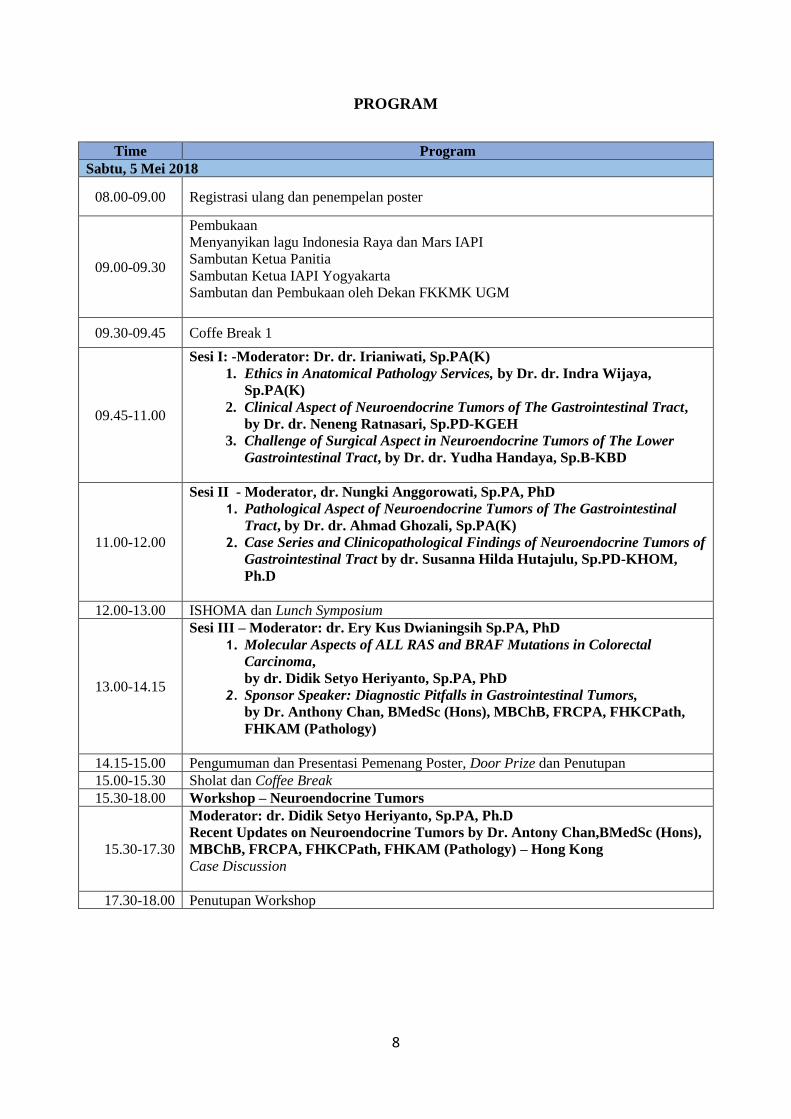
PROGRAM
Time Program
Sabtu, 5 Mei 2018
08.00-09.00 Registrasi ulang dan penempelan poster
09.00-09.30
Pembukaan
Menyanyikan lagu Indonesia Raya dan Mars IAPI
Sambutan Ketua Panitia
Sambutan Ketua IAPI Yogyakarta
Sambutan dan Pembukaan oleh Dekan FKKMK UGM
09.30-09.45 Coffe Break 1
09.45-11.00
Sesi I: -Moderator: Dr. dr. Irianiwati, Sp.PA(K)
1. Ethics in Anatomical Pathology Services, by Dr. dr. Indra Wijaya,
Sp.PA(K)
2. Clinical Aspect of Neuroendocrine Tumors of The Gastrointestinal Tract,
by Dr. dr. Neneng Ratnasari, Sp.PD-KGEH
3. Challenge of Surgical Aspect in Neuroendocrine Tumors of The Lower
Gastrointestinal Tract, by Dr. dr. Yudha Handaya, Sp.B-KBD
11.00-12.00
Sesi II - Moderator, dr. Nungki Anggorowati, Sp.PA, PhD
1. Pathological Aspect of Neuroendocrine Tumors of The Gastrointestinal
Tract, by Dr. dr. Ahmad Ghozali, Sp.PA(K)
2. Case Series and Clinicopathological Findings of Neuroendocrine Tumors of
Gastrointestinal Tract by dr. Susanna Hilda Hutajulu, Sp.PD-KHOM,
Ph.D
12.00-13.00 ISHOMA dan Lunch Symposium
13.00-14.15
Sesi III – Moderator: dr. Ery Kus Dwianingsih Sp.PA, PhD
1. Molecular Aspects of ALL RAS and BRAF Mutations in Colorectal
Carcinoma,
by dr. Didik Setyo Heriyanto, Sp.PA, PhD
2. Sponsor Speaker: Diagnostic Pitfalls in Gastrointestinal Tumors,
by Dr. Anthony Chan, BMedSc (Hons), MBChB, FRCPA, FHKCPath,
FHKAM (Pathology)
14.15-15.00 Pengumuman dan Presentasi Pemenang Poster, Door Prize dan Penutupan
15.00-15.30 Sholat dan Coffee Break
15.30-18.00 Workshop – Neuroendocrine Tumors
15.30-17.30
Moderator: dr. Didik Setyo Heriyanto, Sp.PA, Ph.D
Recent Updates on Neuroendocrine Tumors by Dr. Antony Chan,BMedSc (Hons),
MBChB, FRCPA, FHKCPath, FHKAM (Pathology) – Hong Kong
Case Discussion
17.30-18.00 Penutupan Workshop
9
P1
ETIKA PELAYANAN PATOLOGI ANATOMIK (Ethics in Anatomic
Pathology Service)
Indra Wijaya (IAPI Cabang Semarang)
Departemen Patologi Anatomi Fakultas Kedokteran Universitas Diponegoro
Abstrak
Pesatnya dinamika ilmu pengetahuan dan teknologi kedokteran global, perkembangan
kehendak masyarakat serta dinamika antisipasi perubahan sistem kesehatan nasional, termasuk
afirmasi kepada upaya pelayanan kesehatan masyarakat bersifat promotif dan preventif,
khususnya subsistem pembiayaan pelayanan kesehatan pada era Jaminan Kesehatan Nasional,
berpengaruh terhadap etika pelayanan dokter, khususnya dokter spesialis Patologi Anatomik.
Di Indonesia, perumusan norma dan penerapan nyata etika kedokteran kepada
perseorangan pasien/klien atau kepada komunitas/masyarakat dalam segala bentuk fasilitas
pelayanan kesehatan/kedokteran juga didasarkan atas azas-azas ideologi bangsa dan negara
Pancasila serta Undang-Undang Dasar 1945.
Seorang Dokter Spesialis Patologi Anatomik wajib selalu melakukan pengambilan
keputusan profesional secara independen dan mempertahankan perilaku profesional dalam
ukuran tertinggi, untuk itu perlu penyegaran pengetahuan kita tentang Kode Etik Patologi
Indonesia, yang mungkin sudah mulai pudar, seiring dengan bertambahnya usia.
Di samping pengetahuan kita tentang Kode Etik Patologi Indonesia, dalam pelayanan
Patologi Anatomik sehari-hari perlu pula kita segarkan pengetahuan tentang Standar Profesi
Dokter Spesialis Patologi Anatomik, agar dapat menyesuaikan diri dengan perkembangan ilmu
kedokteran, serta tetap mempertahankan mutu pelayanan Patologi Anatomik yang prima.
10
P2
Clinical Aspect of Neuroendocrine Tumors of the Gastrointestinal Tract
Neneng Ratnasari
Division of Gastroentero-Hepatology, Department of Internal Medicine, Faculty of Medicine, Universitas
Gadjah Mada/Dr Sardjito Hospital, Yogyakarta
Neuroendocrine Tumors (NETs) account for about 0.5% of all newly diagnosed
malignancies. The incidence is approximately 5.86/100,000 per year, with a female
preponderance of around 2.5:1. NETs manifest in the gastrointestinal tract mainly as carcinoid
and pancreatic islet-cell tumors. They comprise an interesting group of rare neoplasms that are
derived from neuroendocrine cells interspersed within the gastrointestinal system amd
throughout the body. There are some tumors that are classified as Nets: carcinoid, gastrinoma,
insulinoma, glucagonoma, somatostatinoma, vipomas and pancreatic polypeptideoma. The
variation of clinical appearance of NETs depended on the origine of the tumors, sized and
metastasis.
Carcinoid tumors are the most common neuroendocrine tumors. They arise from
neuroendocrine cells located primarily in the submucosa of the intestine but can also arise in
the main bronchi. Gastrinoma is growth from ulceration in GI tracts. Insulinomas are the most
common type of islet-cell tumor. Glucagonomas are rare alpha-cell tumors of the pancreas that
occur in people between 50 and 70 years old and mostly malignant, with metastases.
Somatostasin-secreting tumors (somatostatinoma) are the majority tumor that was found in the
pancreas or duodenum, generally malignant. Vipomas are the tumor in which mediated by a
hormone called vasoactive intestinal polypeptide (VIP) and other peptides secreted by
malignant islet-cell tumors in adults and by benign ganglio- neuroblastomas in children, are
located in the pancreas and are usually large and solitary. Pancreatic polypeptideoma is located
primarily within the pancreas, where it is synthesized and released from PP cells, 50% to 75%
of nonfunctioning endocrine tumors can be classified as pancreatic polypeptideomas.
11
P3
Challenge of Surgical Aspect in Neuroendocrine Tumors of The Lower
Gastrointestinal Tract
Yuda Handaya
Digestive Surgery Division, Department of Surgery, Faculty of Medicine, Universitas Gadjah Mada/Dr.
Sardjito Hospital, Yogyakarta, Indonesia
Neuroendocrine neoplasms in the digestive system are generically referred as
gastroenteropancreatic tumors (GEP- NETs). The lower digestive system NET or Colorectal
neuroendocrine tumors (CRNETs) are a group of heterogeneous neoplasms traditionally
referring to carcinoid tumors. The annual incidence of rectal neuroendocrine tumors (NET) is
approximately 0.86/100,000. Despite the obviously increasing incidence of CRNETs in recent
decades, these tumors remain uncommon, accounting for ~20% of all NETs. Moreover, the
overall incidence of CRNETs is slightly higher in males than in females. Neuroendocrine
tumors originate from neuroendocrine cells of endodermal origin. Most of the NET’s
express chromo- granin A and synaptophysin. Serum Chromagranin A is a useful marker
in many neu- roendocrine tumors but of limited use in non-metastatic rectal NETS. The
management of these lesions depends upon the size of the lesion, involvement of the
muscularis, location, and presence of metastatic disease. Small lesions (1 cm) can often be
treated locally, either endoscopically or transanally. However, larger lesions (> 2 cm)
require a formal oncologic resection. risk fac- tors for metastatic disease are tumor size >1
cm, muscularis propria invasion, high proliferation index and lymphovas- cular invasion,
The prognosis of patients with metastatic disease is poor. Challenge of Surgical Aspect in
Neuroendocrine Tumors of The Lower Gastrointestinal Tract is a problem of early diagnosis
and screening so that lower gastrointestinin tumors of NET or other tumors are difficult,
the risk of complications is high and the prognosis is not good because of the advanced
stage.
Keywords: Neuroendocrine tumor, Carcinoid, Lower gastointetinal
12
P4
ABSTRACT
RECENT UPDATE ON GASTROINTESTINAL TRACT NEUROENDOCRINE
NEOPLASM
Ahmad Ghozali
Department Anatomical Pathology, Faculty of Medicine, Gadjah Mada University / RSUP Dr
Sardjito.
Neuroendocrine neoplasms (NENs) are originating from neuroendocrine cell in almost every
epithelial organ. NENs have substantial variations in both tumor biology and clinical
presentation. The biology of each NEN depends on its primary tumor localization, cellular
morphology, mitotic activity, and clinically manifest expression of autonomous hormone
secretion of either a peptide hormone or biogenic amine. Approximately three-quarters of all
NENs originate from the gastrointestinal tract. This review will focus on the latest WHO 2017
classification and grading system of NENs in gastrointestinal tract.
13
P5
CASE SERIES OF NEUROENDOCRINE TUMORS OF THE
GASTROINTESTINAL TRACT
Susanna Hilda Hutajulu
Division of Hematology and Medical Oncology, Department of Internal Medicine, Faculty of Medicine,
Universitas Gadjah Mada/Dr Sardjito Hospital, Yogyakarta
Neuroendocrine tumors of the gastrointestinal tract represent uncommon neoplasm among
other malignancies. They are included in a heterogenous tumor group originating from the
diffuse neuroendocrine system. Recently, their incidences have increased due to the increased
availability of diagnostic tools as well as public and medical awareness. Despite global
advances in early detection and management, the prognosis remain poor. Moreover, only few
publications have come from the local institutions. This situation limitted the experiences of
local clinicians in term of disease management. The present series provided cases of NET of
the gastrointestinal, either from local setting or from the international literature. This series
aimed to bring updated information of the disease to a broad audience in order to increase
knowledge and awareness among pathologists and oncologists. From international reports we
also learnt the recent development in diagnosis and management. In fact, these complex
neoplasms need to receive more specialized attention. Early detection in order to prevent
morbidity from NETs is also advocated.
14
P6
All-RAS and BRAF Mutation in Metastasis Colorectal Carcinoma: What
the Clinicians Need and the Pathologists Perform
Didik Setyo Heriyanto
Department of Anatomical Pathology
Faculty of Medicine, Public Health, and Nursing
Universitas Gadjah Mada
Abstract:
The presence of a RAS mutation is a powerful negative predictor of response to anti-EGFR
monoclonal antibody therapy and worse patient outcome, respectively. RAS mutation is
present in about 40% of colorectal carcinomas. BRAF mutations are recognized as a marker of
very poor prognosis. Overexpression of EGFR, as well as activating mutations in the signaling
pathway (e.g. KRAS and BRAF), is associated with many cancers, including CRC. The human
RAS family consists of three proto-oncogenes, Harvey (H)-, Kirsten (K)- and N-RAS. KRAS
mutations result in constitutive pathway activation, regardless of whether EGFR is inhibited.
About 35-45% of colorectal tumors have at least one activating mutation in the K-RAS gene,
thus constitutively activating cell signaling downstream of EGFR making it resistant to EGFR
targeted therapies. New RAS mutational status shows increased proportion of patients with
mutations compared to wild type mutations. Up to now, the mutations can be identified by
Sanger sequencing and real-time PCR commonly. Even though Sanger sequencing is
considered the gold-standard technique, novel studies showed both sequencing and the real-
time PCR-based assay are reliable tests for KRAS mutation analysis in FFPE colorectal
carcinoma samples. In addition, the real-time PCR based assay is the method of choice in
samples with a tumor cell percentage below 30%. Modern management relies initially on
clinical recognition of suspicious lesions and histopathological assessment and grading
followed by molecular examination which makes the demand for pathologists’ capability to
determine the metastasis and mutation status has risen. Therefore, constructing a good
communication between clinicians and pathologists become a critical component in clinical
practice, the neglect of which can lead to missed diagnosis and contribute to inappropriate
therapy with potentially disastrous consequences.
15
P7
DIAGNOSTIC PITFALLS IN GASTROINTESTINAL TUMORS
Antony Chan
Associate Professor, Department of Anatomical & Cellular Pathology Prince of Wales Hospital Hong
Kong
Gastrointestinal polyps and tumors are one of the commonest tissue specimens encountered in
our routine pathology practices. Most of them are simple and straightforward, but a small
proportion can be challenging with potential diagnostic pitfalls. These diagnostic pitfalls may
range from benign mimics of malignancy (e.g., pseudo-invasion of adenomatous polyp and
mucosal prolapse, atypical cellular infiltrate in the lamina propria), lesions with overlapping
histological appearance or confusing terminology (e.g. colonic polyps with serrated
architecture), polyps in setting of polyposis syndrome, to unusual lesions (e.g. polypoid
mesenchymal neoplasm, ectopic tissue or metastatic tumor in gastrointestinal tract). This
lecture uses various illustrative examples to demonstrate and discuss these potential pitfalls.
Better recognition and understanding of these pitfalls are essential to avoid misdiagnosis and
more importantly minimize over- or under-treatment to our patients.
16
P8
WORKSHOP ON GASTROENTEROPANCREATIC
NEUROENDOCRINE TUMORS
Antony Chan
Associate Professor, Department of Anatomical & Cellular Pathology Prince of Wales Hospital Hong
Kong
Neuroendocrine tumors (NETs) are an uncommon epithelial neoplasm, and three-quarters of
them arise from the gastrointestinal tract. Ever-changing and inconsistent nomenclature of NET
is one of the challenges to clinicians and pathologists. Pathologists play a critical role in
providing the proper diagnostic terminology and grade of NET and recognizing precursor
lesions and associated syndromes, which are crucial for appropriate clinical management for
patients. Differentiation between NET and its morphological mimics, and appreciation of
aberrant immunophenotypes of NET are important for pathologists to achieve an accurate,
reliable diagnosis. Through different demonstrative examples, this interactive workshop helps
the participants to handle diagnostic challenges in NETs.
17
R1
ASSOCIATION BETWEEN AGE AND SEX WITH HISTOLOGICAL
TYPE OF CARCINOMA THYROID IN MARGONO SOEKARJO
HOSPITAL PURWOKERTO
Nadila Nur Pratiwi1, Hidayat Sulistyo2, Dody Novrial2, Gita Nawangtantrini2, Taufan Hidayat3
1Faculty of Medicine, Jenderal Soedirman University, Purwokerto, Central Java, Indonesia 2Department of
Anatomical Pathology, Faculty of Medicine, Jenderal Soedirman University, Purwokerto, Central Java,
Indonesia 3Department of Surgery, Margono Soekarjo Hospital
Background: Thyroid carcinoma is a malignancy of the endocrine glands that are common
and the incidence increases every year. The number of events varies worldwide, about 0.5-10
people per 100,000 population. Thyroid carcinoma has several types of histological, each of
which has a different prognosis. Age and sex are known to be factors associated with
histological types that can determine the prognosis of thyroid carcinoma. Objective: The purpose of this study was to investigate the relationship between age and sex
on histological type of thyroid carcinoma at Anatomy Pathology Laboratory of Margono
Soekarjo Hospital in 2007-2016.
Method: The design of this study was cross sectional study. The sample used total sampling
technique, where the research sample is all medical record data of patients diagnosed with
thyroid carcinoma at Anatomy Pathology Laboratory of Margono Soekarjo Hospital in 2007-
2016. Bivariate analysis using chi square.
Results: The sample age is divided into 3 categories; <20 years old, 20-40 years old and > 45
years old. While histological type of carcinoma thyroid were papillary carcinoma, follicular
carcinoma, and anaplastic carcinoma. The youngest age was found to be a 10-year-old with
papillary carcinoma, whereas the oldest age was 90 years with papillary carcinoma as well. In
addition, the highest number of patients with thyroid carcinoma is women. Age was associated
with histological type with p = 0.029 (p <0.005) and sex was not related to histological type
with p = 0.387 (p <0.05).
Conclusion: Age is associated with histological type of carcinoma thyroid; papillary
carcinoma, follicular carcinoma, and anaplastic carcinoma. Sex is not associated with
histological type of carcinoma thyroid; papillary carcinoma, follicular carcinoma, and
anaplastic carcinoma.
Keywords: thyroid carcinoma, age, sex, histological
18
R2
BASAL CELL CARCINOMA IN YOGYAKRTA, INDONESIA: A
CLINICAL AND PATHOLOGICAL STUDY
Wafa Luthfiananda1; Dyah Ayu Mira Oktarina2, Ery Kus Dwianingsih3; Hanggoro Tri
Rinonce3
1Faculty of Medicine, Public Health, and Nursing, Universitas Gadjah Mada, Yogyakarta, Indonesia;
2Departement of Dermatology and Venerology, Faculty of Medicine, Public Health, and Nursing, Universitas
Gadjah Mada, Dr. Sardjito Hospital, Yogyakarta,
Background: Basal cell carcinoma (BCC) is the most common malignancy of the skin which
commonly presents in the sun exposed areas such as the facial region. The risk factors of BCC
are ultraviolet exposure, fair skin color, male gender, older age, and family history of skin
cancer. Ultraviolet exposure is the main risk factor for the development of BCC. People living
in a tropical country such as Indonesia get relatively high ultraviolet exposure. However,
published data regarding clinical and pathological profile of BCC are limited.
Objectives: To elucidate the clinicopathological profile of BCC in Dr. Sardjito Hospital,
Yogyakarta, Indonesia. Method: Data were collected from the medical record of patient with
BCC in Dr. Sardjito Hospital from 2011 to 2015. A total of 124 cases were diagnosed as BCC
during that period. Their clinical and pathological data were analyzed.
Result and Disscusion: Generally, the number of BCC case increased year by year.
Interestingly, BCC was more commonly found in female (62.40%), different from previous
studies from other countries. It mostly occurred in 61 to 80-year-old people (62.90%). The
most common site was the head and neck (93.80%), with nasal (20.93%), buccal (19.38%),
and orbita (17.05%) regions were the most common affected anatomical location respectively.
Nodular subtype was found to be the most frequent subtype (39%). Most lesions had the size
equal or less than 2 cm (42.74%) and 54.72% of cases had incomplete resection. Twenty four
cases (19%) were recurrent. However, no metastasis cases were found among the patients.
Conclusion: In Yogyakarta, Indonesia, BCC is more commonly found in female. Even though
the size of the tumor is relatively small, most of the tumor excised incompletely. However,
only few numbers of cases are recurrent and no metastatic cases were identified among the
patients. Further detailed study is needed to know the cause of that results.
Keyword: basal cell carcinoma, Yogyakarta, Indonesia
19
R3
CASE SERIES: INCIDENT NEUROENDOCRINE TUMOUR IN
Dr.KARIADI GENERAL HOSPITAL
Hasnul Ramadhani1, Devia Eka Listina2
Departement of Anatomical Pathology, medical Faculty of Diponegoro University RSUP Dr. Kariadi Semarang
1. Resident of Anatomical Pathology Department. Faculty of Medicine, Diponegoro University.Semarang,
Indonesia 2. Lecturer of Anatomical Pathology Department. Faculty of Medicine, Diponegoro University,
Semarang, Indonesia.
Introduction: Over 60% of all neuroendocrine tumours are found in the gastrointestinal tract.
Gastrointestinal neuroendocrine tumours are rare and constitute <2% of all gastrointestinal
cancers. Most tumours are slow growing and asymptomatic, although metastatic lesion may be
the presenting feature in some patients. Material and methods: Data was collected retrospectively over a 3 year period from 2015
until 2017. All patients with proven histopathology of neuroendocrine tumour were included.
Results: There were 6 patients with histopathology positive of neuroendocrine tumour. 3 males
and 3 females with the age range of 8-88 years. The commonest presenting symptoms were
anemia, diarrhea, weight loss and abdominal pain. The duration of symtoms before the
diagnosis was made ranged between 2 months and 5 years.
Conclusion: Mixed adenoneuroendocrine carcinoma, composite tumor, mixed tumor,
colorectal, stomach, caecum, maspin, carcinoembryonic, antigen, keratin 7. The duration of
symptoms before the diagnosis was made ranged between 2 months and 5 years.
Keyword: adenoneuroendocrine carcinoma
20
R4
CLINICOPATHOLOGICAL PROFILE OF WILMS TUMOR IN DR.
SARDDJITO GENERAL HOSPITAL, YOGYAKARTA, INDONESIA
Natasha1, Bambang Ardianto2, Eko Purnomo3, Hanggoro Tri Rinonce4
1Faculty of Medicine, Public Health, and Nursing, Universitas Gadjah Mada, Yogyakarta, Indonesia;
2Department of Pediatric, Faculty of Medicine, Public Health, and Nursing, Universitas Gadjah Mada, Dr.
Sardjito Hospital, Yogyakarta, Indonesia; 3Departement of Surgery, Faculty of Medicine, Public Health, and
Nursing, Universitas Gadjah Mada, Dr. Sardjito Hospital, Yogyakarta, Indonesia; 4Departement of Anatomical
Pathology, Faculty of Medicine, Public Health, and Nursing, Universitas Gadjah Mada, Dr. Sardjito Hospital,
Yogyakarta, Indonesia
Background: Wilms tumor is the most common kidney cancer in children, which is one of the
leading causes of death in children. About 90% of Wilms tumor cases arise sporadically and
unilaterally. The peak age of presentation is during the third year of life. The most common
clinical presentation is abdominal mass. Multimodality approach to therapy gives 90% 2-year
survival rate. However, mortality rate in the developing country is still high. Research about
clinicopathological profile of Wilms tumor in Indonesia is very limited.
Objective: The aim of this study is to understand the clinicopathological profile of Wilms
tumor in Dr. Sardjito General Hospital, Yogyakarta, Indonesia.
Method: This study was a descriptive observational study using cross-sectional design.
Clinical and pathological data were collected from patient's medical records in Dr. Sardjito
General Hospital, from 2011 to 2016.
Result and Discussion: There were 25 patients diagnosed with Wilms tumor. The tumor
occurred more in female (52%). The mean age at the first diagnosis was 38 months. All patients
(100%) have the unilateral tumor. The tumor size was mostly (67%) equal or more than 10 cm.
The most frequent sign and symptom were abdominal mass. Ninety-two percent patients have
favorable histology. The most cases (68%) had triphasic morphology. The most common
metastasis site of tumor was liver (40%), followed by lung (30%), skeletal bone (20%), and
spleen (10%). Eighty-four percent patients received chemotherapy, 80% received surgery, and
28% received radiotherapy. The gender distribution and the most common metastasis site in
this study were different with previous study.
Conclusion: The clinicopathological profile of Wilms tumor in Yogyakarta, Indonesia,
generally is same with other studies from other countries, except the gender distribution and
the most common metastasis site. Further prospective study regarding the prognosis of the
patients is needed.
Keywords: clinicopathological profile; Wilms tumor; pediatric.
21
R5
CORRELATION BETWEEN GENDER, AGE AND TOPOGRAPHY
WITH HISTOLOGICAL TYPE OF COLORECTAL CANCER (A cross
sectional study in Margono Soekarjo Hospital Purwokerto)
Nadya Hasna Rasyida DA1, Dody Novrial2, Kamal Agung Wijayana3
1Faculty of Medicine, Jenderal Soedirman University, Purwokerto, Central Java, Indonesia
2Department of Anatomical Pathology, Faculty of Medicine, Jenderal Soedirman University,
Purwokerto, Central Java, Indonesia 3Department of Surgery, Margono Soekarjo Hospital
Purwokerto
Background: Colorectal cancer (CRC) is a major burden worldwide. It is an emerging public
health problem in Indonesia and ranks among the three highest cancers. Gender, age, and
topography of colon has been discussed in several literature as risk factors of CRC.
Unfortunately, the results are still remain a controversy. Objective: This study aimed to analyze correlation between gender, age, and topography with
histological type of CRC. Methods: Using the anatomical pathology laboratory database, we analyze histologic features
of 726 samples of CRC. CRC features were divided into carcinoma, sarcoma, and lymphoma.
Gender was categorized as male and female. Age criteria was separated into 2 groups, 40
year old and > 40 year old. Topography of colon was divided into proximal (caecum-transverse
colon) and distal (descended colon-rectum). Chi-square ( 2) test was used to correlate the variables. Results: Of 726 CRCs, 682 (93.94%) were carcinomas, 15 (2.06%) were sarcomas, and 29
(4%) were lymphomas. There were no significant correlation between gender and age with
histological type of CRC (p > 0.05). Otherwise, there was significant correlation between
topography and histological type of CRC (p < 0.05). Of 682 carcinomas, 589 (86.4%) were
found in distal colon. Of 15 sarcomas, 11 (73.3%) were found in distal colon. Of 29
lymphomas, 25 (86.2%) were found in proximal colon. Conclusion: Histological type of CRC is similar in male and female, also in young and older
patients. Incidence of carcinoma and sarcoma are higher in distal colon while majority of
lymphoma is found in proximal colon.
Keyword: Colorectal cancer, gender, age, topography.
22
R6
DIAGNOSTIC VALUE OF FINE NEEDLE ASPIRATION BIOPSY IN
BREAST LESIONS AT dr. SOERADJI TIRTONEGORO HOSPITAL,
KLATEN
Aditya Bagas Caswita1, Ery Kus Dwianingsih2, Sumadi Lukman Anwar3, Bidari
Kameswari4, Hanggoro Tri Rinonce2
1Faculty of Medicine, Public Health, and Nursing, Universitas Gadjah Mada, Yogyakarta, Indonesia;
2Department of Anatomical Pathology, Faculty of Medicine, Public Health, and Nursing, Universitas Gadjah
Mada, Dr. Sardjito Hospital, Yogyakarta, Indonesia
Background: Breast lesion is the most common lesion in woman. Histopathological
examination of open biopsy specimen is used as the gold standard for diagnosing breast lesion.
However, open biopsy is an invasive procedure. Therefore, fine needle aspiration biopsy
(FNAB) is preferred to be used as an alternative by many clinicians. However, some clinicians
argue that FNAB has low diagnostic value.
Objective: The purpose of this study is to find out the diagnostic value of FNAB in breast
lesions at dr. Soeradji Tirtonegoro Hospital, Klaten, Central Java. Method: The medical records
of patients underwent FNAB and open biopsy for breast lesions form January 2015 to
December 2016 was reviewed. The result of FNAB cytology examination was compared to
histopathological examination result. The sensitivity, specificity, positive predictive value,
negative predictive value, and accuracy were analyzed respectively.
Result and Discussion: FNAB was conducted to 145 patients consisted of 144 women and 1
man. Based on examination of FNAB specimens, 91 cases were benign lesions, whereas 54
cases were malignant lesions. However, in histopathological examination, there were 80 benign
cases and 65 malignant cases. The FNAB cytology examination showed 76.9% of sensitivity,
95% of specificity, 92.6% of positive predictive value, 83.5% of negative predictive value, and
86.9% of accuracy. The sensitivity was relatively low compare to the other studies from other
centers. Most of the false negative results were caused by inadequate sampling.
Conclusion: FNAB can be the good alternative procedure for diagnosing breast lesions in
clinical setting. However, its sensitivity should be improved by implementing ultrasound or
computed tomography guided FNAB.
Keyword: diagnostic value, fine needle aspiration biopsy, breast lesion, Klaten, Central Java
23
R7
HISTOPATHOLOGICAL PATTERNS OF GALL BLADDER DISEASES
WITH SPECIAL REFERENCE TO INCIDENTAL CASES IN
BOYOLALI PANDAN ARANG GENERAL HOSPITAL
Anita Setiyawati
Department of Pathology of Pandan Arang General Hospital of Boyolali Regency
Background: Cholecystectomy for gall bladder dieases is commonly performed surgical
procedure worldwide. Routine examination of the gall bladder after surgery showed interesting
possibilities including carcinoma. Objectives: The study was aimed at assessing the need for histopathological examination in
all cholecystectomy specimens, and to quantify the various outcomes in a type C level hospital. Methods: A total of 81 cases of cholecystectomy specimens were studied to evaluate the
histopathological patterns. The specimens were sent to the department of Pathology of from
department of surgery. The age, sex, and other hospital details were recorded from the month
of March 2015 to March 2018.The specimens were examined grossly and processed routinely.
Sections were stained with haematoxylin and eosin. The gross and microscopic findings
examined and noted. Results and discussions: In our study out of 81 cases of which 56 cases were female and 25
cases were male. The Male: Female ratio was 1:2.2 in our study. Age distribution showed
between 21-30 there were 2 patients, between 31-40 years 12 patients, between 41-50 years
there were 21 patients, between 51-60 years 22 patients and above 60 years 24 patients.
Neoplastic to nonneoplastic cases is 1:39,5 and percentage of neoplastic cases is 2,47% in our
study. Histomorphological variants of the 81 cases in our study showed that maximum cases
were of chronic cholecystits with cholelithiasis (75 cases), included follicularis and glandularis
type, followed by chronic supurative cholecystitis with cholelithiasis (4 cases), and 2 cases of
adenocarcinoma, which 1 case were detected incidentally. Conclusions: Our study strongly recommends routine histopathological examination of all
cholecystectomy specimens for detection of various variants of chronic cholecystits and also
of incidental carcinoma of gall bladder which helps in their treatment and prognosis.
Keyword: Cholecystitis, Incidental adenocarcinoma of gall bladder
24
R8
MOLECULAR TYPES ON VARIOUS CHARACTERISTICS OF
BREAST CARSINOMA PATIENTS AT GENERAL HOSPITAL
CENTER OF DR. KARIADI IN 2015-2017
Ledisda Apriana, Finot, Dik Puspasari
Anatomical Pathology Department, Faculty of Medicine, Diponegoro University, General Hospital Center of
Dr. Kariadi, Semarang, Indonesia
Background: Immunohistochemistry examination is important for determining the molecular
type as the prognosis of malignancy of the breast. Some of the biomarkers in breast carcinoma
examination are Estrogen Receptor (ER), Progesterone Receptor (PR), Human Epidermal
Growth Factor Receptor-2 (HER-2), and Ki-67 proliferation index. Objective: The purpose of this study was to present the profile of immunohistochemistry
examination of ER, PR, HER-2, and Ki-67 associated with thats molecular type in various
characteristics of breast carcinoma patients at General Hospital Center of Dr. Kariadi in 2015-
2017. Methods: This study using retrospective cross-sectional and descriptive observational method.
The population of this study are all patients with breast carcinoma based on histopathology and
immunohistochemistry examination at anatomical pathology laboratory of General Hospital
Center of Dr. Kariadi in 2015-2017. Results and Discussion: The results of the study are almost all histopathological types of
breast carcinoma is Invasive Carcinoma of no Special Type (NST) with 369 cases (86,6%) and
most of them are <50 years old, the most frequent histopathological grade is grade II with 320
cases (75,1%), and Luminal B as the most common molecular type of breast cancer with 159
cases (54,9%). Conclusion: Luminal B type are the most common molecular type of breast cancer that’s more
often found in patients <50 years of age and in grade II. While luminal A is often at age >50
years and grade II, HER-2 positive often at <50 years and grade II, and basal-like often at <50
years and grade III.
Keyword: immunohistochemistry examination, breast carcinoma, molecular type, invasive ductal carcinoma, luminal b
25
C1
APPENDICEAL NEUROENDOCRIN TUMOR GRADE I : A CASE
REPORT
Theresia Hening, Laila Wahyuningsih, Ery Kus Dwianingsih, FX. Ediati Triningsih
Department of Anatomical Pathology, Faculty of Medicine, Public Health, and Nursing, Universitas Gadjah
Mada/ RSUP Dr. Sardjito Yogyakarta
Background: Neuroendocrine tumors (NETs) are rare neoplasm but it is the most common
tumors in appendix, accounting for 50-70% of all appendiceal neoplasms and for 19% of all
gastrointestinal NET. Appendiceal NETs are either asymptomatic or present as acute
appendisitis, which is then diagnosed incidentially as appendiceal NETs during or after surgical
procedure.
Case description: We reports 18 years old female patient, presented with acute right lower
abdominal pain. She was diagnosed with acute appendicitis and undergone appendectomy
procedure. Microscopic examination revealed tumor nests arranged in tubular and solid
patterns. Tumor cells were small, relatively monomorf, with salt and pepper appearance,
infiltrating through serous layer of appendic wall. Immunohistochemical (IHC) examination
showed strong expression of chromogranin and NSE, and Ki 67 staining showed 1% of
positivity, giving solid diagnosis of NET Grade I. Other organ involment is not obeserved.
Discussion: Appendiceal NETs are usually benign, while certain cases may also show potential
of malignancy and metastasis. Precise diagnosis is important to determine its prognosis and
treatment. In this case, diagnosis of appendiceal NET grade I was established by its
morphological features which supported by IHC staining.
Keywords: Neuroendocrine tumor Grade I, appendix
26
C2
BENIGN CHONDROID SYRINGOMA OF THE ORBIT: A RARE
CAUSE OF EXOPHTHALMOS
M. Sonny Yanuar, Finot, Ika Pawitra Miranti and Indra Wijaya
Departement of Anatomical Pathology, Medical Faculty of Diponegoro/RSUP Dr. Kariadi, Semarang,
Indonesia
Background: Chondroid syringoma (CS) of the orbit is an extremely rare benign neoplasm
that exhibit both epithelial and mesenchymal components, originating form sweat glands and
most often seen in the head-and-neck region of patients in the sixth to seventh decade with a
male preponderance.
Case Description: We report a case of a 58-year-old woman with orbital CS associated with
exopthalmos of left eye develope slowly over 8 years and painful. Visual acuity was 1/60 of
left eye and 3/60 of right eye. Grossly measuring 3x3x2 cm and 3x3x1.5 cm, tan and firm.
Discusion: Chondroid syringoma present as a slow-growing, non-tender, non-ulcerated mass
with smooth, firm, area on the head and neck about 60-80% of cases, reported sites were eye-
lid, orbit and brain. Due to the rare and unremarkable clinical presentation, chondroid
syringoma could be overlooked and misdiagnosis as skin lession.
Conclusion: Chondroid syringoma of retro-orbit are rare benign clinical entities, Careful
clinical and histological evaluation is required to obtain diagnosis, follows with anterior
orbitomy of the left eye.
Keyword: Chondroid syringoma, exopthalmos, intra-orbital tumor
27
C3
CASE REPORT A RARE CASE OF MEDULLARY THYROID
CARCINOMA
Adi Arianto1, Saiful Ilham Nurmansyah1,Devia Eka Listiana2, Kasno3
1Resident of Anatomical Pathology, Medical Faculty of Diponegoro/RSUP Dr. Kariadi, Semarang, Indonesia
2Lecturer of Anatomical Pathology, Medical Faculty of Diponegoro/RSUP Dr. Kariadi, Semarang, Indonesia
3Consultant of Anatomical Pathology, Medical Faculty of Diponegoro/RSUP Dr. Kariadi
Background: Medullary thyroid carcinoma (MTC) is a rare malignant tumour of the thyroid
gland originating in parafollicular C cells. MTC accounts for 2–3 % of all thyroid malignancies.
About 75% of MTCs are sporadic, while the remainders are hereditary. The 10-year survival
rate for patients with MTC is 75%–85%.
Case Description: A 50 year-old male patient with a 10-year history of painless nodule on
dextra lobe of thyroid. The patient undergo a thyroid ultrasound, thyroid function tests and
radionuclide scanning. A thyroidectomy was done. Macroscopically consist of a solid mass.
Microscopically it is composed of proliferation of thyroid follicles and groups of malignant
cells with lobular, trabecular, papillary and solid pattern separated by fibrous connective tissue
with focally calcified amyloid deposits. The tumour cells showed positivity for chromogranin,
synaptophysin, and CK7 by immunohistochemistry.
Discussion: MTC is associated with mutation in the Rearranged during Transfection (RET)
proto-oncogene. Most patients present with a painless thyroid mass. As many as 70% of
patients who present with a papable thyroid nodule have cervical nodal metastases and 10%
have distant metastases. Metastases occur in the liver, lung, bone, and brain. MTC can be cured
only by complete resection of the thyroid tumour and any loco-regional metastases.
Conclusion: Based on the clinical examination, thyroid ultrasound, thyroid function tests,
radionuclide scanning, macroscopic, microscopic and immunohistochemical test, the patients
was diagnosed Medullary thyroid carcinoma of lobe dextra.
Keyword: Medullary carcinoma, thyroid, sporadic, diagnosis.
28
C4
CASE REPORT A RARE CASE OF NEUROENDOKRINE TUMOR IN
DISTAL PANKREAS
Yuliawaty1, Sofa Primatir1, Siti Amarwati2, Indra Wijaya3
1Resident of Anatomical Pathology, Medical Faculty of Diponegoro/RSUP Dr. Kariadi, Semarang, Indonesia 2Lecturer of Anatomical Pathology, Medical Faculty of Diponegoro/RSUP Dr. Kariadi, Semarang, Indonesia
Background: Neuroendocrine tumors are a rare form of neoplasma which secreting hormones
in variable clinical syndromes. Despite the relatively low incidence, NETS area clinical
challange because of varying clinical presentation and no effective initial imaging modalities.
Case description: The patient was a 46 year old was admitted with abdominal pain, icterus
and loss of weight. Macroscopic’s examination showed tissue size 16x15x9,5 cm, bump,
brownies, firm and rubbery. Resection of spesimen show mass white, hemorrhage, necrosis
and calcification. Microscopic’s examination showed the tumours are composed group of
monoton malignant cell with round to oval nuclei with coarsely chromatin, hiperchromatic,
prominent nucleoli, abundant and granular cytoplasma, and moulding. Infiltrating to hyperemic
connective tissue of stroma. Mitoses 5/10 hpf.
Discussion and conclusion: Neuroendocrine tumors are a rare form of neoplasma and appear
with various clinical variations. Diagnosis can be done with hystopathology and
immunohistochemistry. Difficulty in terms of action and high mortality due the almost all
patient come in an advanced tumor state. Based on clinical examination, macroscopic,
microscopic and immunohistochemical test, the patient was diagnosed Neuroendocrine tumors
in distal pancreas.
Keywords: Neuroendocrine tumor (NETS), malignant, pancreas
29
C5
CHILDHOOD SIGNET RING CELL CARCINOMA OF THE COLON :
A RARE CASE REPORT
Nimah Hayati, Intannuary Paringga, Ery Kus Dwianingsih, Indrawati
Department of Anatomical Pathology, Faculty of Medicine, Public Health and Nursing, Universitas Gadjah
Mada, Yogyakarta, Indonesia
Background: Signet ring cell carcinoma (SRCC) of colon and rectum is a rare tumor,
characterized by marked number of signet ring cells, accounting for 0.8% of colorectal
carcinoma. SRCC predominantly occurs in adult and male population, only few cases are
reported in children. This lesion has aggressive behavior, as it often presents with distant
metastasis.
Case Description: A 15-year-old girl was referred to Sardjito hospital with chief complaint of
recurrent abdominal pain and distension. She showed bowel habit change and weight loss since
one year ago. Abdominal MSCT-scan and chest Xray demonstrated multiple inhomogen
lesions in sigmoid wall, size 10x10x11 cm, with ascites and pleural effusion. Colon biopsy
revealed solid pattern growth, with abundant appearance of signet ring cells. Patient was
diagnosed with signet ring cell carcinoma confirmed with positive staining of PAS and CK20.
Unfortunately, total resection of tumor was unable to be performed since it was already
metastasized to peritoneum.
Discussion and Conclusion: Based on WHO Classification, signet ring cell should account
for more than 50% of tumor cells to meet the diagnosis criteria of SRCC. Given the extreme
rarity and non-specific signs and symptoms of SRCC, this tumor is unexpected to occur in
children with advanced stage and higher tumor grade at presentation. SRCC in children has
poor prognosis with median survival rate about 9 to 11 months. Therefore, early detection and
careful histological examination play important role in improving the survival rates.
Keyword: Signet ring cell carcinoma, sigmoid colon, children.
30
C6
COLON ADENOCARCINOMA, CRIBRIFORM COMEDO-TYPE
Atikah1, Shinta Rahma Yoeda2, Udadi Sadhana3
Departement of Anatomical Pathology, Medical Faculty of Diponegoro University/RSUP Dr. Kariadi,
Semarang
Background: Colon adenocarcinoma cribriform comedo-type is rare tumour has extensive
large cribriform glands with central necrosis, represent 1% of all colonic adenocarcinoma, in
median age of 56,3 year. Case Description: Women 60-years-old with 25 days history of pain in the lower right
abdomen, the pain feels like hot and intermitten, constipation, nausea and vomitus. Gross
description, intestinal tissue with length 15,5 cm, the mass 3 cm in diameter, reddish, white
color, grows eksofitik into the lumen, 17 pieces of nodules. Histologic description, the mass
consisting of a mucous layer coated with a columnar epithelial layer, goblet, groups of
malignant cells with a round nucleus oval, pleomorphic, hyperchromatic, coarse chromatin,
composed largely of cribriform and other forms of glandular structure, tunica serosa invading,
6 lymph node metastasis and lymphangioinvasi. Discussion: Colon adenocarcinoma Cribriform comedo-type is recognized by the WHO as a
colorectal cancer subtype with morphologic features reminiscent of breast carcinoma in situ.
Adenocarcinoma cribriform comedo-type are composed of expansile sheets of malignant
glands with a fused growth pattern with a central area of comedo-like necrosis. In this case, we
show extensive lymphovascular invasion, and lymph node metastasis. Surgical resection is
generally required unless small tumour. Adjuvant therapy given for patients with lymph node
metastases. Base on journal, 90% have >5 lymph node metastasis, 89% limphovascular
invasion and subserosal, serosa invasion.
Conclusion: We here in reported a case of Adecarcinoma Colon Cribriform Comedo-type,
based on the results of histopathologic examination.
Keywords: Cancer, colorectal, cribriform, gastrointestinal.

31
C7
CELLULAR CONGENITAL MESOBLASTIC NEPHROMA: A RARE
CASE OF MESENCHYMAL RENAL TUMOR
Sofia Pranacipta, Rovi Panji Mustiko Aji, Intannuary Paringga, Hanggoro Tri Rinonce
Departement of Anatomical Pathology, Faculty of Medicine, Public Health, and Nursing, Universitas Gadjah
Mada, Yogyakarta, Indonesia
Background: Congenital mesoblastic nephroma (CMN) is the renal tumour which accounts
for 3–5% of all pediatric renal neoplasms. CMN is uncommon occurs more than two years and
generally occurs before three months of age. The cellular variant tends to be more aggressive
compare to the classic variant. The survival rate of the both variants is 85% and 100%
respectively. The both variants show variable immunoreactivity of triple fibroblastic marker.
Case Description: A 2-years-old male child has an abdominal enlargement since the last three
months. Ultrasonography examination showed a solid mass in right kidney, and the diagnosis
of nephroblastoma was suggested. The patient underwent radical right nephrectomy. Gross
examination showed a mass that extend from the renal cortex to the renal hilum, measuring
12.9 x 10.7 x 9.5 cm, with myxomatous-appearing cut surface, tan color, and some cystic parts.
Microscopic evaluation showed a cellular tumor composed of sheet of small plump cells with
vesicular nuclei and minimal cytoplasm. The tumor had expansile-pushing-border where it
meets normal kidney. Immunohistochemical staining showed positivity of vimentin, and
negativity of SMA, desmin, and CD34.
Discussion and Conclusion: Based on microscopic characteristics, CMN is divided into the
classic, cellular, and mixed subtypes. This presented case is classified into the cellular subtype,
which is the most common subtype (65%). However, this presented case is more suitable for
the classic variant, because it has a bulging cut surface with area of hemorrhage and cystic
degeneration. It is also very rare because the tumor cells are only positive for vimentin. It
remains a diagnostic challenge for pathologists due to its similarity with other pediatric kidney
neoplasms.
Keyword : congenital mesoblastic nephroma, classic, cellular, vimentin
32
C8
GASTRIC NEUROENDOCRINE TUMOR: A RARE CASE REPORT
Deflen Jumatul Sastri, Noviana Nugrohowati, Hanggoro Tri Rinonce, Irianiwati
Department of Anatomical Pathology, Faculty of Medicine, Public Health, and Nursing, Universitas Gadjah
Mada, Dr. Sardjito Hospital, Yogyakarta, Indonesia
Background: Gastric neuroendocrine tumor (gNET) is the uncommon neoplasm that may
present with or without clinical symptoms. NETs of the stomach comprise about 0.1 - 0.6 % of
all gastric cancers and represent about 7 - 8 % off all NETs. It arises from enterochromaffin-
like cells of the stomach and is characterized by hypergastrinemia. The incidence has been
increasing in recent decades. The widespread endoscopic screening and the increasing
awareness of the neuroendocrine histology have contributed to the early detection of tumors.
Prognosis of patients is better if the tumor is diagnosed at early stage.
Case Description: A 49-years-old woman complained difficulty of swallowing since the last
two weeks. Endoscopy examination revealed Barret’s esophagus. Biopsy was done.
Histopathological examination showed a tumor with the glandular patterns composed of cells
with bland nuclei and salt and pepper chromatin. The tumor showed positive expression of
chromogranin A and synaptophysin on immunohistochemistry analysis. The Ki-67 index was
< 3 %. The diagnosis of gNET G1 was determined.
Discussion and Conclusion: Gastric neuroendocrine tumors comprise different subtypes with
distinct management and prognosis. Correct management of patients with gNETs can only be
proposed when the tumor has been classified by an accurate clinical and pathological
evaluation. Immunohistochemistry examination with neuroendocrine marker is mandatory to
rule out other mimicker neoplasm, such as gastric adenocarcinoma, particularly in small biopsy
specimens. Ki-67 labeling index is important to determine the grade of the tumor.
Keyword: gNET, synaptophysin, chromogranin
33
C9
INFLAMMATORY MYOFIBROBLASTIC TUMOR COLON: A RARE
CASE REPORT
Ika Fiila sari, Fita Trisnawati, Fitria, Nungki Anggorowati
Department of Anatomical Pathology, Faculty of Medicine, Public Health and Nursing Universitas Gajah Mada
Background: Inflammatory myofibroblastic tumors (IMTs) also known as inflammatory
pseudotumors or inflammatory fibrosarcomas, are uncommon mesenchymal tumors composed
of myofibroblastic spindle cells admixed with lymphocytes, plasma cells and eosinophils. This
tumor has aggressive behavior and recurrence tendency. It is commonly reported in the lungs
and it can be present in extrapulmonary sites including GIT as shown in this case.
Case description: A 38 year old woman presented with acute abdominal pain and anemia.
Radiologic examination indicated ileus paralytic. Histopathological examination revealed fatty
tissue and connective tissue with the proliferation of dense spindle cells infiltrating the
muscular layer, lymphocytes, multiple PMN leukocytes and plasma cells with bleeding areas,
blood vessel dilation and extensive necrosis.
Discussion and conclusion: Inflammatory myofibroblastic tumor is a rare neoplasm that most
commonly found in the pulmonary system. The most frequent sites of extrapulmonary
inflammatory myofibroblastic tumor is colon. Patients with IMTs in the GI tract usually have
non speci?c symptoms, such as anemia, abdominal pain. IMTs in the colon and rectum present
the same clinicopathologic features as in colorectal carcinoma. Histopathological examination
is a way to distinguish it and immunohistochemistry important to confirm diagnosis. Complete
surgical resection is considered the treatment of choice for IMTs.
Keyword: Inflammatory myofibroblastic tumor (IMT), colon
34
C10
LACRIMAL GLAND ADENOID CYSTIC CARCINOMA
Erry Aries Afrian1, Shinta Rachma Yoeda1, Udadi Sadhana2
1Resident Department of Anatomical Pathology, Faculty of Medicine, Diponegoro University/Dr.Kariadi
hospital 2Lecturer Department of Anatomical Pathology, Faculty of Medicine, Diponegoro
University/Dr.Kariadi Hospital Semarang-Indonesia
Background: Adenoid Cystic Carcinoma (ACC) is rare tumor type, grows slowly malignancy
of the secretory glands with a prolonged clinical course. Base on the data, ACC rises in the eye
account for only 1,8% of total patients, among who the lacrimal gland is most commonly
involved in over 80%. Due to the malignant behavior and complex orbital anatomy location of
the tumors, early detection and complete resection are very difficult, which results in frequent local recurrence and outcome poorly. Given the rarity of ACCs in the lacrimal gland, very little
has been published on this disease. Case description: We report the case a 14-years-old girl admitted with one year history of
increased periorbital pressure and progressive swelling of her left eye. On gross description,
the specimen consisted of brownish, rubbery, 4x2x1,7 cm and 0,5 cm in diameter. On histologic
description, showed basaloid cell with round to ovoid nuclei, pleomorphic, hyperchromatic,
formed tubular and structured cribriform. Discussion: Adenoid cystic carcinoma generally affects adults, but can arise in children.
Patients with adenoid cystic carcinoma of the lacrimal glands present with histories of eye-
related symptoms such as pain, disturbed motility. The majority of lacrimal gland adenoid
cystic carcinomas are high grade phenotype which translate into a more aggressive clinical
course and shorter median survival of only 2.5 years compared to other sites of origin. Indeed,
even with aggressive therapy, recurrence rate is as high as 75 percent. Conclusion: We herein reported a case of Lacrimal gland adenoid cystic carcinoma, based on
the results of microscopic and histopathologic examination.
Keyword: Adenoid cystic carcinoma, lacrimal gland
35
C11
LIPOBLASTOMA: A RARE CASE REPORT
Rovi Panji M. A., Hanggoro Tri Rinonce, Irianiwati
Departement of Anatomical Pathology, Faculty of Medicine, Public Health, and Nursing, Universitas Gadjah
Mada, Yogyakarta, Indonesia
Background: Lipoblastoma is a rare benign mesenchymal tumor of embryonal fat, comprising
of adipocytes and lipoblasts It occurs in infants and young children (between 5 days to 6 years).
it may attain large size and cause compression symptoms, example if lesion is located near
spinal cord patient presents with neurological symptoms.
Case Description: A 1 years old female child complained for a fast growing mass on back of
the head or occipital region, mobile, and no pain. She was in surgery on regional hospital.
Macroscopically showed a mass in 2x1,5x 1 cm, capsulated, white tan in color and ruberry.
Microscopic feature showed a tumor with restricted connective tissue and myxoid background
which consists of cells with centered hipercromatic small nuclei and abundant vacuolated
cytoplasma, difficult to find mitosis.
Discussion & Conclusion: Based on clinical, and histopathological findings, patient was
diagnosed with lipoblastoma.. Lipoblastoma is derived from proliferation of embrional white
fat. It can be miss diagnosed with well differentiated liposarcoma, rhabdomyosarcoma and
lipoma in frozen section. Lipoblastoma is definitively treated by complete resection.
Recurrence rate is high in incomplete resection. Recurrence rates are reported in the range of
13% to 20%. The prognosis is excelent depend on large tumor size and local invasion.
Keyword: Lipoblastoma, infant and young children
36
C12
METAPLASTIC BREAST CARCINOMA WITH CARCINOSARCOMA
DIFFERENTIATION: A RARE CASE REPORT WITH REVIEW OF
LITERATURE
Noviana Nugrohowati, Hanggoro Tri Rinonce, Irianiwati, Ahmad Ghozali
Department of Anatomical Pathology, Faculty Medicine, Public Health and Nursing, Universitas of Gadjah
Mada
Background : Metaplastic breast carcinoma (MBC) with carcinosarcoma differentiation is a
rare entity malignancy and accounting for less than 1% of all breast carcinomas, which is often
composed of epithelial and mesenchymal components within the same tumor. It has usually
aggressive clinical behavior and poorly prognosis with triple negative receptor status and is
characterized by a rapid increase in size. The majority are with high grade and larger in size
than epithelial breast cancer.
Case Description : A 69-year-old post menopause woman presented with a lump in the right
breast with rapid growth in two months. The fine needle aspiration cytology was revealed a
diagnosis of malignant phylloides tumor. The mass 7x7x4.5 cm in size was totally resected and
histopathological examination showed MBC with carcinosarcoma differentiation. Ki67
proliferation index was found 40%. Tumor cells were positive for cytokeratin and vimentin,
and negative for ER, PR, Her2.
Discussion : The clinical and pathologic features of MBC are important to distinguish this rare
tumor from other types of uncommon breast malignancies. The origin of MBC is still unclear.
Carcinosarcoma shows the myoepithelial origin and histopathologically, consists of both
carcinomatous and sarcomatous components. Immunohistochemistry examination is
mandatory to determine the definitive diagnosis.
Conclusion : MBC is a rare case among breast carcinomas. The diagnosis of MBC is difficult
in some cases and immunohistochemistry is necessary to rule out other malignancies.
Obtaining an accurate diagnosis of MBC is essential in order to choose the best treatment.
Keyword : MBC, carcinosarcoma, immunohistochemistry, triple negative, poorly diagnosis
37
C13
MALIGNANT HEMANGIOPERICYTOMA (HPC) ON REGION
ANTEBRACHII DEXTRA
Deschairul1, Saiful Ilham Nurmansyah1, Udadi Sadhana2.
1Resident Department of Anatomical Pathology, Faculty of Medicine, Diponegoro University/Dr.Kariadi
hospital 2Lecturer Department of Anatomical Pathology, Faculty of Medicine, Diponegoro
University/Dr.Kariadi hospital Semarang-Indonesia
Background: HPC are rare soft tissue neoplastic lesion that can arise in all of part the body.
They are mesenchymal tumors that account for 3-5% of all soft tissue sarcomas and 1% of all
vascular tumors. They originate in extravascular cell (pericytes).The adult ranged from 18 to
70 years and diameter of tumors range 1-20 cm.These tumors generally develop in the
limbs,pelvis,head and neck, and most frequencies in muscle tissue.Surgery remain the mainstay treatment.Prognosis worse in adult type. Late relapses may accur by incompleting resection
and require for long term follow up. Case description: A 20-year-old man complained a connective tissue mass, swelling and no
pain on antebrachii dextra region .MSCT revealed inhomogen solid mass on region distal
antebrachii dextra which measured (AP 4.7x CC 5.28x LL 4.35 ) cm. Gross: lobulated,
brown,firm and circumscribe mass measure ( 8.5x6.5x5 ) cm . Histopathologic: the specimen
contain a group of malignat cells with oval round shape, pleomorphic, hyperchomatic, rough
chromatin, prominent nucleotide, atypical mitoses , separated by thick binding tissue septa with
fibrovascular stalk, including small cleft of blood vessels with pericystic pattern. CD34
(+),EMA(+),SMA(+) and Ki67(+) >30% with Imunohistochemical show positively. Discussion: HPC is solitary fibrous tumor.HPC is fibroblastic mesenchymal neoplasm
characteristically featuring a prominent branching staghorn vascular pattern.There are 2 type:
infantile and adult.There is no gender prelilection.The etiology undescribed, it’s associated
with trauma, long time steroid consumption, pregnance and hypertensive.Survival rate is about
2-5 years (86%-93%). Conclusion: Based on pathological examination, Immunohistochemistry, clinicy condition,
MSCT feature, malignant hemangiopericytoma was rendered.
Keyword: Hemangiopericytoma, vascular tumor, MSCT
38
C14
METASTASIS OF HEPATOCELLULAR CARCINOMA IN MANDIBLE,
A VERY RARE CASE REPORT
Ery Kus Dwianingsih, Dewa Nyoman Murti Adyaksa, Irianiwati
Department of Anatomical Pathology, Faculty of Medicine, Public Health, and Nursing, Universitas Gadjah
Mada, Yogyakarta, Indonesia.
Background: Hepatocellular carcinoma (HCC) is the most common malignancy in liver, and
one-thirds of HCC will metastasize frequently to the lungs. HCCs is extremely rare to
metastasize to the oral cavity region. HCC with oral cavity metastasis has poor prognosis
because of impeding diagnosis. Common cancer to metastasize to the oral cavity are usually
from bone, lungs, breast and kidney. Metastatic malignancies in oral cavity are very rare,
accounting for only 1–4% of all oral malignancies, which may occure in the jaw bones, soft
tissues or both. Up to our knowledge, this is the first case of metastasis of HCC in mandible to
be reported in Indonesia.
Case Description: A 67-year-old male presented with painful right mandible mass for 2 weeks.
Three months prior to hospital admission, patient was diagnosed with Hepatocellular
Carcinoma (HCC) based on MSCT and laboratory result, showing 10 cm diameter of
inhomogeneous mass in liver and high serum AFP (923.80IU/L). Histopathological
examination as gold standard of diagnosis was not performed. Current imaging result revealed
another destructive mass in right mandible, size 6,9 x 5,07 x 3,5 cm. Mandible mass resection
was performed and revealed malignancy with pleomorphic polygonal cells arranged in
trabecular pattern, infiltrating surrounding fibrous tissue and lamellar bone, suggesting
metastasis of HCC. Immunostaining of glypican and AFP confirmed the diagnosis as it showed
strong expression in tumor cells.
Discussion & Conclusion: The valve less Batson s paravertebral plexus (BPP) is the pathway
for distant metastasis into the oral cavity. The spread of HCC from the liver to the mandible
has occurred bypassing the lungs, via the Batson s plexus. HCC clinically appears as a tumor
mass in the oral cavity may mimic an odontogenic tumor. Histologically, metastatic lesion may
comprise of strandslike patterns composed of cells resembling hepatocytes. IHC markers are
useful to establish diagnosis and confirm the microscopic diagnosis. Early diagnosis of
metastatic oral lesions is quite challenging because patients may come to oral physician first
for treatment and need sequential investigations to detect primary lesion.
Keyword: Hepatocellular carcinoma, mandible mass, immunostaining, oral metastasis
39
C15
METASTATIC SIGNET RING CELL CARCINOMA WITH NEGATIVE
RAS AND RAF PROFILE IN 10-YEAR-OLD BOY: POSSIBILITY OF
ANTI-EGFR USE
Naomi Yoshuantari, Didik Setyo Heriyanto, Irianiwati
Department of Anatomical Pathology, Faculty of Medicine, Public Health, and Nursing, Universitas Gadjah
Mada/ RSUP Dr. Sardjito Yogyakarta
Background: Primary childhood gastrointestinal malignancy are rare entities, less than 5% of
all pediatric neoplasms. The most often types are lymphoma, colorectal carcinoma (CRC), and
carcinoid tumors. Colorectal carcinoma is rarely diagnosed in children, therefore remains
unsuspected. Delay in diagnosis and advanced stage of disease lead to poor prognosis.
Case Description: A 10-year-old boy was referred to Sardjito Hospital for abdominal wall
mass. In 2016 he complained of vomiting, abdominal pain, and mass, diagnosed as
appendicitis. Appendectomy was performed, albeit two weeks after, the mass persisted. Second
exploratory surgery discovered colonic mass of which histopathology revealed signet ring cell
carcinoma (SRCC). Subsequent chemotherapy regimen was prescribed. In 2017, third
exploratory surgery of abdominal wall mass revealed signet ring cell histology as metastatic
spread. Ancillary tests with CK20 confirmed strong expression in tumor cells with negative
RAS nor RAF mutation.
Discussion and Conclusion: Childhood SRCC is rare, with poor prognosis due to its
aggressive behavior. The majority of SRCC occur as primary tumor in stomach. Colorectal
SRCC commonly occur in adults, median age of 59 years. We present SRCC in 10-year-old
boy spreading to abdominal wall after chemotherapy. Immunohistochemical staining with
CK20 confirmed colorectal primary site. Negative RAS and RAF testing do not necessarily
indicate different tumor behavior but better for therapy. Anti-EGFR therapy is possible for such
cases, even in pediatrics. The diagnosis of CRC should be considered in children with acute
abdominal signs for early diagnosis and eventually improve prognosis.
Keyword: Signet ring cell carcinoma, colon carcinoma, childhood malignancy, RAS-RAF
40
C16
NEUROENDOCRINE CARCINOMA ARISING IN GLUTEUS ON FOUR
YEAR OLD BOY
Vienna Alodia Lesmana, Djamila Zakaria, Udadi Sadhana, DikPuspasari
Department of Anatomical Pathology, Faculty of Medicine Diponegoro University, Semarang.
Background: Neuroendocrine neoplasms are neoplasm arising from neuroendocrine cells or
neurosecretory cells of neural crest origin, characterized by the presence of neurosecretory
granules often producing biogenic amines and hormones. Neuroendocrine neoplasms are
considered rare, annual incidence of these tumors is approxi-mately 6.5 - 5 per 500,000. Most
commonly occur in the gastrointestinal tract (30.4%), in the lung (25%) and other parts of the
body (13.7%). Primary soft tissue NEC is excessively rare. Case Description: 4-years-old boy, with enlarged mass quickly growing at the right gluteus,
look anorexic, anemicand dypsneic.MSCT shown gluteal mass with destruction to the sacrum
and multiple para aortic lymphadenopathy and multiple nodule on the thoracal vertebrae. The
cliniciandiagnosed as rhabdomyosarcoma. Histopathological examination showed round to
oval, spindle and bizarre nuclei, coarse chromatin, some with moulding nuclei, with scanty
cytoplasm, plenty of mitosis (25/10HPF). Immunohistochemistry stain showed positive results
for Chromogranin, Synaptophysin, and negative results for CK, Desmin and CD99. Discussion and Conclussion: Neuroendocrine carcinoma refferred to neoplasm with high
mitotic index (>20) and high proliferation index (Ki67 >30%). In this case we found 4 years
old boy with gluteal mass. The histopathology consistent with High Grade neuroendocrine
Carcinoma with differential diagnosis was Rhabdomyosarcoma. The immunohistochemistry
stain (Chromogranin, Synaptophysin, Ki67, CK, Desmin and CD99) revealed that it was a High
Grade Neuroendocrine Carcinoma.
Keyword: Neuroendocrine Carcinoma, Neuroendocrine Soft Tissue
41
C17
POSSIBLE PROGRESSION OF OLIGODENDROGLIOMA TO
ANAPLASTIC OLIGODENDROGLIOMA: A CASE REPORT
Budiarsinta, Urfan Mafazan, Rita Cempaka
Department of Anatomical Pathology, Faculty of Medicine, Public Health and Nursing, Universitas Gadjah
Mada, Jogjakarta, Indonesia
Background: Anaplastic Oligodendroglioma (AODs) WHO grade 3 are uncommon case.
Oligodendrogliomas (OD) gradually become more anaplastic and progress from low-grade
glioma into high-grade glioma with anaplastic features. The morphologic changes, which are
characteristic of high-grade glioma, appear gradually within a glioma, the exact delineation of
low- and high-grade (or anaplastic) oligodendroglioma is unclear. OD may also present as
anaplastic tumors, without clinically manifest low-grade precursor lesion. AODs composing
approximately 1.2% of all primary brain tumours. In populationbased series, 20–35% of
oligodendroglial tumours are AODs. This tumors occur most commonly in adults, the peak age
of onset is 35 to 44 years.
Case Description: We report a 45-year-old male complaining headache and seizure. CT
imaging revealed mass at the left frontal lobe. Patient was reffered to Sardjito Hospital, subtotal
resection of the mass performed, gross examination showed ±7 cc, white tan, fragmented tissue,
with soft consistency. Histopathologic features revealed tumor cell with fried egg appearance,
hyperchromatic nuclei, vascular proliferation, and numerous mitosis. Immunohistochemical
examination revealed AODs.
Discussion and Conclusion: AODs are diffusely infiltrating, well-differentiated glioma of
adults, typically located in the cerebral hemispheres, composed of neoplastic cells
morphologically resembling oligodendroglia and often harbouring deletions of chromosomal
arms 1p and 19q, with focal or diffuse histological features of malignancy and a less favourable
prognosis. Our case shows histopathology pattern and imunohistochemical staining positive
for GFAP and Ki67, thus the diagnosis of AODs was confirmed.
Keyword: Anaplastic Oligodendroglioma; GFAP; Ki67
42
C18
PRIMARY MALIGNANT MELANOMA OF THE BREAST
Wahyu Tri Widayati, Yosinta Snak, Novan Adi Setyawan, Brian Wasita, Bakri
Departement of Anatomical Pathology, Karanganyar County General Hospital, Central Java, Indonesia
Background: Malignant melanoma of the breast has four predomi¬nant manifestations: 1)
Primary malignant melanoma of the breast skin; 2) malignant melanoma metastasis to the
breast; 3) in transit metastases to breast tissue and skin; and 4) primary malignant melanoma
of the breast gland. Primary malignant melanoma of the breast gland is considerably rare. The
incidence is <5% of all melanomas and <0.5% of malignant breast tumors.
Case Description: A 36 years old woman presents with right breast lump. Physical and
radiological examination suspicious for hemangioma. There was no lymphnode enlargement
and other lession detected. Macroscopic examination showed 12x10x1,5-2 cm encapsulated
firm mass. Cut surface showed white tan mass with brown patches. On microscopic
examination, those brown patches proven as tumour nests, arranged in solid and trabecular
nests, infiltrated into surrounding tissues. Tumour cells were polymorphic, with coarse
chromatine and a lot of brown pigmen in the cytoplasm. Immunohistochemistry (IHC) analysis
of S100 and HMB45 were positive.
Discussion and Conclusion: Diagnosis of primary malignant melanoma of the breast require
carefully observation of the clinical pathological features and immunohistochemical staining
methods. Surgical resection is the commonly adopted treatment method for malignant
mela¬noma, be equipped by chemo, radio and immunotherapy treatments resulting in a
comprehensive treatment strategy. Due to the fact that the tumor was completely resected and
there was no lymphnode enlargement and other lession detected, no adjuvant therapy was
necessary based on guidelines’ recommendations.
Keywords: Malignant melanoma, breast, primary, S100, HMB45
43
C19
PRIMARY CUTANEOUS BLASTOMYCOSIS: HISTOPATHOLOGY
AND HISTOCHEMISTRY APPROACH
Ilhami Romus1, Alida Widyawati2
1 Departmen of Anatomical Pathology, Faculty of medicine, Universitas Riau, Riau, Indonesia. 2 Departmen of
Dermatology and Venerology , Faculty of medicine, Universitas Riau, Riau, Indonesia
Blastomycosis is a chronic granulomatous and suppurative mycosis caused by the fungus
Blastomyces dermatitidis. Primary cutaneous Blastomycosis is rare and occurs following direct
inoculation of the organism into the skin. Secondary cutaneous lesions occur in the course of
disseminated disease. We report a case of cutaneous blastomycosis from 58 years old men with
no evidence of systemic involvement. He was a farmer which frequent soil contact. Multiple
lesion on upper extremity presents as verrucous nodules, ulcerated plaques and widespread
pustular eruptions. Clinically, diagnosed as Tuberculosis verrucosa with differential diagnosed
as Chromoblastomycosis, Blastomycosis, Lupus vulgaris, Morbus Hansen and Squamous cell
carcinoma. Skin biopsy specimen was stained with hematoxylin and eosin, periodic acid-Schiff
staining (PAS) and Fite-Faraco Staining (FF). Histopathologic examination showed
pseudoepitheliomatous hyperplasia and a granulomatous inflammation with round to oval
organisms, with refractile cell walls in the cytoplasm of giant cells. PAS staining showed
Blastomyces organisms. FF staining was negative. Radiological examination of the patient
showed no evidence of systemic involvement. Our case may represent the rare primary
cutaneous inoculation blastomycosis which frequent soil contact as risk factor. Clinically, this
case was not specific. Histopathology with histochemistry examination made it clear.
Keyword: Skin, Blastomycosis, histopathology, PAS, FF
44
C20
RECIDIVE OF PAPILLARY THYROID CARCINOMA WITH BRAF
STATUS MUTATION IN JUVENILE BOY
Sri Murtiningrum
Department of Anatomical Pathology, Faculty of Medicine, Public Health and Nursing Universitas Gadjah
Mada
Background: Papillary thyroid carcinoma (PTC) is the most common malignant neoplasm of
the thyroid, comprising about 80% of all thyroid cancers. It occurs in all ages, including
children and reaches the incidence peak in the third to fourth decade. Thyroid cancer occurs
more in female compared to male. Some of the risk factors include exposure to radiation,
genetic factors and nodular hyperplasia.
Case presentation: A 12-year-old boy was presented with a lump in the left neck since three
months before. He was diagnosed with PTC 2 years ago and underwent ablation therapy. Neck
ultrasound-guided FNA showed high cellularity cells with polymorphic cells and ground glass
nuclei that are of PTC characteristics. The molecular examination showed BRAF mutation.
Discussion: BRAF gene mutation may cause PTC by creating aberrant activation. BRAF
mutation may be associated with aggressive character in PTC, reducing differentiation of the
cancer and radioactive iodine (RAI) retention capacity by decreased expression of the sodium-
iodide symporter.
Conclusion: BRAF mutation is a criterion for tumor aggressiveness in PTC studies.
Keyword: Juvenile boy, papillary carcinoma, BRAF mutation, FNAB
45
C21
RECTAL MIXED ADENONEUROENDOCRINE CARCINOMA: A
RARE CASE REPORT
Dewa Nyoman Murti Adyaksa, Ery Kus Dwianingsih, Irianiwati
Department of Anatomical Pathology, Faculty of Medicine, Public Health, and Nursing, Universitas Gadjah
Mada/ RSUP Dr. Sardjito Yogyakarta
Background: Mixed adenoneuroendocrine carcinoma (MANEC) is a rare malignancy of
gastrointestinal tract, composed from dual morphologies: adenocarcinomatous and
neuroendocrine differentiation, with each component representing at least 30% of the tumor.
Diagnosis is mainly based on the specific dual tumor architecture, being supported by the
immunostains specific for neuroendocrine markers (chromogranin, synaptophysin, CD56,
NSE) combined with non-endocrine differentiation markers (CK20, CK7, CDX2, CEA). Due
to its rarity, few aspects regarding the origin and preferred therapeutic modalities are known.
Case description: A 61-years-old female was admitted to the hospital with symptoms
representing rectal obstruction. Rectal biopsy was performed and histopathological
examination revealed epithelial tumor that infiltrated until muscular layer. Two population of
tumor cells were observed; atypical large cells with vacuolated cytoplasm and prominent nuclei
arranged in tubular pattern, and less atypical small cell with bland chromatin arranged in solid
pattern. Immunostaining was performed to further differentiate the dual tumor architectures.
Tumor with tubular pattern was positively stained with CK20, indicated adenocarcinomatous
component. Solid pattern part was stained with chromogranin, P40 and P63 to distinguish
neuroendocrine and basal cell differentiation, and it demonstrated positive expression of
chromogranin, indicated neuroendocrine carcinoma. Thus, the case was diagnosed with
MANEC.
Discussion and Conclusion: MANEC is frequently occur in woman, with majority of the cases
present with an aggressive behaviour and a high risk for liver metastasis. Some reports suggest
that clinical behaviour of MANEC depends on the grade of the neuroendocrine component,
however some of them reveal that the characteristics of adenocarcinomatous part influence the
outcome in well-differentiated neuroendocrine components. In our case, it is also occurred in
female and showed aggressive behaviour even though metastasis is not yet detected.
Identification of the neuroendocrine components have therapeutic relevance, therefore a very
attentive evaluation may warrant a better outcome and early detection of distant metastases.
Keyword: MANEC, adenoneuroendocrine, rectal carcinoma
46
C22
RECTOSIGMOID ENDOMETRIOSIS CLINICALLY MIMICKING
COLORECTAL MALIGNANT TUMOUR: A RARE CASE REPORT
Fenny Halim, Hermawan Istiadi, Dik Puspasari
Anatomical Pathology Department, Faculty of Medicine, Diponegoro University, Semarang, Indonesia
Background: Gastrointestinal endometriosis is relatively rare, around 3.8–37 %; its
differentiation from malignancy is often difficult. Cases of colon endometriosis were only 3%
of all endometriosis cases.
Case Description: We report a case of rectosigmoid endometriosis that clinically mimicking
malignant tumour in gastrointestinal tract. A 38-year-old woman was referred to Surgery
Department with a suspected of malignant tumour in rectosigmoid. Her chief symptoms were
bloody slimy stool with abdominal pain. Colonoscopy showed an intraluminal mass in
rectosigmoid, the biopsy shown proctitis. She was undergone a laparoscopic anterior resection;
during operation the mass was thought as malignant tumour in rectosigmoid with infiltration
to left ovary. Rectosigmoid endometriosis was diagnosed pathologically based on the endometrial
stromal and glands that are found in rectosigmoid tissue, confirmed with
immunohistochemistry, i.e positive ER and CD10 in endometrial stroma with positive ER and
CK7 in endometrial glands. The histopathology examination of the left ovary sample also
showed an endometriosis of left ovary. Discussion & Conclusion: A clinical diagnosis of gastrointestinal endometriosis is often
difficult because the symptoms, radiological findings and the view in endoscopy mimics
malignant tumour in gastrointestinal tract. Biopsy sometimes gives unsatisfied result based on
the location of specimen sampling. This case gives some consideration of the theory of
endometriosis’ origin, whether the regurgitation, benign metastases, metaplastic or extra
uterine stem theories. We can conclude that endometriosis in gastrointestinal tract could give
symptoms mimicking malignant tumour in gastrointestinal tract. Histopathology examination
is important, may be followed by immunohistochemistry test in misdoubt cases.
Keyword: Rectosigmoid endometriosis, colon endometriosis, bowel endometriosis
47
C23
ROLE & PITFALL OF IMMUNOHISTOCHEMISTRY IN
DIAGNOSING SMALL ROUND BLUE CELL TUMOR: LESSON
LEARNED FROM DIFFICULT CASE OF EXTRASKELETAL EWING
SARCOMA PREVIOUSLY DIAGNOSED AS T CELL LYMPHOMA
Fikar Arsyad Hakim1, Naomi Yoshuantari1, Nungki Anggorowati1
1Universitas Gadjah Mada, Yogyakarta, Indonesia
Background: Immunohistochemistry (IHC) has a pivotal role in diagnosing small round blue
cell tumor. Accuracy of immunohistochemistry staining is very important to establish diagnosis
& decide theraphy.
Case Description: We present a case of extraskeletal Ewing sarcoma arising in right medial
ankle of 17 years old boy Morphological analysis from biopsy specimen revealed small round
blue cells mimicking non-Hodgkin lymphoma. Patient was referred to Sardjito hospital.
Immunohistochemistry staining from previous specimen was positive for CD3, and negative
for CK & CD20. The patient underwent chemotheraphy regiment for T cell lymphoma for
several months, but the mass got bigger. Immunohistochemistry staining of CD3 was repeated
and showed ambiguous staining pattern. Clinicopathological conference was held and
suggested to repeat the biopsy & process the specimen with highest standard to minimize false
diagnosis. Latest histopathological specimen showed sheets of densely packed monotonous
round cells, with scanty cytoplasm, indistinct cell membranes, round, vesicular nuclei with
irregular nuclear contours, coarse chromatin & inconspicuous nucleoli. Mitoses can be seen.
Immunohistochemistry from latest specimen showed positive staining for CD99, whereas
negative for LCA, CK, S100, synaptophysin, desmin, CD5. Finally, diagnosis of Ewing
sarcoma was established.
Discussion & Conclusion: Despite its advantage, immunohistochemistry staining from
substandard specimen can be confusing. Preanalytical, analytical, and postanalytical phases
should fulfill quality assurance standard.
Keyword: Immunohistochemistry, Pitfall, Small round blue cell tumor, Extraskeletal Ewing
sarcoma
48
C24
SMALL CELL NEUROENDOCRINE CARCINOMA OF LARYNX A
CASE REPORT
Urfan Mafazan, Laila Wahyuningsih, Nurwestri Herdyastuti, Endang Soetristi
Department of Anatomical Pathology, Faculty of Medicine, Gadjah Mada University
Background: Laryngeal carcinoma is the second most common head and neck cancer after
oral cancers.. There are 4 different types of laryngeal neuroendocrine tumours, consist of
paraganglioma, typical carcinoid, atypical carcinoid tumor and small cell neuroendocrine
carcinoma. Small cell neuroendocrine carcinoma have poor prognosis, with 5 year survival
rates of 5%. It is difficult to distinguish between neuroendocrine carcinoma with squamous
carcinoma.
Case description: We report a 68-year old man with hoarseness, hemoptysis and colli dextra
mass. Endoscopic examination showed larynx mass. Patient was referred to Dr Sardjito
hospital for further treatment.. Macroscopic showed fragmented tissue +/-3 cc, white tan.
Histopathology examination revealed solid tumours arranged of small pleomorphic cells with
scanty cytoplasm, round to spindle nuclei, coarse chromatin with salt and pepper features and
numerous mitoses were observed. Immunohistochemical staining examination showed
expression in chromogranin, synapthopysin, and NSE
Discussion and Conclusion: In this case diagnosis of small cell neuroendocrine carcinoma
was establised by histopathologycal and immunohistochemical examination. To distinguish
squamous carcinoma with small cell carcinoma using immunohistochemical stainning, which
P40 stain not express, NSE, Synapthophysin and Chromogranin are positive.
Keyword: Keywords: Small cell neuroendocrine carcinoma, larynx, immunohistochemical.

49
C25
SPINDE TYPE NEUROENDOCRINE OF MULTIPLE METASTATIC
RECTAL CARCINOMA: A RARE CASE REPORT
Nita Sahara, Dina Hernawati, Naomi Yoshuantari, Ahmad Ghozali
Department of Anatomical Pathology, Faculty of Medicine, Public Health, and Nursing, Universitas Gadjah
Mada/ RSUP Dr. Sardjito, Yogyakarta, Indonesia
Background: Rectal neuroendocrine carcinoma (NEC) is a rare, poorly differentiated
neoplasm which belongs to the poorest prognostic subgroup among primary colorectal
neoplasms. Diagnosis based on clinical clues are challenging since tumor can be present in
various organs and difficult to distinguish from other rectal tumors especially when the
morphology can resembles other entity. NEC demonstrate aggressive clinical course and
commonly metastasize to lymph nodes, liver, and bone.
Case Description: A 62-year-old female presented with anal bleeding, altered bowel habits,
and weight loss for the past two months. Computed tomography scan of the chest, abdomen,
and pelvis revealed multiple masses in the rectum, liver, and lung. Biopsy of rectal, liver mass
and mesorectal lymph nodes was performed. Histopathology of both masses identified solid
tumors composed of polymorphic mostly spindle cells showing salt and pepper features,
extensive necrosis, with mitotic index more than 20/10 HPF. Subsequent
immunohistochemistry with chromogranin A and synaptophysin stained positive on tumor
cells with Ki-67 labeling index (LI) of 52%.
Discussion & Conclusion: Histopathology examination of the masses exclude Poorly
Differentiated Adenocarcinoma but difficult to distinguish with Hemangioma Pericytoma so
Chromogranin A and synaptophysin staining established neuroendocrine origin of the tumor
as well as confirm presence of metastases in liver and mesorectal lymph nodes. Mitotic count
is essential to determine tumor grade. High Ki-67 LI of this case indicates poor prognosis. KI-
67 LI is recommended as an objective classification tool and prognostic factor. We report a
case of spindle type rectal neuroendocrine carcinoma with multiple metastases.
Keyword: Neuroendocrine neoplasms, Ki-67, Rectal tumors, metastases
50
C26
THE ROLE OF RAPID ON-SITE EVALUATION (ROSE) WITH
CYTOHISTOPATHOLOGICAL CORRELATION IN DIAGNOSIS OF
CHOLANGIOCARCINOMA: AN EFFORT TO IMPROVE DIAGNOSIS
ACCURACY AND ADEQUACY
Fita Trisnawati, Intannuary Paringga, Didik Setyo Heriyanto
Department of Anatomical Pathology, Faculty of Medicine, Public Health and Nursing, Universitas Gadjah
Mada
Background: Cholangiocarcinoma is a malignancy from cholangiocytes and can be arise from
different anatomical location. Early detection of cholangiocarcinoma is difficult, because its
variable clinical manifestation’s and may go unnoticed until late stages. The diagnosis of
cholangiocarcinoma can be a challenge only with ultrasound-guided core needle biopsy
because the specimen adequacy from this prosedures still undetermined. Rapid on-site
evaluation (ROSE) is useful technique to rapidly assess the adequacy material obtained by core
needle biopsy.
Case description: A 33-year old man with liver enlargement and multiple nodules suspicious
for malignancy on abdominal ultrasound, Computerized Tomography (CT) scan and Possitron
Emission Tomography (PET) scan. Core needle biopsy was performed with ultrasound-guided
and ROSE, and then cytomorphology revealed some clusters of cholangiocarcinoma cells. The
histopathology also finding the same tumour structure.
Discussion and conclusion: ROSE can help to confirm the histopathology diagnosis for
ultrasound guided core needle biopsy on cholangiocarcinoma, because its cytomorphology
findings. ROSE also can reducing the number of repeated procedure for nondiagnostic
specimen because its specimen adequacy assessed rapidly.
Keywords: Cholangiocarcinoma, Rapid on-site evaluation, Ultrasound guided core needle
biopsy
51
C27
THE USE OF FINE NEEDLE ASPIRATION CYTOLOGY SPECIMEN
FOR DETERMINING PROGNOSIS IN THE CASE OF
HEPATOBLASTOMA: AN APPLICATION OF microRNA PROFILING
(miR-122, miR-195, miR-210, miR-214, and miR-221) AS A BIOMARKER
Faizah Dwi Tirtasari, Fitria, Didik Setyo Heriyanto
Department of Anatomical Pathology, Faculty of Medicine, Public Health and Nursing, Universitas Gadjah
Mada, Special Region of Yogyakarta, Indonesia
Background: Liver cancers are rare in children, representing only 1.1% of malignancies,
incidence rate of 1.5 cases per million. Deregulated expression of miRNAs has been showed
to contribute to carcinogenesis. miRNA profiling is a potential use as a prognostic marker in
hepatoblastoma (HB). It has been known that expression of miR-122, miR-195, miR-210, miR-
214 and miR-221 are able to predict the progress of HB.
Case description: An 8-month-old boy, with an enlarged and palpable stomach. Radiology
examination found solid cystic mass in retroperitoneal inferolateroanterior from ren dextra that
suggestive for hepatoblastoma. Ultrasound guided FNA liver showed high cellularity of round-
poligonal cells with rosette formation. Scrapping cells from slide was performed, subsequently
miRNA extraction was performed. A quantitative RT-PCR showed over expression of miR-
122, miR-195, miR-210, miR-214 and miR-221 compared to internal control. The expression
of thoses microRNA correlated with disease progression and poor prognosis of HB.
Discussion and conclusion: In this case, profil of miR-122, miR-195, miR-210, miR-214, and
miR-221 showed over expression. This miRNAs can be proposed to be a biomarker of disease
progression dan poor prognosis in HB.
Keyword: Hepatoblastoma, FNAB examination, over expression miRNAs, cytology specimen
52
C28
UROTHELIAL CARCINOMA WITH GLANDULAR
DIFFERENTIATION: A HISTOPATHOLOGICAL CHALLANGE OF
UNUSUAL VARIANT
Maria Reynelda Santoso, Urfan Mafazan, Rovi Panji Mustiko Aji, Hanggoro Tri Rinonce
Department of Anatomical Pathology, Faculty of Medicine, Public Health and Nursing, Universitas Gadjah
Mada, Yogyakarta, Indonesia
Background: Urothelial carcinomas are the commonest neoplasm of the urinary bladder.
Many variants of urothelial carcinoma have been described. Urothelial carcinoma with
glandular differentiation is difficult to distinguish from other tumors such as prostate
adenocarcinoma. Distinguishing these malignancies is necessary due to the therapeutic and
prognostic purpose. We describe an rare case of urothelial carcinoma with glandular
differentiation along with its immunohistochemical profile arising in elderly male.
Case Description: A 63-year-old man complained of back pain and history of hematuria,
urinary retention, and weight loss of 20 kg. He had history of transurethral resection of the
prostate (TURP) for three times and a bladder urothelial carcinoma. He had received
chemotherapy for eight times and referred to our hospital for radical cystectomy. MSCT
showed bladder carcinoma that infiltrates the prostate. Histopathological features showed solid
tumors with tubular gland formation infiltrating surrounding connective tissue. The cells were
polymorphic, with partially vacuolated cytoplasm, and prominent nucleoli. The tumor
expressed CK7 and p63, but did not express CK20. Histopathological features and
immunohistochemistry profile were consistent with the diagnosis of urothelial carcinoma with
glandular differentiation.
Discussion and Conclusion: The presented case highlights that urothelial carcinoma with
glandular differentiation is an aggressive variant that should be separated from the common
mimicking lesions such as prostatic adenocarcinoma owing to its poor prognosis.
Distinguishing these malignancies is morphologically difficult and relies on
immunohistochemical staining. In immunohistochemistry staining, urothelial carcinoma
usually shows the positive result for CK7, CK20, and p63, whereas prostatic adenocarcinoma
shows the negative result for CK7, CK20, and p63.
Keyword: urothelial carcinoma, glandular differentiation, prostatic adenocarcinoma, p63
53
C29
VARICELLA MIMICKING BULLOUS PEMPHIGOID IN
IMMUNOCOMPROMISED PATIENT
Jessica Winoto1, Hermawan Istiadi1, Meira Dewi Kusuma Astuti2
Department of Anatomical Pathology, Medical Faculty of Diponegoro University RSUP Dr. Kariadi Semarang
Background: Varicella is characterized by a disseminated vesicular eruption which is varying
stages are present at any one time. Children generally most susceptible. In the absence of a
vaccination programme, varicella affects nearly every person. More severe symptoms tend to
occur in immunocompromised patient. Case description: A 15 year old male patient, presented with blisters on his face, lip, palate,
head, trunk, abdomen, and extremities, since three days before admitted to the hospital. The
patient had craniotomy surgery for meningioma ten days before,and given acetaminophen and
cephadroxyl as a home medicine after surgery. Skin biopsy from back and thigh region was
performed. Discussion and Conclusion: Methylprednisolon was given to the patient during
hospitalization. The immune system contributes to the severity of the disease. On physical
examination, there were tense blisters in erythematous base, hyperpigmentation macules,
erosions, and crusts. The blisters measure 0.3 up to 1 centimeters in diameter. Microscopically,
epidermal layer with keratinized complex squamous epithelium, intraepidermal bullae,
subepidermal bullae, and intranuclear inclusion bodies. Perivascular and periadnexal
inflammatory infiltrates of lymphocytes, histiocytes, neutrophils, and eosinophils were seen.
This patient was diagnosed as vesicobulllous skin lesion caused by varicella infection.
Keyword: varicella, intraepidermal bullae, subepidermal bullae, inclusion bodies
54
C30
METASTASIS DETECTION OF RECTAL NEUROENDOCRINE
CARCINOMA IN THE BONE
Emilia Theresia, Naomi Yoshuantari, Ery Kus Dwianingsih, Irianiwati
Department of Anatomical Pathology, Faculty of Medicine, Public Health, and Nursing, Universitas Gadjah
Mada, Yogyakarta, Indonesia.
Background: Poorly differentiated neuroendocrine tumors (NET) are rare tumors with
incidence between 0.1% and 3.9% of all colorectal malignancies. These uncommon tumors are
highly aggressive since most patients developing metastatic disease at early stage of disease.
Lack of effective therapy of the disease also leads to poor prognosis.
Case Description: We report a case of a 65-year-old female presented with history of pain and
lumps in right upper arm and left breast. Needle aspiration on the humerus showed atypical
cells, suggesting a metastazised malignancy. Series of radiographical examination revealed
multiple masses in right humerus, breast, lung, and rectum. Sigmoidoscopy confirmed a
partially obstructing rectal mass, located 3 to 8 cm from anal verge. Biopsy of bone and rectum
were performed and consistently revealed neuroendocrine carcinoma. Immunostaining of
chromogranin A, synapthophysin, and CD56 of both samples showed strong positivity,
confirmed the histopathological diagnosis. The tumor was classified into grade 3 or poorly
differentiated neuroendocrine carcinomas since the Ki-67 index was >20%.
Discussion & Conclusion: Rectal neuroendocrine carcinomas are rare and have a significantly
worse prognosis than adenocarcinomas. Higher stage and metastatic disease are likely to be
found at time of diagnosis. It is important to distinguish poorly differentiated neuroendocrine
carcinomas from adenocarcinomas histopathologically due to its benefits from alternative
cytotoxic chemotherapeutic regimens.
Keyword : Neuroendocrine carcinoma, rectum, immunostaining, metastasis
55
I1
ASPEK HUKUM DAN ETIKA PROFESI AHLI TEKNOLOGI
LABORATORIUM MEDIK DALAM TATA LAKSANA
PEMERIKSAAN SAMPEL
Yanuar Amin1
ABSTRAK
Ahli Teknologi Laboratorium Medik adalah tenaga kesehatan yang mempunyai kompetensi
dalam melakukan upaya kesehatan dalam rangka menegakkan diagnosa melalui pemeriksaan
laboratorium. Pokok bahasan dalam paper ini menganalisis tentang aspek hukum dan etika
profesi ahli teknologi laboratorium medik dalam tata laksana pemeriksaan sampel. Melalui
pendekatan yuridis normatif berdasarkan pada kajian baik melalui bahan hukum primer,
sekunder dan tersier serta didukung oleh berbagai referensi tentang permasalahan yang
disajikan. Berbagai permasalahan dan tantangan tentang pelayanan laboratorium medik baik
dari aspek regulasi maupun pengembangan sumber daya manusia didasarkan pada hal
mendasar yaitu penerapan etika profesi ATLM. Proses pelayanan laboratorium medik dimulai
dari tahap pra analitik, analitik dan pasca analitik akan menghasilkan informasi kesehatan
secara individu yang akan menjadi dasar bagi klinisi menentukan diagnosa dan terapi lainnya.
60 – 68% penegakan diagnosa klinis didasarkan pada hasil pemeriksaan laboratorium medik.
Dari aspek hukum selain melalui Undang-Undang tentang Kesehatan maupun Undang-Undang
Tenaga Kesehatan, secara teknis telah diatur tentang kewenangan Ahli teknologi laboratorium
medik dalam menjalankan tugas dan profesinya secara prinsip diatur dalam Peraturan Menteri
Kesehatan Republik Indonesia Nomor 42 Tahun 2015 tentang Izin dan Penyelenggaraan
Praktek Ahli Teknologi Laboratorium Medik. Aspek hukum dan etika profesi ATLM akan
menjadi dasar dalam pertanggungjawaban hukum ahli teknologi laboratorium medik dapat
dibedakan berdasarkan bidang hukum yaitu secara Hukum Perdata, Hukum Pidana dan Hukum
Administrasi Negara. Pertanggungjawaban secara hukum perdata bersumber pada perbuatan
melawan hukum atau wanprestasi. Pertanggungjawaban secara hukum pidana akan bersumber
terhadap persyaratan untuk dapat dimintai pertanggungjawaban hukum, bentuk
pertanggungjawaban adalah mandiri dan langsung sesuai dengan fungsi sanksi. Sementara
pertanggungjawaban secara hukum administrasi negara bersumber dari kewenangan yang
diperoleh dan dihubungkan dengan fungsi ahli teknologi laboratorium medik dalam
menjalankan profesinya. Kewenangan atribusi yang melekat pada fungsi independen dimana
ahli teknologi laboratorium medik menjalankan tugasnya berdasarkan kewenangan yang
diperolehnya melalui peraturan perundangan-undangan.
Kata kunci: ahli teknologi laboratorium medik, etika profesi, aspek hukum, fasilitas pelayanan
kesehatan
1 Sekretaris Jenderal DPP PATELKI 2017 – 2021
56
12
PERAN DAN TANGGUNG JAWAB ATLM DI LABORATORIUM
PATOLOGI ANATOMI
Bowo Yuniarto
ATLM Laboratorium Patologi Anatomi CITO Yogyakarta
Menurut Peraturan Menteri Kesehatan Republik Indonesia Nomor 42 Tahun 2015
tentang Izin dan Penyelenggaraan Praktek Ahli Teknologi Laboratorium Medik, bahwa
seorang ATLM mempunyai kewenangan melakukan pemeriksaan dalam bidang hematologi,
kimi klinik, imunologi, imunohematologi, mikrobiologi, parasitologi, mikologi, virologi,
toksikologi, histoteknologi, sitoteknologi.
Pelayanan Patologi Anatomi adalah Pelayanan yang dilakukan untuk menegakkan
diagnosis dari suatu penyakit, melalui pemeriksaan jaringan / sel yang diperoleh dari tindakan
biopsi / operasi secara makroskopik dan mikroskopik. Jenis pemeriksaan Patologi Anatomi :
Histopatologi, sitopatologi, Histokimia, Imunohistokimia, Patologi Molekular, Biopsi jarum
halus (BAJaH) atau FNAB, Potong beku, Autopsi klinik.
ATLM mempunyai peran dan tanggung jawab yang besar dalam kegiatan pelayanan di
laboratorium Patologi Anatomi dari proses Pra analitik, analitik, sampai pasca analitik. Dengan
membuat sediaan yang baik dan berkualitas, maka akan memudahkan dokter spesialis Patologi
Anatomi dalam menegakkan diagnosis PA dengan tepat. Hasil diagnosis ini akan digunakan
oleh Klinisi untuk menentukan arah dan tindakan selanjutnya terhadap pasien yang sedang
ditangani. Sehingga penanganan pasien bisa paripurna
57
I3
PENANGANAN SAMPEL DAN PANEL PEMERIKSAAN SPESIMEN
BIOPSI GINJAL
Hanggoro Tri Rinonce
Departemen Patologi Anatomik, Fakultas Kedokteran, Kesehatan Masyarakat, dan Keperawatan, Universitas
Gadjah Mada, RSUP Dr. Sardjito, Yogyakarta, Indonesia
Biopsi ginjal merupakan tindakan pengambilan sampel jaringan ginjal yang bertujuan
untuk menegakkan diagnosis pasti pada pasien dengan berbagai penyakit yang mengenai
ginjal. Diagnosis pasti tersebut sangat diperlukan untuk menentukan terapi yang paling tepat.
Pada saat dilakukan biopsi, perlu dinilai kecukupan jumlah glomerulus pada spesimen biopsi.
Penilaian tersebut dapat dilakukan dengan menggunakan mikroskop cahaya biasa pada
spesimen yang sudah digenangi dengan larutan NaCl. Apabila jumlah glomerulus yang
diperoleh belum mencukupi, maka dilakukan pengambilan sampel ulang. Pada umumnya,
cukup dilakukan pemeriksaan dengan pengecatan rutin hematoksilin eosin (HE), pengecatan
histokimia, dan imunofloresen untuk penegakan diagnosis. Apabila diperlukan, dapat
dilakukan pemeriksaan menggunakan mikroskop elektron. Namun, pemeriksaan tersebut
sangat mahal dan hanya sedikit laboratorium yang mempunyai peralatan yang dibutuhkan.
Pengecatan histokimia yang perlu dilakukan antara lain pengecatan periodic acid-Schiff
(PAS), Masson’s trichrome, Congo red, orcein, dan Von Kossa. Pada pemeriksaan dengan
mikroskop floresen, dilakukan pengecatan imunofloresen dengan antibodi IgM, IgG, IgA,
komplemen, dan fibrinogen. Jaringan yang akan digunakan untuk pengecatan HE dan
pengecatan histokimia difiksasi dengan formalin buffer 10%, sedangkan jaringan segar yang
akan digunakan untuk pengecatan imunofloresen direndam dalam NaCl kemudian dipotong
menggunakan cryo-cut. Untuk pemeriksaan spesimen biopsi ginjal diperlukan potongan
jaringan yang sangat tipis setebal 2 mikrometer.
Agar diagnosis dapat ditegakkan dengan tepat, diperlukan penanganan dan pengecatan
sampel biopsi ginjal dengan baik. Peran analis patologi anatomik sangat penting dalam semua
tahapan, mulai dari penanganan sampel, pengecatan spesimen, sampai tersedianya sediaan
yang layak untuk dievaluasi.
58
I4
PENTINGNYA ASPEK PRA ANALITIK PADA IMUNOHISTOKIMIA
Agustina Supriyanti
Lab Patologi Anatomi RSUP Dr Sardjito Yogyakarta
Tujuan persiapan pra analitik untuk menggantikan komposisi air dari jaringan dengan
substansi,seperti parafin,agar jaringan nenjadi lebih keras dan dapat dipotong menjadi bagian
yang tipis.
Fase pra analitik dari proses jaringan terdiri dari empat tahap proses meliputi
fiksasi,dehidrasi,pembersihan dan infiltrasi.
Teknisi lab.PA sebaiknya mengembangkan validasi dan mengikuti protokol setiap
tahap dari fase pra analitik proses jaringan untuk menghasilkan pewarnaan IHC jaringan yang
dapat dipercaya.
59
I5
PEMBUATAN CELL BLOCK PADA SEDIAAN SITOLOGI YANG
BAIK DAN BENAR DI ERA BIOMOLEKULER
Didik Setyo Heriyanto
Departemen Patologi Anatomik
Fakultas Kedokteran, Kesehatan Masyarakat, dan Keperawatan Universitas Gadjah Mada, RSUP Dr. Sardjito,
Yogyakarta, Indonesia
Pemeriksaan sitologi baik dari cairan pleura, ascites, urin dan cairan tubuh lainnya efusi telah
diterima dengan baik untuk kepentingan diagnosis, bahkan akhir-akhir ini dipertimbangkan
dapat memberikan diagnosis yang definitif, sehingga membantu dalam menetapkan
manajemen pasien dalam keganasan. Selain itu, Pada era “personalized therapy” seperti
sekarang ini, dimana pemeriksaan lanjutan berbasis molekular mutlak dilakukan sehingga
dibutuhkan arsip dari sampel yang dapat digunakan berkali-kali sebagai bahan pemeriksaan.
Blok sel dipertimbangkan sebagai salah satu bentuk arsip sampel yang dapat diandalkan dalam
pemeriksaan sitopatologi. Blok sel masih mengandung sel-sel yang masih dapat diperiksa dan
diteruskan ke pemeriksaan molekular dengan mengambil DNA, RNA, maupun mikro-RNA.
Masalah diagnostikpun terkadang timbul dalam praktek sehari-hari, misalnya untuk
membedakan antara sel-sel mesothelial reaktif dengan sel ganas jenis adenokarsinoma,
sehingga dibutuhkan pemeriksaan lanjutan berbasis imunositokimia yang dapat dipenuhi
dengan blok sel. Seorang teknisi yang handal hendaknya mampu untuk menyediakan blok
melalui proses-proses yang baku yang akhirnya peran teknisi dalam era “personalized therapy”
ini benar-benar kontribusinya dibutuhkan.
60
S1
FINE NEEDLE ASPIRATION BIOPSY (FNAB) DIAGNOSTIC TEST
WITH HISTOPATHOLOGY CHECK AS GOLD STANDARD IN
BREAST CANCER PATIENT IN RSUD DR. SOEHADI PRIJONEGORO
SRAGEN
Aris Soeryati
Unit Patologi Anatomi RSUD dr. Soehadi Prijonegoro Sragen Jawa Tengah
Cancer is the most common type of disease that found in the breast. In Indonesia the number
of illnes and mortality of breast cancer is still on the second rank of malignancy in women after
cervical cancer. Some ways used to detect breast cancer, like Fine Needle Aspiration Biopsy
(FNAB). This study aims to determine the diagnostic value of FNAB examination in patients
with breast cancer. The study design used was analytic observational diagnostic test method using retrospective
data taken from January 2011 - December 2013. This study was conducted in RSUD dr.Soehadi
Prijonegoro Sragen, the sample is 50 samples, the examination of FNAB compared to the gold
standard histopathological examination showed 31 positive cases, for the examination of
FNAB positive and negative histopathology there are 3 cases of breast cancer, and for
histopathological examination of the positive and negative FNAB 2 cases, while the negative
results on both tests, there were 14 cases. The results of the analyst examination of FNAB diagnostic sensitivity is 93.90%; specificity
of 82.35%; predictive value of positive results of 91.18%; Negative predictive value of 87.5%
results; positive probability ratio 5.17; Negative probability ratio of 0.08; and accuracy 90%.
Keyword: Diagnostic test; FNAB; breast cancer
61
S2
GAMBARAN MIKROSKOPIS CARCINOMA MAMMAE YANG
DIFIKSASI DENGAN NETRAL BUFFER FORMALIN 10% DAN
ALKOHOL 70% PADA PEWARNAAN HEMATOXYLIN - EOSIN
Juliati
Laboratorium Patologi Anataomi RSUP.Dr.Kariadi Semarang.
Tujuan penelitian ini adalah untuk mengetahui gambaran mikroskopis jaringan Ca mammae
yang difiksasi dengan buffer netral formalin 10% dan Alkohol 70% dengan melihat penyerapan
dan keseragaman warna pada inti dan sitoplasma menggunakan pewarnaan Hematoxylin
Eosin. Pemilihan larutan alkohol 70% dilakukan karena larutan ini lebih banyak dijumpai
dipasaran dan lebih mudah dibuat. Penelitian analitik dengan desain cross sectional ini
menggunakan 60 sampel yang difiksasi 2 larutan yaitu buffer netral formalin 10% 30 sampel
dan alkohol 70% 30 sampel dibuat preparat histology dengan pewarnaan Hematoxylin Eosin
kemudian dinilai gambaran sediaan. Hasil pengamatan gambaran mikroskopis Ca mammae
yang difiksasi dengan Buffer netral formalin 10% didapatkan 73% hasil yang baik dan 27%
hasil kurang baik. Fiksasi Alkohol 70% didapatkan 23% hasil baik.77 % hasil kurang baik,
dengan demikian dilihat dari total skor pada tabel penilaian gambaran mikroskopis
menunjukkan hasil yang berbeda pada kedua fiksasi, Disimpulkan Buffer netral formalin 10%
lebih baik untuk memfiksasi jaringan khususnya jaringan Ca Mammae.
Keyword: Gambaran mikroskopis,Ca Mammae, fiksasi, Netral Buffer Formalin 10%, Alkohol
70%, , pewarnaan Hematoxylin – Eosin.
62
S3
PENGGUNAAN BLOK SEL DARI SPESIMEN CAIRAN PLEURA
PADA KASUS KARSINOMA PARU BUKAN SEL KECIL
JENIS ADENOKARSINOMA: OPTIMASI PULASAN
IMUNOHISTOKIMIA PDL-1
Fiqi Nor Rohmah, Agustina Supriyanti, Didik Setyo Heriyanto
Instalasi Patologi Anatomi RSUP DR Sardjito
Pada umumnya efusi pleura merupakan komplikasi dari kanker paru stadium lanjut. Efusi
pleura dapat terjadi pada pasien dengan karsinoma paru dari semua jenis histologi, namun lebih
sering terjadi pada jenis adenokarsinoma (40% dari semua kasus efusi pleura).
Adenokarsinoma adalah salah satu jenis karsinoma bukan sel kecil yang paling umum, selain
itu jenis histologi ini lebih sering tumbuh pada bagian perifer paru sehingga sering melibatkan
pleura. Untuk mengetahui efusi pleura tersebut mengandung sel ganas atau tidak, maka
dilakukan pemeriksaan sitopatologi. Selain smear (hapusan), blok sel juga dapat dibuat menjadi
materil tambahan untuk mengembangkan diagnosis sitopatologi yang lebih akurat. Spesimen
blok sel juga dapat untuk menetapkan diagnosis primer dan analisis imunohistokimia, salah
satunya adalah anti PDL-1 yang merupakan biomarker untuk imunoterapi kanker paru. Maka
dari itu kami bertujuan untuk mengoptimasi imunohistokimia PDL-1 dari blok sel pada
karsinoma paru bukan sel kecil. Di Laboratorium Patologi Anatomi RSUP DR Sardjito
dilakukan pemeriksaan Imunohistokimia PDL-1 dengan reagen Skytex. Sampel yang diperiksa
bukan berasal dari jaringan melainkan dari cairan yang sudah dibuat menjadi blok sel. Blok sel
yang dibuat harus bebas dari alkohol sehingga akan dapat terpulas dengan baik.
Keyword: Blok sel, PDL-1, cairan pleura
63
S4
PEWARNAAN HISTOKIMIA PADA JARINGAN BIOPSI GINJAL
Datik Ratih Wijayanti1, Fiqi Nor Rohmah 1, Hanggoro Tri Rinonce2
1Instalasi Patologi Anatomi, Rumah Sakit Umum Pusat Dr. Sardjito, Yogyakarta, Indonesia; 2Departemen
Patologi Anatomi, Fakultas Kedokteran, Kesehatan Masyarakat, dan Keperawatan, Universitas Gadjah Mada,
Rumah Sakit Umum Pusat Dr. Sardjito, Yogyakarta
Latar Belakang: Penegakan diagnosis pasti penyakit ginjal non-tumor sangat diperlukan
untuk penentuan terapi dan prognosis pasien. Penegakan diagnosis pasti dapat dilakukan
dengan pemeriksaan histopatologi dari spesimen biopsi ginjal. Sampai saat ini, pemeriksaan
histopatologi masih menjadi baku emas untuk penegakan diagnosis pasti penyakit ginjal non-
tumor. Dalam pemeriksaan histopatologi diperlukan pewarnaan histokimia untuk melihat
gambaran komponen sel tertentu dalam jaringan yang kurang bisa diamati pada pemeriksaan
rutin dengan pewarnaan hematoksilin eosin.
Metode: Pada tulisan ini akan dipaparkan berbagai metode dan interpretasi pewarnaan
histokimia yang dilakukan di Instalasi Patologi Anatomi RSUP Dr. Sardjito, yang diperlukan
untuk menunjang penegakan diagnosis. Pewarnaan histokimia tersebut adalah Masson’s
trichrome, periodic acid-Schiff (PAS), Congo red, orcein, dan Von Kossa.
Hasil dan Diskusi: Pewarnaan rutin hematoksilin eosin digunakan untuk melihat struktur
jaringan secara umum. Selanjutnya diperlukan pewarnaan histokimia untuk mengidentifikasi
struktur-struktur khusus pada jaringan. Pewarnaan Masson’s trichrome digunakan untuk
melihat struktur kolagen, sehingga dapat dinilai ada tidaknya fibrosis. PAS untuk mendeteksi
adanya kandungan mukopolisakarida pada jaringan, misalnya pada membran basal gomerulus
dan membran basal pembuluh darah glomerulus. Perwarnaan Congo red digunakan untuk
melihat ada tidaknya timbunan amiloid di jaringan, sedangkan orcein digunakan untuk
memvisualisasikan serabut elastis. Pewarnaan Von Kossa digunakan untuk mengidentifikasi
deposit kalsium dalam jaringan. Semua pewarnaan tersebut diperlukan untuk menunjang
pemeriksaan histopatologi rutin dan memastikan diagnosis penyakit non-tumor pada ginjal.
Kesimpulan: Pewarnaan histokimia mudah dilakukan dan sangat penting untuk menegakkan
diagnosis histopatologik pada spesimen biopsi ginjal.
Keyword: pewarnaan histokimia, biopsi ginjal, Masson’s trichrome, PAS, Congo red
64
S5
PROSEDUR PRA ANALITIK PEMERIKSAAN K/N RAS PADA KASUS
KANKER KOLOREKTAL
Nur Eka Wiraditya
DEPARTEMEN PATOLOGI ANATOMIK FKKM UGM
Protein RAS merupakan sebuah GTPase dan merupakan salah satu molekul penting dalam jalur
sinyal downstream pada reseptor epidermal growth factor (EGFR). Protein RAS mentransduksi
sinyal dari reseptor yang melekat di membrane sel melalui banyak jalur efektor downstream
sehingga mempengaruhi proses-proses seluler yang fundamental, meliputi: proliferasi,
apoptosis, dan diferensiasi. Secara total, mutasi yang menyebabkan teraktifasinya KRAS dan
NRAS terjadi pada 20-50% dan 1-6% dari kejadian kanker kolorektal, umumnya di exon 2, 3,
atau 4. Status mutasi gen RAS bersifat relevan terhadap resistensi obat primer dari kanker
kolorektal yang diterapi dengan antibody monoclonal anti-EGFR. Pasien dengan gen KRAS
dan NRAS wild-type dapat merespon dengan Erbitux (Cetuximab) atau Vectibix
(Panitumumab) sedangkan pasien dengan gen mutan KRAS dan NRAS tidak menunjukkan
respon baik dengan terapi tersebut. Pengujian RAS direkomendasikan oleh NCCN Clinical
Practice Guideline in Oncology and European Drug Administration Organization pada proses
seleksi pasien kanker kolorektal yang akan diberi terapi antibody anti-EGFR. Metode
pemeriksaan K/N RAS menggunakan kit komersial untuk purifikasi DNA dan menggunakan
kit komersial untuk mutase deteksi K/N Ras pada exon 2, 3, dan 4. Pemeriksaan mutasi menggunakan alat qRT-PCR. Sebelum dilakukan
purifikasi DNA, dilakukan terlebih dahulu pemilihan sel kanker. Pemilihan sel kanker
dilakukan oleh dokter spesialis Patologi Anatomi, kemudian diambil bagian yang mengandung
tumor. Pemilihan hasil ini sangat berpengaruh pada hasil akhir qRT-PCR karena dapat
menyebabkan hasil negative atau positif palsu.
Keyword: KRAS NRAS PRA ANALITIK

65