Embed Size (px)
Citation preview
Protein structureVisualization
Molecular Story
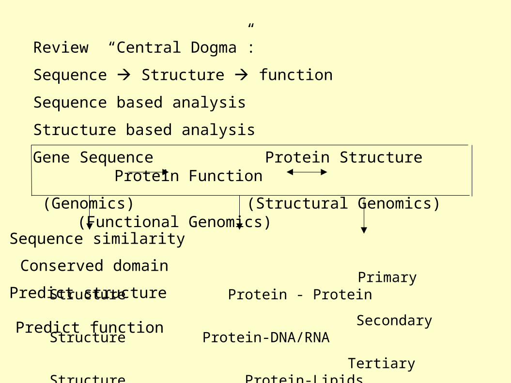
Review “Central Dogma”:
Sequence Structure function
Sequence based analysis
Structure based analysis
Gene Sequence Protein Structure Protein Function
(Genomics) (Structural Genomics) (Functional Genomics)
Primary Structure Protein - Protein
Secondary Structure Protein-DNA/RNA
Tertiary Structure Protein-Lipids
Quaternary Structure Protein-Small Molecules Determine Structure Drug Development
Sequence similarity
Conserved domain
Predict structure
Predict function
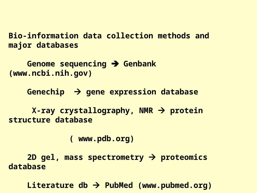
Bio-information data collection methods and major databases
Genome sequencing Genbank (www.ncbi.nih.gov)
Genechip gene expression database
X-ray crystallography, NMR protein structure database ( www.pdb.org)
2D gel, mass spectrometry proteomics database
Literature db PubMed (www.pubmed.org)
Inherent genetic disease db OMIM (www.ncbi.nih.gov)

Structure based analysis Molecular profile and story
Protein structure: Primary Secondary Tertiary -Folding quaternaryProtein structure determination: X-ray crystallography NMR spectroscopyProtein structure prediction: Secondary structure prediction Homology modelingProtein structure analysis: The structure character of the individual protein The structural basis of the protein function Protein family (based on conserved domain, motifs to predict their functions)Write the molecular profile and story
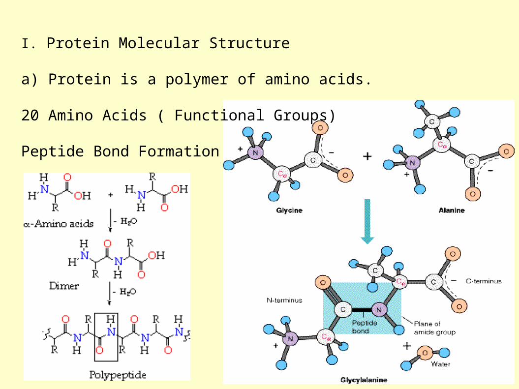
I. Protein Molecular Structure
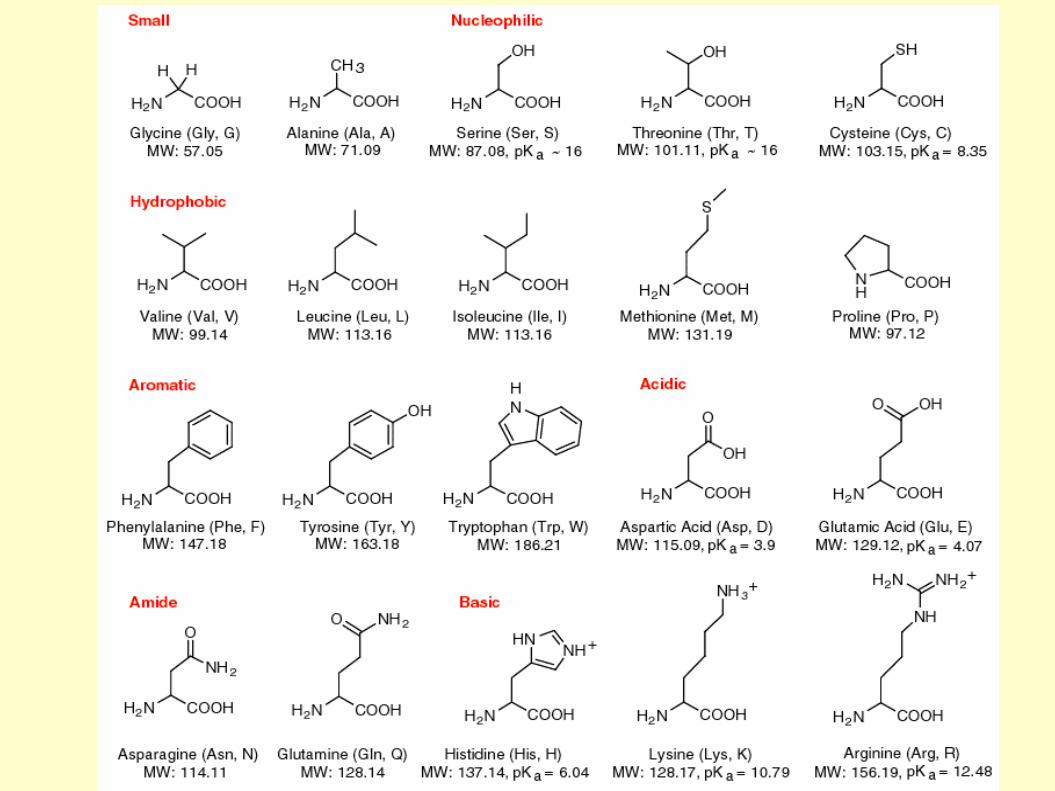
a) Protein is a polymer of amino acids.
20 Amino Acids ( Functional Groups)
Peptide Bond Formation
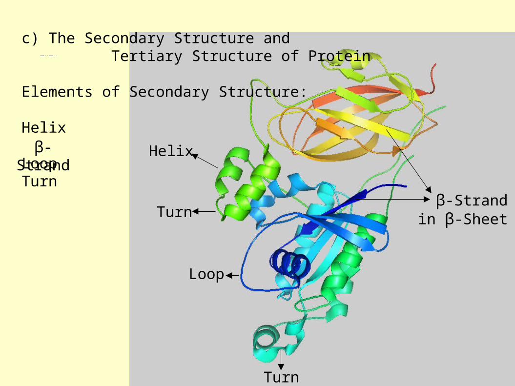
c) The Secondary Structure and Tertiary Structure of Protein
Elements of Secondary Structure:
Helix
LoopTurn
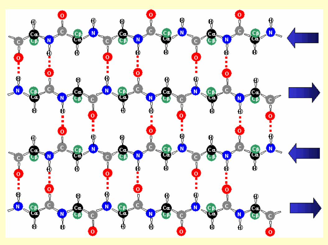
β-Strandin β-Sheet
β-Strand Helix
Turn
Loop
Turn
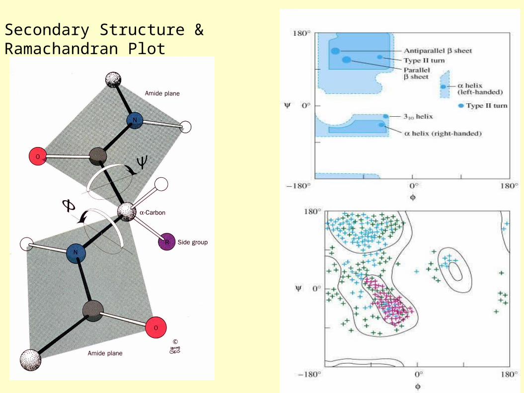
Secondary Structure & Ramachandran Plot

Ramachandran Plot of CDC42-RhoGDI Complex [PDB Code 1DOA]
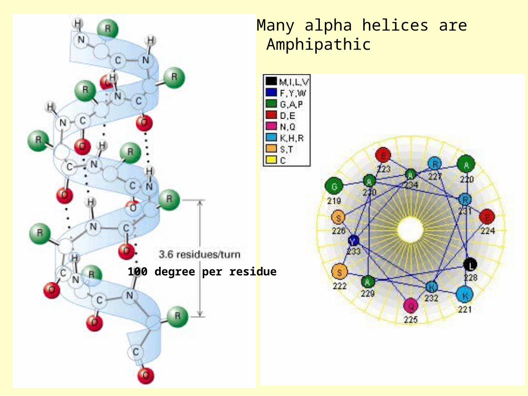
Many alpha helices are Amphipathic
100 degree per residue

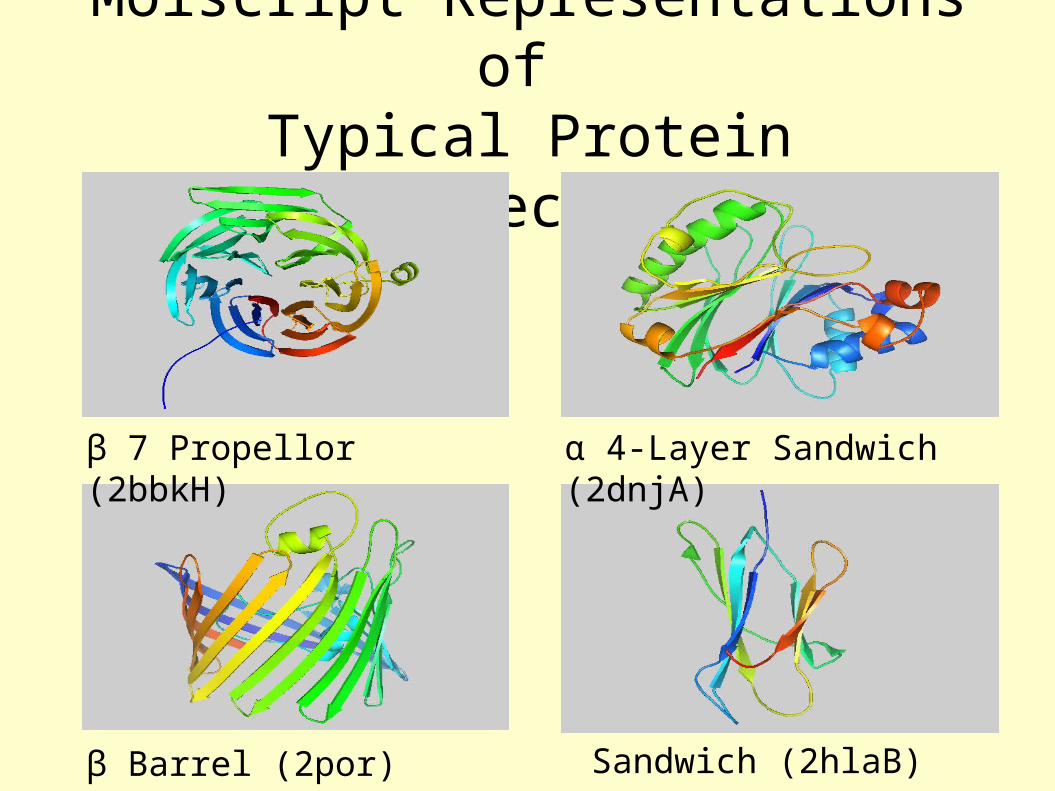
Molscript Representations of Typical Protein Architectures.
β Barrel (2por)
β 7 Propellor (2bbkH) α 4-Layer Sandwich (2dnjA)
Sandwich (2hlaB)

Molscript Representations of Typical Protein Architectures.
α β Barrel (4timA) α Helix Bundle (2ccy)
2 Solenoid (1tsp) α Horseshoe (1bnh)
b a
c da b
d c
--b
a----b
a-----b
a
--d c---d c---d c
-
b
a
dc
a b
c dc
d
a
b
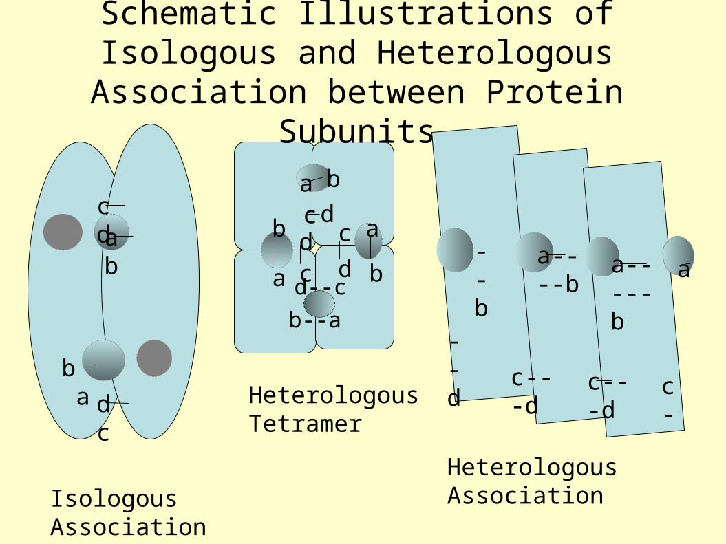
Isologous Association
Heterologous Tetramer
Heterologous Association
Schematic Illustrations of Isologous and Heterologous Association between Protein
Subunits
b--a
d--c
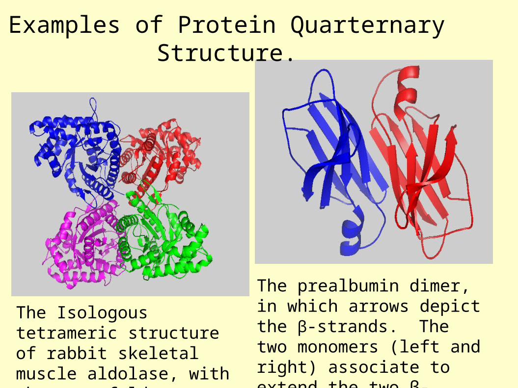
The Isologous tetrameric structure of rabbit skeletal muscle aldolase, with three twofold symmetry axes (1ado).
The prealbumin dimer, in which arrows depict the β-strands. The two monomers (left and right) associate to extend the two β-sheets (1bm7).
Examples of Protein Quarternary Structure.

DNA Quadruplex & G Quartets
K+

NMR SpectroscopySample-Spectrum-Structure
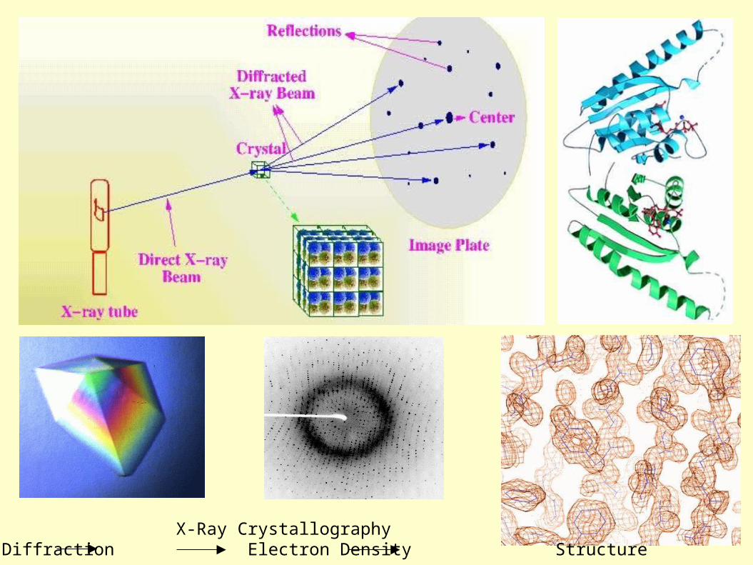
X-Ray CrystallographyCrystal Diffraction Electron Density Structure
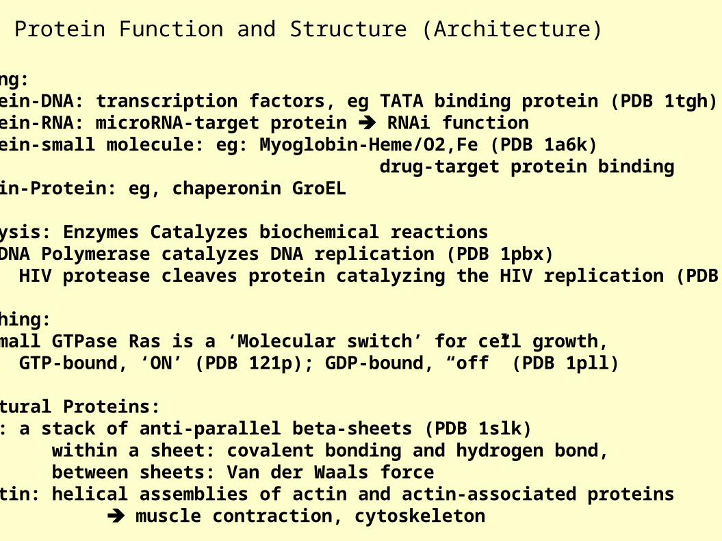
Protein Function and Structure (Architecture)
a) Binding: Protein-DNA: transcription factors, eg TATA binding protein (PDB 1tgh) Protein-RNA: microRNA-target protein RNAi function Protein-small molecule: eg: Myoglobin-Heme/O2,Fe (PDB 1a6k) drug-target protein binding Protein-Protein: eg, chaperonin GroEL
b) Catalysis: Enzymes Catalyzes biochemical reactions eg: DNA Polymerase catalyzes DNA replication (PDB 1pbx) HIV protease cleaves protein catalyzing the HIV replication (PDB 1a8k)
c) Switching: eg: small GTPase Ras is a ‘Molecular switch’ for cell growth, GTP-bound, ‘ON’ (PDB 121p); GDP-bound, “off” (PDB 1pll)
d) Structural Proteins: Silk: a stack of anti-parallel beta-sheets (PDB 1slk) within a sheet: covalent bonding and hydrogen bond, between sheets: Van der Waals force F-actin: helical assemblies of actin and actin-associated proteins muscle contraction, cytoskeleton
The Second Project
Go to www.pdb.org,
a) Type a protein (or a disease) name, search for the structures
b) Download the structure text file, save it as a text file with .pdb extension.
c) Input the xxx.pdb file to Pymol (download from www.pymol.org) to see the molecular structure, notice its structural characters.d) Read the major reference paper (from www.pubmed.org) for this
protein structure and the corresponding gene, summarize the connection of the structure and the protein function. Write the molecular story for this gene.