Embed Size (px)
Citation preview
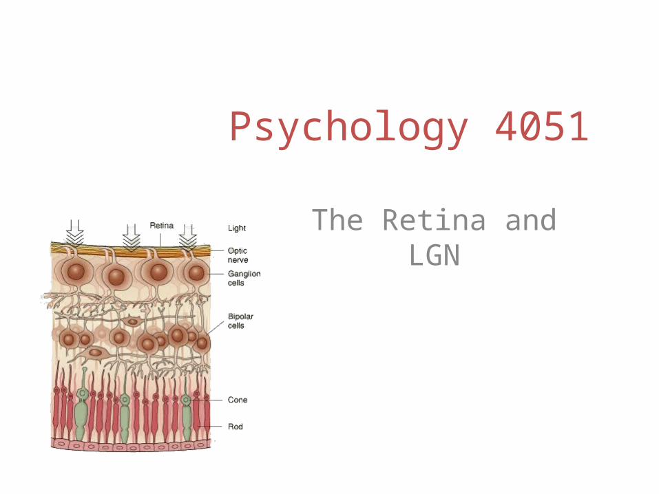
Psychology 4051
The Retina and LGN
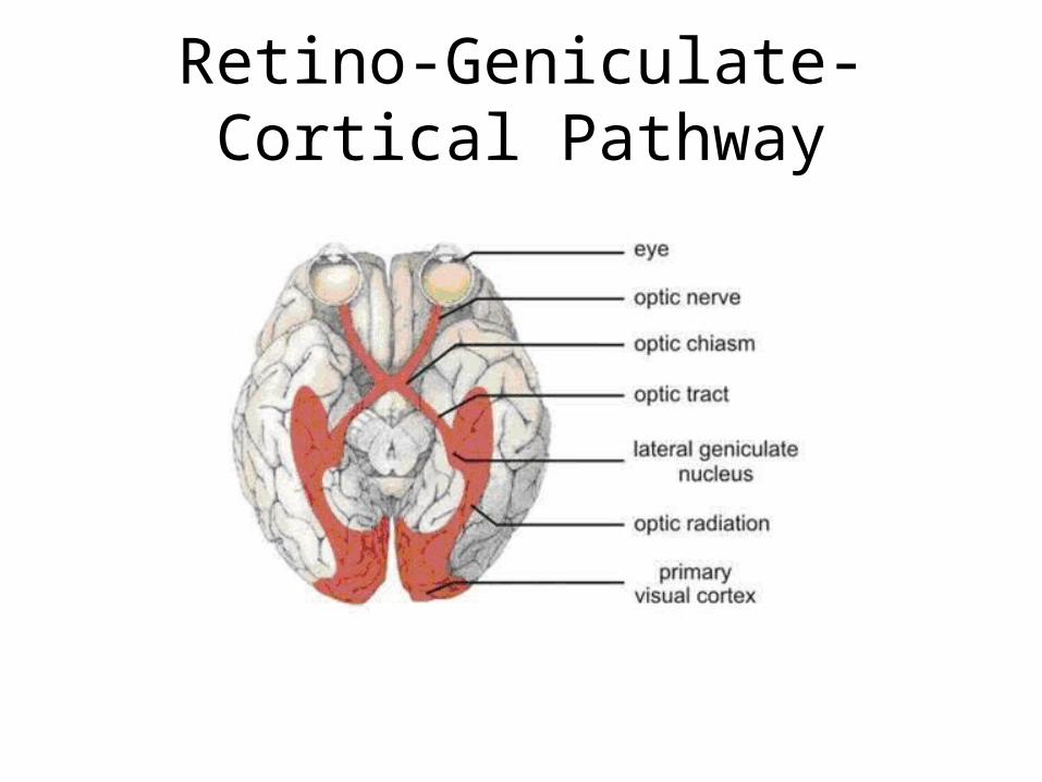
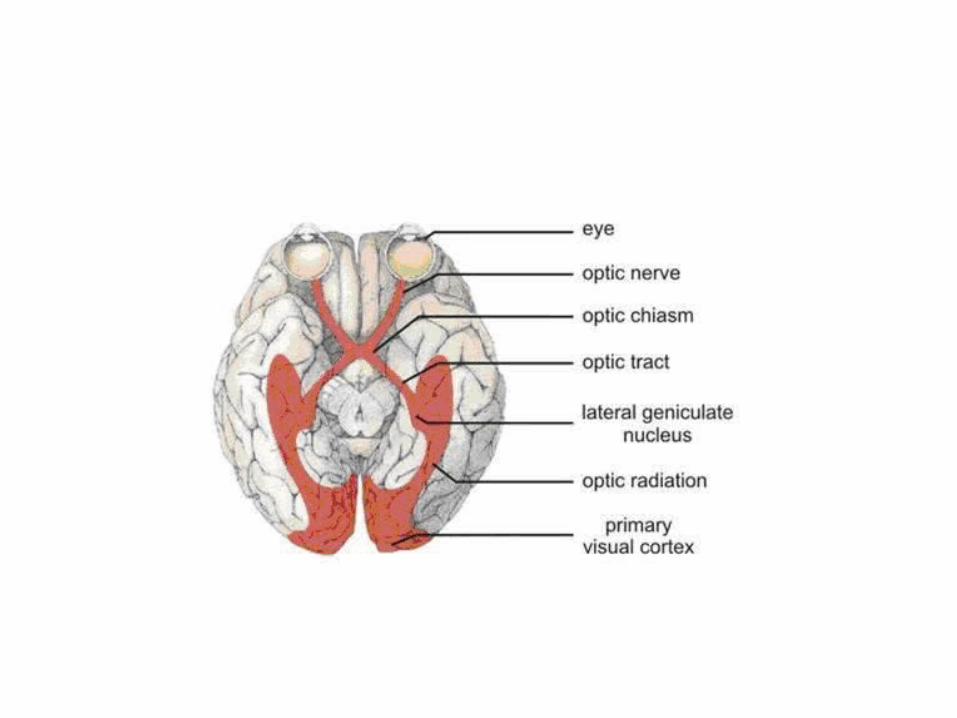
Retino-Geniculate-Cortical Pathway
The Retina
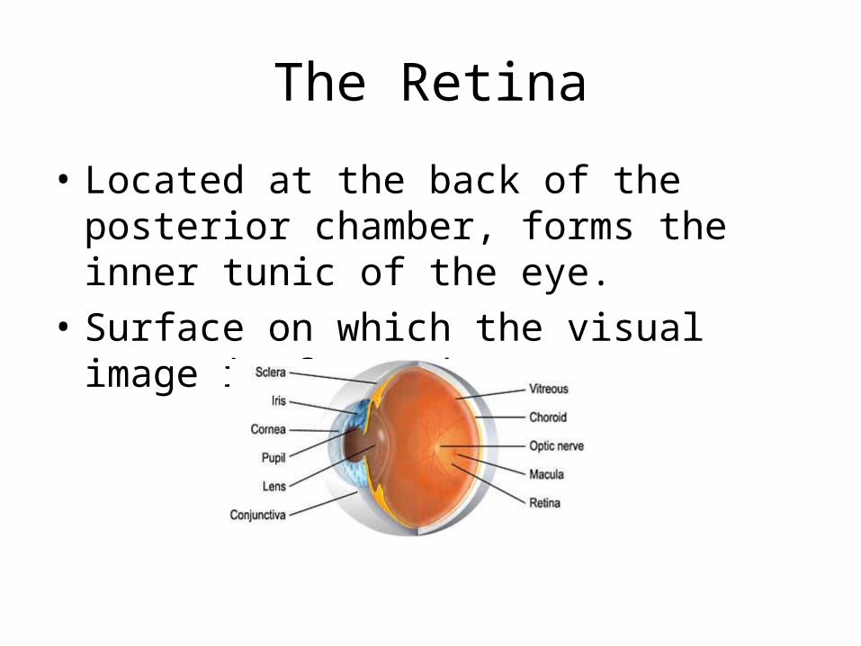
• Located at the back of the posterior chamber, forms the inner tunic of the eye.
• Surface on which the visual image is focused

The Retina
• A laminar tissue with multiple layers.– Pigment epithelium, photoreceptor layer, external
limiting membrane, outer nuclear layer, outer plexiform layer, inner nuclear layer, inner plexiform layer, ganglion cell layer, optic nerve layer, internal limiting membrane.
• Transduction takes place in the photoreceptors.– Rods and cones
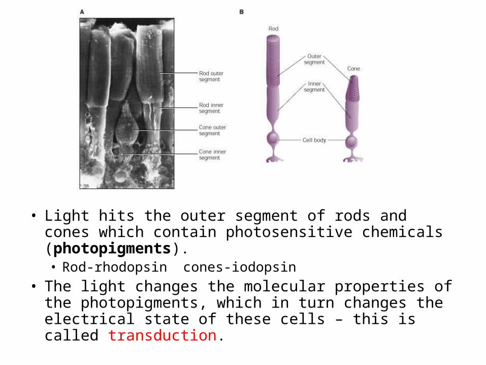
• Light hits the outer segment of rods and cones which contain photosensitive chemicals (photopigments). • Rod-rhodopsin cones-iodopsin
• The light changes the molecular properties of the photopigments, which in turn changes the electrical state of these cells – this is called transduction.
The Retina
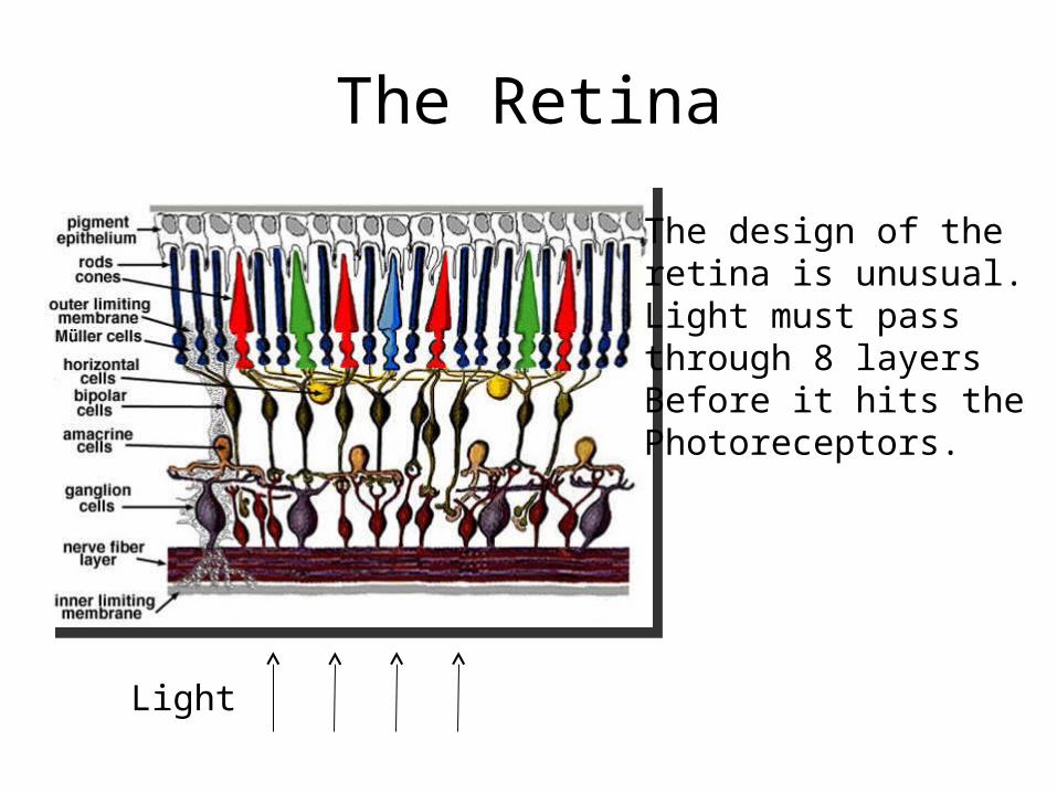
Light
The design of the retina is unusual.Light must pass through 8 layers Before it hits the Photoreceptors.
The Retina
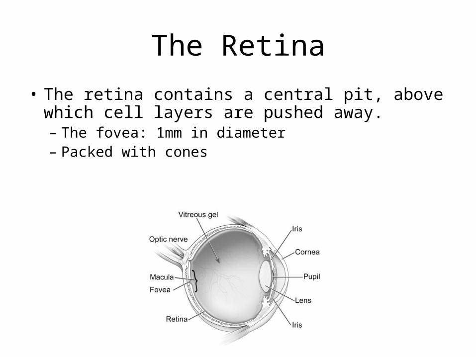
• The retina contains a central pit, above which cell layers are pushed away. – The fovea: 1mm in diameter– Packed with cones
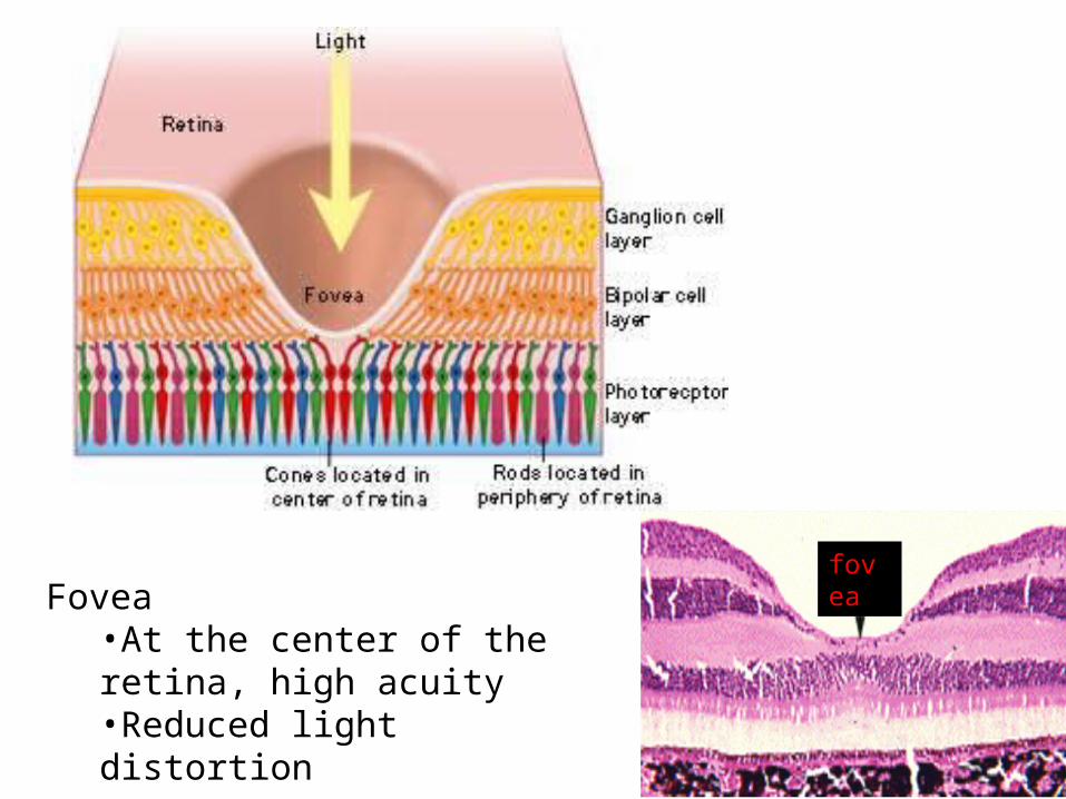
foveaFovea•At the center of the retina, high acuity•Reduced light distortion
The Retina
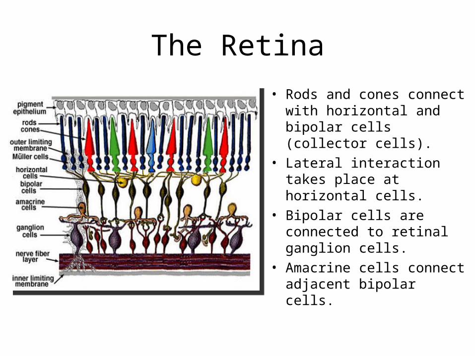
• Rods and cones connect with horizontal and bipolar cells (collector cells).
• Lateral interaction takes place at horizontal cells.
• Bipolar cells are connected to retinal ganglion cells.
• Amacrine cells connect adjacent bipolar cells.
Rods
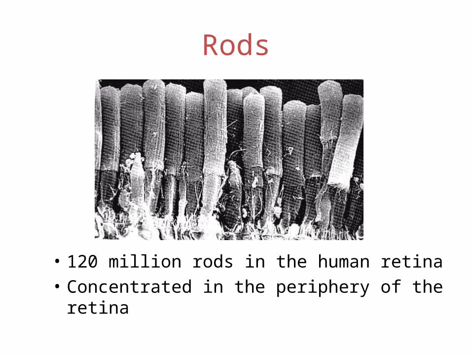
• 120 million rods in the human retina• Concentrated in the periphery of the retina
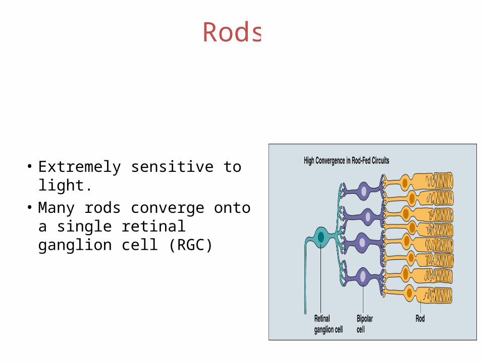
• Extremely sensitive to light.• Many rods converge onto a
single retinal ganglion cell (RGC)
Rods
Rods – good light sensitivity
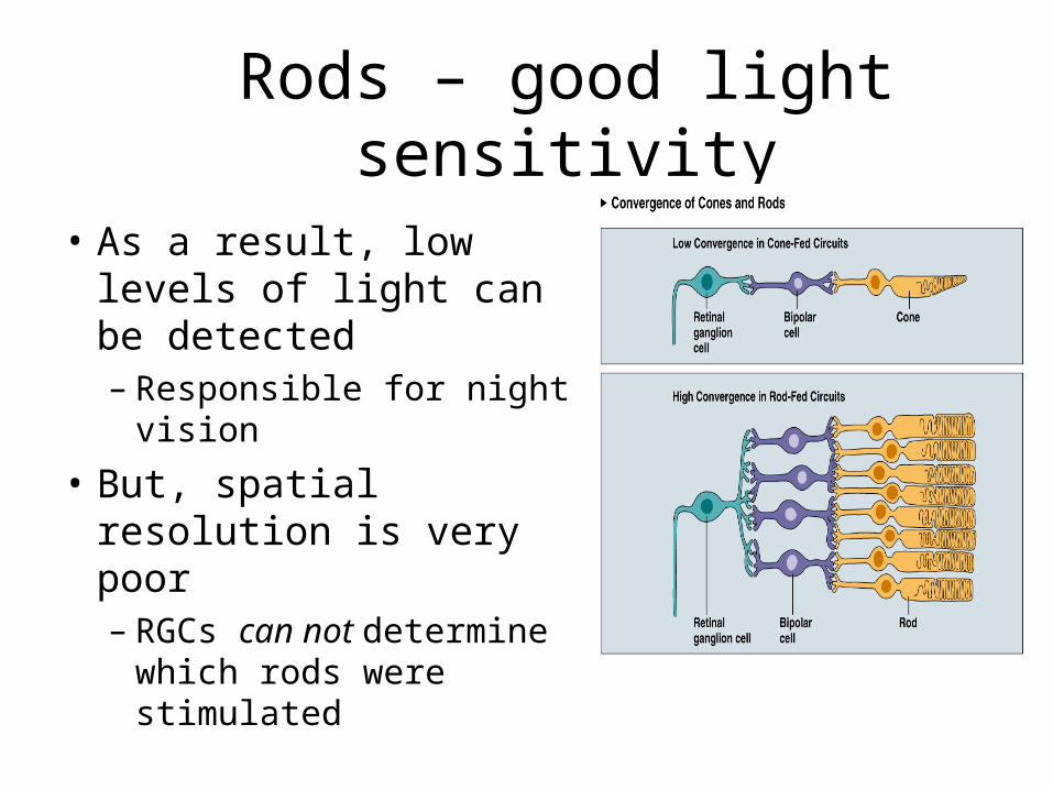
• As a result, low levels of light can be detected– Responsible for night
vision
• But, spatial resolution is very poor– RGCs can not determine
which rods were stimulated
Cones
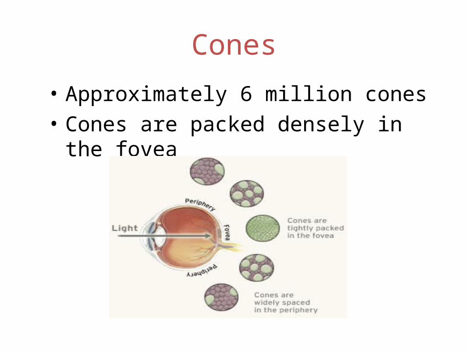
• Approximately 6 million cones• Cones are packed densely in the fovea
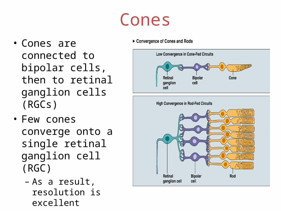
Cones• Cones are
connected to bipolar cells, then to retinal ganglion cells (RGCs)
• Few cones converge onto a single retinal ganglion cell (RGC)– As a result,
resolution is excellent
Cones
• Light sensitivity is poor– Day vision
Cones
• Humans have 3 different cone types, each with a different photopigment.
• Photopigments are maximally sensitive to specific wavelengths.– Short wavelength sensitive– Mid-wavelength sensitive– Long wavelength sensitive
Retinal Ganglion Cells
• Retinal ganglion cells possess receptive fields that are responsive to light stimulation.
• Receptive field – that area of the retina over which a ganglion cell is sensitive to light stimulation (area of the retina that the RGC monitors).
Receptive Fields
• Their receptive fields are designed in an antagonistic fashion
• A single receptive field has an on-center/off- surround arrangement or an off-center/on-surround arrangement.
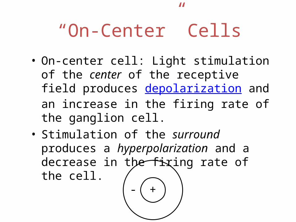
“On-Center” Cells• On-center cell: Light stimulation of the center of
the receptive field produces depolarization and an increase in the firing rate of the ganglion cell.
• Stimulation of the surround produces a hyperpolarization and a decrease in the firing rate of the cell.
+-
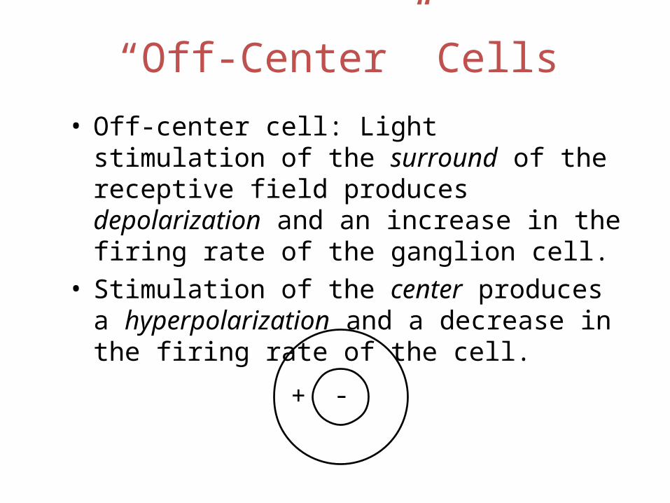
“Off-Center” Cells• Off-center cell: Light stimulation of the surround
of the receptive field produces depolarization and an increase in the firing rate of the ganglion cell.
• Stimulation of the center produces a hyperpolarization and a decrease in the firing rate of the cell.
-+
Retinal Ganglion Cells
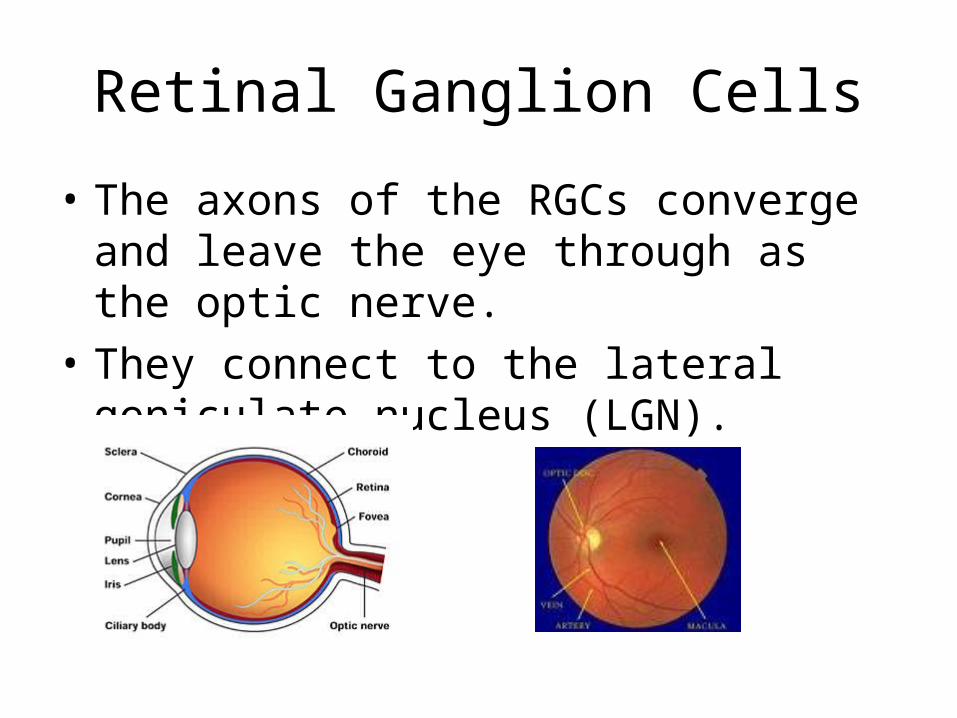
• The axons of the RGCs converge and leave the eye through as the optic nerve.
• They connect to the lateral geniculate nucleus (LGN).

Retinal Ganglion Cells
• Most RGCs fall into two functional classes, M and P cells
• M cells – project to the magnocellular layers of the lateral geniculate nucleus (LGN)
• P cells – project to the parvocellular layers of the LGN
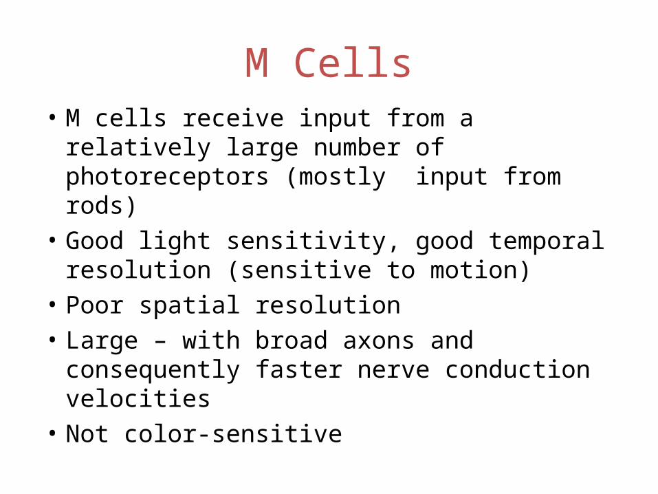
M Cells• M cells receive input from a relatively large
number of photoreceptors (mostly input from rods)
• Good light sensitivity, good temporal resolution (sensitive to motion)
• Poor spatial resolution• Large – with broad axons and consequently
faster nerve conduction velocities• Not color-sensitive
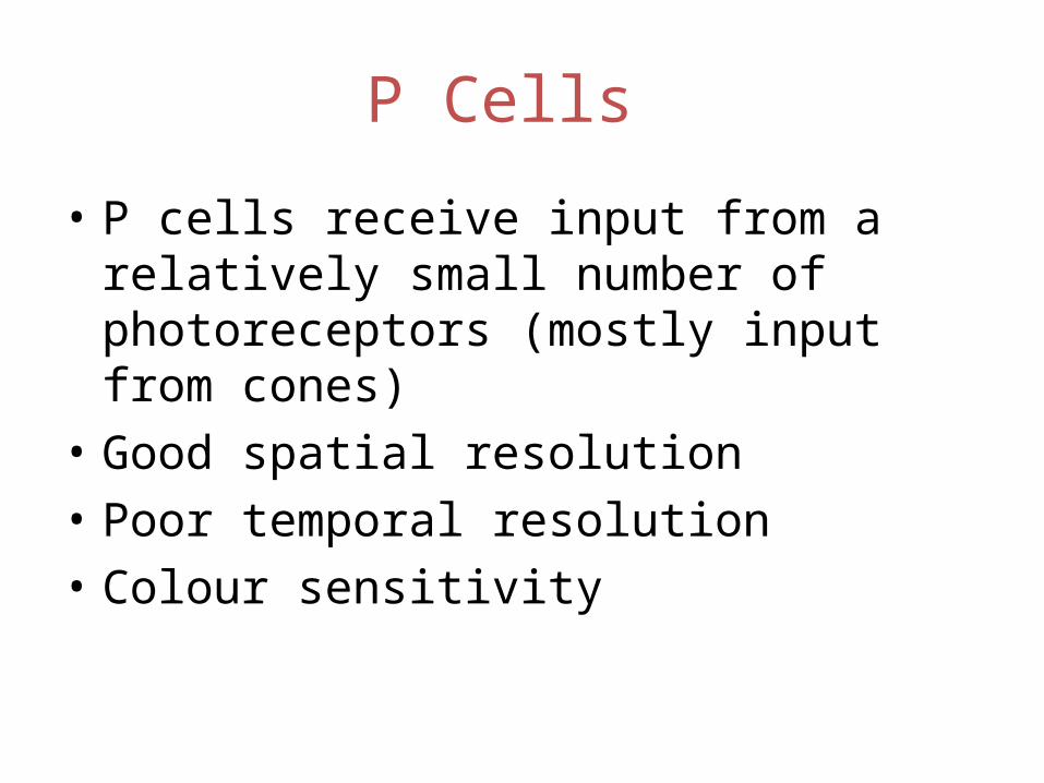
P Cells
• P cells receive input from a relatively small number of photoreceptors (mostly input from cones)
• Good spatial resolution• Poor temporal resolution• Colour sensitivity
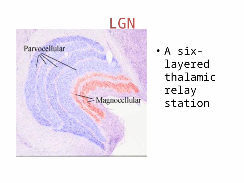
LGN
• A six-layered thalamic relay station
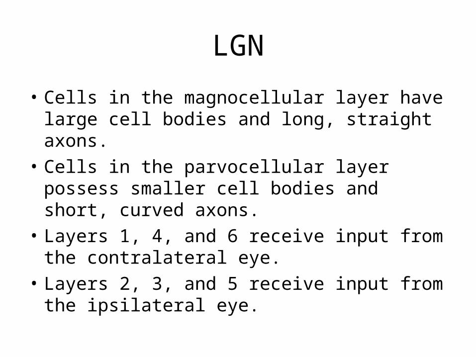
LGN
• Cells in the magnocellular layer have large cell bodies and long, straight axons.
• Cells in the parvocellular layer possess smaller cell bodies and short, curved axons.
• Layers 1, 4, and 6 receive input from the contralateral eye.
• Layers 2, 3, and 5 receive input from the ipsilateral eye.
LGN
• Most parvocellular cells appear to be responsive to color and have center/surround receptive fields.