Embed Size (px)
Citation preview
CASE REPORT
Pucker sign in proximal humeral fractures: implicationson management
Nipun Jindal • Parmanand Gupta • Ravi Kumar Gupta •
Amit Kumar • Ankush Jindal
Received: 8 March 2013 / Accepted: 23 May 2013 / Published online: 5 June 2013
� The Author(s) 2013. This article is published with open access at Springerlink.com
Abstract Fracture of the surgical neck of humerus in
young patients is a relatively rare injury. We reviewed the
available material on the topic and identified puckering at the
shoulder in high-energy fracture of the surgical neck as a
finding which has been reported infrequently but signifies a
need for open reduction. We present a review of the literature
on the subject and our similar experience in two young males
who had puckering and ecchymosis at the shoulder.
Keywords Surgical neck humerus fracture � Puckering �Dimpling � Buttonholing � Open reduction
Introduction
Fracture of surgical neck of humerus accounts for 12.7 % of
proximal humeral fractures [1] with majority of cases
occurring in elderly as fragility fractures. However, these
fractures can also occur in young patients following high-
energy trauma. The energy of trauma together with the force
generated by surrounding muscles may result in buttonholing
of distal fragment through the deltoid muscle causing
puckering on the skin. Skin puckering as a sign of proximal
humeral fractures has been described as a physical sign in
only two reports worldwide [2, 3]. We review the available
literature and consider the implications of such a sign on the
treatment protocol observed in such injuries. We also present
our experience with two cases of fracture of surgical neck
of humerus in young males with buttonholing through the
deltoid muscle and subsequent skin puckering.
Case 1
A 17 year old male presented with pain and inability to move
right shoulder after being involved in a road traffic accident.
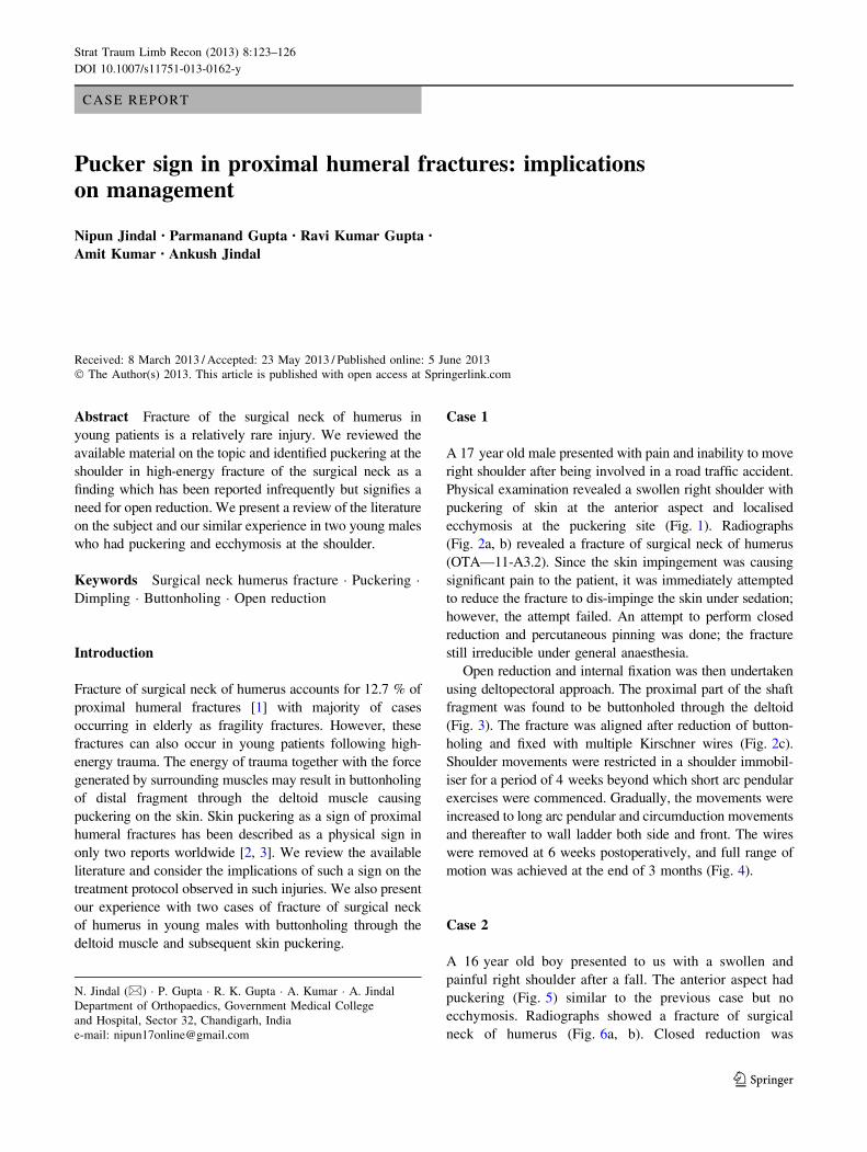
Physical examination revealed a swollen right shoulder with
puckering of skin at the anterior aspect and localised
ecchymosis at the puckering site (Fig. 1). Radiographs
(Fig. 2a, b) revealed a fracture of surgical neck of humerus
(OTA—11-A3.2). Since the skin impingement was causing
significant pain to the patient, it was immediately attempted
to reduce the fracture to dis-impinge the skin under sedation;
however, the attempt failed. An attempt to perform closed
reduction and percutaneous pinning was done; the fracture
still irreducible under general anaesthesia.
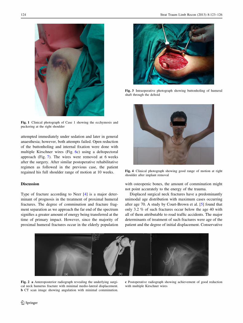
Open reduction and internal fixation was then undertaken
using deltopectoral approach. The proximal part of the shaft
fragment was found to be buttonholed through the deltoid
(Fig. 3). The fracture was aligned after reduction of button-
holing and fixed with multiple Kirschner wires (Fig. 2c).
Shoulder movements were restricted in a shoulder immobil-
iser for a period of 4 weeks beyond which short arc pendular
exercises were commenced. Gradually, the movements were
increased to long arc pendular and circumduction movements
and thereafter to wall ladder both side and front. The wires
were removed at 6 weeks postoperatively, and full range of
motion was achieved at the end of 3 months (Fig. 4).
Case 2
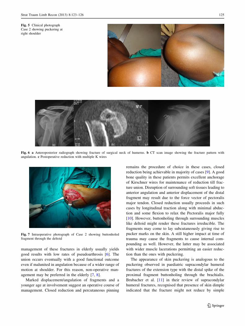
A 16 year old boy presented to us with a swollen and
painful right shoulder after a fall. The anterior aspect had
puckering (Fig. 5) similar to the previous case but no
ecchymosis. Radiographs showed a fracture of surgical
neck of humerus (Fig. 6a, b). Closed reduction was
N. Jindal (&) � P. Gupta � R. K. Gupta � A. Kumar � A. Jindal
Department of Orthopaedics, Government Medical College
and Hospital, Sector 32, Chandigarh, India
e-mail: [email protected]
123
Strat Traum Limb Recon (2013) 8:123–126
DOI 10.1007/s11751-013-0162-y
attempted immediately under sedation and later in general
anaesthesia; however, both attempts failed. Open reduction
of the buttonholing and internal fixation were done with
multiple Kirschner wires (Fig. 6c) using a deltopectoral
approach (Fig. 7). The wires were removed at 6 weeks
after the surgery. After similar postoperative rehabilitative
regimen as followed in the previous case, the patient
regained his full shoulder range of motion at 10 weeks.
Discussion
Type of fracture according to Neer [4] is a major deter-
minant of prognosis in the treatment of proximal humeral
fractures. The degree of comminution and fracture frag-
ment separation as we approach the far end of the spectrum
signifies a greater amount of energy being transferred at the
time of primary impact. However, since the majority of
proximal humeral fractures occur in the elderly population
with osteopenic bones, the amount of comminution might
not point accurately to the energy of the trauma.
Displaced surgical neck fractures have a predominantly
unimodal age distribution with maximum cases occurring
after age 70. A study by Court-Brown et al. [5] found that
only 3.2 % of such fractures occur below the age 40 with
all of them attributable to road traffic accidents. The major
determinants of treatment of such fractures were age of the
patient and the degree of initial displacement. Conservative
Fig. 1 Clinical photograph of Case 1 showing the ecchymosis and
puckering at the right shoulder
Fig. 2 a Anteroposterior radiograph revealing the underlying surgi-
cal neck humerus fracture with minimal medio-lateral displacement.
b CT scan image showing angulation with minimal comminution.
c Postoperative radiograph showing achievement of good reduction
with multiple Kirschner wires
Fig. 3 Intraoperative photograph showing buttonholing of humeral
shaft through the deltoid
Fig. 4 Clinical photograph showing good range of motion at right
shoulder after implant removal
124 Strat Traum Limb Recon (2013) 8:123–126
123
management of these fractures in elderly usually yields
good results with low rates of pseudoarthrosis [6]. The
union occurs eventually with a good functional outcome
even if malunited in angulation because of a wider range of
motion at shoulder. For this reason, non-operative man-
agement may be preferred in the elderly [7, 8].
Marked displacement/angulation of fragments and a
younger age at involvement suggest an operative course of
management. Closed reduction and percutaneous pinning
remains the procedure of choice in these cases, closed
reduction being achievable in majority of cases [9]. A good
bone quality in these patients permits excellent anchorage
of Kirschner wires for maintenance of reduction till frac-
ture union. Disruption of surrounding soft tissues leading to
anterior angulation and anterior displacement of the distal
fragment may result due to the force vector of pectoralis
major tendon. Closed reduction usually proceeds in such
cases by longitudinal traction along with minimal abduc-
tion and some flexion to relax the Pectoralis major fully
[10]. However, buttonholing through surrounding muscles
like deltoid might render these fractures irreducible. The
fragments may come to lay subcutaneously giving rise to
pucker marks on the skin. A still higher impact at time of
trauma may cause the fragments to cause internal com-
pounding as well. However, the latter may be associated
with wider muscle lacerations permitting an easier reduc-
tion than the ones with puckering.
The appearance of skin puckering is analogous to the
puckering observed in paediatric supracondylar humeral
fractures of the extension type with the distal spike of the
proximal fragment buttonholing through the brachialis.
Brubacher et al. [11] in their review of supracondylar
humeral fractures, recognised that presence of skin dimple
indicated that the fracture might not reduce by simple
Fig. 5 Clinical photograph
Case 2 showing puckering at
right shoulder
Fig. 6 a Anteroposterior radiograph showing fracture of surgical neck of humerus. b CT scan image showing the fracture pattern with
angulation. c Postoperative reduction with multiple K wires
Fig. 7 Intraoperative photograph of Case 2 showing buttonholed
fragment through the deltoid
Strat Traum Limb Recon (2013) 8:123–126 125
123
manipulation alone. However, a search of literature did not
give the exact percentage of cases with positive puckering
sign which did require an open reduction.
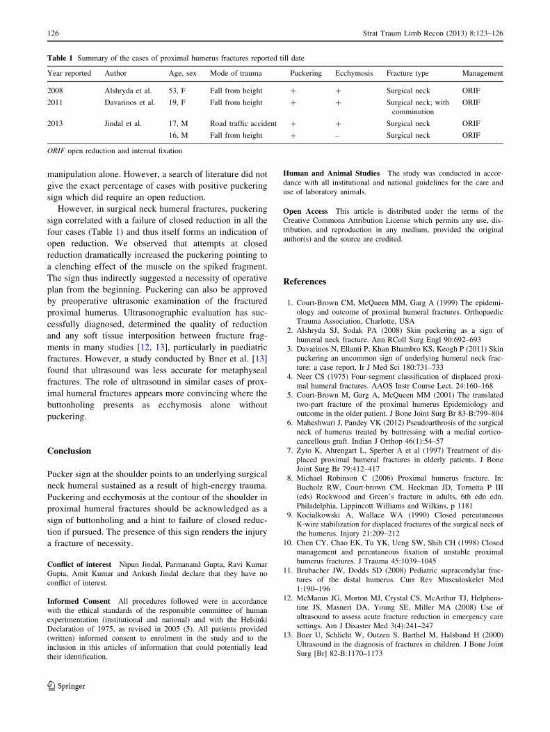
However, in surgical neck humeral fractures, puckering
sign correlated with a failure of closed reduction in all the
four cases (Table 1) and thus itself forms an indication of
open reduction. We observed that attempts at closed
reduction dramatically increased the puckering pointing to
a clenching effect of the muscle on the spiked fragment.
The sign thus indirectly suggested a necessity of operative
plan from the beginning. Puckering can also be approved
by preoperative ultrasonic examination of the fractured
proximal humerus. Ultrasonographic evaluation has suc-
cessfully diagnosed, determined the quality of reduction
and any soft tissue interposition between fracture frag-
ments in many studies [12, 13], particularly in paediatric
fractures. However, a study conducted by Bner et al. [13]
found that ultrasound was less accurate for metaphyseal
fractures. The role of ultrasound in similar cases of prox-
imal humeral fractures appears more convincing where the
buttonholing presents as ecchymosis alone without
puckering.
Conclusion
Pucker sign at the shoulder points to an underlying surgical
neck humeral sustained as a result of high-energy trauma.
Puckering and ecchymosis at the contour of the shoulder in
proximal humeral fractures should be acknowledged as a
sign of buttonholing and a hint to failure of closed reduc-
tion if pursued. The presence of this sign renders the injury
a fracture of necessity.
Conflict of interest Nipun Jindal, Parmanand Gupta, Ravi Kumar
Gupta, Amit Kumar and Ankush Jindal declare that they have no
conflict of interest.
Informed Consent All procedures followed were in accordance
with the ethical standards of the responsible committee of human
experimentation (institutional and national) and with the Helsinki
Declaration of 1975, as revised in 2005 (5). All patients provided
(written) informed consent to enrolment in the study and to the
inclusion in this articles of information that could potentially lead
their identification.
Human and Animal Studies The study was conducted in accor-
dance with all institutional and national guidelines for the care and
use of laboratory animals.
Open Access This article is distributed under the terms of the
Creative Commons Attribution License which permits any use, dis-
tribution, and reproduction in any medium, provided the original
author(s) and the source are credited.
References
1. Court-Brown CM, McQueen MM, Garg A (1999) The epidemi-
ology and outcome of proximal humeral fractures. Orthopaedic
Trauma Association, Charlotte, USA
2. Alshryda SJ, Sodak PA (2008) Skin puckering as a sign of
humeral neck fracture. Ann RColl Surg Engl 90:692–693
3. Davarinos N, Ellanti P, Khan Bhambro KS, Keogh P (2011) Skin
puckering an uncommon sign of underlying humeral neck frac-
ture: a case report. Ir J Med Sci 180:731–733
4. Neer CS (1975) Four-segment classification of displaced proxi-
mal humeral fractures. AAOS Instr Course Lect. 24:160–168
5. Court-Brown M, Garg A, McQueen MM (2001) The translated
two-part fracture of the proximal humerus Epidemiology and
outcome in the older patient. J Bone Joint Surg Br 83-B:799–804
6. Maheshwari J, Pandey VK (2012) Pseudoarthrosis of the surgical
neck of humerus treated by buttressing with a medial cortico-
cancellous graft. Indian J Orthop 46(1):54–57
7. Zyto K, Ahrengart L, Sperber A et al (1997) Treatment of dis-
placed proximal humeral fractures in elderly patients. J Bone
Joint Surg Br 79:412–417
8. Michael Robinson C (2006) Proximal humerus fracture. In:
Bucholz RW, Court-brown CM, Heckman JD, Tornetta P III
(eds) Rockwood and Green’s fracture in adults, 6th edn edn.
Philadelphia, Lippincott Williams and Wilkins, p 1181
9. Kocialkowski A, Wallace WA (1990) Closed percutaneous
K-wire stabilization for displaced fractures of the surgical neck of
the humerus. Injury 21:209–212
10. Chen CY, Chao EK, Tu YK, Ueng SW, Shih CH (1998) Closed
management and percutaneous fixation of unstable proximal
humerus fractures. J Trauma 45:1039–1045
11. Brubacher JW, Dodds SD (2008) Pediatric supracondylar frac-
tures of the distal humerus. Curr Rev Musculoskelet Med
1:190–196
12. McManus JG, Morton MJ, Crystal CS, McArthur TJ, Helphens-
tine JS, Masneri DA, Young SE, Miller MA (2008) Use of
ultrasound to assess acute fracture reduction in emergency care
settings. Am J Disaster Med 3(4):241–247
13. Bner U, Schlicht W, Outzen S, Barthel M, Halsband H (2000)
Ultrasound in the diagnosis of fractures in children. J Bone Joint
Surg [Br] 82-B:1170–1173
Table 1 Summary of the cases of proximal humerus fractures reported till date
Year reported Author Age, sex Mode of trauma Puckering Ecchymosis Fracture type Management
2008 Alshryda et al. 53, F Fall from height ? ? Surgical neck ORIF
2011 Davarinos et al. 19, F Fall from height ? ? Surgical neck; with
comminution
ORIF
2013 Jindal et al. 17, M Road traffic accident ? ? Surgical neck ORIF
16, M Fall from height ? – Surgical neck ORIF
ORIF open reduction and internal fixation
126 Strat Traum Limb Recon (2013) 8:123–126
123