Embed Size (px)
Citation preview
Pulmonary Atelectasis Presented by Kang, ting-jui
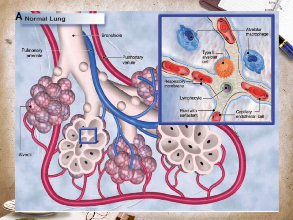
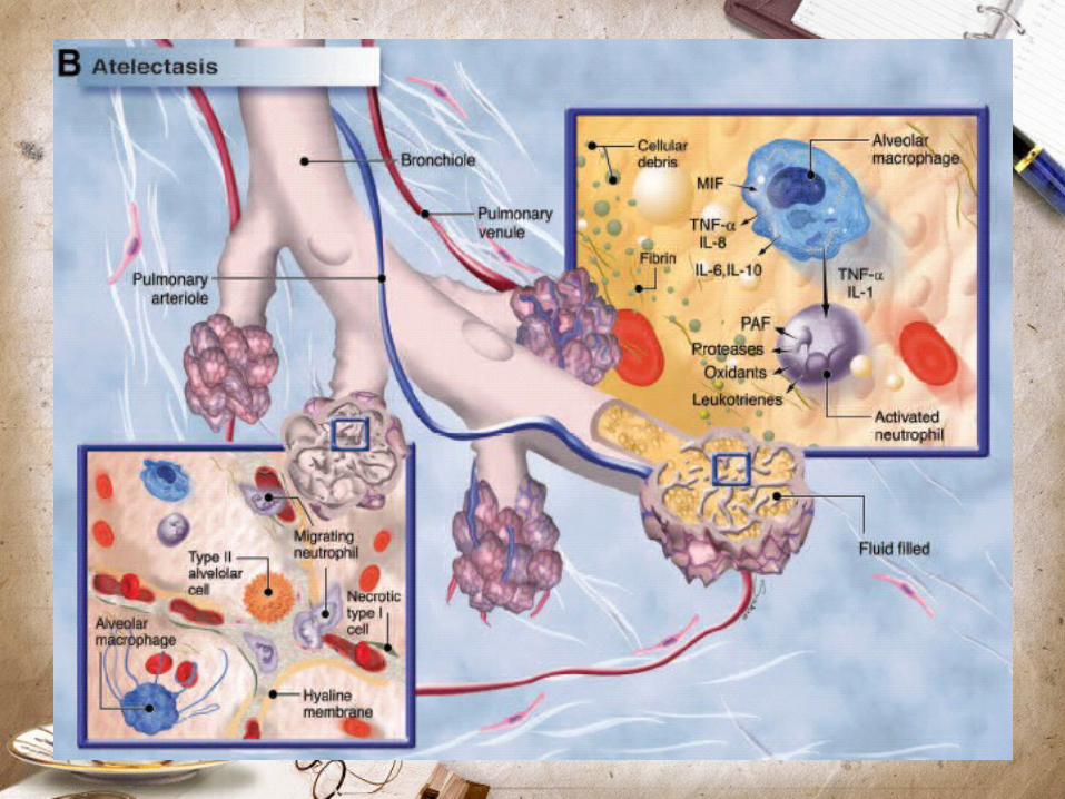
IntroductionGeneral anesthesia is associated with impaired oxygenation pulmonary atelectasis was suspected as the major causeDecrease in lung compliance and the partial pressure of arterial oxygen (PaO2)Atelectasis occurs in the most dependent parts of the lung of 90% of patients who are anesthetized gas exchange abnormalities and reduced static compliance associated with acute lung injury perioperative morbidity
Physiologic causes of atelectasis
Mechanisms:Compression of lung tissueAbsorption of alveolar airImpairment of surfactant function
Compression atelectasisOverall cephalad diaphragm displacement after anesthesia, the diaphragm is relaxed Differential regional diaphragmatic changes
In an anesthetized patient breathing spontaneously the diaphragm moves the most in the lower, dependent portion
During paralysis and positive-pressure ventilation the passive diaphragm is displaced by the positive pressure preferentially in the upper, nondependent portion
Compression atelectasis
Shift of thoracic central vascular blood into the abdomen additional dependent pressure arising from the abdomenAltered diaphragmatic dynamics phrenic nerve stimulation versus isovolumic conditions in anesthetized patients
Gas resorptionResorption atelectasis occur by two mechanisms
After complete airway occlusion gas trapped gas uptake by the blood continues and gas inflow is prevented gas pocket collapses; increases with elevation of FI02
Low ventilation relative to perfusion (low [VA/Q] ratio) have a low partial pressure of alveolar oxygen (PAO2) when FIO2 increased, PAO2 increases oxygen moves from alveolar to blood greatly lung unit progressively smaller
Surfactant impairmentStabilizing function of surfactant may be depressed by anesthesia
Reduction in percent maximum lung volume was proportional to the concentration of both chloroform and halothane
Halothane anesthesia combination with high oxygen concentration, caused increased permeability of the alveolar– capillary barrier in rabbit lungsIncreased tidal volume cause release of surfactant
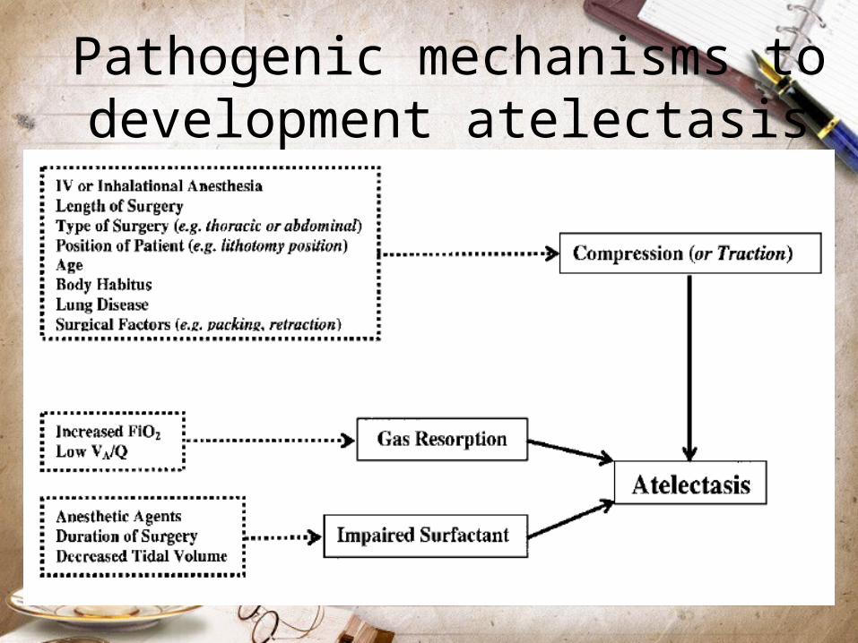
Pathogenic mechanisms to development atelectasis
Factors modulating the formation of atelectasis
Type of anesthesiaAtelectasis develops with both intravenous and inhalational anesthesia, whether the patient is breathing spontaneously or is paralyzed and mechanically ventilatedKetamine: not produce atelectasis when used aloneRegional anesthesia: depend on the type and extension of motor blockade
Reduces inspiratory capacity by up to 20%, and expiratory reserve volume approaches zero
Less extensive blockade closing capacity and FRC remain unchanged
Impact of timeThe maximum decrease in FRC seems to occur within the first few minutes of general anesthesiaDuring anesthesia for surgical operations on the limb FRC is not influenced further by depth or duration of anesthesiaDuring abdominal or thoracic surgery, pulmonary gas exchange deteriorates progressively during the course of the operation unable to determine the independent impact of time on atelectasis as opposed to surgical manipulation
Effects of position
Changing from the upright to the supine position results in a decrease of 0.5L in FRC to 1.0L, even in the awake stateAfter anesthesia, FRC is reduced by a further 0.5L to 0.7LTrendelenburg position resulting in further decrease in FRCLateral decubitus position usually slight increase in total lung FRCProne position may increase FRC slightly, although this may not decrease atelectasis
Effects of positionDistribution of ventilation is more uniform in anesthetized patients in the prone position; improves oxygenation in patients with ARDSAtelectasis is more prominent after cardiac surgery with cardiopulmonary bypass; not related to increasing in pulmonary endothelial permeabilityLung recruitment strategy and PEEP improves oxygenation in patients after CPBIntentionally inflate the patient’s lungs before coming off CPB and directly visualize equal expansion of the lungs
Inspired oxygenHigh oxygen concentration has been associated with atelectasis formationIncreasing FIO2 at the end of surgery to 1.0 before extubation also causes additional atelectasisDuring routine induction of general anesthesia, 80% oxygen caused minimal atelectasis, but the time margin before desaturation occurred was significantly shortened compared with that of 100% oxygen
Inspired oxygenNo difference in the incidence of postoperative atelectasis if nitrous oxide in oxygen was used or if air in oxygen was usedThe very rare possibility of acute hypoxemia in the event of difficulty with airway management versus the common and predictable — but generally mild—impact of hyperoxia-induced atelectasis on later intraoperative gas exchangeA lower FIO2 to replace preoxygenation has not been recommended
Effects of ageProgressive age is not associated with increased propensity for development of atelectasisYoung children (aged 1–3 yr) develop atelectasis more readily greater thoracic wall compliance, less outwardly directed lung distension forcesChildren younger than 2yr prone to respiratory failure and fatigue: type I and II muscle fibers are not fully developedInfant at greater risk for atelectasis because the elastic supporting structure of the lung is incompletely developed
Body habitusObesity markedly reduced FRC and lung compliance development of atelectasis worsens arterial oxygenationThe weight of the torso and abdomen make diaphragmatic excursions more difficult— especially when recumbent or supine—the FRC decreases, intensified in paralysis with neuromuscular blockadePregnancy also potentiates atelectasis
Tidal volumeThe use of low tidal volume in patients with ARDS reduces stretch-induced lung injury in patients with ARDS, improved patient survivalLow tidal volume increases atelectasis in the absence of lung injuryThe specific “low-tidal-volume” approach was actually associated with the development of intrinsic PEEPPressure-controlled ventilation results in smaller delivered tidal volumes when respiratory system compliance is decreased may lead to atelectasis and may go undiagnosed
Preexisting lung disease
Smokers and patients with lung disease show more pronounced gas exchange impairment than healthy subjectsOnly a small shunt and almost no atelectasis develops in these patients, but they may have severe VA/Q mismatchCOPD may make them resist collapseLarge regions with low VA/Q ratios that can result, over time, in resorption atelectasis
Effects of atelectasis
Decreased complianceImpaired oxygenationPulmonary vascular resistance increaseLung injury
Postoperative periodAtelectasis can persist for 2 days after major surgeryThe lung dysfunction is often transient; may be related to reduction in FRCPostoperative mechanical respiratory abnormality after abdominal or thoracic surgery is a restrictive pattern with severely reduced inspiratory capacity, vital capacity, and FRC pain control in preventing postoperative atelectasisAtelectasis and pneumonia are often considered together because the changes associated with atelectasis may predispose to pneumonia
Detection of atelectasis
Conventional chest radiographyComputed tomographyMagnetic resonance imagingUltrasonographyIntravital microscopyCytokine profile
Prevention / reversal of atelectasisHealthy lungs
Reversible by passive hyperinflation (i.e., three successive inflations: a pressure of 20cmH2O for 10s; then a pressure of 30cm H2O for 15s; and third, a pressure of 40 cm H2O sustained for 15s)
High initial pressures are needed to overcome the anesthesia-induced collapse and that PEEP of 5cm H2O or more is required to prevent collapse
No evidence of barotrauma or pulmonary complications occurred in the high initial airway pressure
Prevention / reversal of atelectasisInjured lungs
Large tidal volumes, high peak airway pressures, and end-expiratory alveolar collapse with cyclic reopening have all been proposed as deleterious
Calculated the inflection point from a pressure–volume curve, and PEEP was preset at 2cm H2O above the inflection point
Recruiting maneuver: continuous positive airway pressures of 35–40cm H2O for 40 s followed by a return to previous PEEP levels
Optimum overall cardiopulmonary interactionOnly in patients with early ARDS
Treating atelectasis in the postoperative period
Encourage or force patients to inspire deeplyMethod: intermittent positive-pressure breathing, deep-breathing exercises, incentive spirometry, and chest physiotherapyA simple posture change from supine to seatedThoracic or sternal traction, the use of intravenous aminophylline as successful treatments of atelectasis in a number of case reports

HAVE A HAVE A NICE DAY NICE DAY