Embed Size (px)
Citation preview

Exp Brain Res (2007) 183:271–281
DOI 10.1007/s00221-007-1045-6RESEARCH ARTICLE
QuantiWcation of reXex activity in stroke survivors during an imposed multi-joint leg extension movement
iian Black · Diane Nichols · Marlena Pelliccio · Joseph Hidler
Received: 3 February 2006 / Accepted: 22 June 2007 / Published online: 21 July 2007© Springer-Verlag 2007
Abstract The goal of this study was to compare short-and long-latency reXex responses in eight major lower-extremity muscle groups following an imposed multi-jointleg movement between a group of 14 chronic (>1 year)stroke survivors and 10 healthy age-matched controls, andto investigate the inXuence of joint velocities and muscleexcitation levels on these reXex responses in each respec-tive group. Subjects were seated with their foot anchored toa sliding footplate that could extend their leg. Prior to theleg being moved, subjects were instructed to pre-activatehip and knee Xexors and extensors. Feedback of jointtorque was used to help subjects activate muscles over arange of excitation levels. Following pre-activation, thesubject’s leg was passively extended so the knee or hipjoint rotated at one of three diVerent speeds (30, 60, and120°/s). In general, it was found that the magnitude ofstroke survivors’ reXex response was greater compared tocontrols’ in certain biarticular muscles, notably the gastroc-nemius and medial hamstring, and the uniarticular adductorlongus, and that the long-latency reXex component(between 40 and 150 ms post-movement) accounted formost of the observed diVerences. Furthermore, while reXexresponse amplitudes increased in both groups with increasing
movement speed, the rate of increase was signiWcantlylarger in stroke subjects than in controls. Clinically, theseWndings may help explain why stroke survivors walk slowlysince it is under these conditions that reXex responses betteremulate those of their able-bodied counterparts.
Keywords Stroke · Spasticity · Hemiparesis · Electromyography · ReXex · Pre-activation
Introduction
The literature is replete with evidence that reXex responsesexhibited in stroke survivors are exaggerated compared totheir able-bodied counterparts in both the lower (Ibrahimet al. 1993; Lamontagne et al. 2001; Knutsson et al. 1997;Knutsson 1980) and upper limbs (Dewald et al. 1996; Powerset al. 1988, 1989; Schmit et al. 1999). It is reasonable toinfer that these exaggerated reXexes, commonly referred toas “spasticity” in clinical settings, may be implicated as areason for why stroke survivors move more slowly thantheir able-bodied counterparts since faster walking speedsmay trigger involuntary and unexpected reXex-mediatedmuscle contractions. However, reXex excitability is modu-lated by a host of speed-independent parameters including,but not limited to, whether movement is generated pas-sively or actively (Dietz et al. 1991, 1993), the extent of amuscle’s level of background excitation (Capaday et al.1994; Duysens et al. 1990; Kearney et al. 1999), whether aperson is able to anticipate loads by visual cues (Yamamotoand Ohtsuki 1989), or whether the person is in a supine,sitting or upright posture (Perry 1980).
Although lower limb spasticity in hemiparetic strokesurvivors has been the focus of extensive study, it stillremains unclear what role it plays in gait and balance
i. Black (&) · D. Nichols · M. Pelliccio · J. HidlerCenter for Applied Biomechanics and Rehabilitation Research (CABRR), National Rehabilitation Hospital, 102 Irving Street, NW, Washington, DC 20010, USAe-mail: [email protected]
D. Nichols · M. PelliccioPhysical Therapy Service, National Rehabilitation Hospital, 102 Irving Street, NW, Washington, DC 20010, USA
J. HidlerDepartment of Biomedical Engineering, Catholic University, 620 Michigan Ave., NE, Washington, DC 20064, USA
123

272 Exp Brain Res (2007) 183:271–281
impairment. One possible reason for this is that the major-ity of studies have only evaluated spasticity following a sin-gle-joint imposed movement (Engardt et al. 1995;Knutsson 1985; Vattanasilp et al. 2000; Ada et al. 1998;Galiana et al. 2005). Executing a normal gait patterninvolves the synergistic movement of multiple joints usingboth uniarticular and biarticular muscles. As a result, spas-tic responses observed during single joint perturbationsmay not be representative of those exhibited during gait ormay be diYcult to interpret in the context of walking.
A number of recent studies have looked at spasticity andabnormal muscle activation patterns following multi-jointleg movements (Lamontagne et al. 2001; Brown and Kautz1999). For example, Lamontagne et al. (2001) looked at theelectromyography (EMG)-velocity slopes of the gastrocne-mius muscle during the stance and swing phases of gait instroke and control subjects. They found that two-thirds of thestroke subjects (20/30) exhibited velocity-dependent EMGresponses in their paretic leg that were not present in thenon-paretic or control legs, concluding that spasticity may bean underlying factor impairing gait. Brown and Kautz (1999)found prolonged activation of the vastus medialis during acycling paradigm in stroke subjects and attributed this to avelocity-dependent spastic response. While these studiesdemonstrated that stroke subjects exhibit velocity-dependentabnormal muscle activation patterns that may be interpretedas spasticity, they were evaluated during a dynamic task thatinvolved continuous descending drive. As a result, separat-ing the feed-forward and feedback contributions to the netEMG response is diYcult if not impossible.
The purpose of this study was to quantify multi-jointreXex responses in hemiparetic stroke survivors in a well-controlled setting that would allow for the eVects of voli-tional input on muscle excitability to be systematically seg-regated from reXex-mediated inputs triggered duringmovement. We were interested in identifying the contribu-tions of movement velocity and muscle excitation levelsprior to movement on reXex excitability during a stereo-typed multi-joint leg extension. We hypothesized thatstroke survivors would demonstrate enhanced reXexresponses compared to age-matched control subjects. Wealso expected that increased joint velocities and pre-activa-tion levels would enhance reXex activity more in chronicstroke survivors compared to controls. Portions of thiswork have been previously reported (Black et al. 2005).
Methods
Subjects
Fourteen chronic stroke survivors (mean age: 56.7 §11.2 years) and ten age-matched control subjects (mean
age: 59.8 § 4.8 years) with clinical features described inTable 1 were recruited for the study from the staV and out-patient population at the National Rehabilitation Hospitalas well as stroke support group centers located in theWashington, DC, metropolitan area. Stroke survivors wereadmitted to the study if they presented with a unilaterallesion of the cortical or sub-cortical white matter as con-Wrmed by a CT, PET, or MRI and whose date of cerebralvascular accident (CVA) was at least 1 year prior to testing.Most subjects tested were beyond two years post CVA (seeTable 1). Subjects were excluded from the study if theypresented with severe osteoporosis, contracture limitingrange of motion, cardiac arrhythmia, or cognitive or com-munication impairments which could impede the under-standing of the purpose or procedures of the study asindicated by a score of less than 24 on the Folstein Mini-Mental State Examination. All experimental procedureswere approved by the Institutional Review Board of MedstarResearch Institute and the Research Review Committee atthe National Rehabilitation Hospital. Informed consent wasobtained for each subject prior to each test session.
Motor function in the paretic lower limb was assessed bya physical therapist using the lower extremity portion of theFugl-Meyer (FM) scale (Fugl-Meyer et al. 1975) rangingfrom a score of 0 to 34, where a score of 34 indicated noobservable functional deWcits. The ModiWed AshworthScale (Ashworth et al. 1964) was used to quantify tone inthe aVected lower extremity. Here, the physical therapistrapidly stretched the subject’s ankle, knee, and hip whereeach stretch was assigned a score of between 0 and 5 (“0”indicated no increase in muscle tone and a “5” indicatedtotal rigidity at the joint). The individual scores for eachstretch were summed to give a Lower Extremity AshworthSum (LEAS) with scores ranging from 0 to 20.
Instrumentation
As illustrated in Fig. 1, subjects were seated and strappedinto a Biodex Dynamometer System 3 (Biodex MedicalSystems Inc., Shirley, NY) with their knee Xexed. The legwas laterally braced with vertical uprights to prevent side-to-side motion of the knee and also to make it unnecessaryfor subject’s to exert out-of-plane torques to stabilize theleg during trials. The vertical leg supports did not impedeleg motion in the sagittal plane and foam padding was usedto reduce the contact friction between the subject’s leg andthe support structure. The foot of the leg being tested wasrigidly attached to a custom footplate (Turn-Key Automa-tion, Raymond, Maine) instrumented with a six degree-of-freedom (DOF) load cell (JR3 Inc, Woodland, CA). Thecoupled footplate and load cell assembly was designed toslide on a horizontal track (Biodex closed-chain attach-ment) to produce passive leg extension movements at
123

Exp Brain Res (2007) 183:271–281 273
controlled velocities. Hip and knee joint torques were cal-culated from the forces and moments measured by the loadcell located under the footplate using a static analysisapproach (Craig 1989). Joint torques were displayed on acomputer screen and updated at approximately 30 Hz toenable subjects to modulate torque levels at one of thesejoints in real-time.
Surface EMGs were recorded diVerentially from thegastrocnemius, medial hamstring, vastus medialis, vastuslateralis, rectus femoris, adductor longus, gluteus medius,
and gluteus maximus using a Bagnoli-8 EMG system(Delsys Inc., Boston, MA). Each muscle was palpated andthe electrode placed over the muscle belly. Forces andtorques from the load cell, Biodex dynamometer torque,velocity and position, and all EMG data were anti-aliasWltered at 500 Hz prior to sampling at 1000 Hz using a 16-bit data acquisition board (Measurement Computing, PCI-DAS 6402, Middleboro, MA) and custom data acquisitionsoftware written in Matlab (Mathworks Inc., Natick,MA).
Table 1 Clinical characteristics of subjects
a Five out of 14 (36%) stroke survivors tested were females compared to 6 out of 10 (60%) for controlsb Six out of 14 stroke survivors had left leg paresis. Five out of 10 controls’ left legs were testedc All stroke survivors are chronic (meaning subjects’ CVAs occurred at least 1 year prior to participating in this study)
Group Subject Gendera Age (years)
Leg testedb
CVA location
Years post-strokec Fugl–Meyer score
Lower extremity Ashworth sum
Stroke Survivors
KL M 68 L RCVA 1 year 7 months 10 1
PW M 71 L RCVA 15 years 12 months 19 7
ST M 59 L RCVA 1 year 1 month 15 1
DS M 56 R LCVA 2 years 6 months 16 7
TO M 50 R LCVA 16 years 2 months 18 3
ME M 44 R LCVA 12 years 5 months 28 1
LK F 36 R LCVA 2 years 3 months 30 2
FP M 54 R LCVA 2 years 11 months 23 7
BC F 56 L RCVA 2 years 2 months 18 2
JM M 49 R LCVA 2 years 5 months 19 2
EJ F 47 L RCVA 10 years 0 month 18 5
JB M 58 L RCVA 1 year 12 months 16 3
IP F 77 R LCVA 7 years 11 months 23 4
LS F 68 R LCVA 6 years 9 months 27 4
Stroke average: 9 males/5 females
56.64 (§11.35) 8 right legs/6 left legs
6 RCVA/8 LCVA 6 years 2 months 20 3.5
Control average: 4 males/6 females
59.6 (§4.72) 5 right legs/5 left legs
Fig. 1 Experimental setup showing leg orientation, electrode placement, direction of footplate movement to produce passive leg extension, bio-feedback screen with target zone displayed, and all signals feeding into the data acquisition computer
123

274 Exp Brain Res (2007) 183:271–281
Protocol
Once situated in the Biodex, the subject’s leg was moved toits start position with the knee bent at an angle of approxi-mately 105° with the ankle and hip angles being approxi-mately 85 and 40°, respectively. In this position subjectswere asked to produce maximum Xexion and extensiontorques about the hip or knee joints for approximately 4 s,during which time verbal encouragement was used to helpfacilitate the maximum eVort. Maximum voluntary torques(MVTs) generated for each Xexion and extension exertionat each joint were used to normalize target torques duringsubsequent reXex test trials. Additionally, the maximumEMG signal recorded for a given muscle during any one ofthese maximum Xexion or extension isometric exertionswas used to normalize the corresponding EMG signalobtained for that muscle during subsequent reXex test trials(see below).
During reXex test trials, subjects were instructed to pro-duce sub-maximal isometric Xexion or extension torques atthe hip or knee joint using the visual biofeedback screen tomodulate torque levels. To facilitate this task, subjects wereshown a red target zone on a computer screen centered at10, 20, or 30% of their maximum torque with the upper andlower limits of the zone set to §5%, yielding torque targetzones with ranges 5–15%, 15–25%, and 25–35%, respec-tively. Subjects were instructed to produce enough torque ateither the hip or knee joint to move a cursor into the ini-tially red target zone so that it turned green. The cursor thatthe subject controlled was only sensitive to either the kneeor hip torque, depending on which joint was being tested.When subjects stabilized isometric torque levels within thetarget zone for approximately 3 s, the footplate and, there-fore, the subject’s foot was moved away from the subject’sbody causing the hip, knee, and ankle joints to extendsimultaneously. On average, the ankle, knee, and hip start-ing and ending angles were 85–100°, 105–140°, and 40–20°, respectively, where the ankle angle was measured asthe angle formed between the shank and the surface of thefootplate, the knee was measured as the angle formedbetween the thigh and shank, and the hip was measured asthe angle formed between a horizontal line passing throughthe thigh and the center of rotation of the hip.
Footplate movement proWles were designed to produceconstant angular velocities of 30, 60, or 120°/s (§5°/s) atthe hip or knee joint, which was a function of thigh length,shank length, and the lateral displacement of the subject’sfoot. Using these relationships, the Biodex dynamometerwas programmed speciWcally for each subject, where thesubject’s foot was translated along the closed-chain attach-ment (CCA) track to a new position every thousandth of asecond so as to keep the desired hip or knee angle changingby a constant amount over each time interval. In this way,
nearly constant hip and knee angular velocities wereachieved using a simple one-degree-of-freedom transla-tional actuator.
In all, a total of 36 trials were performed (e.g. 2joints £ 2 exertion types (Xexion or extension) £ 3 pre-activation levels £ 3 test speeds) for each subject. Restperiods of 1 min or more were utilized between trials tohelp minimize fatigue. It should be emphasized that thesole purpose of having subjects target match speciWc kneeand hip joint torques was to encourage subjects to activatehip and knee Xexors and extensors over a wide range ofmuscle activation levels.
In general, a given subject’s thigh and shank were simi-lar in length, with length diVerences being less than 4 cmon average. This inherent symmetry resulted in imposedknee angular velocities of 30, 60, or 120°/s simultaneouslycorresponding to hip angular velocities of approximately15, 30, and 60°/s, respectively, while imposed hip angularvelocities of 30, 60, or 120°/s simultaneously correspondedto knee angular velocities of approximately 60, 120, and240°/s. These relationships resulted in the emergence offour distinct hip and knee (hip-knee) angular velocity pair-ings: 15–30, 30–60, 60–120, and 120–240°/s–°/s, respec-tively. Electromyographic data obtained during the slowestvelocity trials of 15–30°/s was eliminated in the analysissince this speed category was found to be too slow to pro-duce consistent reXex responses in either group. Variabilityin dynamometer performance resulted in ranges of hip-kneevelocity pairs that clustered around the remaining threevelocity pairs of 30–60, 60–120, and 120–240°/s–°/s. Theseclusters were labeled “slow”, “medium”, and “fast” withtheir associated velocity ranges depicted in Table 2 for eachjoint. Figure 2 shows experimental knee angular velocitiesplotted against corresponding hip angular velocity for alltrials illustrating the nearly 2:1 relationship between kneeand hip angular velocities for all subjects and the dispersionof velocity pairs within each “slow”, “medium”, and “fast”group.
In order to determine the time window during whichEMG activities could be attributed to non-volitional reXexresponses, the minimum voluntary reaction time for eachsubject was identiWed. Here, subjects were presented with a
Table 2 DeWnition of speed categories
a Only trials in which the knee and hip angular velocities simulta-neously met the speciWed ranges were included in a particular speedcategory
Speed category
Angular velocity ranges (°/s) fora:
Hip Knee
Slow 22 · hip velocity < 41 40 · knee velocity < 75
Medium 41 · hip velocity < 86 75 · knee velocity < 160
Fast 86 · hip velocity 160 · knee velocity
123

Exp Brain Res (2007) 183:271–281 275
target zone ranging from 5 to 15% of their knee MVT onthe screen and asked to produce suYcient torque within thisrange for approximately 3 s to trigger the dynamometer. Assoon as movement was detected, subjects were instructed toforcibly push down or pull back against the footplate ashard as possible. This action produced a sudden increase inforce, or “force spike”, which was preceded by a corre-sponding spike in the sum of the rectiWed EMG signals ofall muscles. The time lapse between the onset of footplatemovement and the Wrst indication of a rise in the summedrectiWed EMG response was used as a measure of a sub-ject’s volitional response time. With the exception of onecontrol subject whose reaction time was 120 ms, all othersubjects reacted to the movement with latencies of 170 msor longer. Therefore, a representative volitional responsetime of 150 ms was adopted for this analysis and muscleactivities occurring prior to 150 ms were regarded as beingpurely reXex mediated.
Analysis
Raw EMG signals were rectiWed by taking the root-mean-square of each signal and then low-pass Wltered using afourth-order Butterworth with a cutoV frequency at 10 Hz.This produced a rectiWed-Wltered EMG (rfEMG). EachrfEMG signal was divided by the maximum rfEMGobtained when subjects produced maximum voluntary hipand knee Xexion and extension torques at the start of theexperiment. This yielded a rectiWed-Wltered-normalizedEMG (rfnEMG) that was comparable across subjects sinceeach EMG signal was expressed as a percentage of themaximum rfEMG produced during the earlier MVT trials.
Figure 3 illustrates the processing of a typical EMG sig-nal. The middle and bottom traces represent the raw EMGand processed hamstring rfnEMG, respectively, during one
trial, while the top trace is the normalized velocity signal ofthe dynamometer motor. The total reXex response, deWnedas the area under the rfnEMG over the entire 150 ms periodfollowing the onset of movement, was compared betweengroups (Fig. 4). Because the overall reXex response is madeup of reXexes mediated by short-latency segmental path-ways and long-loop supraspinal pathways, the overall reXexwas divided into short latency (0 < t · 40 ms) and longlatency (41 · t · 150 ms) responses to see if there wereany signiWcant diVerences in the magnitude of the reXexresponse in these time frames between groups. The shortand long latency responses were also examined acrossspeed categories to determine whether movement velocityimpacted reXex responses diVerently between stroke survi-vors and controls. Finally, reXex responses were binnedinto categories based on pre-activation levels for each mus-cle group just prior to movement onset. Pre-activationranges of 1–10%, 10–20%, and 20–100% of MVC wereselected as binning criteria since the number of trialsbetween subject groups within a given pre-activation rangeand between ranges within a given subject group wereapproximately equal. Mean reXex magnitudes within eachpre-activation group were compared to determine whetherbackground excitation levels in each muscle prior to move-ment impacted reXex responses diVerently between groups.
In order to gain a better understanding of how theimposed motion of the leg translated into changes in musclelengths, SIMM (Musculographics, Evanston, IL) softwarewas used to model the lengthening and shortening proWlesof the lower extremity muscles instrumented in this study.
Statistical analysis
A single-factor ANOVA was used to determine whether themean short latency, long latency, and total reXex response
Fig. 2 Relationship between knee and corresponding hip angular velocities during trials. The regression line slope of 0.53 (r2 = 0.96) illustrates the nearly 2:1 ratio of knee to hip angular velocities. Trials falling into each of the “slow”, “medium”, and “fast” speed categories (see Table 2) are circled
y = 0.53x - 0.16, R2 = 0.96
0
10
20
30
40
50
60
70
80
90
100
110
120
130
0 10 20 30 40 50 60 70 80 90 100 110 120 130 140 150 160 170 180 190 200 210 220 230 240 250
knee velocity (deg/s)
)s/ge
d( yticolev
pih
123

276 Exp Brain Res (2007) 183:271–281
taken over all trials (all speed categories and pre-activationlevels) were statistically diVerent (� · 0.05) betweengroups. A single-factor ANOVA was used to determinewhether the mean short and long latency responses withineach speed category were statistically diVerent betweengroups (� · 0.05). A single-factor ANOVA was used tocompare mean short and long latency responses within eachpre-activation range and determine whether means werestatistically diVerent between groups (� · 0.05). Spearmannonparametric correlation analysis was used to determinewhether stroke subjects’ Fugl–Meyer and Lower ExtremityAshworth Sum scores were signiWcantly correlated withreXex magnitude in any of the muscles examined.
Results
Overall reXex response
Figure 4 shows groups’ mean total (0 < t < 150 ms) reXexresponses averaged over all trials. On average, the totalreXex response of stroke survivors were larger than controlsfor the gastrocnemius, hamstring, and adductor longusmuscles, while the rectus femoris demonstrated less reXexactivity (P · 0.05). The vastus medialis, vastus lateralis,
gluteus medius, and gluteus maximus showed no statisticaldiVerences between groups (P > 0.05). Table 3 lists eachmuscle’s short latency, long latency, and total reXexresponse for each group. Stroke survivors’ mean total gas-trocnemius reXex was almost four times controls’ meantotal reXex, while hamstring and adductor longus responseswere approximately three and two times as large as con-trols’, respectively (P · 0.05). The stroke group’s rectusfemoris mean total reXex was about half the magnitude ofcontrols (P · 0.05).
While the overall magnitudes of the short latencyresponses were quite low compared to the long latency
Fig. 3 The upper trace depicts the normalized dynamometer spindlevelocity in units of percent of peak velocity. The middle trace showsthe raw EMG response. The lower right trace depicts the processedEMG signal (rfnEMG) after rectifying, Wltering at a low-pass cutoVfrequency of 10 Hz, and normalizing by the muscle’s MVC obtained atthe beginning of the trial. The reXex response was computed as the areabounded by the rfnEMG and the nominal EMG level, represented bythe horizontal dashed line which is the average of the rfnEMG one sec-ond prior to the onset of movement, here indicated by the location ofthe Wrst dashed vertical line. The short latency reXex response is inte-grated over the Wrst 40 ms following movement onset, while the long-latency component is integrated over the next 110 ms (40 < t <150 ms)
Fig. 4 Average reXex responses computed for each group across alltrials. Asterisk (*) indicates means are statistically diVerent (P · 0.05).The mean reXex responses in hamstring, gastrocnemius, and adductorlongus were greater in stroke subjects compared to controls while therectus femoris response was less. No statistical diVerences were foundbetween group’s mean gluteus medius, gluteus maximus, vastus med-ialis, or vasti lateralis responses. Units of reXex response are percent-seconds (i.e. %-s) which represents the rectiWed, Wltered, and normal-ized reXex response (rfnEMG) integrated over the Wrst 150 ms follow-ing onset of movement
123

Exp Brain Res (2007) 183:271–281 277
responses, Table 3 shows stroke survivors’ short latencyreXexes were greater than controls in every muscle tested(P · 0.05 for all muscles except the gluteus maximus).Trends in the long latency reXexes mirrored those found forthe total reXex response, namely, the gastrocnemius, ham-string, and adductor longus showed higher reXexes amongstroke survivors compared to controls (P · 0.05) while thevasti and gluteal long-latency responses showed no diVer-ences between groups (P > 0.05). Similar to the overallreXex response, the rectus femoris showed reduced reXexactivity among stroke survivors compared to controls(P · 0.05). In general, the total reXex response was domi-nated by the long latency response, primarily since the timeinterval over which the long-latency response was inte-grated was approximately 3 times as large as the shortlatency interval, but also because the long latency responsehad larger magnitudes over its time interval compared tothe short latency reXex component.
InXuence of speed on reXex responses
Figures 5 and 6 illustrate the inXuence of increasing move-ment velocities on the short and long latency reXex compo-nents. Stroke survivors’ short latency gastrocnemius,hamstring, adductor longus, and gluteus medius reXexeswere consistently larger across speed categories comparedto control’s responses, but the diVerences between group’smeans remained nearly constant at each speed (Fig. 5). Thisdemonstrated that diVerences between group’s short latencyreXexes were not signiWcantly impacted by changes inmovement velocity. The short latency vastus lateralis, vastusmedialis, rectus femoris, and gluteus maximus responsesshowed little group diVerences across speeds. In contrast,the long latency gastrocnemius, hamstring, and adductorlongus reXexes showed marked divergence between groupsacross speed categories. The rate of increase in the strokesurvivor’s gastrocnemius response with increasing speedwas signiWcantly higher than the control subjects, particu-larly when transitioning from “slow” to “medium” speeds,
while the hamstring and adductor longus reponses showeda marked increase when transitioning from “medium” to“fast” speeds (Fig. 6). The long latency gluteus maximus,gluteus medius, vastus lateralis, and vastus lateralis showedno diVerences between groups. Control’s long latency rectusfemoris response, while consistently exceeding stroke survi-vor’s, were nearly the same across all speed categories.
Muscle pre-activation and reXex responses
Since subjects were required to exert some backgroundlevel of muscle activity prior to movement onset, we wereinterested in determining whether the level of backgroundactivity inXuenced the magnitude of the reXex response,and how this compared across subject groups. To do this,we binned all responses into one of three background pre-activation groups, where the pre-activation range for eachgroup was deWned as 1–10%, 10–20%, and 20–100% ofMVC, and then compared stroke and control’s mean reXexresponses in each bin. In all muscles tested, there were nocorrelations between muscle pre-activation level and reXexresponses within or across groups. Most subjects pre-acti-vated muscles at levels below 20% MVC which mayexplain why diVerences were not observed.
ReXex responses related to clinical scales
Table 4 lists Spearman correlation coeYcients obtained bycorrelating stroke survivor’s mean reXex activity at eachspeed category and in general over all speeds with their cor-responding Fugl–Meyer and Lower-Extremity AshworthSum scores. The Fugl–Meyer score was not signiWcantlycorrelated with reXex activity in any of the muscles at anyspeed category or in general over all speeds (P > 0.05). Thegluteus maximus response co-varied with the Ashworthscore (P · 0.05) but only at fast speeds. None of theremaining muscles’ reXex activities correlated signiWcantlywith stroke subjects’ Ashworth scores at any speed cate-gory or in general over all speeds (P > 0.05).
Table 3 Average short latency, long latency, and total reXex responses over all trials
Short (0–40 ms) Long (41–150 ms) Total (0–150 ms)
Ctrl (%-s) Stroke (%-s) Ctrl (%-s) Stroke (%-s) Ctrl (%-s) Stroke (%-s)
gmax 2.01(1.12) 3.83(1.52) 1.71(2.65) 3.14(2.58) 3.72(3.69) 6.98(4.02)
gmed 0.04(0.62) 2.94(1.69)* 2.32(1.92) 0.14(3.06) 2.36(2.42) 3.07(4.65)
add l 0.08(0.18) 2.06(0.52)* 7.44(1.21) 11.63(1.7)* 7.52(1.25) 13.68(2.04)*
rf 0.04(0.12) 0.36(0.19)* 8.8(1.82) 2.85(0.91)* 8.84(1.83) 3.21(1.01)*
vmed 0.2(0.11) 0.63(0.36)* 0.34(0.47) ¡0.93(1.12) 0.54(0.5) ¡0.3(1.32)
vlat 0.14(0.19) 1.61(1.11)* ¡0.88(0.56) ¡0.39(1.72) ¡0.73(0.67) 1.21(2.69)
ham 0.14(0.18) 1.69(0.51)* 5.57(0.94) 14.43(1.95)* 5.71(1.04) 16.11(2.2)*
gast 0.81(0.25) 2.86(0.49)* 4.16(0.64) 15.71(1.87)* 4.97(0.77) 18.57(2.12)*
*P < 0.05 between subject groups
95% conWdence intervals for each mean are included in parentheses
123

278 Exp Brain Res (2007) 183:271–281
Discussion
The results from this study demonstrate that during animposed multi-joint leg movement, the combined short andlong latency reXex responses were greater in stroke subjectsthan in age-matched control subjects in at least three legmuscles, most notably the biarticular gastrocnemius andhamstring muscles and the uniarticular adductor longusmuscle. Furthermore, movement velocity impacted reXexresponses in these muscles to a greater degree in stroke sur-vivors compared to controls.
While, the long latency reXex showed greater speeddependency than the short latency component, this Wndingcould be due to the inability of the Biodex dynamometer toaccelerate the leg and reach constant joint angular velocityover a time window shorter than the 0–40 ms time windowassociated with the short latency reXex component. In spiteof this limitation with our hardware, on average, every mus-cle’s short latency component was larger in stroke survivors
compared to controls. This Wnding suggests that the restingpotentials of segmental circuits appear to be in an elevatedstate, such that for perturbations resulting in little or noresponse in controls, a heightened response is observed instroke subjects. Similar eVects have been reported in indi-viduals with spinal cord injury (Hidler and Rymer 2000).
The long latency responses of the hamstring, gastrocne-mius, and adductor longus showed signiWcant speed depen-dency among stroke survivors, whereas the reXex activityof controls was more speed-independent, with responsesstaying relatively constant across the diVerent speed cate-gories. We found the gastrocnemius long latency responseincreased markedly in stroke survivors during the transitionfrom “slow” to “medium” speeds, while the hamstring andadductor longus muscles demonstrated a similar rampingup eVect at faster speeds (i.e. during the transition from“medium” to “fast” speed categories). While it is only pos-sible to speculate on the clinical impact heightened reXexactivity would have on function, it is not unreasonable to
Fig. 5 Short-latency responses, 0 < t · 40 ms. Control (solid) and stroke (dashed) reXex responses are binned according to speed category. Asterisk (*) indicates means are statistically diVerent between groups (p · 0.05). The reXex response, represented in units of percent-seconds (i.e. %-s), is the rectiWed, Wltered, and normalized EMG signal (rfnEMG) integrated over the Wrst 40 ms following onset of movement
123

Exp Brain Res (2007) 183:271–281 279
hypothesize that exaggerated reXex activity in the ham-string, gastrocnemius, and adductor longus might produceunpredictable movements of the leg at faster movementvelocities, and that slow gait in stroke survivors could be acontainment strategy designed to reduce the inXuence ofinvoluntary reXex-mediated muscle contractions when exe-cuting a multi-joint movement.
ReXexes in shortening muscles
The passive leg extension used here produced little or nonet elongation of the biarticular muscles crossing the hipand knee joints. That is, the joint angles of a typical subjectcalculated during a typical trial were input to SIMM(Musculographics, Evanston, IL) to determine changes in
Fig. 6 Long-latency responses, 41 < t · 150 ms. With the exception of a larger y-axis range all plots are formatted identically to those in Fig. 5. In these plots, the reXex response, again expressed in units of percent-seconds (%-s), is the rectiWed, Wltered, and normalized EMG signal (rfnEMG) integrated over a time window of 41–150 ms following the onset of movement
Table 4 Correlation of the mean reXex activity at each speed with Fugl–Meyer and Ashworth scores
Fugl–Meyer Ashworth
Slow Medium Fast All speeds Slow Medium Fast All speeds
gmax ¡0.36 ¡0.23 ¡0.53 ¡0.2 ¡0.23 ¡0.02 0.78* ¡0.13
gmed 0.38 0.36 ¡0.13 0.31 ¡0.32 ¡0.07 0.23 ¡0.23
add l 0.42 0.47 0.26 0.49 ¡0.08 0.17 ¡0.35 ¡0.03
rf 0.16 0.34 0.54 0.37 0.17 0.26 0.09 0.15
vmed ¡0.08 0.25 0.36 0.14 0.02 ¡0.29 ¡0.39 ¡0.2
vlat ¡0.1 0.31 ¡0.01 0.07 0.13 ¡0.23 ¡0.1 ¡0.07
ham ¡0.16 0.25 0.3 0.25 0.43 0.53 0.18 0.45
gast 0.09 0.06 0.45 0.03 0.24 ¡0.43 ¡0.03 ¡0.28
*P < 0.05
Subject’s Fugl–Meyer and Ashworth scores showed little covariance (r = ¡0.03, P = 0.91) suggesting the two scales were independent measures of function
123
280 Exp Brain Res (2007) 183:271–281
musculotendon lengths. Simulated average musculotendonlengths as a function of leg orientation indicated that thehamstring experienced approximately a 4% elongationwhile the gastrocnemius experienced a 2% shortening andthat nowhere in the movement proWle was the musculoten-don length of the gastrocnemius muscle found to beincreasing. Within the accuracies of the model, a surprisingWnding of this study is that the gastrocnemius, in spite ofgetting shorter, demonstrated enhanced reXex activity in thelegs of stroke subjects compared to controls. Contrary tothe implication made by Lance (1980) and others that exag-gerated reXex activity is due to hyper-excitability of thetonic stretch reXex, the results of this study suggest reXexactivity is mediated by parallel mechanisms separate andapart from the tonic stretch reXex mechanism and that theseparallel pathways can produce substantial reXex activity inshortening muscles during a multi-joint movement.
If the mono-synaptic stretch reXex was solely responsi-ble for the reXex responses observed in these studies, thenuniarticular muscles such as the vasti and gluteal muscles,all of which shorten during the leg extension trials con-ducted in this experiment, would have been expected toremain nearly zero in both subject populations. However,stroke survivor’s shortening uniarticular vasti and glutealmuscles all demonstrated heightened short latency reXexactivity compared to controls. Similar results have beenrecently reported in individuals with spinal cord injury(Schmit and Benz 2002; Schmit et al. 2002). In these stud-ies, it was found that signiWcant ankle and knee extensormuscle contractions could be triggered by an imposed hipextension movement during which time the ankle and kneejoints were held Wxed in place preventing the articulatingmuscles from changing length at these joints (Schmit et al.2002). Similarly, imposed ankle movements were able togenerate hip Xexion responses, where again, the hip jointwas held in a Wxed position. Results from the present studydemonstrate that signiWcant reXex responses can be trig-gered through polysynaptic segmental circuits even in non-elongating muscles or in muscles that remain unperturbedwhen movement occurs about other joints.
ReXex excitability and function
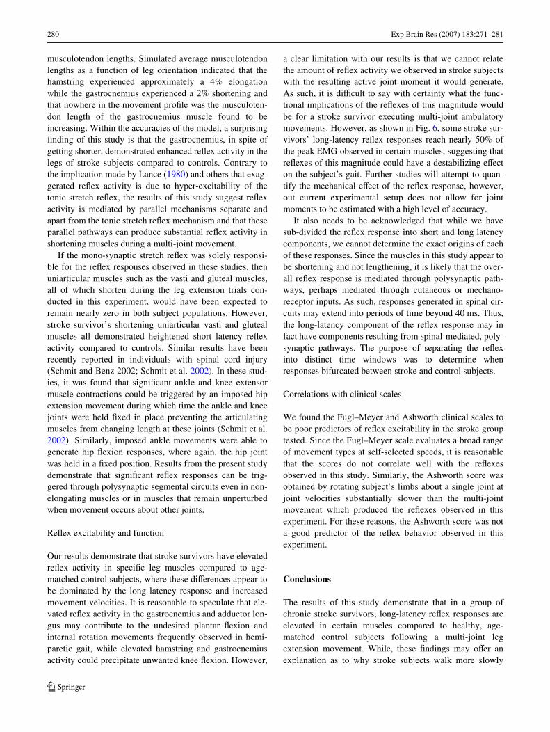
Our results demonstrate that stroke survivors have elevatedreXex activity in speciWc leg muscles compared to age-matched control subjects, where these diVerences appear tobe dominated by the long latency response and increasedmovement velocities. It is reasonable to speculate that ele-vated reXex activity in the gastrocnemius and adductor lon-gus may contribute to the undesired plantar Xexion andinternal rotation movements frequently observed in hemi-paretic gait, while elevated hamstring and gastrocnemiusactivity could precipitate unwanted knee Xexion. However,
a clear limitation with our results is that we cannot relatethe amount of reXex activity we observed in stroke subjectswith the resulting active joint moment it would generate.As such, it is diYcult to say with certainty what the func-tional implications of the reXexes of this magnitude wouldbe for a stroke survivor executing multi-joint ambulatorymovements. However, as shown in Fig. 6, some stroke sur-vivors’ long-latency reXex responses reach nearly 50% ofthe peak EMG observed in certain muscles, suggesting thatreXexes of this magnitude could have a destabilizing eVecton the subject’s gait. Further studies will attempt to quan-tify the mechanical eVect of the reXex response, however,out current experimental setup does not allow for jointmoments to be estimated with a high level of accuracy.
It also needs to be acknowledged that while we havesub-divided the reXex response into short and long latencycomponents, we cannot determine the exact origins of eachof these responses. Since the muscles in this study appear tobe shortening and not lengthening, it is likely that the over-all reXex response is mediated through polysynaptic path-ways, perhaps mediated through cutaneous or mechano-receptor inputs. As such, responses generated in spinal cir-cuits may extend into periods of time beyond 40 ms. Thus,the long-latency component of the reXex response may infact have components resulting from spinal-mediated, poly-synaptic pathways. The purpose of separating the reXexinto distinct time windows was to determine whenresponses bifurcated between stroke and control subjects.
Correlations with clinical scales
We found the Fugl–Meyer and Ashworth clinical scales tobe poor predictors of reXex excitability in the stroke grouptested. Since the Fugl–Meyer scale evaluates a broad rangeof movement types at self-selected speeds, it is reasonablethat the scores do not correlate well with the reXexesobserved in this study. Similarly, the Ashworth score wasobtained by rotating subject’s limbs about a single joint atjoint velocities substantially slower than the multi-jointmovement which produced the reXexes observed in thisexperiment. For these reasons, the Ashworth score was nota good predictor of the reXex behavior observed in thisexperiment.
Conclusions
The results of this study demonstrate that in a group ofchronic stroke survivors, long-latency reXex responses areelevated in certain muscles compared to healthy, age-matched control subjects following a multi-joint legextension movement. While, these Wndings may oVer anexplanation as to why stroke subjects walk more slowly
123
Exp Brain Res (2007) 183:271–281 281
(von Schroeder et al. 1995) and have less postural stability(Collen 1995) when compared to able-bodied individuals ofsimilar age, further studies run under more functionalbehaviors (e.g. gait) are necessary to determine whetherspasticity impairs walking and balance.
References
Ada L, Vattanasilp W, O’Dwyer NJ, Crosbie J (1998) Does spasticitycontribute to walking dysfunction after stroke? J Neurol Neuro-surg Psychiatry 64:628–635
Ashworth B (1964) Preliminary trial of carisoprodal in multiple scle-rosis. Practitioner 192:540–542
Black i, Nichols D, Pelliccio M, Hidler JH (2005) QuantiWcation ofreXex activity in stroke survivors during an imposed mulit-jointleg extension movement. In: Proceedings of the 35th annual meet-ing of the society for neuroscience in Washington, DC, SessionNo. 869
Brown D, Kautz S (1999) Speed dependent reductions of force outputin people with post stroke hemiparesis. Phys Ther 79:919–930
Capaday C, Forget R, Milner T (1994) A re-examination of the eVectsof instruction on the long-latency stretch reXex response of theXexor pollicis longus muscle. Exp Brain Res 100(3):515–521
Collen FM (1995) The measurement of standing balance after stroke.Physiother Theory Pract 11:109–182
Craig JJ (1989) An introduction to robotics: mechanics and control.Addison-Wesley Publishing, Reading, MA
Dewald JP, Given JD, Rymer WZ (1996) Long-lasting reductions ofspasticity induced by skin electrical stimulation. IEEE TransRehabil Eng 4(4):231–242
Dietz V, Ibrahim IK, Trippel M, Berger W (1993) Spastic paresis:reXex activity and muscle tone in elbow muscles during passiveand active motor tasks. In: Thilmann AF et al (eds) Spasticity:mechanisms and management. Springer, Berlin–Heidelberg,pp 251–265
Dietz V, Trippel M, Berger W (1991) ReXex activity and muscle toneduring elbow movements in patients with spastic paresis. AnnNeurol 30:767–779
Duysens J, Trippel M, Horstmann GA, Dietz V (1990) Gating andreversal of reXexes in ankle muscles during human walking. ExpBrain Res 82(2):351–358
Engardt M, Knutsson E, Jonsson M, Sternhag M (1995) Dynamic mus-cle strength training in stroke patients: eVects on knee extensiontorque, electromyographic activity, and motor function. ArchPhys Med Rehabil 76(5):419–425
Fugl-Meyer A, Jaasko L, Leyman I, Olsson S, Seglind S (1975) Thepost-stroke hemiplegic patient: a method of evaluation of physicalperformance. Scand J Rehabil Med 7:13–31
Galiana L, Fung J, Kearney R (2005) IdentiWcation of intrinsic and reX-ex ankle stiVness components in stroke patients. Exp Brain Res165(4):422–434
Hidler JM, Rymer WZ (2000) Limit cycle behavior in spasticity: anal-ysis and evaluation. IEEE Trans Biomed Eng 47:1565–1575
Ibrahim IK, Berger W, Trippel M, Dietz V (1993) Stretch-inducedelectromyographic activity and torque in spastic elbow muscles.DiVerential modulation of reXex activity in passive and activemotor tasks. Brain 116(Pt 4):971–989
Kearney RE, Lortie M, Stein RB (1999) Modulation of stretch reXexesduring imposed walking movements of the human ankle. J Neu-rophysiol 81(6):2893–2902
Knutsson E (1980) Restraint of spastic muscles in diVerent types ofmovement. In: Feldman RG, Young RR, Werner KP (eds) Spas-ticity: disordered motor control. Year Book, Chicago, pp 123–132
Knutsson E, Martensson A, Gransberg L (1997) InXuences of musclestretch reXexes on voluntary, velocity-controlled movements inspastic paraparesis. Brain 120(Pt 9):1621–1633
Knutsson E (1985) Studies of gait control in patients with spastic pare-sis. Clin Neurophysiol Spasticity 174–183
Lamontagne A, Malouin F, Richards CL (2001) Locomotor-speciWcmeasure of spasticity of plantarXexor muscles after stroke. ArchPhys Med Rehabil 82(12):1696–1704
Lance JW (1980) Pathophysiology of spasticity and clinical experiencewith baclofen. In: Feldman RG, Young RR, Koella WP (eds)Spasticity: disordered motor control. Year Book Medical Publish-ers, Chicago, pp 185–203
Perry J (1980) Rehabilitation of spasticity. In: Feldman RG, YoungRR, Werner KP (eds) Spasticity: disordered motor control. YearBook, Chicago, pp 87–99
Powers RK, Campbell DL, Rymer WZ (1989) Stretch reXex dynamicsin spastic elbow Xexor muscles. Ann Neurol 25(1):32–42
Powers RK, Marder-Meyer J, Rymer WZ (1988) Quantitative relationsbetween hypertonia and stretch reXex threshold in spastic hemi-paresis. Ann Neurol 23(2):115–124
Schmit BD, Benz EN, Rymer WZ (2002) AVerent mechanisms for thereXex response to imposed ankle movement in chronic spinal cordinjury. Exp Brain Res 145:40–49
Schmit BD, Dhaher Y, Dewald JP, Rymer WZ (1999) ReXex torque re-sponse to movement of the spastic elbow: theoretical analyses andimplications for quantiWcation of spasticity. Ann Biomed Eng27(6):815–829
Vattanasilp W, Ada L, Crosbie J (2000) Contribution of thixotropy,spasticity, and contracture to ankle stiVness after stroke. J NeurolNeurosurg Psychiatry 69(1):34–39
von Schroeder HP, Coutts RD, Lyden PD, Billings E Jr, Nickel VL(1995) Gait parameters following stroke: a practical assessment.J Rehabil Res Dev 32:25–31
Yamamoto C, Ohtsuki T (1989) Modulation of stretch reXex by antic-ipation of the stimulus through visual information. Exp Brain Res77(1):12–22
123