Embed Size (px)
Citation preview

APPTED MICROBIOLOGY, Aug. 1969, p. 214-220 VoL 18, No. 2Copyright © 1969 American Society for Microbiology Printed in U.S.A.
Quantitation of Microorganisms in SputumP. W. MONROE, H. G. MUCHMORE, F. G. FELTON, AND J. K. PIRTLE
Departments ofMedicine and Microbiology, The University ofOklahoma Medical Center andVeterans Administration Hospital, Oklahoma City, Oklahoma 73104
Received for publication 25 April 1969
A method of quantitating microbial cultures of homogenized sputum has beendevised. Possible application of this method to the problem of determining theetiologic agent of lower-respiratory-tract infections has been studied to determineits usefulness as a guide in the management of these infections. Specimens wereliquefied by using an equal volume of 2% N-acetyl-L-cysteine. The liquefied sputumsuspension was serially diluted to 10-1, 10-3, 10-5, and 10-7. These dilutions wereplated on appropriate media byusing anO.01-ml calibrated loop; they were incubatedand examined by standard diagnostic methods. Quantitation of fresh sputum frompatients with pneumonia prior to antimicrobial therapy revealed that probablepathogens were present in populations of 107 organisms/ml or greater. Normaloropharyngeal flora did not occur in these numbers before therapy. Comparison ofmicrobial counts on fresh and aged sputum showed that it is necessary to use onlyfresh specimens, since multiplication or death alters both quantitative and qualita-tive findings. Proper collection and quantitative culturing of homogenized sputumprovided information more directly applicable to patient management than didqualitative routine methods. Not only was the recognition of the probable patho-genic organism in pneumonia patients improved, but serial quantitative cultureswere particularly useful in recognizing the emergence of superinfections and inevaluating the efficacy of antimicrobial therapy.
Routine qualitative microbial cultures of spu-tum are widely utilized in medicine to determinethe etiology of lower-respiratory-tract infectionsand as a guide in the administration of appro-priate antimicrobial agents. However, these cul-tures often fail to give adequate information,because the contamination of the specimen byoral flora (3, 7, 13) and the irregular distributionof organisms within the sample (8) make correctrecognition of the true pathogen difficult. Insuch cases, quantitative microbial analyses ofhomogenized sputum would be helpful if theycould provide better pathogen recognition.Studies of other infectious diseases, e.g., urinarytract infections, have demonstrated that deter-minations of the numbers of organisms presentmay be of considerable value in deciding whichof these is causing disease and which is presentas incidental colonization or recent contamina-tion during specimen collection (4, 10, 12).Quantitation of organisms in sputum presentsmore problems than in urine specimens, becausethe former normally contains many bacteriawhich, of necessity, will be enumerated and be-cause the sputum contains several substances,such as mucus, which must be liquefied beforethorough mixing can be achieved. This study
was undertaken to develop a reliable method forobtaining a true estimate of the numbers of thevarious species of microorganisms present insputum, and, if possible, to relate the numbers ofresident microbial populations with respect totheir significance as agents of lower-respiratory-tract infections.
MATERIALS AND METHODSClinical specimens. Specimens used in the study
were of three types. (i) Sputum specimens collectedand submitted to the clinical microbiology laboratoryof the Veterans Administration Hospital, OklahomaCity, were used. The time between specimen collectionand its culture in the laboratory was approximately4 hr, more or less. These specimens are referred to asaged sputum. (ii) Fresh sputum specimens were col-lected from 19 patients with clinical pneumonia whohad received no antibiotics. These patients wereselected by resident physicians assigned to the Infec-tious Disease section of the Department of Medicine,University of Oklahoma School of Medicine, Okla-homa City. Specimens were collected prior to andduring antimicrobial therapy after rinsing the mouthwith fresh tap water. The specimens were either proc-essed immediately or refrigerated at 4 C for no longerthan 1 hr. (iii) Unstimulated saliva specimens wereobtained from subjects who had no clinical symptomsof lower respiratory disease.
214
on March 2, 2020 by guest
http://aem.asm
.org/D
ownloaded from
QUANTITATION OF MICROORGANISMS IN SPUTUM
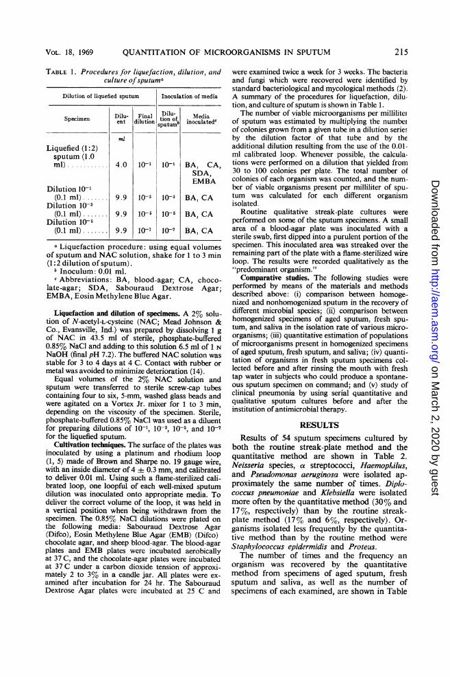
TABLE 1. Procedures for liquefaction, dilution, andculture ofsputuma
Dilution of liquefied sputum Inoculation of media
Specimen Dilu- Final Dilu- MediaSpecimen D-ent dilution tion ofb inoculated"sputum
ml
Liquefied (1:2)sputum (1.0ml) ..........0.4010- 10'- BA, CA,
SDA,EMBA
Dilution 10-1(0.1 ml).. 9. 9l0-3 1o-3 BA, CA
Dilution 10-3(0.1 ml).. 9.9 10- 10 BA, CA
Dilution10-X(0.1 ml). 9.9 10-7 10-7 BA, CA
a Liquefaction procedure: using equal volumesof sputum and NAC solution, shake for 1 to 3 min(1:2 dilution of sputum).
1 Inoculum: 0.01 ml.c Abbreviations: BA, blood-agar; CA, choco-
late-agar; SDA, Sabouraud Dextrose Agar;EMBA, Eosin Methylene Blue Agar.
Liquefaction and dilution of specimens. A 2% solu-tion of N-acetyl-L-cysteine (NAC; Mead Johnson &Co., Evansville, Ind.) was prepared by dissolving 1 gof NAC in 43.5 ml of sterile, phosphate-buffered0.85% NaCl and adding to this solution 6.5 ml of 1 NNaOH (final pH 7.2). The buffered NAC solution wasstable for 3 to 4 days at 4 C. Contact with rubber ormetal was avoided to minimize deterioration (14).
Equal volumes of the 2% NAC solution andsputum were transferred to sterile screw-cap tubescontaining four to six, 5-mm, washed glass beads andwere agitated on a Vortex Jr. mixer for 1 to 3 min,depending on the viscosity of the specimen. Sterile,phosphate-buffered 0.85% NaCl was used as a diluentfor preparing dilutions of 10-1, 10-s, 10-5, and 10-7for the liquefied sputum.
Cultivation techniques. The surface of the plates wasinoculated by using a platinum and rhodium loop(1, 5) made of Brown and Sharpe no. 19 gauge wire,with an inside diameter of 4 i 0.3 mm, and calibratedto deliver 0.01 ml. Using such a flame-sterilized cali-brated loop, one loopful of each well-mixed sputumdilution was inoculated onto appropriate media. Todeliver the correct volume of the loop, it was held ina vertical position when being withdrawn from thespecimen. The 0.85% NaCl dilutions were plated onthe following media: Sabouraud Dextrose Agar(Difco), Eosin Methylene Blue Agar (EMB) (Difco)chocolate agar, and sheep blood-agar. The blood-agarplates and EMB plates were incubated aerobicallyat 37 C, and the chocolate-agar plates were incubatedat 37 C under a carbon dioxide tension of approxi-mately 2 to 3% in a candle jar. All plates were ex-amined after incubation for 24 hr. The SabouraudDextrose Agar plates were incubated at 25 C and
were examined twice a week for 3 weeks. The bacteriaand fungi which were recovered were identified bystandard bacteriological and mycological methods (2).A summary of the procedures for liquefaction, dilu-tion, and culture of sputum is shown in Table 1.The number of viable microorganisms per milliliter
of sputum was estimated by multiplying the numberof colonies grown from a given tube in a dilution seriefby the dilution factor of that tube and by theadditional dilution resulting from the use of the 0.01-ml calibrated loop. Whenever possible, the calcula-tions were performed on a dilution that yielded from30 to 100 colonies per plate. The total number ofcolonies of each organism was counted, and the num-ber of viable organisms present per milliliter of spu-tum was calculated for each different organismisolated.
Routine qualitative streak-plate cultures wereperformed on some of the sputum specimens. A smallarea of a blood-agar plate was inoculated with asterile swab, first dipped into a purulent portion of thespecimen. This inoculated area was streaked over theremaining part of the plate with a flame-sterilized wireloop. The results were recorded qualitatively as the"predominant organism."
Comparative studies. The following studies wereperformed by means of the materials and methodsdescribed above: (i) comparison between homoge-nized and nonhomogenized sputum in the recovery ofdifferent microbial species; (ii) comparison betweenhomogenized specimens of aged sputum, fresh spu-tum, and saliva in the isolation rate of various micro-organisms; (iii) quantitative estimation of populationsof microorganisms present in homogenized specimensof aged sputum, fresh sputum, and saliva; (iv) quanti-tation of organisms in fresh sputum specimens col-lected before and after rinsing the mouth with freshtap water in subjects who could produce a spontane-ous sputum specimen on command; and (v) study ofclinical pneumonia by using serial quantitative andqualitative sputum cultures before and after theinstitution of antimicrobial therapy.
RESULTSResults of 54 sputum specimens cultured by
both the routine streak-plate method and thequantitative method are shown in Table 2.Neisseria species, a streptococci, HaemopIilus,and Pseudomonas aeruginosa were isolated ap-proximately the same number of times. Diplo-coccus pneumoniae and Klebsiella were isolatedmore often by the quantitative method (30% and17%, respectively) than by the routine streak-plate method (17% and 6%, respectively). Or-ganisms isolated less frequently by the quantita-tive method than by the routine method wereStaphylococcus epidermidis and Proteus.The number of times and the frequency an
organism was recovered by the quantitativemethod from specimens of aged sputum, freshsputum and saliva, as well as the number ofspecimens of each examined, are shown in Table
VOL. 18, 1969 215
on March 2, 2020 by guest
http://aem.asm
.org/D
ownloaded from

MONROE ET AL.
TABLE 2. Comparison of results obtained by theliquefaction-dilution method and the routinestreak-plate methodfor 54 sputum specimens
Liquefaction- Routine streak-dilution method plate method
OrganismNo. of No. oftimes Per cent times Per cent
isclated isolated
Staphylococcusepidermidis ......... 6 11 14 26
Neisseria spp.......... 40 74 37 69a Streptococci ....... 25 46 28 52Diplococcus pneu-moniae ............. 16 30 9 17
Haemophilus spp...... 6 11 5 9Klebsiella sp .......... 9 17 3 6Proteus spp ........ 4 7 6 11Pseudomonas aeru-ginosa .............. 5 9 5 9
3. Except for Neisseria species in the aged sputumspecimens, the isolation rates among the orga-nisms of the normal oral flora were very similarin three studies. D. pneumoniae was isolated 10%
more often and Haemophilus was isolated 30%more often when the sputum was fresh than whenit had been held at room temperature beforeculturing. Whether the sputum was fresh or agedappeared to make no difference in the recoveryrates of Klebsiella and Candida. Certain orga-nisms, namely D. pneumoniae, Haemophilus, andEscherichia coli, were not isolated from any of thesaliva specimens. In general, there were fewerkinds of organisms isolated per specimen fromaged sputum than from fresh sputum or saliva(Table 4).Except for Klebsiella and Haemophilus, larger
populations of organisms were recovered fromaged sputum than from fresh sputum and saliva.The mean numbers for those organisms whichare considered to be members of the normal oralflora, i.e., S. epidermidis, Neisseria species, and et
streptococci, were very similar for fresh sputumand saliva (Table 5).
Comparative results of quantitative sputumcultures before and after the mouth was rinsedwith fresh tap water are presented in Table 6. Atotal of six specimens were examined. No S.epidermidis, E. coli, or Candida were isolated
TABLE 3. Comparision of the number of isolates from aged sputum specimens,fresh sputum specimens, and saliva specimens
Aged sputum Fresh sputum Saliva(78 specimens)a (103 specimens) (20 specimens)
Organism _ _Times Per cent *Times Per cent Times Per cent
isolated ssolated isolated
Staphylococcus epidermidis ....... 14 18 14 14 3 15Neisseria spp .............. 32 41 76 74 16 80axStreptococci ............. 62 80 80 78 17 81Diplococcus pneumoniae............. 16 20 31 30 0 0Haemophilus spp .................... 6 8 39 38 0 0Klebsiella sp .............. 17 22 17 17 7 35Enterobacter sp ............ 3 4 27 26 5 25Escherichia coli ............ 4 5 16 15 0 0Candida spp .............. 15 19 14 21 2 10
a Incubated at room temperature for approximately 4 hr or longer before being cultured.
TABLE 4. Comparison of the number of species of microorganisms isolated per specimenfrom aged sputum, fresh sputum, and saliva
No. of microbial species per specimen" Mean no. MedianNo. of ofseis no.of
Type of specimen specimens per speciesspecsmens -. ~~~~~~~~~~~~~~~~~~~perieper1 2 3 4 5 | specimen
Aged sputum. 78 13 (17%) 28 (36%) 25 (32%) 7 (9%) 5 (6%) 2.4 2Fresh sputum ... 103 11 (11%) 27 (26%) 33 (32%) 20 (20%) 12 (11%) 3.0 3Saliva .......... 20 0 (0%) 5 (25%) 7 (35%) 7 (35%) 1 (5%) 3.2 3
a Number and frequency of specimens containing given number of microbial species.
216 APPL. MICROBIOL.
on March 2, 2020 by guest
http://aem.asm
.org/D
ownloaded from

QUANTITATION OF MICROORGANISMS IN SPUTUM
TABLE 5. Quantitation of organisms in aged sputum, fresh sputum,and saliva (logio organisms/ml)
Aged sputum Fresh sputum Saliva(78 specimens) (103 specimens) (20 specimens)
Organism
Range Median Mean Range Median Mean Range Median Mean
Staphylococcus epidermidis. 5-10 7 8 5-6 6 6 3-6 3 5Neisseria spp.6-11 8 9 3-8 6 6 3-8 6 7a Streptococci.7-10 8 9 3-9 6 7 3-7 6 6Diplococcuspneumoniae............7-11 8 10 3-10 6 8 0 0 0Haemophilus spp.9-11 10 11 4-12 8 10 0 0 0Klebsiella sp.3-10 5 8 3-10 3 8 3-4 3 3Enterobacter sp.3-11 5 10 3-7 3 6 3 3 3Escherichia coli.5-8 6 7 3-4 3 3 0 0 0Candida spp.3-13 4 11 3-5 4 4 3-4 3 3
TABLE 6. Quantitative results from sputum cultures before and afterrinsing mouth with fresh tap water
Before rinsing After rinsing
Organism _ Per cent changeTimes Organisms/ml Times Organisms/mlisolated (mean) isolated (mean)
Haemophilus spp..................... 5 4 X 10¹⁰ 5 1 X 10¹⁰ -75%Diplococcus pneumoniae.............. 3 3 X 10⁹ 4 2 X 10¹⁰ +85%a Streptococcus..................... 4 3 X 10⁶ 4 2 X 10⁵ -93%Neisseria spp........................ 6 9 X 10⁷ 6 2 X 10⁷ -78%Staphylococcus epidermidis .......... 1 8 X 10⁷ 0 0 -100%Escherichia coli..................... 1 3 X 10³ 0 0 -100%Candida spp......................... 1 2 X 10³ 0 0 -100%
TABLE 7. Comparison between quantitative androutine sputum cultures in pneumonia
(before treatment)
No. of times isolated
Organism Quantitative Routine(107 or (predom-greater) inant
organism)'
Diplococcus pneumonia 7 4Haemophilus spp.......... 8 1Both of the above ........ 3 0Staphlococcus aureus1......ITotal isolated/total pa-
tients ................... 19/19 6/17
a Two patients had no routine sputum cultureson admission.
after the mouth was rinsed, as compared withone isolate of each before rinsing. In three in-stances (Haemophilus, a streptococci, and Neis-seria species), the number of times an organismwas isolated remained the same before and afterrinsing. However, D. pneumoniae was isolated
three times before mouth rinsing but was foundfour times after rinsing, and was present ingreater numbers. Even though some of the orga-nisms were still present after rinsing the mouth,their numbers were significantly reduced. Popu-lations of a streptococci and Neisseria specieswere decreased 93% and 78%, respectively,whereas the count of Haemophilus was decreased75%.
Quantitative cultures on admission sputumspecimens from 19 patients with pneumoniarevealed D. pneumoniae to be present in greatestnumbers in seven patients, Haemophilus ineight, and both D. pneumoniae and Haemophiluspresent in largest numbers in three; in onepatient, S. aureus was the most numerous (Table7). These organisms in high numbers wererecovered from all 19 patients by use of the quan-titative method, whereas by the routine qualita-tive streak-plate method they were the predomi-nating organisms in only 6 of 17, approximately35 %. Routine streak-plate cultures on admissionwere not done on two patients. In all instances,the organisms noted above were present in thesputum specimens in counts of 107 organisms/ml
VOL. 18, 1969 217
on March 2, 2020 by guest
http://aem.asm
.org/D
ownloaded from

218
108
MONROE ET AL.
| KEFLIN 4 GM/ D IV I
I.156 ~~~~~~~~~~~~Li
0 105
2 ........--'...
i...... .
._
0 2 6
DAYS
FIG. 1. Serial quantitative sputum cultures from apneumonia patient prior to (day 0) and during anti-microbial therapy. D. pneumoniae is the apparentpathogenic organism and is the only organism occur-ring in numbers greater than 107/ml. This level ofapparent pathogenic significance is indicated by thehorizontal line at that point.
| PENICILLIN 2.4 M.U./D IM
DAYS
FIG. 2. Serial quantitative sputum cultures from apatient with pneumonia apparently due to two orga-nisms, D. pneumoniae and H. influenzae. Day 0 andhorizontal line ofsignificance are as in Fig. 1.
or greater. None of the other organisms occurredin concentrations this high. Quantitative cultureswere repeated at intervals during the patients'hospital treatment. These results on four patientsare shown in Fig. 1-4.
APPL. MICROBIOL.
h. ~TETRACYCLINE GM D-
06I
00
00o
LnIo
1i3
0 3DAYS
FIG. 3.IFSerial quantitative counts on sputum inpneumonia with two potential pathogens, H. parain-fluenzae and D. pneumoniae. Day 0 and line ofsignifi-cance are as in Fig. 1.
I-.------ PENICILUN 200.MU. /D IV- |i---AMPICILUN 6GM, cD IV --43C49-KAAKMYCIN GM/ D - |750 MGf
10i0 .FCOUSTIN4100 MG/
#- D. PNEUMONIAE
109 A- A^_ ^H. INFLUENZAEP. AERUGINOSA
I'1 ........ KLEBSIELLA SP.
0 I8
O71
1,060
0 1 2 4 5 7 a 9 10 II
DAYS
FIG. 4. Serial quantitative sputum cultures from a
patient with pneumonia followed by superinfectionswith Klebsiella and P. aeruginosa. Day 0 and line ofsignificance are as in Fig. 1.
-
0
z
0v0
020
1031
108
a . . a a
on March 2, 2020 by guest
http://aem.asm
.org/D
ownloaded from

QUANTITATION OF MICROORGANISMS IN SPUTUM
DISCUSSIONCertain obvious differences were evident in the
results of culturing the same sputum specimenby both the quantitative method and the routinestreak-plate method. May (8) pointed out thatto obtain accurate, viable culture results it wasnecessary to use techniques which took intoaccount the irregular distribution of pathogensin sputum specimens. The quantitative methodused in the present study produced a uniformlyhomogeneous specimen, and this homogeneitymight have accounted for the higher isolationrates of D. pneumoniae and Klebsiella than werefound by routine plating. It is possible that thecapsule characteristic of these two organismscaused clumping and uneven distribution through-out the specimen.The investigations of Richardson and Jones
(11) have shown that in saliva the quantitativerange of S. epidermis is 10 to 104 organisms/ml.Since an 0.01-ml calibrated loop was used in thepresent study to streak plates from the serialdilution tubes, the number of organisms permilliliter in a sputum specimen would have tohave been present in a minimal concentration of103 organisms/ml to be observed (10-1 dilutiontube X 10-2 calibrated loop). This fact wouldaccount for the smaller number of S. epidermidisisolates by the quantitative method than by theroutine streak-plate method. There was no ap-preciable difference in the isolation rate ofNeisseria species and a streptococci by the twomethods. Their populations are reported to begreater than 104 organisms/ml (6, 11), and theywould, therefore, be isolated as often by thequantitative method as by routine methods.The isolation rate of D. pneumoniae and
species of Haemophilus from aged sputum wasless than that of fresh sputum. May and Delves(9) compared the isolation rates of D. pneumoniaeand H. influenzae from fresh specimens ofsputum with those from specimens sent to thelaboratory by mail. The isolation rates for bothorganisms from mailed specimens were approxi-mately half those from fresh specimens.The results of the present study revealed that
fewer kinds of organisms were isolated perspecimen from aged sputum than from freshsputum or saliva. The most plausible reason forthis may be that certain fastidious organismscannot remain viable for long periods of timeoutside their natural environment before beingcultured on appropriate media. For example,certain organisms, such as the pneumococcus,often undergo autolysis. Also, sputum and salivacontain antimicrobial substances, such as lyso-zyme, which can destroy organisms, and some
organisms produce substances which haveantimicrobial properties toward other microbialspecies. These factors are possibly related to thedecrease in the number of microbial species inaged sputum specimens.The fact that quantitative counts of various
bacterial species in aged sputum were higherthan those in fresh sputum or saliva indicatesthat the organisms continued to multiply aftercollection. Even though D. pneumoniae and H.influenzae had a tendency to be nonviable in theaged sputum specimens, when they were isolatedtheir numbers were as high as those of some ofthe other organisms, such as S. epidermidis anda streptococci. This suggests that if the organismsare able to survive, they usually multiply.The results showed that rinsing the mouth
with fresh tap water before obtaining a sputumspecimen decreased the bacterial counts of thenormal oral flora organisms. Organisms whosecolony counts are reported to be low in saliva(11, 15), i.e., S. epidermidis, E. col, and Candida,were reduced below minimal numbers recognizedby this method. As was noted previously, how-ever, the number of D. pneumoniae isolates, aswell as the quantitative count, was increasedafter mouth rinsing. Reasons for this increaseare not clear. However, rinsing the mouth shouldreduce the concentration of any inhibiting sub-stance in the saliva. Also, rinsing would reducethe populations of a streptococci, allowing D.pneumoniae to be recognized more easily, sincetheir colonies are very similar in macroscopicappearance. Thus, if the mouth is rinsed beforeobtaining a sputum specimen to reduce thenumbers of a streptococci, the possibility ofconfusing these colonies should be greatlydiminished.The patients with pneumonia whose sputum
yielded one organism in high numbers usually
a 25uiI-
:520
ui 15
1=1-b 10
z <2Li;NON-PATHOGENS
IPATHOGENS
mRl~3 4 5 6 7 8 9 in 11
LOGIO ORGANISMS/ ML.
FIG. 5. Quantitative distribution of pathogens andnonpathogens cultured from the sputum of 19 pneu-monia patients before antimicrobial therapy.
II IIM am.MM1
VOL. 18, 1969 219
on March 2, 2020 by guest
http://aem.asm
.org/D
ownloaded from

MONROE ET AL.
responded within a few days to appropriateantimicrobial therapy and had no problem withsuperinfection (Fig. 1). Pneumonia caused bytwo different organisms responded similarly whentherapy was directed against susceptible orga-nisms (Fig. 2).e. The results obtained from culturing serialsputum specimens from the 19 patients withpneumonia by using the quantitative methodshow that this method may provide betterbacteriological data to the physician, thusleading to more effective patient care in lower-respiratory-tract infections. The presence ofpotential pathogens in sputum may be ignoredif they occur in low numbers, e. g., Candida inFig. 1 and Enterobacter in Fig. 2. The administra-tion of antimicrobial agents to eliminate theseorganisms from the sputum would have beenuseless and possibly detrimental to the patient.Quantitative counts appeared to establish whichwas the etiologic agent if two different potentialpathogens were present in the sputum, e.g.,Haemophilus and D. pneumoniae in Fig. 3. Onepatient died due to a superinfection first withKlebsiella followed by P. aeruginosa (Fig. 4).The clinical course of this patient might havebeen altered if the significance of the high colonycounts of P. aeruginosa, indicating a truesuperinfection, had been realized sooner, andappropriate change of antibiotics had been madeearlier.By this technique, the organism associated with
a given lower-respiratory-tract infection, whetherit be primary pneumonia or a superinfection, ispresent in concentrations of 107 or more orga-nisms/ml of sputum. Normal mouth flora andopportunists are found in sputum before therapyin concentrations of 106 organisms/ml or less(Fig. 5).
Quantitative cultures performed on serialspecimens in identical fashion will reveal adecrease or disappearance of the pathogenicmicroorganisms in response to appropriatetherapy and, moreover, may provide the earliestindication of the emergence of superinfectionbefore there is any apparent worsening of thepatient's clinical condition. Quantitative countsabove 107 of normal flora organisms during
antimicrobial therapy could not be related tosuperinfection in this study (Fig. 3).
This study shows that quantitative microbialcultures of sputum provide the physician withuseful information regarding infections of thelower respiratory tract and that they are a betterguide than routine culture in the selection of theproper antibiotic to administer to the patient.
ACKNOWLEDGMENTS
This investigation was supported by Public Health Serviceresearch grant AI-07618 and training grant I-TI-AI-326 from theNational Institute of Allergy and Infectious Diseases, by researchgrant CC-00081 fiom the National Communicable Disease Cen-ter, and by research funds from the Veterans AdministrationHospital, Oklahoma City, Okla.
LITERATURE CITED
1. American Public Health Association. 1960. Standard methodsfor the examination of dairy products, 11th ed. Amer.Publ. Health Ass., New York.
2. Bailey, W. R., and E. G. Scott. 1966. Diagnostic micro-biology, 2nd ed. C. V. Mosby Co., Saint Louis.
3. Brumfit, W., M. L. N. Willoughby, and L. L. Bromley. 1957.An evaluation of sputum examination in chronic bronchitis.Lancet 2:1306-1309.
4. Dineen, P. 1964. The importance of the route of infection inexperimental biliary tract obstruction. Surg. Gynecol.Obstet. Int. Abstr. Surg. 119:1001-1008.
5. Hoeprick, P. D. 1960. Culture of the urine. J. Lab. Clin.Med. 56:899-907.
6. Kraus, F. W., and C. Gaston. 1956. Individual constancyof numbers among the oral flora. J. Bacteriol. 71:703-707.
7. Lapinski, E. M., E. D. Flakes, and B. C. Taylor. 1964. Anevaluation of some methods for culturing sputum frompatients with bronchitis and emphysema. Amer. Rev.Resp. Dis. 89:760-763.
8. May, J. R. 1953. The bacteriology of chronic bronchitis.Lancet 2:543-547.
9. May, J. R., and D. M. Delves. 1964. The survival of Haemo-philus influenzae and pneumococci in specimens of sputumsent to the laboratory by post. J. Clin. Pathol. 17:254-256.
10. Okinaka, A. J., and P. Dineen. 1968. Bacterial colony countson bronchial washings. Ann. Surg. 167:47-50.
11. Richardson, R. L., and M. Jones. 1958. A bacteriologiccensus of human saliva. J. Dent. Res. 37:697-709.
12. Sanford, J. P., C. B. Favour, F. H. Mao, and J. H. Harrison.1956. Evaluation of positive urine cultures. Amer. J. Med.20:88-93.
13. Shulman, J. A., L. A. Phillips, and R. G. Petersdorf. 1965.Errors and hazards in the diagnosis and treatment of bac-terial pneumonias. Ann. Int. Med. 62:41-58.
14. Woodhams, A. W., and G. R. Mead. 1965. A comparisonbetween pancreatin and N-acetyl-L-cysteine as sputumliquefying agents for the culture of organisms. Tubercle46:224-226.
15. Young, G., H. G. Resca, and M. T. Sullivan. 1951. The yeastsof the normal mouth and their relation to salivary acidity.J. Dent. Res. 30:426-430.
220 APPL. MICROBIOL.
on March 2, 2020 by guest
http://aem.asm
.org/D
ownloaded from