Embed Size (px)
Citation preview

Quiz
1. Bacteria of which phase are used for most experiments? Why?
Which phase today?
2. What are the three properties that a plasmid has to have to be useful for cloning purposes?
And guess the fourth property to be used for protein expression purposes?

If you repeat the “bacterial growth curve” experiment, which step would you do different than you have done last week? (To perform one of your data collection step correctly and easier)

Plasmids
Usually occur naturally in bacteria
Circular, ds1-400 kb1- 100/1000 copies per cellSingle OR
Stringent : Replicates only when the host chromosome replicates Relaxed: Replicates independently
Conjugation, a mechanism of horizontal gene transfer.

Vectors
• Plasmids used in genetic engineering (as vectors)
• Antibiotic resistance gene
• MCS (multiple cloning site)
• OR (origin of replication)
• Commercially available for cloning purposes
• Commonly used to multiply (make many copies of) or
express particular genes
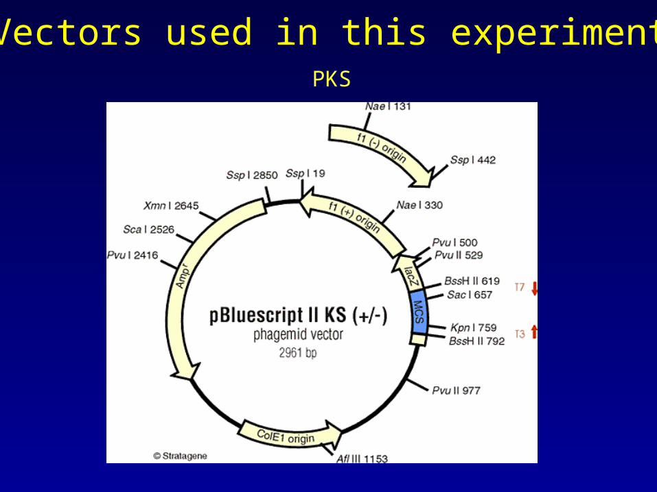
Vectors used in this experimentPKS
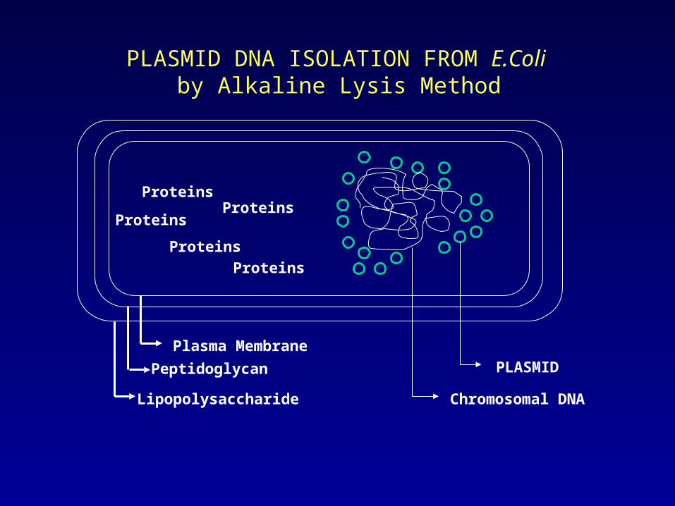
PLASMID DNA ISOLATION FROM E.Coli by Alkaline Lysis Method
Lipopolysaccharide
Peptidoglycan
Plasma Membrane
PLASMID
Chromosomal DNA
Proteins
Proteins
Proteins
Proteins
Proteins

Plasmid DNA Isolation
(Alkaline Lysis Method)
o/n culture of cells containing plasmids (liquid medium)
Cell extract preparation by SDS Lysis
Deproteinization by Phenol Extraction
Removal of salts and concentrating the DNA
Miniprep (ng)---2 ml
Midiprep (μg)---10ml
Maxiprep (mg)---50ml

Table 1. Techniques used for the physical disruption of cells.
Lysis Method Description Apparatus
MechanicalWaring BlenderPolytron
Rotating blades grind and disperse cells and tissues
Liquid Homogenization
Dounce HomogenizerPotter-Elvehjem HomogenizerFrench Press
Cell or tissue suspensions are sheared by forcing them through a narrow space
Sonication SonicatorHigh frequency sound waves shear cells
Freeze/ThawFreezer or dry ice/ethanol
Repeated cycles of freezing and thawing disrupt cells through ice crystal formation
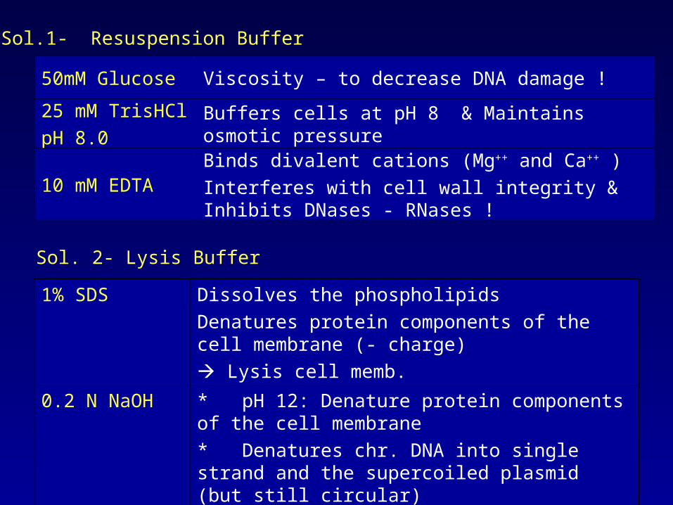
Sol.1- Resuspension Buffer
50mM Glucose Viscosity – to decrease DNA damage !
25 mM TrisHCl
pH 8.0Buffers cells at pH 8 & Maintains osmotic pressure
10 mM EDTABinds divalent cations (Mg++ and Ca++ )
Interferes with cell wall integrity & Inhibits DNases - RNases !
1% SDS Dissolves the phospholipids
Denatures protein components of the cell membrane (- charge)
Lysis cell memb.
0.2 N NaOH * pH 12: Denature protein components of the cell membrane
* Denatures chr. DNA into single strand and the supercoiled plasmid (but still circular)
Sol. 2- Lysis Buffer
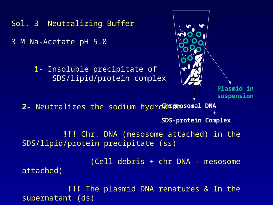
Sol. 3- Neutralizing Buffer
3 M Na-Acetate pH 5.0
1- Insoluble precipitate of SDS/lipid/protein complex
Plasmid in suspension
Chromosomal DNA +SDS-protein Complex
2- Neutralizes the sodium hydroxide
!!! Chr. DNA (mesosome attached) in the SDS/lipid/protein precipitate (ss)
(Cell debris + chr DNA – mesosome attached)
!!! The plasmid DNA renatures & In the supernatant (ds)

Phenol-chloroform-isoamylalcohol extraction (25:24:1)
Phenol: Dissociate proteins from nucleic acids
Chloroform: Protein and lipid denaturation
Isoamylalcohol: Prevents foaming
Biphasic mixture
Organic phase : Proteins
Aqueous phase (upper): Nucleic acids (+ other contaminants such as salts, sugars, etc.)

Ethanol Precipitation of DNA & Concentrating
DNA is polar, soluble in water & Insoluble in less polar ethanol
1. 100 % ethanol & -70
Centrifugation
Precipitate DNA & salts that form ionic bonds with DNA
!!! Ethanol interaction with the water Less water molecules are free to dissolve DNA
2. 70% ethanol
30 % water solubilizes the salts present in the pellet
!!! Supernatant is removed
DNA is resuspended in TE / dH2O

Quantification of the Plasmid DNA
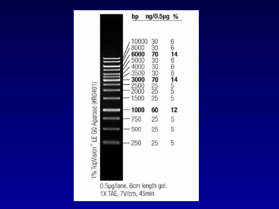
Agarose gel: Comparing the intensity of the ladder bands (for linear DNA)
Spectrophotometry:
OD at 260nm
1 OD = 50 µg/ml ds DNA = 37 µg/ml ss DNA = 40 µg/ml ss RNA
Q (µg/ml)= A260 x 50 x Dilution factor

OD260 = 0.2 0,2 x 50 µg/ml = 10 µg/ml
(2 µlDNA + 98 µl H2O) with 0.2 OD260:
10 µg/ml x 50 = 500 µg/ml
Yield (µg) : 500 (µg/ml ) x Total volume (ml)

Purity
Pure dsDNA A260/A280= 1.7-1.9 (1.8)
OD 260/280 < 1.8 protein contamination (Prt absorbs at 280nm)
OD 260/280 > 1.8 RNA or residual organics contamination
OD 260/270 ratio should be ~1.2, if no phenol cont. OD 260/280: 2.0 Phenol
OD 260/230 2 < 2 presence of organics
OD 330 should be “0”

-Nicked-Linear
- Relaxed circular- Supercoiled

1- 1/50 diln in 300 μl dH2O measure OD260!
1μl using Nanodrop Spectrophotometer!
2- 5 μl plasmid DNA + 1 μl 6X agarose loading dye 4 μl 1 kb ladder (Fermentas SM0313)
1% agarose gel Take the image of agarose gel separated plasmids!