Embed Size (px)
Citation preview
155
IRANIAN JOURNAL OF PATHOLOGYVol.11 No.2, Spring 2016
Background: Overexpression of Rad51 protein in many tumor cells has been proven to increase radioresistance and can be related to the resistance of chemosensitivity of tumor cells. This preliminary study was conducted to determine the relationship between the Rad51 expression level in nasopharyngeal carcinoma and the response of the treatment based on the measurement of the tumor reduction.
Methods:Thirteen cases of the NPCs were analyzed. The expression levels of the Rad51 were examined from the pretreatment biopsies. Furthermore, tumor reductions were determined based on the change in sum longest diameter of the nasopharyngeal CT-scan before and after therapy.
Results: The expression level of the Rad51 was associated with the reduction of tumor mass. The P value was 0.049 and the correlation coefficient was 0.479.
Conclusion: The tumor cells Rad51 expression levels may affect the tumor reduction of NPC after the therapy.
Short Communication | Iran J Pathol. 2016; 11(2): 155 - 160
Introduction
The nasopharyngeal carcinoma or NPC is known as a major health problem in Asia (1). This malignancy is the third most commonly found in the growth of malignant among men in Malaysia, where the age-standardized rate or ASR of the NPC for male was 8.5/100 000 (2), while in Indonesia, the incidence rate of the NPC is 6.2/100000. In addition, the nasopharyngeal carcinomas are the highest cases between head and neck malignancies (3).
Etiology factors on NPC i.e. Epstein-Barr virus (EBV) infection, diet, smoking, occupational exposure, and environmental as well as genetic factors, are inter-related to each other (4-6). The EBV infection and genotoxic agents can contribute to DNA damage (4-8). Thus, individuals with defects in DNA repair genes have a tendency to have a higher risk on the NPC. This leads to establish a concept of the induction of the NPC that has a strong link with the genes involvement in DNA-repair mechanism (7, 8).
Rad51 Expression in Nasopharyngeal Carcinoma and Its Association with Tumor Reduction: A Preliminary Study in Indonesia
Dian Cahyanti1, Lisnawati Rachmadi1, Vally Wulani2, Marlinda Adham3
1. Department of Anatomical Pathology, Faculty of Medicine Universitas Indonesia/Cipto Mangunkusumo Hospital, Jakarta, Indonesia
2. Department of Radiology, Faculty of Medicine Universitas Indonesia/Cipto Mangunkusumo Hospital, Jakarta, Indonesia3. Department of Ear Nose Throat Head and Neck Surgery, Faculty of Medicine Universitas Indonesia/Cipto
Mangunkusumo Hospital, Jakarta, Indonesia
Nasopharyngeal carcinomaRad51 expressionTumor reduction
K E Y W O R D S
A R T I C L E I N F O
Corresponding Information: Dr. Dian Cahyanti, Department of Anatomical Pathology, Faculty of Medicine Universitas Indonesia/ Cipto Mangunkusumo Hospital, Jalan Salemba Raya 6, Jakarta 10430. Email: [email protected], Tel: +622-131905888
http://www.ijp.iranpath.org/
Received 13 Jan 2015; Accepted 08 Jul 2015;
©Iran J Pathol. All rights reserved.
A B S T R A C T
Copyright © 2016, IRANIAN JOURNAL OF PATHOLOGY. This is an open-access article distributed under the terms of the Creative Commons Attribution-noncommercial 4.0 International License which permits copy and redistribute the material just in noncommercial usages, provided the original work is properly cited.
156
Vol.11 No.2, Spring 2016IRANIAN JOURNAL OF PATHOLOGY
Rad51 is a protein that plays a fundamental role in DNA-repair mechanism by homologous recombination repair or HRR. This pathway acts when there is a double-strand breaks (DSBs) type of DNA damage because of exposure to ionizing radiation and crosslinking agents (9-12). The increase of Rad51 expression can be characterized by the increase in cellular resistance against radiation and some chemotherapeutic agents (13-16). Moreover, overexpression in Rad51 protein is found in a variety of tumor cell lines and associated with poor prognosis in some malignancies (16-18).
Radiation therapy is the main treatment of the NPCs. Chemotherapy usually is given as concurrent chemotherapy with a platinum-based regimen (19). Both therapeutic agents, work by damaging the DNA of tumor cells and causes the onset of DNA damage DSBs type (9-11). Studies on Rad51 protein in nasopharyngeal carcinoma are sparse. This gives rise to us to do a preliminary study in Indonesia.
In this study, Rad51 expression was investigated in the nasopharyngeal biopsies from patients with nasopharyngeal carcinoma. All of them were treated with the combination of radiation therapy and chemotherapy. The reduction of the tumor mass was evaluated by CT scan of the nasopharynx before and after therapy.
Methods
This cross-sectional study had received the ethical approval from the Faculty of Medicine Universitas Indonesia-Cipto Mangunkusumo Hospital.
The cases of NPC were retrieved from the archive of the Department of Anatomical Pathology between January 2012 and December 2013. The CT-scan information before and after therapy were discovered from the Picture Archiving and Communication System (PACS) in the Department of Radiology in the same period. Total number of patients was calculated by using
the Pearson’s coefficient correlation formula and selected for inclusion based on the availability of the CT-scan data after treatment. None of the patients had undergone chemotherapy and radiation before biopsy.
All patients received a total dose of radiation 70 Gy, and a platinum-based chemotherapy in accordance to their clinical stage. The tumor mass reduction was assessed at 8-12 weeks after completion of radiation.
Immunohistochemical techniqueSlides for immunostaining prepared with 4
μm-thick sections deparaffinized and underwent for rehydration. The slides were processed for heat induced antigen retrieval via microwave in citrate buffer; followed by incubated the slides with primary anti-Rad51 (ab88572, Abcam®) antibody with a dilution of 1:300 for 60 min then biotinylated universal antibody (Starrtrek Universal, Biocare Medical). It continued with a streptavidin-peroxidase complex. The slides were colored with diaminobenzidine substrate. Before proceed for dehydration, the slides were stained with Mayer’s hematoxylin solution.
The scoring of cells was based on nuclear staining via manual cell counting of 200 randomly selected tumor cells. The intensity of nuclear staining (0 to 3+) was scored by comparing the intensity in the negative control slides. This study utilized the H-score scoring system that represented the summation of the positively cells multiplied by a weighted intensity of staining (σ[Pi(i+1)]) (20). The assessment of Rad51 expression levels were done without knowing the clinical history.
Measurement of tumor reductionThe tumor mass reduction measurement were
used the unidimensional measurement method, by means of measuring the longest diameter of the tumor mass area. The value obtained was the percentage of the tumor mass reduction (21).
Rad51 Expression in Nasopharyngeal Carcinoma
157
IRANIAN JOURNAL OF PATHOLOGYVol.11 No.2, Spring 2016
Statistical analysis Statistical analysis was done by the
program SPSS 17.0 (Chicago, IL, USA). The correlation and association between Rad51 expression and the tumor reduction was assessed by Pearson’s correlation.
Results and Discussion
Thirteen cases were obtained in this study. Nasopharyngeal biopsy tissue was analyzed to determine the H-score of Rad51 staining. As known function on DNA repair, the Rad51 protein was quantified in nuclear staining only, although some cytoplasmic staining was noted. The assessments on H-scores of Rad51 expression level were done by two observers. The scoring
method on the intensity (score 1 to 3) was made by agreement. Interpretation of Rad51 staining is shown in Figure 1.
Since the patients who underwent nasopharyngeal CT scan after therapy at Cipto Mangunkusumo Hospital are limited, consequently we included three cases with clinical stage II and III, and one case with non-keratinizing carcinoma feature. The clinical and histopathological characteristics of the patients and tumors are detailed in Table 1.
To determine whether Rad51 expression level of the NPC tumor is associated with its resistance to therapy, the H-scores were compared with radiographic tumor reduction (Figure 2 and 3).
CT scans of the nasopharynx were performed on each patient before and after therapy. For
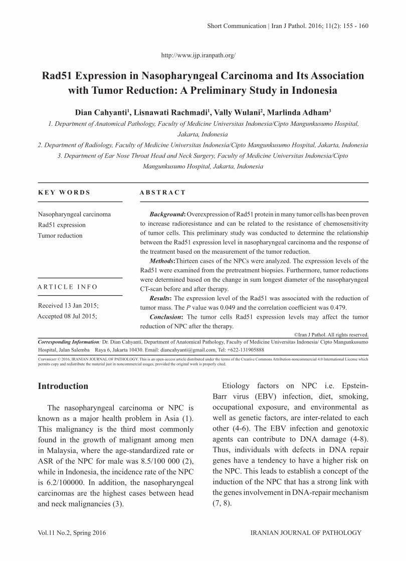
Fig. 1Interpretation of Rad51 immunostaining in high power magnification. Even the cytoplasm of the tumor cells were also stained, the H-scores evaluation was based on the positivity in the tumor cells nuclei only. (A) Score 0 for negative expression, (B) score 1 for mild expression of the tumor cells nuclei, (C) score 2 for moderate expression, and (D) score 3 for strong expression
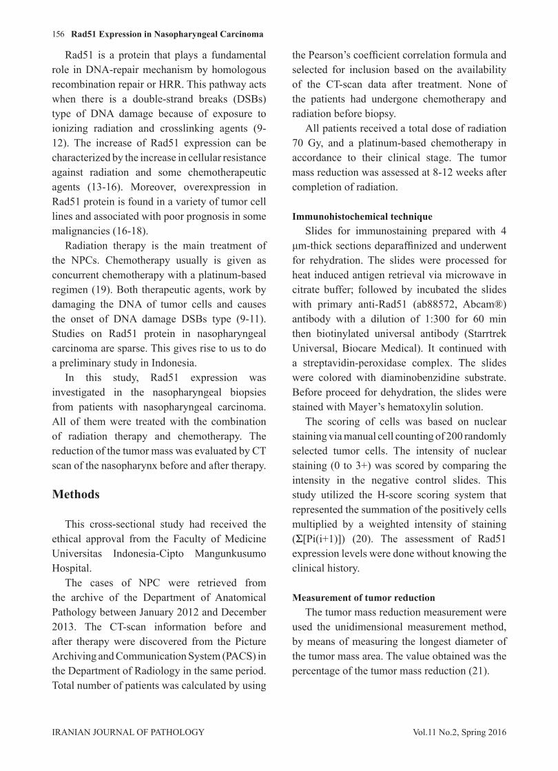
Patients number Sex Age (yr) Histopathology Clinical Stage H-score Persentage of tumor reduction (%)1 M 42 UC II 292 34.52 M 25 UC IVA 302.5 333 M 40 UC IVA 246.5 69.34 M 34 UC IVA 291 23.65 M 51 UC IVB 288.5 32.16 M 64 UC IVA 294 29.57 M 42 NC IVB 295 54.48 M 63 UC III 273.5 58.39 M 56 UC IVA 272 100
10 F 22 UC III 291.5 50.811 M 27 UC IVA 309 44.512 M 39 UC IVA 299.5 17.213 F 44 UC IVB 292 12.3
Table 1Clinical and histopathological characteristics of patients and tumors
*UC: Undifferentiated carcinoma, NC: Nonkeratinizing carcinoma
Cahyanti et al.
158
Vol.11 No.2, Spring 2016IRANIAN JOURNAL OF PATHOLOGY
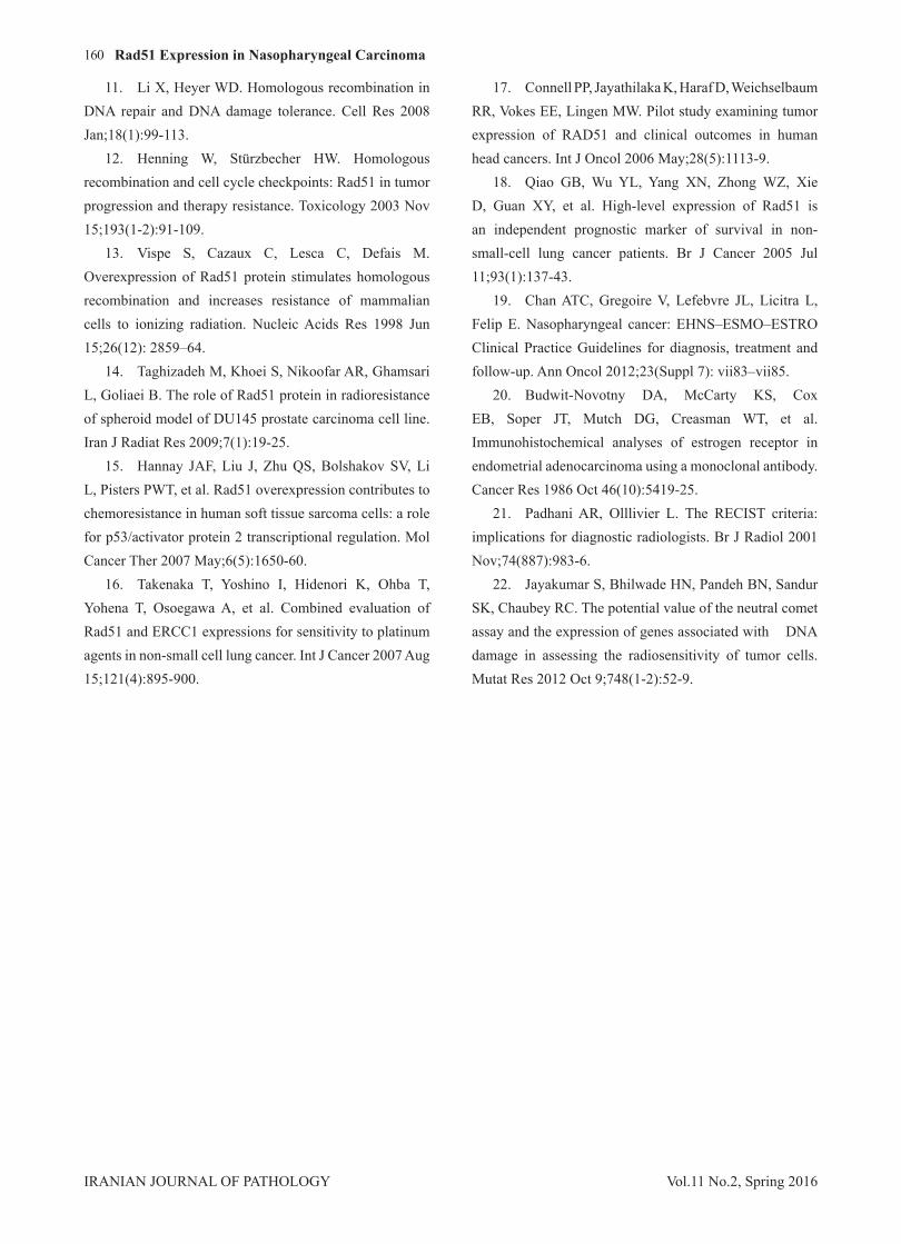
each patient, the tumor mass was selected for unidimensional measurement. The correlation between the H-scores and tumor reduction after therapy is shown in Figure 4.
DNA repair gene RAD51L1 has consistent evidence as genetic factors associated with NPC risk (7, 8). This is the first study in Indonesia to compare the Rad51 overexpression in NPC with the radiographic tumor response. In the end, this study demonstrates a correlation between Rad51
expression level and the percentage of tumor mass reduction. The result indicates that the higher the level of Rad51 expression the tumor cells become more resistant to radiation and chemotherapy.
Several studies have reported Rad51 overexpression in other types of human malignancies. The study of Connel et al. in patients with head and neck cancer stated that those with high expression level of Rad51 protein in their pre-treatment tissues showed poorer survival rate than those without (33.3% vs. 88.9% at 2 years; P=0.025) (17).In addition, there was a trend correlation between Rad51 expression level and tumor responsiveness after platinum-based chemotherapy (17). We assume that Rad51 could be a prognostic marker in order to predict the nasopharyngeal carcinoma response to chemoradiation.
We expect that overexpression of Rad51 protein associated with the poor tumor responsiveness. The elevated in Rad51 expression level results in an increase response to DNA damage (9). These occurrences are presumably due to a consequence from up-regulation at transcriptional level in tumor cells (9, 12).
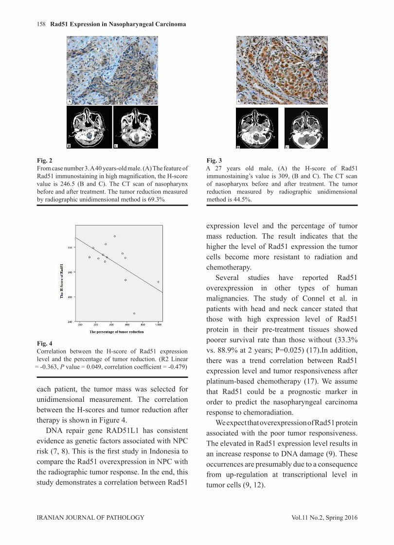
Fig. 2From case number 3. A 40 years-old male. (A) The feature of Rad51 immunostaining in high magnification, the H-score value is 246.5 (B and C). The CT scan of nasopharynx before and after treatment. The tumor reduction measured by radiographic unidimensional method is 69.3%
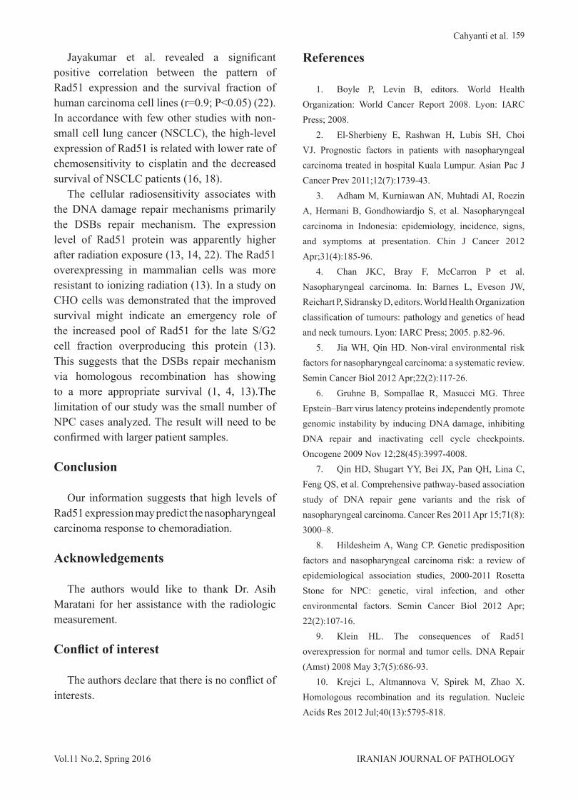
Fig. 3A 27 years old male, (A) the H-score of Rad51 immunostaining’s value is 309, (B and C). The CT scan of nasopharynx before and after treatment. The tumor reduction measured by radiographic unidimensional method is 44.5%.
Fig. 4Correlation between the H-score of Rad51 expression level and the percentage of tumor reduction. (R2 Linear
= -0.363, P value = 0.049, correlation coefficient = -0.479)
Rad51 Expression in Nasopharyngeal Carcinoma
159
IRANIAN JOURNAL OF PATHOLOGYVol.11 No.2, Spring 2016
Jayakumar et al. revealed a significant positive correlation between the pattern of Rad51 expression and the survival fraction of human carcinoma cell lines (r=0.9; P<0.05) (22). In accordance with few other studies with non-small cell lung cancer (NSCLC), the high-level expression of Rad51 is related with lower rate of chemosensitivity to cisplatin and the decreased survival of NSCLC patients (16, 18).
The cellular radiosensitivity associates with the DNA damage repair mechanisms primarily the DSBs repair mechanism. The expression level of Rad51 protein was apparently higher after radiation exposure (13, 14, 22). The Rad51 overexpressing in mammalian cells was more resistant to ionizing radiation (13). In a study on CHO cells was demonstrated that the improved survival might indicate an emergency role of the increased pool of Rad51 for the late S/G2 cell fraction overproducing this protein (13). This suggests that the DSBs repair mechanism via homologous recombination has showing to a more appropriate survival (1, 4, 13).The limitation of our study was the small number of NPC cases analyzed. The result will need to be confirmed with larger patient samples.
Conclusion
Our information suggests that high levels of Rad51 expression may predict the nasopharyngeal carcinoma response to chemoradiation.
Acknowledgements
The authors would like to thank Dr. Asih Maratani for her assistance with the radiologic measurement.
Conflict of interest
The authors declare that there is no conflict of interests.
References
1. Boyle P, Levin B, editors. World Health Organization: World Cancer Report 2008. Lyon: IARC Press; 2008.
2. El-Sherbieny E, Rashwan H, Lubis SH, Choi VJ. Prognostic factors in patients with nasopharyngeal carcinoma treated in hospital Kuala Lumpur. Asian Pac J Cancer Prev 2011;12(7):1739-43.
3. Adham M, Kurniawan AN, Muhtadi AI, Roezin A, Hermani B, Gondhowiardjo S, et al. Nasopharyngeal carcinoma in Indonesia: epidemiology, incidence, signs, and symptoms at presentation. Chin J Cancer 2012 Apr;31(4):185-96.
4. Chan JKC, Bray F, McCarron P et al. Nasopharyngeal carcinoma. In: Barnes L, Eveson JW, Reichart P, Sidransky D, editors. World Health Organization classification of tumours: pathology and genetics of head and neck tumours. Lyon: IARC Press; 2005. p.82-96.
5. Jia WH, Qin HD. Non-viral environmental risk factors for nasopharyngeal carcinoma: a systematic review. Semin Cancer Biol 2012 Apr;22(2):117-26.
6. Gruhne B, Sompallae R, Masucci MG. Three Epstein–Barr virus latency proteins independently promote genomic instability by inducing DNA damage, inhibiting DNA repair and inactivating cell cycle checkpoints. Oncogene 2009 Nov 12;28(45):3997-4008.
7. Qin HD, Shugart YY, Bei JX, Pan QH, Lina C, Feng QS, et al. Comprehensive pathway-based association study of DNA repair gene variants and the risk of nasopharyngeal carcinoma. Cancer Res 2011 Apr 15;71(8): 3000–8.
8. Hildesheim A, Wang CP. Genetic predisposition factors and nasopharyngeal carcinoma risk: a review of epidemiological association studies, 2000-2011 Rosetta Stone for NPC: genetic, viral infection, and other environmental factors. Semin Cancer Biol 2012 Apr; 22(2):107-16.
9. Klein HL. The consequences of Rad51 overexpression for normal and tumor cells. DNA Repair (Amst) 2008 May 3;7(5):686-93.
10. Krejci L, Altmannova V, Spirek M, Zhao X. Homologous recombination and its regulation. Nucleic Acids Res 2012 Jul;40(13):5795-818.
Cahyanti et al.
160
Vol.11 No.2, Spring 2016IRANIAN JOURNAL OF PATHOLOGY
11. Li X, Heyer WD. Homologous recombination in DNA repair and DNA damage tolerance. Cell Res 2008 Jan;18(1):99-113.
12. Henning W, Stürzbecher HW. Homologous recombination and cell cycle checkpoints: Rad51 in tumor progression and therapy resistance. Toxicology 2003 Nov 15;193(1-2):91-109.
13. Vispe S, Cazaux C, Lesca C, Defais M. Overexpression of Rad51 protein stimulates homologous recombination and increases resistance of mammalian cells to ionizing radiation. Nucleic Acids Res 1998 Jun 15;26(12): 2859–64.
14. Taghizadeh M, Khoei S, Nikoofar AR, Ghamsari L, Goliaei B. The role of Rad51 protein in radioresistance of spheroid model of DU145 prostate carcinoma cell line. Iran J Radiat Res 2009;7(1):19-25.
15. Hannay JAF, Liu J, Zhu QS, Bolshakov SV, Li L, Pisters PWT, et al. Rad51 overexpression contributes to chemoresistance in human soft tissue sarcoma cells: a role for p53/activator protein 2 transcriptional regulation. Mol Cancer Ther 2007 May;6(5):1650-60.
16. Takenaka T, Yoshino I, Hidenori K, Ohba T, Yohena T, Osoegawa A, et al. Combined evaluation of Rad51 and ERCC1 expressions for sensitivity to platinum agents in non-small cell lung cancer. Int J Cancer 2007 Aug 15;121(4):895-900.
17. Connell PP, Jayathilaka K, Haraf D, Weichselbaum RR, Vokes EE, Lingen MW. Pilot study examining tumor expression of RAD51 and clinical outcomes in human head cancers. Int J Oncol 2006 May;28(5):1113-9.
18. Qiao GB, Wu YL, Yang XN, Zhong WZ, Xie D, Guan XY, et al. High-level expression of Rad51 is an independent prognostic marker of survival in non-small-cell lung cancer patients. Br J Cancer 2005 Jul 11;93(1):137-43.
19. Chan ATC, Gregoire V, Lefebvre JL, Licitra L, Felip E. Nasopharyngeal cancer: EHNS–ESMO–ESTRO Clinical Practice Guidelines for diagnosis, treatment and follow-up. Ann Oncol 2012;23(Suppl 7): vii83–vii85.
20. Budwit-Novotny DA, McCarty KS, Cox EB, Soper JT, Mutch DG, Creasman WT, et al. Immunohistochemical analyses of estrogen receptor in endometrial adenocarcinoma using a monoclonal antibody. Cancer Res 1986 Oct 46(10):5419-25.
21. Padhani AR, Olllivier L. The RECIST criteria: implications for diagnostic radiologists. Br J Radiol 2001 Nov;74(887):983-6.
22. Jayakumar S, Bhilwade HN, Pandeh BN, Sandur SK, Chaubey RC. The potential value of the neutral comet assay and the expression of genes associated with DNA damage in assessing the radiosensitivity of tumor cells. Mutat Res 2012 Oct 9;748(1-2):52-9.
Rad51 Expression in Nasopharyngeal Carcinoma