Embed Size (px)
Citation preview
Acc
epte
d A
rtic
le
This article has been accepted for publication and undergone full peer review but has not been
through the copyediting, typesetting, pagination and proofreading process, which may lead to
differences between this version and the Version of Record. Please cite this article as doi:
10.1111/tpj.13260
This article is protected by copyright. All rights reserved.
Received Date : 12-Jul-2015
Revised Date : 24-Jun-2016
Accepted Date : 30-Jun-2016
Article type : Original Article
Dimer/monomer status and in vivo function of salt-bridge mutants of the plant UV-B
photoreceptor UVR8
Monika Heilmann1, Christos N. Velanis, Catherine Cloix2, Brian O. Smith, John M. Christie
and Gareth I. Jenkins*
Institute of Molecular, Cell and Systems Biology, College of Medical, Veterinary and Life
Sciences, Bower Building, University of Glasgow, Glasgow G12 8QQ, UK
1 Present address: BASF SE, Crop Protection, Speyerer Str. 2, 67117 Limburgerhof,
Germany
2 Present address: Cancer Research UK Beatson Institute, Garscube Estate, Switchback
Road, Bearsden, Glasgow G61 1BD
* For correspondence (e-mail [email protected])
Acc
epte
d A
rtic
le
This article is protected by copyright. All rights reserved.
Running title: UVR8 salt-bridge mutants
Key words: UV-B, UVR8, photoreceptor, photomorphogenesis, Arabidopsis thaliana
Author for correspondence: Gareth I. Jenkins, Bower Building, University of Glasgow,
Glasgow G12 8QQ, UK. Phone +44 141 330 5906; e-mail [email protected]
SUMMARY
UV RESISTANCE LOCUS8 (UVR8) is a photoreceptor for ultraviolet-B (UV-B) light that
initiates photomorphogenic responses in plants. UV-B photoreception causes rapid
dissociation of dimeric UVR8 into monomers that interact with CONSTITUTIVELY
PHOTOMORPHOGENIC1 (COP1) to initiate signal transduction. Experiments with purified
UVR8 show that the dimer is maintained by salt-bridge interactions between specific
charged amino acids across the dimer interface. However, little is known about the
importance of these charged amino acids in determining dimer/monomer status and UVR8
function in plants. Here we evaluate the use of different methods to examine dimer/monomer
status of UVR8 and show that mutations of several salt-bridge amino acids affect
dimer/monomer status, interaction with COP1 and photoreceptor function of UVR8 in vivo. In
particular, the salt-bridges formed between arginine 286 and aspartates 96 and 107 are key
to dimer formation. Mutation of arginine 286 to alanine impairs dimer formation, interaction
with COP1 and function in vivo, whereas mutation to lysine gives a weakened dimer that is
functional in vivo, indicating the importance of the positive charge of the arginine/lysine
Acc
epte
d A
rtic
le
This article is protected by copyright. All rights reserved.
residue for dimer formation. Notably, a UVR8 mutant in which aspartates 96 and 107 are
conservatively mutated to asparagine is strongly impaired in dimer formation but mediates
UV-B responses in vivo with a similar dose-response relationship to wild-type. The UV-B
responsiveness of this mutant does not correlate with dimer formation and monomerisation,
indicating that monomeric UVR8 has the potential for UV-B photoreception, initiating signal
transduction and responses in plants.
INTRODUCTION
UV-B wavelengths (280-315 nm) in sunlight have a major impact on the biosphere. The high
energy of UV-B radiation has the potential to damage molecules such as DNA, impair
cellular activities and cause cell death. However, plants have evolved effective mechanisms
to protect themselves from damage by UV-B, which enable them to survive constant
exposure to sunlight. In particular, plants synthesize UV-absorbing sunscreen compounds
that are deposited in the outer tissues and they employ efficient cellular systems for
repairing damage by UV-B (Frohnmeyer and Staiger, 2003; Ulm and Nagy, 2005; Jenkins,
2009). The exposure of plants to low doses of UV-B stimulates transcriptional responses
that underpin UV-protection (Ulm et al., 2004; Brown et al., 2005; Favory et al., 2009).
Furthermore, UV-B acts as a regulatory stimulus for other responses in plants, including the
suppression of extension growth (Favory et al., 2009; Hayes et al., 2014), entrainment of the
circadian clock (Feher et al., 2011) and defence against insect herbivory (Ballaré et al.,
2012).
Regulatory responses to UV-B are mediated by the photoreceptor UV RESISTANCE
LOCUS8 (UVR8; Brown et al., 2005; Favory et al., 2009; Rizzini et al., 2011; Tilbrook et al.,
2013; Jenkins, 2014a). Arabidopsis uvr8 mutant plants are defective in photomorphogenic
responses to UV-B and are highly susceptible to damage by UV-B because they are unable
Acc
epte
d A
rtic
le
This article is protected by copyright. All rights reserved.
to stimulate expression of genes concerned with UV-protection (Brown et al., 2005; Favory
et al., 2009; Kliebenstein et al., 2002). UVR8 is a 7-bladed -propeller protein that is present
as a homo-dimer in plants (Rizzini et al., 2011; O’Hara and Jenkins, 2012; Christie et al.,
2012; Wu et al., 2012; Jenkins, 2014b). Unlike other photoreceptors UVR8 does not use a
prosthetic chromophore for light sensing, instead specific tryptophan amino acids of UVR8
act as chromophores for UV-B detection (Rizzini et al., 2011; O’Hara and Jenkins, 2012;
Christie et al., 2012; Wu et al., 2012; Zeng et al., 2015). Photoreception induces rapid
dissociation of the dimer into monomers (Rizzini et al., 2011; Christie et al., 2012; Wu et al.,
2012), which then interact with the CONSTITUTIVELY PHOTOMORPHOGENIC1 (COP1)
protein to initiate signaling and hence regulate transcription of target genes involved in
UVR8-mediated responses (Favory et al., 2009; Rizzini et al., 2011; Cloix et al., 2012). One
of the genes most rapidly induced by UV-B following UVR8 photoreception encodes the
transcription factor ELONGATED HYPOCOTYL5 (HY5). This transcription factor, sometimes
acting with the related HY5 HOMOLOG (HYH) is a key effector of transcriptional responses
regulated by UVR8 (Brown et al., 2005; Oravecz et al., 2005; Brown and Jenkins, 2008;
Favory et al., 2009; Huang et al., 2013). In addition, UVR8 photoreception stimulates
expression of genes encoding the REPRESSOR OF UV PHOTOMORPHOGENESIS1
(RUP1) and RUP2 proteins, which negatively regulate UVR8 responses (Gruber et al.,
2010).
The crystal structure of UVR8 shows that dimer formation is dependent on salt-
bridge interactions between charged amino acids at the interface where monomers come
into contact (Christie et al., 2012; Wu et al., 2012; Jenkins, 2014b). Hydrophobic interactions
between the monomers are negligible. The dimerisation surface of each monomer is rich in
basic amino acids, notably arginine, and acidic aspartate and glutamate residues. These
amino acids are distributed such that patches of complementary electrostatic potential are
aligned opposite each other across the dimer interface. The salt-bridge interactions are
sufficiently strong that the dimer is maintained even in the presence of SDS, as long as the
Acc
epte
d A
rtic
le
This article is protected by copyright. All rights reserved.
protein sample is not boiled (Rizzini et al., 2011; Christie et al., 2012; Wu et al., 2012).
However, monomerisation occurs when salt-bridges are neutralized by low pH and high ionic
strength (Christie et al., 2012; Wu et al., 2012). Moreover, studies with purified UVR8
expressed in E. coli show that mutation of particular salt-bridging amino acids prevents
dimer formation (Christie et al., 2012; Wu et al., 2012).
Inevitably the molecular environment of UVR8 in cells will differ from that in vitro. It is
therefore essential to establish whether findings with the purified protein extend to UVR8 in
plants. Hence the aim of the present study was to determine whether particular salt-bridge
amino acids are important in maintaining UVR8 structure in plants and to assess the
consequences of altered dimer/monomer status on UVR8 function in vivo. We show that
specific arginine and aspartate amino acids at the UVR8 dimerisation surface are required
for dimer formation in transgenic Arabidopsis plants. However, although mutants in some of
these amino acids are impaired in dimer formation both in vitro and in vivo, they are
nevertheless able to functionally complement Arabidopsis uvr8-1 plants. This study
highlights the methodological difficulty of establishing the dimer/monomer status of a UVR8
mutant protein in plants, and provides evidence that UVR8 can perceive UV-B and initiate
signaling even in its monomeric form.
RESULTS
Dimer/monomer status of selected UVR8 salt-bridge mutant proteins
We wished to study the effects of mutations of several charged amino acids at the dimer
interface on UVR8 dimer/monomer status in vivo. The selection of amino acids and
mutations was based on examination of the UVR8 crystal structure and on biochemical
studies of purified UVR8 mutant proteins, which identified amino acids that are critical for
Acc
epte
d A
rtic
le
This article is protected by copyright. All rights reserved.
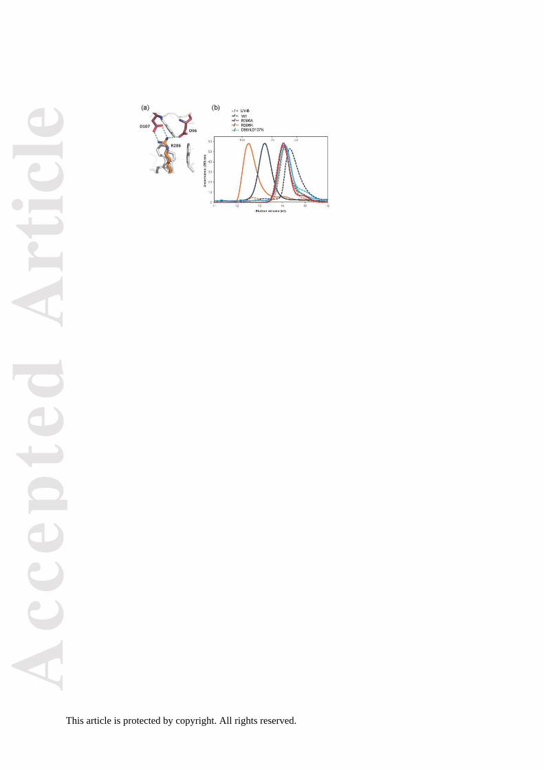
dimer formation in vitro (Christie et al., 2012; Wu et al., 2012). A key residue is arginine
R286, which is adjacent to the principal chromophore tryptophan, W285, and forms double
and single H-bonded salt-bridges respectively with aspartates D107 and D96 (Figure 1a).
Size exclusion chromatography (SEC) shows that purified wild-type UVR8 is a dimer when
not exposed to UV-B and a monomer following UV-B exposure, whereas mutation of either
R286 to alanine (UVR8R286A) or D96 and D107 to asparagine (UVR8D96N,D107N) causes UVR8
to become constitutively monomeric in vitro (Figure 1b; Christie et al., 2012; Wu et al.,
2012). In contrast, UVR8R286K, in which R286 is conservatively mutated to positively charged
lysine, appears dimeric and monomerises in response to UV-B (Figure 1b). The SEC elution
volume of the UVR8R286K dimer differs from that of wild-type UVR8, most likely because of a
change in the hydrodynamic radius (shape) of the protein; this difference is not evident when
the salt concentration is increased (Figure S1). Similar to R286, R146 forms a double H-
bonded salt bridge, in this case with E182 (Figure S2a). However, in contrast to UVR8R286A,
UVR8R146A is a dimer in vitro that monomerises in response to UV-B (Christie et al., 2012;
Wu et al., 2012; Figure S2b). R234, which is adjacent to UV-B chromophore W233, forms an
intra-molecular salt-bridge with E182 (Figure S2a). UVR8R234A adopts a conformation in vitro
that is non-responsive to UV-B (Figure S2c), most likely because the mutation could disrupt
the spatial arrangement of the chromophore tryptophans, impairing UV-B photoreception.
R338 is adjacent to the triad tryptophan W337 and forms a single hydrogen-bonded salt
bridge with D44 and a non hydrogen-bonded ionic interaction with E43 as well as a water
mediated hydrogen bond with its backbone carbonyl (Figure S3a). The UVR8R338A mutant is
reported to be constitutively monomeric in vitro (Wu et al., 2012). However, the
dimer/monomer status of UVR8R338A in vitro is dependent on the salt concentration; it is
monomeric in 500 mM NaCl but appears to be in equilibrium between dimer and monomer in
low salt concentrations (Figure S3b).
Acc
epte
d A
rtic
le
This article is protected by copyright. All rights reserved.
Mutation of key UVR8 salt-bridge amino acids impairs dimer formation in plants
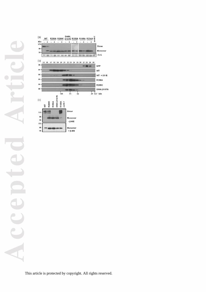
The mutant UVR8 proteins described above were expressed as GFP fusions in the null
uvr8-1 mutant. Several transgenic lines were obtained for each mutant and compared to a
control GFP-UVR8 fusion that was shown previously to functionally complement uvr8-1
(Brown et al., 2005; Kaiserli and Jenkins, 2007; Figure S4). The dimer/monomer status of
the mutant UVR8 proteins was examined initially using SDS-PAGE with non-boiled samples.
This assay shows that wild-type GFP-UVR8 is a dimer that monomerises after UV-B
exposure, but in contrast each mutant protein appears constitutively monomeric (Figure 2a).
However, this method very sensitively detects even slight weakening of the dimer and does
not rigorously determine dimer/monomer status; for instance, purified UVR8 mutant proteins,
such as UVR8R146A and UVR8R286K, which appear dimeric in the absence of UV-B when
examined by SEC (Figure S2b; Figure 1b) appear constitutively monomeric in the SDS-
PAGE assay (Figure S5). We therefore used additional methods to examine dimer/monomer
status of the mutant proteins.
We expressed each GFP-tagged UVR8 mutant protein transiently in Nicotiana
leaves, immunoprecipitated the protein from an extract and used SEC to determine its
dimer/monomer status. In this assay, wild-type GFP-UVR8 protein is dimeric in the absence
of UV-B but is monomeric following exposure of the protein extracts to UV-B (Figure 2b). In
contrast, each of the mutant proteins is constitutively monomeric in this assay. However, the
conditions used to obtain immunoprecipitated UVR8 could promote monomerisation of
mutants with weak dimers and so the results may not reflect the dimer/monomer status of
the mutant proteins in planta.
We therefore used cross-linking with dithiobis(succinimidylpropionate) (DSP), to
establish whether UVR8 mutant proteins are dimeric or monomeric in plants. This method
has been used previously to show that wild-type UVR8 forms a dimer that dissociates into
monomers following UV-B exposure (Rizzini et al., 2011). In the experiment shown in Figure
Acc
epte
d A
rtic
le
This article is protected by copyright. All rights reserved.
2c, the cross-linking agent was added to protein extracts of plants not exposed to UV-B.
Wild-type GFP-UVR8 is dimeric in this assay, as is GFP-UVR8R146A; GFP-UVR8R286K
appears as a dimer with some monomer present, consistent with SEC of the purified protein
at elevated salt concentrations (Figure S1). However, for both GFP-UVR8R286A and GFP-
UVR8D96N,D107N, only monomeric protein is detectable.
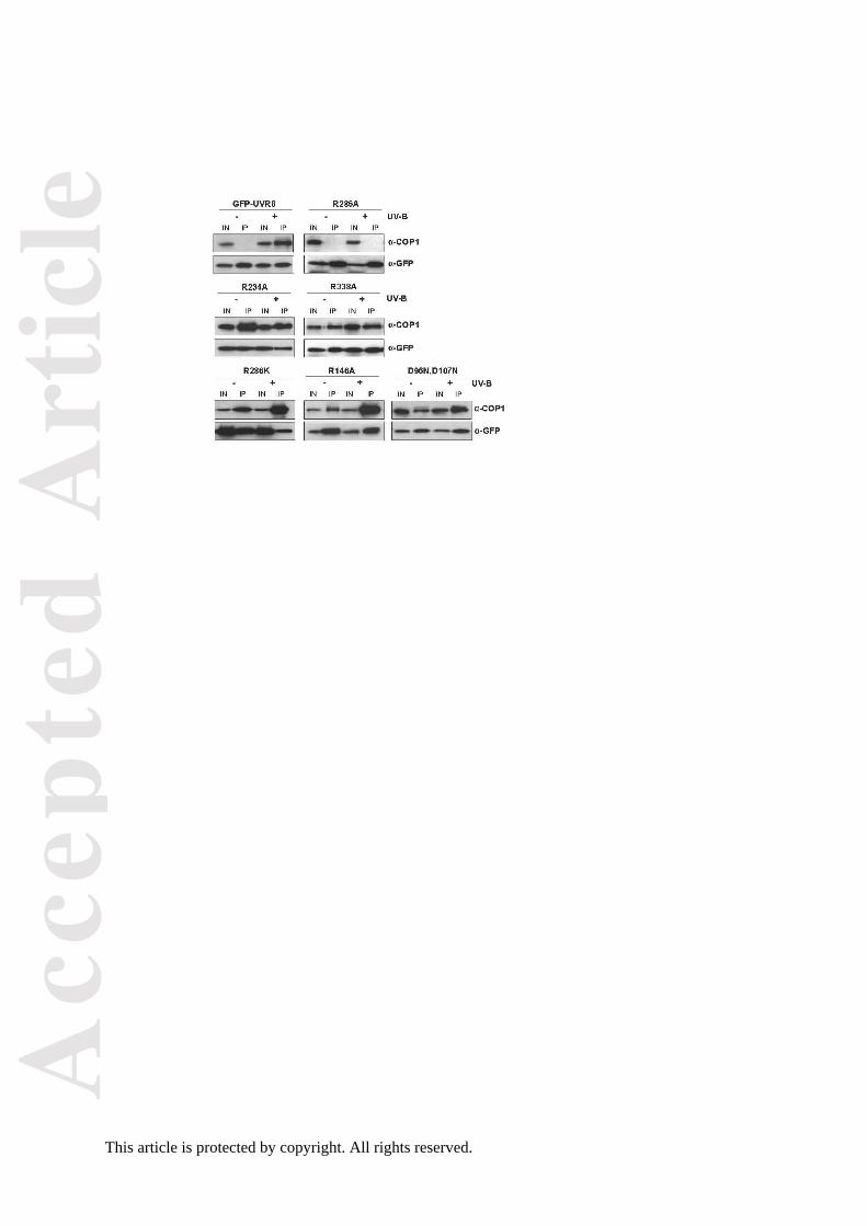
Some, but not all, salt-bridge mutants interact constitutively with COP1
UV-B photoreception stimulates monomerisation and interaction of UVR8 with COP1 to
initiate signaling (Rizzini et al., 2011). COP1 interacts with a 27 amino acid region near the
C-terminus of UVR8 and also with the -propeller core of the protein (Cloix et al., 2012; Yin
et al., 2015). It is proposed that the C-terminus becomes accessible to COP1 following UV-B
exposure of UVR8 (Cloix et al., 2012). However, some tryptophan mutants of UVR8 bind
COP1 in the absence of UV-B (O’Hara and Jenkins, 2012; Heijde et al., 2013; Huang et al.,
2013), suggesting that these mutations expose the C-terminus. Since several of the salt-
bridge mutants have weakened dimers we examined their interaction with COP1, which
impacts on their potential ability to function.
We examined whether the salt-bridge mutants were able to interact with COP1 using
a co-immunoprecipitation assay. As shown in Figure 3, GFP-UVR8 interacted with COP1 in
the presence but not the absence of UV-B, as reported previously (Favory et al., 2009; Cloix
et al., 2012). However, GFP-UVR8R286A did not interact with COP1 at all. In contrast, each of
the other mutants tested interacted with COP1 in both the presence and absence of UV-B.
For GFP-UVR8R286K and GFP-UVR8R146A, which have weakened dimers that monomerise in
response to UV-B, and monomeric GFP-UVR8D96N,D107N, more COP1 was consistently co-
immunoprecipitated from UV-B exposed plants.
Acc
epte
d A
rtic
le
This article is protected by copyright. All rights reserved.
Some salt-bridge mutants are functional in plants
To test whether the UVR8 mutants are functional in initiating photomorphogenic responses
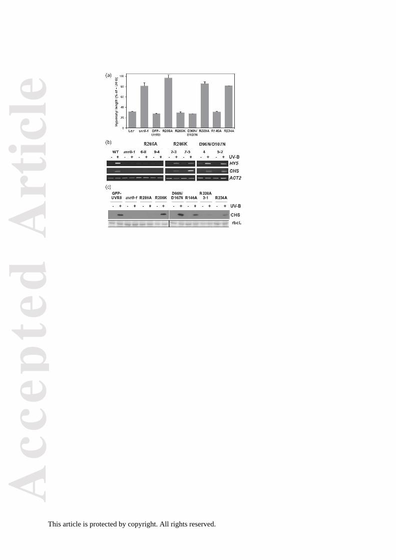
to UV-B we examined both the suppression of hypocotyl extension and the induction of gene
expression, assaying specifically HY5 and CHS transcript levels and CHS protein
accumulation.
As shown in Figure 4a, hypocotyl growth is suppressed by a low fluence rate of
narrowband UV-B in wild-type plants and in the control GFP-UVR8 transgenic line, but the
response is impaired in uvr8-1 plants (Favory et al., 2009; Cloix et al., 2012). The GFP-
UVR8R286A, GFP-UVR8R338A and GFP-UVR8R234A mutants do not show growth suppression
and are of similar length to uvr8-1 under UV-B. In contrast, GFP-UVR8R286K, GFP-UVR8R146A
and GFP-UVR8D96N,D107 show very similar hypocotyl growth suppression to wild-type and
GFP-UVR8 plants and are evidently functional in the UV-B response. Very similar results
were obtained for the induction of gene expression. HY5 and CHS transcript accumulation
are induced by UV-B in two independent lines of the GFP-UVR8R286K and GFP-
UVR8D96N,D107N mutants, but the GFP-UVR8R286A mutant fails to show induction (Figure 4b).
Equivalent results were obtained for CHS protein accumulation in these mutants (Figure 4c).
GFP-UVR8R146A also shows UV-B induction of CHS whereas GFP-UVR8R338A has little if any
response, consistent with the hypocotyl suppression data. GFP-UVR8R234A shows a small
response to UV-B in CHS induction that is not apparent in hypocotyl growth suppression.
The molecular and UV-B response phenotypes of the UVR8 mutants are summarized in
Table S1.
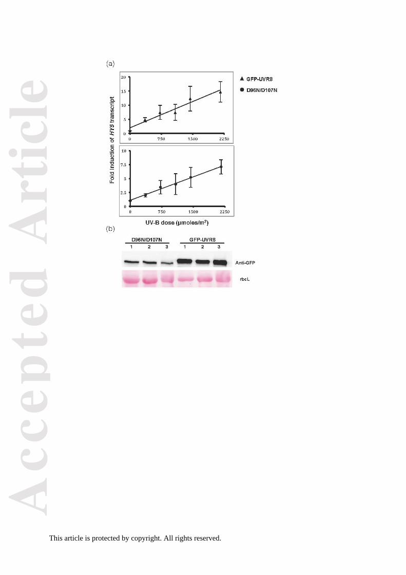
To further test whether the putatively monomeric GFP-UVR8D96N,D107N mutant is
similarly responsive to UV-B as wild-type GFP-UVR8, we examined the dose-response
relationship for HY5 transcript accumulation. We previously reported that this UV-B
response shows reciprocity between exposure duration and fluence rate (Brown et al.,
2009), and used this information to select treatment conditions for the present study. The
Acc
epte
d A
rtic
le
This article is protected by copyright. All rights reserved.
results (Figure 5) show that the GFP-UVR8D96N,D107N mutant and GFP-UVR8 both respond to
UV-B with linear increases in HY5 transcript levels over the same fluence range. The mutant
has a lower fold-induction of HY5 transcripts, but this is likely because the plants used for
these experiments expressed lower amounts of photoreceptor protein than the GFP-UVR8
control (Figure 5b). It is known that the magnitude of response mediated by UVR8 is related
to its level of expression (Favory et al., 2009).
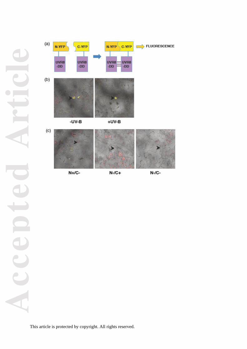
Since the putatively monomeric GFP-UVR8D96N,D107N mutant mediated responses to
UV-B similarly to wild-type GFP-UVR8, we further examined whether the mutant fails to form
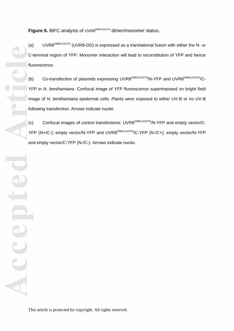
dimers in vivo. We used a sensitive bimolecular fluorescence complementation (BiFC) assay
(Walter et al., 2004) in which the protein is transiently expressed in Nicotiana leaves as a
fusion with either the N- or C-terminal region of YFP (Figure 6a). Dimer formation is
expected to permit reconstitution of YFP and hence generate a fluorescence signal, whereas
no signal should be seen if the mutant protein is constitutively monomeric. Expression of
either the N- or C-terminal fusion protein together with the complementary expression vector
containing no fusion protein is used as a control. As shown in Figure 6b, a fluorescence
signal is detected for UVR8D96N,D107N predominantly in the nucleus under both minus and plus
UV-B conditions, whereas no fluorescence is seen with the empty-vector controls (Figure
6c). Similar results were obtained for UVR8D96N,D107N in 6 independent experiments.
DISCUSSION
Mutations of UVR8 salt bridge amino acids affect dimer stability to varying extents. However,
the method used to assess dimer/monomer status is critical. SDS-PAGE with non-boiled
samples is convenient for monitoring the dimer/monomer status of wild-type UVR8 in
response to different treatments (e.g. Rizzini et al., 2011; Christie et al., 2012; Heilmann and
Jenkins, 2013; Heijde and Ulm, 2013; Huang et al., 2014), but it has limited value for
Acc
epte
d A
rtic
le
This article is protected by copyright. All rights reserved.
characterizing mutant proteins because it very sensitively detects any reduction in affinity in
the dimer. Thus, all salt-bridge mutants examined to date appear constitutively monomeric
when analysed by this method, either as purified proteins (Christie et al., 2012; Wu et al.,
2012; Figure S5) or in plant extracts (Figure 2a). In contrast, SEC with purified proteins
shows that some salt-bridge mutants (UVR8R286K; UVR8R146A) are dimeric (Figure 1b; Figure
S2b) and are converted to monomers upon UV-B illumination. Nevertheless, caution is
required with the SEC analysis because some salt-bridge mutations affect the hydrodynamic
radius of the protein, and hence the elution volume (e.g. UVR8R286K; Figure 1b), most likely
by altering the position of the flexible C-terminal region. Moreover, the conformation of some
mutants (e.g. UVR8R338A; Figure S3b) is significantly affected by ionic strength. It would be
valuable to develop a quantitative method to evaluate the relative strength of the UVR8
dimer in different mutants by measuring a dissociation constant, but this has not yet been
done for purified protein and extending the method to plant extracts would be difficult.
To assess the dimer/monomer status of salt-bridge mutants in vivo we attempted to
employ SEC with immunoprecipitated, transiently expressed proteins. Using this method,
wild-type UVR8 appears as a dimer that monomerizes after UV-B exposure, as expected. In
contrast, the salt-bridge mutants appear constitutively monomeric. While this observation
could suggest that the molecular environment in vivo impairs dimerisation of salt-bridge
mutants, it is more likely that the high pH used to elute the immunoprecipitated proteins
causes dissociation of the weakened mutant dimers. A more reliable method to assess
dimer/monomer status in vivo is cross-linking of proteins, since it does not employ a
chemical treatment that will promote monomerisation. The results obtained using cross-
linking are consistent with the SEC data for purified mutant proteins. In the absence of UV-B
GFP-UVR8R146A is predominantly a dimer, whereas GFP-UVR8R286K is a mixture of dimer
and monomer. In contrast, GFP-UVR8R286A and GFP-UVR8D96N,D107N are monomeric with no
detectable dimer, again in agreement with the in vitro data. Nonetheless, although the data
obtained with cross-linking concur with those obtained for purified proteins, the assay is
Acc
epte
d A
rtic
le
This article is protected by copyright. All rights reserved.
performed with protein extracts rather than intact cells, and it is important to determine the
dimer/monomer status of the protein where it functions in cells.
The BiFC analysis of UVR8D96N,D107N (Figure 6) contradicts the cross-linking data in
that dimers are detected in the nucleus. A possible explanation is that the amount of dimer
formed by the mutant protein is below the limit of detection of the cross-linking assay;
nuclear UVR8 is estimated to be approximately 10% of the total UVR8 in wild-type cells
(Kaiserli and Jenkins, 2007) and probably an even smaller fraction of the UVR8D96N,D107N
protein in the nucleus is in the dimeric form. On the other hand, BiFC is very sensitive and
the YFP interaction is irreversible, so the fluorescence observed may represent trapping of
short-lived, weak interaction between monomers of the mutant proteins. Thus, although
BiFC shows that the UVR8D96N,D107N mutant is capable of being trapped in a YFP linked state
in vivo, the method may over-state the extent and stability of dimer formation. The other
methods employed all indicate that UVR8D96N,D107N is constitutively monomeric, within their
limits of detection, and it is therefore likely that this mutant forms very little dimer in vivo, and
the dimers present are likely to be unstable and short lived.
We therefore conclude that mutations that affect the ability of purified UVR8 to form a
dimer have an equivalent effect in vivo despite evident differences in the cellular
environment, not least in ionic composition and the presence of proteins that could
potentially influence dimer/monomer status. Furthermore, the in vivo experiments highlight
the importance of specific charged amino acids in maintaining the dimer structure. In
particular, the R286-D96/D107 salt-bridges are crucial for maintaining the dimer both in vivo
and in vitro. The UVR8D96N,D107N and UVR8R286A mutants are strongly impaired in dimer
formation whereas UVR8R286K forms a weakened dimer, indicating that the positive charge of
the arginine/lysine residue is critical for dimerisation.
Assays of hypocotyl growth suppression and gene expression show that some salt
bridge mutants are functional in response to UV-B exposure. The impaired responses of
Acc
epte
d A
rtic
le
This article is protected by copyright. All rights reserved.
GFP-UVR8R234A, GFP-UVR8R286A and GFP-UVR8R338A to UV-B may be because the
mutations impact on photoreception. R234, R286 and R338 are adjacent to the triad
tryptophans involved in UV-B sensing and the introduction of the small non-polar alanine in
place of arginine is likely to disrupt the spatial relationships of the tryptophans as well as
charge-based interactions between the tryprophans and adjacent arginines that are probably
important in photoreceptor function (Wu et al., 2012). In contrast, photoreception is not
impaired in the GFP-UVR8R146A, GFP-UVR8R286K and GFP-UVR8D96N,D107N mutants and the
hypocotyl growth suppression and gene expression responses to UV-B are very similar to
those of wild-type UVR8. These mutations should have little impact on UV-B photoreception
by UVR8. R146 is not close to the tryptophan triad and UVR8R286K is a conservative
mutation. D96 and D107 are not adjacent to the chromophore tryptophans and are not part
of the charge network that surrounds them (Christie et al., 2012; Wu et al., 2012), and since
aspartate and asparagine are very similar in size the UVR8D96N,D107N mutations will not cause
spatial disruption within the monomer.
Interaction of UVR8 with COP1 is necessary to initiate signaling and transcriptional
responses. The GFP-UVR8R286A mutant is unable to bind COP1 and is non-functional,
consistent with the findings of Huang et al. (2014). A possible explanation is that this
mutation prevents UV-B induced conformational changes that make the C-terminus, and
potentially other regions of the protein, accessible for binding to COP1. In contrast, the
constitutive binding of COP1 to GFP-UVR8D96N,D107N is likely caused by exposure of residues
involved in binding. Similarly, the binding of COP1 to the weakened dimers of GFP-
UVR8R146A and GFP-UVR8R286K is presumably the result of physical exposure of the
region(s) interacting with COP1 in the non-illuminated photoreceptor as a result of the
mutations. Although several of the mutants studied here bind COP1 constitutively, none
initiate responses in the absence UV-B, indicating that interaction with COP1 is not sufficient
to initiate signaling. Similarly, the constitutive binding of COP1 to GFP-UVR8R234A and GFP-
UVR8R338A is not sufficient for function. These results are consistent with those reported
Acc
epte
d A
rtic
le
This article is protected by copyright. All rights reserved.
previously for alanine mutants of triad tryptophans (O’Hara and Jenkins, 2012). In contrast,
strong over-expression of UVR8W285A gives a cop mutant phenotype, likely because of
sequestration of COP1, and the plants show constitutive activation of UV-B signaling (Heijde
et al. 2013). A similar phenotype is reported for UVR8R338A (Huang et al., 2014), in contrast
to the data in Figure 4. A possible explanation of these different findings is that the lower
level of transgenic expression employed in this study and previously (O’Hara and Jenkins,
2012; Kaiserli and Jenkins, 2007) is too low to promote obvious constitutive activation.
The finding that the in vivo UV-B responses of GFP-UVR8D96N,D107N are very similar to
those of wild-type GFP-UVR8 is particularly interesting, because the mutant is evidently
strongly impaired in dimer formation. The dose-response analysis (Figure 5) indicates that
the mutant and wild-type photoreceptor proteins are similarly responsive to UV-B over a
fluence range where the response is not saturated. The lower fold-induction of HY5
transcripts in GFP-UVR8D96N,D107N can be explained by the smaller amount of photoreceptor
protein in the plants used for the experiments. Evidence from over-expression lines (Favory
et al., 2009) indicates that an increased level of UVR8 protein gives an increased magnitude
of response.
The established model of UVR8 action is that the dimer acts in UV-B photoreception,
which causes monomerisation, and then the monomer interacts with COP1 to initiate
signaling and hence responses (Tilbrook et al., 2013; Jenkins, 2014a). This model is
inadequate to explain the in vivo UV-B response of GFP-UVR8D96N,D107N. There is at most,
only a very low concentration of dimer present in GFP-UVR8D96N,D107N plants, whereas
responsiveness to UV-B is similarly efficient to that of wild-type GFP-UVR8. Clearly, the
physiological response of GFP-UVR8D96N,D107N does not correlate with monomer formation
through UV-B induced dimer dissociation. The simplest explanation is that the monomeric
protein is competent in photoreception, which initiates signalling and UV-B responses in
vivo. Thus, the molecular and physiological phenotype of GFP-UVR8D96N,D107N indicates that
dimer formation is not essential for photoreception by UVR8. UV-B photoreception could
Acc
epte
d A
rtic
le
This article is protected by copyright. All rights reserved.
activate monomeric UVR8D96N,D107N bound to COP1 by, for example causing a
conformational change to the protein that initiates signalling. Recent studies show that
photochemical events associated with UV-B photoreception are detectable in both dimeric
and monomeric UVR8 (Mathes et al. 2015), indicating that monomeric UVR8 is capable of
UV-B photoreception, at least in vitro.
The findings with the GFP-UVR8D96N,D107N mutant raise the question of whether
monomeric UVR8 could be active in photoreception in wild-type plants to initiate responses.
Recent research shows that UVR8 does not behave as a simple dimer/monomer UV-B
switch under photoperiodic conditions, but establishes a photo-equilibrium between the
dimer and monomer forms (Findlay and Jenkins, 2016). Monomeric UVR8 is present in
plants even at low ambient levels of UV-B and could potentially be active in photoreception
to initiate responses. At present, dimer and monomer photoreception cannot be
distinguished in plants by distinct biophysical signals or associated physiological responses,
and therefore further research is needed to test the intriguing possibility that monomeric
UVR8 is active in photoreception in wild-type plants.
EXPERIMENTAL PROCEDURES
Experiments with purified proteins
Site-directed mutagenesis was carried out using the QuikChange Site-directed Mutagenesis
kit (Stratagene, USA) following the manufacturer’s instructions and verified by sequencing.
The primers used for site-directed mutagenesis are listed in Table S2. Proteins were
expressed in E. coli and purified as described previously (Christie et al. 2012). UV-B
exposure of proteins was undertaken with a narrowband UV-B source with maximal
emission at 311 nm (Philips TL20W/01RS; spectrum shown in Cloix et al. 2012).
Acc
epte
d A
rtic
le
This article is protected by copyright. All rights reserved.
Purified proteins were exposed to 1.5 µmol m-2 s-1 narrowband UV-B on ice for 1 h.
Protein samples were prepared for electrophoresis without boiling as described (Christie et
al. 2012) and loaded on a 7.5 % SDS-PAGE gel. Gels were stained with Coomassie Blue.
Analytical SEC was performed on a Superdex 200 HR10/30 column (GE Healthcare)
equilibrated with wash buffer containing 50 mM Tris pH 7.5, 150 mM NaCl (or 500 mM for
high-salt samples), 1mM β-mercaptoethanol and 0.02% sodium azide, and run at a flow rate
of 0.5 ml/min at 4°C on an AKTA FPLC system (Christie et al. 2012). Aldolase, albumin and
ovalbumin were used as standards.
Plant material
Seeds of wild-type Arabidopsis thaliana ecotype Landsberg erecta (Ler) were obtained from
the Nottingham Arabidopsis Stock Center. Seeds of the uvr8-1 mutant allele (Ler
background; Kliebenstein et al., 2002) were obtained from Dr Dan Kliebenstein (University of
California, Davis). The uvr8-1/UVR8pro:GFP-UVR8 transgenic line 6-2 was described by
Kaiserli and Jenkins (2007).
Mutant UVR8 proteins were expressed in uvr8-1 using Agrobacterium mediated
transformation. Mutant UVR8 sequences were sub-cloned into the pEZR(K)L-C vector
downstream of eGFP and the CaMV 35S promoter (Brown et al. 2005; Cloix et al. 2012).
DNA sequencing confirmed that the fusions were made correctly. At least 3 independent
transgenic lines were selected for each fusion with a level of transgene expression
comparable to that of the control GFP-UVR8 fusion (Figure S4).
Acc
epte
d A
rtic
le
This article is protected by copyright. All rights reserved.
Arabidopsis experiments
Except where indicated below, plants were grown on agar plates containing half-strength
Murashige and Skoog (MS) salts under 100 µmol m-2 s-1 constant white light (warm white
fluorescent tubes Osram) at 21°C for 7 to 10 d and then either placed in darkness for 16 h
or, for RT-PCR experiments, transferred to 20 µmol m-2 s-1 constant white light for 4 days.
Plant protein extracts were made and exposed to UV-B using the above narrowband
source as described in (Heilmann and Jenkins, 2013). UVR8 dimer/monomer status was
examined by SDS-PAGE with non-boiled samples (Heilmann and Jenkins, 2013).
Immunoblots were incubated with an anti-GFP antibody (Clontech) or anti-UVR8 antibody
(Kaiserli and Jenkins, 2007), as indicated in the figure legends.
For cross-linking of proteins, proteins were extracted in PBS containing protease
inhibitor cocktail tablets (complete, Roche). Samples were then centrifuged for 10 min at
16,000 g at 4°C and the supernatant transferred to a fresh tube. Dithiobis (succinimidyl
propionate) (4 mM final concentration) (DSP; Thermo Scientific) was then added to the
extract and incubated on ice for 30 min. Immediately afterwards, protein sample buffer
without reducing agent (-mercaptoethanol, -ME) or with -ME (5% final concentration to
reverse crosslinking) was added, and samples were boiled for 10 min before separation on a
10% SDS PAGE and subsequent immunodetection using anti-UVR8 antibody.
Interaction of wild-type and mutant GFP-UVR8 fusions with COP1 was examined by
co-immunoprecipitation (Cloix et al. 2012). Plants were grown on agar plates as described
above and put in darkness for 16 h. The plants were treated for 3 h with 3 mol m-2 s-1
narrowband UV-B. Whole cell extracts were prepared as described in (Kaiserli and Jenkins,
2007) in the absence or presence of 3 mol m-2 s-1 narrowband UV-B. The co-
immunoprecipitation assays were carried out in the same light conditions using anti-GFP
microbeads (Macs, 130-091-370, Miltenyi Biotec) as described previously (Cloix et al.
2012). The ‘input’ samples applied to the microbead columns and the immunoprecipitate
Acc
epte
d A
rtic
le
This article is protected by copyright. All rights reserved.
eluates were analysed by SDS-PAGE followed by western blotting and immunodetection
using anti-GFP (Clontech) and anti-COP1 (kindly provided by Dr Nam-Hai Chua; Jang et al.
2010) antibodies.
RT-PCR assays of HY5 and CHS transcript levels in plant RNA samples (Figure 4b)
were measured as described previously (Cloix et al. 2012) with the primers stated in Brown
and Jenkins (2008). Plants grown as above were exposed, or not in controls, to 3 mol m-2 s-
1 broadband UV-B (Q-panel UV-B-313 fluorescent tubes covered with cellulose acetate;
spectrum shown in Cloix et al. 2012) for 4 h. Transcript levels of ACTIN2 were assayed in
the same cDNA samples as a control. For each gene, PCR was monitored over a range of
cycle numbers to select optimal conditions for visualization of the PCR product and
quantification. Transcript levels in different RNA samples were compared using cycle
numbers within the linear range of amplification.
For the dose-response experiments (Figure 5) plants were grown on agar plates as
above in a 16 h light (60 µmol m-2 s-1) / 8 h dark cycle for 14 days and transferred to
darkness for 16 h prior to exposure to narrowband UV-B at different doses as described by
Brown et al. (2009): 20 and 40 min treatments with 0.3, 0.6 and 0.9 µmol m-2 s-1 followed by
transfer to darkness so that all tissue was harvested 2 h after the start of illumination. qRT-
PCR assays of HY5 transcripts were undertaken similarly to Brown et al. (2009), but using
different primers (HY5: forward GGCTGAAGAGGTTGTTGAGGAAC; reverse
AGCATCTGGTTCTCGTTCTGAAGA. ACTIN2: forward GTATTGTGCTGGATTCTGGTG;
reverse GAGGTAATCAGTAAGGTCACG).
For measurements of hypocotyl length, seedlings were grown for 4 days on agar
plates containing half-strength Murashige and Skoog (MS) salts in 1.5 mol m-2 s-1 white
light supplemented, or not in controls, with 1.5 mol m-2 s-1 narrowband UV-B (Cloix et al.
2012). For the analysis of CHS protein, seedlings were grown under the same conditions for
7 days. Whole cell extracts were made and protein samples boiled prior to electrophoresis
Acc
epte
d A
rtic
le
This article is protected by copyright. All rights reserved.
on a 7.5 % SDS-PAGE gel. Immunoblots were probed with an anti-CHS (Santa Cruz)
antibody. Immunoblots were stained with Ponceau S to reveal the Rubisco large subunit
(rbcL), which was used as a loading control.
Similar results for dimer/monomer status, COP1 interaction, hypocotyl growth
suppression and gene expression were obtained for several lines expressing a particular
fusion. Unless indicated otherwise, the transgenic lines used in the experiments shown are
as follows: GFP–UVR8 6-2; GFP–UVR8R286A 6-8; GFP–UVR8R286K 2-3; GFP–UVR8D96N/D107N
74-2 or 5-2 (for the dose-response experiments); GFP–UVR8R234A 16-5; GFP–UVR8R338A 9-3,
and GFP–UVR8R146A line 9. The data presented are representative of at least three
independent experiments.
Transient expression in Nicotiana benthamiana for SEC
A single colony from Agrobacterium cells freshly transformed with the desired plasmid DNA
was inoculated in 10 ml of LB medium with appropriate antibiotics and grown overnight at
28°C under constant shaking (200 rpm). When cultures had reached an OD600 of about 0.6
- 1.0, cells were pelleted by centrifugation at 2,000 g for 10 min. The cells were then
resuspended in 10 mM MgCl2, 10 mM MES pH 6.5 and 200 μM acetosyringone at an
OD600 of 0.2 and incubated at room temperature for 3 hours. The Agrobacterium medium
was infiltrated into the lower side of N. benthamiana leaves using a syringe. The infiltrated
plants were moved back into the growth room at 28°C and left for 2-3 days before examining
gene expression by confocal microscopy and preparation of protein extracts.
For protein extraction, N. benthamiana leaf segments were frozen in liquid nitrogen
and ground with a mortar and pestle. A spatula of polyvinyl-pyrrolidone (PVP), an effective
absorbent for phenolic compounds, was added as soon as the liquid nitrogen had
evaporated. Once ground, the plant material was transferred to a microcentrifuge tube and
approximately one volume of extraction buffer (1 mM EDTA, 10% glycerol, 5 mM DTT, 0.1%
Acc
epte
d A
rtic
le
This article is protected by copyright. All rights reserved.
v/v Triton, 25 mM Tris-HCl, pH 7.5) was added and vortexed to mix. Samples were
centrifuged at 16,000 g for 15 min at 4°C and the supernatant was transferred to a fresh
tube. The transiently expressed GFP fusions were immunoprecipitated using anti-GFP
microbeads (Macs, 130-091-370, Miltenyi Biotec) as described previously (Cloix et al.
2012). The immunoprecipitated proteins were examined by SEC, using the same method as
for the purified proteins, followed by standard SDS-PAGE and immunodetection using an
anti-GFP antibody (Clontech).
BiFC experiments
The GFP–UVR8D96N/D107N fusion was cloned into the pSPYNE and pSPYCE vectors (Walter
et al., 2004) containing the N- and C-terminal regions of YFP respectively. Agrobacteria
containing the plasmids were grown overnight as above, and resuspended together in 10
mM MgCl2, 10 mM MES pH 6.5 and 200 μM acetosyringone at an equivalent OD600 of 0.1
for each culture. The Agrobacterium suspension was incubated at room temperature for 2
hours before infiltration into N. benthamiana leaves as described above. Plants were
exposed to low fluence rate (1 mol m-2 s-1) narrowband UV-B in a growth chamber at 21°C
for approximately 60 hours; controls were kept under a UV-B cutoff filter. Leaves were
examined for YFP fluorescence in at least 3 fields of view using a Zeiss LSM confocal
microscope.
ACKNOWLEDGEMENTS
MH was supported by a grant from The Leverhulme Trust to GIJ, JMC and BOS, and CC
and CNV by grants from the UK Biotechnology and Biological Sciences Research Council to
GIJ and JMC (BB/J008494/1) and GIJ, JMC and BOS (BB/K00932X/1).
Acc
epte
d A
rtic
le
This article is protected by copyright. All rights reserved.
SUPPORTING INFORMATION
Additional Supporting Information may be found in the online version of this article.
Figure S1. Dimer/monomer status of UVR8R286K examined by SEC.
Figure S2. Dimer/monomer status of UVR8R146A and UVR8R234A examined by SEC.
Figure S3. Dimer/monomer status of UVR8R338A examined by SEC.
Figure S4. Expression levels of GFP-UVR8 mutants in transgenic lines.
Figure S5. Dimer/monomer status of purified mutant proteins examined by SDS-PAGE with
non-boiled samples.
Table S1. Summary of phenotypes of UVR8 salt-bridge amino acid mutants.
Table S2. Primers used for site-directed mutagenesis.
REFERENCES
Ballaré, C.L., Mazza, C.A., Austin, A.T. and Pierik, R. (2012) Canopy light and plant health.
Plant Physiol. 160, 145-155.
Brown, B.A., Cloix, C., Jiang, G.H., Kaiserli, E., Herzyk, P., Kliebenstein, D.J. and
Jenkins, G.I. (2005) A UV-B-specific signaling component orchestrates plant
UV protection. Proc. Natl. Acad. Sci. USA 102, 18225-18230.
Brown, B.A. and Jenkins, G.I. (2008) UV-B signaling pathways with different fluence-rate
response profiles are distinguished in mature Arabidopsis leaf tissue by requirement
for UVR8, HY5, and HYH. Plant Physiol. 146, 576-588.
Acc
epte
d A
rtic
le
This article is protected by copyright. All rights reserved.
Brown, B.A., Headland, L.R. and Jenkins, G.I. (2009) UV-B action spectrum for UVR8-
mediated HY5 transcript accumulation in Arabidopsis. Photochem. Photobiol. 85,
1147-1155.
Christie, J.M., Arvai, A.S., Baxter, K.J., Heilmann, M., Pratt, A.J., O’Hara, A., Kelly, S.M.,
Hothorn, M., Smith, B.O., Hitomi, K., Jenkins, G.I. and Getzoff, E.D. (2012) Plant
UVR8 photoreceptor senses UV-B by tryptophan-mediated disruption of cross-dimer
salt bridges. Science 335, 1492-1496.
Cloix, C., Kaiserli, K., Heilmann, M., Baxter, K.J., Brown, B.A., O’Hara, A., Smith, B.O.,
Christie, J.M. and Jenkins, G.I. (2012) The C-terminal region of the UV-B
photoreceptor UVR8 initiates signaling through interaction with COP1. Proc. Natl.
Acad. Sci. USA 109, 16366-16370.
Favory, J.J., Stec, A., Gruber, H., Rizzini, L., Oravecz, A., Funk, M., Albert, A., Cloix, C.,
Jenkins, G.I., Oakeley, E.J., Seidlitz, H.K., Nagy, F. and Ulm, R. (2009) Interaction of
COP1 and UVR8 regulates UV-B-induced photomorphogenesis and stress acclimation
in Arabidopsis. EMBO J. 28, 591-601.
Feher, B., kozma-Bognar, L., Kevei, E., Hajdu, A., Binkert, M., Davis, S.J., Schäfer, E., Ulm,
R. and Nagy, F. (2011) Functional interaction of the circadian clock and UV
RESISTANCE LOCUS 8-controlled UV-B signaling pathways in Arabidopsis thaliana.
Plant J. 67, 37-48.
Findlay, K.M.W. and Jenkins, G.I. (2016) Regulation of UVR8 photoreceptor dimer/monomer
photo-equilibrium in Arabidopsis plants grown under photoperiodic conditions. Plant
Cell Environ. doi: 10.1111/pce.12724.
Frohnmeyer, H. and Staiger, D. (2003) Ultraviolet-B radiation-mediated responses in plants.
Balancing damage and protection. Plant Physiol. 133, 1420-1428.
Acc
epte
d A
rtic
le
This article is protected by copyright. All rights reserved.
Grüber, H., Heijde, M., Heller, W., Albert, A., Seidlitz, H.K. and Ulm, R. (2010)
Negative feedback regulation of UV-B-induced photomorphogenesis and stress
acclimation in Arabidopsis. Proc. Natl. Acad. Sci. USA 107, 20132-20137.
Hayes, S., Velanis, C.N., Jenkins, G.I. and Franklin, K.A. (2014) UV-B detected by the UVR8
photoreceptor antagonizes auxin signaling and plant shade avoidance. Proc. Natl.
Acad. Sci. USA 111, 11894-11899.
Heilmann, M. and Jenkins, G.I. (2013) Rapid reversion from monomer to dimer regenerates
the ultraviolet-B photoreceptor UV RESISTANCE LOCUS8 in intact Arabidopsis
plants. Plant Physiol. 161, 547-555.
Heijde, M. and Ulm, R. (2013) Reversion of the Arabidopsis UV-B photoreceptor UVR8 to
the homodimeric ground state. Proc. Natl. Acad. Sci. USA 110, 1113-1118.
Heijde, M., Binkert, M., Yin, R., Ares-Orpel, F., Rizzini, L., Van De Slijke, E., Persiau,
G., Nolf, J., Gevaert, K., De Jaeger, G. and Ulm, R. (2013) Constitutively active
UVR8 photoreceptor variant in Arabidopsis. Proc. Natl. Acad. Sci. USA 110,
20326–20331.
Huang, X., Ouyang, X., Yang, P., Lau, O.S., Chen, L., Wei, N. and Deng. X-W. (2013)
Conversion from CUL4-based COP1–SPA E3 apparatus to UVR8–COP1–SPA
complexes underlies a distinct biochemical function of COP1 under UV-B. Proc. Natl.
Acad. Sci. USA 110, 16669-16674.
Huang, X., Yang, P., Ouyang, X., Chen, L. and Deng, X-W. (2014) Photoactivated UVR8-
COP1 Module Determines Photomorphogenic UV-B Signaling Output in Arabidopsis.
PLOS Genetics 10:e1004218.
Acc
epte
d A
rtic
le
This article is protected by copyright. All rights reserved.
Jang, I-C., Henriques, R., Seo, H.S., Nagatani, A. and Chua, N-H. (2010) Arabidopsis
PHYTOCHROME INTERACTING FACTOR proteins promote phytochrome B
polyubiquitination by COP1 E3 ligase in the nucleus. Plant Cell 22, 2370-2383.
Jenkins, G.I. (2009) Signal transduction in responses to UV-B radiation. Ann. Rev. Plant
Biol. 60, 407-431.
Jenkins, G.I. (2014a) The UV-B photoreceptor UVR8: from structure to physiology. Plant
Cell 26, 21-37.
Jenkins, G.I. (2014b) Structure and function of the UV-B photoreceptor UVR8. Curr. Op.
Struct. Biol. 29, 52-57.
Kaiserli, E. and Jenkins, G.I. (2007) UV-B promotes rapid nuclear translocation of
the UV-B-specific signaling component UVR8 and activates its function in the
nucleus. Plant Cell 19, 2662-2673.
Kliebenstein, D.J., Lim, J.E., Landry, L.G. and Last, R.L. (2002) Arabidopsis UVR8
regulates ultraviolet-B signal transduction and tolerance and contains sequence
similarity to human Regulator of Chromatin Condensation 1. Plant Physiol. 130,
234-243.
Mathes, T., Heilmann, M., Pandit, A., Zhu, J., Ravensbergen, J., Kloz, M., Fu, Y.,
Smith, B.O., Christie, J.M., Jenkins, G.I. and Kennis, J.T.M. (2015) Proton-
coupled electron transfer constitutes the photoactivation mechanism of the
plant photoreceptor UVR8. J. Am. Chem. Soc. Doi:
O’Hara, A. and Jenkins, G.I. (2012) In vivo function of tryptophans in the Arabidopsis
UV-B photoreceptor UVR8. Plant Cell 24, 3755-3766.
Acc
epte
d A
rtic
le
This article is protected by copyright. All rights reserved.
Oravecz, A., Baumann, A., Máté, Z., Brzezinska, A., Molinier, J., Oakeley, E.J., Ádám, É.,
Schäfer, E., Nagy, F. and Ulm, R. (2006) CONSTITUTIVELY
PHOTOMORPHOGENIC1 is required for the UV-B response in Arabidopsis. Plant Cell
18, 1975-1990.
Rizzini, L., Favory, J-J., Cloix, C., Faggionato, D., O’Hara, A., Kaiserli, E., Baumeister, R.,
Schäfer, E., Nagy, F., Jenkins, G.I. and Ulm, R. (2011) Perception of UV-B by the
Arabidopsis UVR8 protein. Science 332, 103-106.
Tilbrook, K., Arongaus, A.B., Binkert, M., Heijde, M., Yin, R. and Ulm, R. (2013) The UVR8
UV-B photoreceptor: perception, signaling and response. The Arabidopsis Book,
American Society of Plant Biologists, June 11 2013: e0164. Doi: 10.1199/tab.0164.
Ulm, R. and Nagy, F. (2005) Signalling and gene regulation in response to ultraviolet
light. Curr. Opinion Plant Biol. 8, 477-482.
Ulm, R., Baumann, A., Oravecz, A., Mate, Z., Adam, E., Oakeley, E.J., Schäfer, E. and
Nagy, F. (2004) Genome-wide analysis of gene expression reveals function of the
bZIP transcription factor HY5 in the UV-B response of Arabidopsis. Proc. Natl. Acad.
Sci. USA 101, 1397-1402.
Walter, M., Chaban, C., Schütze, K., Batistic, O., Weckermann, K., Näke, C, Blazevic, D.,
Grefen, C., Schumacher, K., Oecking, C., Harter, K. and Kudla, J. (2004) Visualization
of protein interactions in living plant cells using bimolecular flurorescence
complementation. Plant J. 40, 428-438.
Wu, D., Hu, Q., Yan, Z., Chen, W., Yan, C., Huang, X., Zhang, J., Yang, P., Deng, H., Wang,
J., Deng, X. and Shi, Y. (2012) Structural basis of ultraviolet-B perception by UVR8.
Nature 484, 214-219.
Acc
epte
d A
rtic
le
This article is protected by copyright. All rights reserved.
Yin, R., Arongaus, A.B., Binkert, M. and Ulm, R. (2015) Two distinct domains of the
UVR8 photoreceptor interact with COP1 to initiate UV-B signaling in
Arabidopsis. Plant Cell 27, 202-213.
Zeng, X., Ren, Z., Wu, Q., Fan, J., Peng, P-P., Tang, K., Zhang, R., Zhao, K-H. and
Yang, X. (2015) Dynamic crystallography reveals early signalling events in
ultraviolet photoreceptor UVR8. Nature Plants doi: 10.1038/NPLANTS.2014.6.
FIGURE LEGENDS
Figure 1. Dimer/monomer status of purified UVR8 salt bridge mutant proteins.
(a) PyMol image showing inter-monomer salt bridges formed between R286 and D96
and D107.
(b) SEC on a Superdex 200 column of purified wild-type UVR8 and the
UVR8R286A, UVR8R286K and UVR8D96N,D107N mutant proteins exposed (dashed line) or
not (solid line) to 1.5 µmol m-2 s-1 narrowband UV-B for 1 h. Elution points of marker
proteins (in kDa) are shown at the top.
Figure 2. Dimer/monomer status of UVR8 salt bridge mutant proteins expressed in
plants.
(a) Western blot of whole cell extracts from uvr8-1 plants expressing either GFP-UVR8
or GFP-UVR8 salt-bridge mutants exposed (+) or not (-) to 4 µmol m-2 s-1 narrowband UV-B
for 30 min. SDS-loading buffer was added and samples were run on a 7.5% SDS-PAGE gel
without boiling. An immunoblot was probed with anti-UVR8 antibody. Ponceau staining of
Acc
epte
d A
rtic
le
This article is protected by copyright. All rights reserved.
Rubisco large subunit (rbcL) is shown as a loading control. The GFP-UVR8 dimer and
monomer are indicated.
(b) SEC profiles of immunoprecipitated wild-type GFP-UVR8 (WT) and salt-bridge
mutant fusions expressed in N. benthamiana plants. Vector with GFP alone was used as a
control. For wild-type GFP-UVR8, extracts were illuminated (or not) with 4 µmol m-2 s-1
narrowband UV-B for 30 min. All other samples were not exposed to UV-B. Eluates of
immunoprecipitation assays with anti-GFP beads were loaded onto a Superdex 200 column
and fractions 15 to 30 were used for standard SDS-PAGE and immunoblotting with an anti-
GFP antibody.
(c) Western blot of whole cell extracts from uvr8-1 plants expressing either GFP-
UVR8 (WT) or GFP-UVR8 salt-bridge mutants not exposed to UV-B incubated with
the cross-linking reagent DSP in the absence (upper panel) or presence (lower
panel) of -mercaptoethanol. SDS-loading buffer was added and samples were run
on a 10% SDS-PAGE gel without boiling. An immunoblot was probed with anti-UVR8
antibody. The UVR8 dimer and monomer are indicated.
Figure 3. Interaction of UVR8 mutants with COP1 in vivo.
Co-immunoprecipitation of GFP-UVR8 and COP1 in whole cell extracts obtained from uvr8-1
plants transformed with either GFP-UVR8 or GFP-UVR8 salt-bridge mutants exposed (+) or
not (-) to 3 µmol m-2 s-1 narrowband UV-B for 3 h. Co-immunoprecipitation assays were
performed under the same conditions. Input samples (15 µg, IN) and eluates (IP) were run
on a SDS-PAGE gel, and an immunoblot was probed with anti-COP1 and anti-GFP
antibodies.
Figure 4. Functional complementation of uvr8-1 by UVR8 salt-bridge mutant proteins.
Acc
epte
d A
rtic
le
This article is protected by copyright. All rights reserved.
(a) Hypocotyl lengths (+ S.E., n = 10) for 4 day-old wild-type Ler, uvr8-1, GFP-
UVR8 and the indicated transgenic lines of UVR8 salt-bridge mutant seedlings
grown in 1.5 mol m-2 s-1 white light (- UV-B) supplemented with 1.5 mol m-2 s-1
narrowband UV-B (+ UV-B).
(b) RT-PCR assays of HY5, CHS and control ACTIN2 transcripts in Ler (WT), uvr8-1,
and independent transgenic lines expressing either UVR8R286A, UVR8R286K or UVR8D96N,D107N
grown under 20 mol m-2 s-1 white light (-) and exposed to 3 mol m-2 s-1 broadband UV-B for
4 h (+).
(c) Expression of CHS protein in GFP-UVR8, uvr8-1 and the indicated transgenic lines
of UVR8 salt-bridge mutant plants grown and illuminated as in (b) for 7 days. Proteins
extracts were run on standard SDS-PAGE and an immunoblot was probed with anti-CHS
antibody. Ponceau staining of Rubisco large subunit (rbcL) is shown as a loading control.
Figure 5. UV-B dose-response of HY5 transcript accumulation in UVR8D96N,D107N
mutant compared to GFP-UVR8.
(a) qRT-PCR measurements of HY5 transcripts in GFP-UVR8 and GFP-UVR8D96N,D107N
plants exposed to a range of doses of narrowband UV-B. HY5 transcripts were normalized
to control ACTIN2 transcript levels and fold-induction is relative to the pre-illumination dark
transcript level. The data are the means (+ S.D.) of 3 independent experiments.
(b) Abundance of GFP-UVR8D96N,D107N and GFP-UVR8 proteins in plants used for the
three replicate experiments in (a). Protein extracts were subjected to SDS-PAGE and an
immunoblot was probed with anti-GFP antibody. Ponceau staining of Rubisco large subunit
(rbcL) is shown as a loading control. Quantification of relative band intensities indicates that
the mean level of GFP-UVR8D96N,D107N is approximately 25% that of GFP-UVR8.
Acc
epte
d A
rtic
le
This article is protected by copyright. All rights reserved.
Figure 6. BiFC analysis of UVR8D96N,D107N
dimer/monomer status.
(a) UVR8D96N,D107N (UVR8-DD) is expressed as a translational fusion with either the N- or
C-terminal region of YFP. Monomer interaction will lead to reconstitution of YFP and hence
fluorescence.
(b) Co-transfection of plasmids expressing UVR8D96N,D107N/N-YFP and UVR8D96N,D107N/C-
YFP in N. benthamiana. Confocal image of YFP fluorescence superimposed on bright field
image of N. benthamiana epidermal cells. Plants were exposed to either UV-B or no UV-B
following transfection. Arrows indicate nuclei.
(c) Confocal images of control transfections: UVR8D96N,D107N/N-YFP and empty vector/C-
YFP (N+/C-); empty vector/N-YFP and UVR8D96N,D107N/C-YFP (N-/C+); empty vector/N-YFP
and empty vector/C-YFP (N-/C-). Arrows indicate nuclei.
Acc
epte
d A
rtic
le
This article is protected by copyright. All rights reserved.
Acc
epte
d A
rtic
le
This article is protected by copyright. All rights reserved.
Acc
epte
d A
rtic
le
This article is protected by copyright. All rights reserved.
Acc
epte
d A
rtic
le
This article is protected by copyright. All rights reserved.
Acc
epte
d A
rtic
le
This article is protected by copyright. All rights reserved.
Acc
epte
d A
rtic
le
This article is protected by copyright. All rights reserved.