Embed Size (px)
Citation preview
Redox Potential and Equilibria in the Reductive Half-Reaction ofVibrio harVeyiNADPH-FMN Oxidoreductase†
Benfang Lei,‡ He Wang,§ Yimin Yu,| and Shiao-Chun Tu*
Department of Biology and Biochemistry, UniVersity of Houston, Houston, Texas 77204-5001
ReceiVed September 22, 2004; ReVised Manuscript ReceiVed October 22, 2004
ABSTRACT: Vibrio harVeyi NADPH:FMN oxidoreductase P (FRPVh) is a homodimeric enzyme having abound FMN per enzyme monomer. The bound FMN functions as a cofactor of FRPVh in transferringreducing equivalents from NADPH to a flavin substrate in the absence ofV. harVeyi luciferase but as asubstrate for FRPVh in the luciferase-coupled bioluminescent reaction. As part of an integral plan to elucidatethe regulation of functional coupling between FRPVh and luciferase, this study was carried out to characterizethe equilibrium bindings, reductive potential, and the reversibility of the reduction of the bound FMN inthe reductive half-reaction of FRPVh. Results indicate that, in addition to NADPH binding, NADP+ alsobound to FRPVh in either the oxidized (Kd 180 µM) or reduced (Kd 230 µM) form. By titrations withNADP+ and NADPH and by an isotope exchange experiment, the reduction of the bound FMN by NADPHwas found to be readily reversible (Keq ) 0.8). Hence, the reduction of FRPVh-bound FMN is not thecommitted step in coupling the NADPH oxidation to bioluminescence. To our knowledge, such an aspectof flavin reductase catalysis has only been clearly established for FRPVh. Although the reductive potentialsand some other properties of a R203A variant of FRPVh and an NADH/NADPH-utilizing flavin reductasefrom Vibrio fischeri are quite similar to that of the wild-type FRPVh, the reversal of the reduction ofbound FMN was not detected for either of these two enzymes.
A growing number of two-component flavin-dependentmonooxygenases have been shown to function in a widescope of biochemical processes such as bacterial biolumi-nescence (1, 2), biosynthesis of the antibiotic agents valani-mycin (3) and actinorhodin (4), and desulfurization of fossilfuel (5). More examples and descriptions of the biochemicalproperties of selected systems can be found in a recent review(6). Each two-component monooxygenase system consistsof a monofunctional monooxygenase and an NAD(P)H-flavinoxidoreductase (or flavin reductase). The former enzymecatalyzes the hydroxylation of a substrate by molecularoxygen utilizing reduced flavin as a cosubstrate. The requiredreduced flavin is provided by the corresponding flavinreductase, which catalyzes the reduction of flavin at theexpense of NAD(P)H.
Because free reduced flavin is subject to autoxidation (7-9), it is intriguing how the various monofunctional mo-nooxygenases efficiently acquire their necessary reducedflavin substrate from the flavin reductases. However, mech-anisms of reduced flavin transfer from donor to acceptorenzymes, in two-component monooxygenases or any otherreduced flavin-requiring systems, have long evaded elucida-tion. Only recently, a few selected two-component monooxy-
genases have been subjected to mechanistic investigationswith respect to reduced flavin transfer (4, 10, 11). Amongthem, the bacterial luciferase-flavin reductase system hasbeen studied considerably more extensively.
Bacterial luciferase catalyzes a reaction in which FMNH21
and a long-chain aldehyde are oxidized by molecular oxygento generate FMN, carboxylic acid, water, and biolumines-cence. The required FMNH2 is provided in vivo by flavinreductases, of which theVibrio harVeyi NADPH-preferringflavin reductase P (FRPVh) (10, 12-18) and theVibriofischeriNADH/NADPH-utilizing general flavin reductase G(FRGVf) (10, 16, 19-23) have been characterized in con-siderable detail. Both FRPVh and FRGVf are homodimericflavoenzymes containing a bound FMN per enzyme mono-mer. Their crystal structures have been determined (14, 15,22). Both FRPVh and FRGVf undergo a monomer-dimerequilibrium (13, 23). In the case of FRPVh, the monomericform is the dominant species in vivo and only the monomerbinds to luciferase to form a functional complex (17, 24).
The bound FMN in FRPVh and FRGVf holoenzymes hasdual functionalities. In the absence of bacterial luciferase,the bound FMN functions as a genuine cofactor, and bothreductases follow a ping-pong mechanism (10, 19), whichinvolves the sequential events of NAD(P)H binding, reduc-tion of FMN cofactor, NAD(P)+ release, binding andsubsequent reduction of a flavin substrate by the reduced
† Supported by Grant GM25953 from the National Institutes ofHealth and Grant E-1030 from The Robert A. Welch Foundation.
* Corresponding author: telephone, 713-743-8359; fax, 713-743-8351; e-mail, [email protected].
‡ Current address: Department of Veterinary Molecular Biology,Montana State University, Bozeman, MT 59717.
§ Current address: FM:Systems Inc., Raleigh, NC 27609.| Current address: Church & Dwight, Inc., 326 Half Acre Road,
Cranbury, NJ 08512.
1 Abbreviations: FRPVh and LVh, NADPH-preferring flavin reductaseP and luciferase, respectively, fromVibrio harVeyi; FRGVf and LVf,NADH/NADPH-utilizing general flavin reductase G and luciferase,respectively, fromVibrio fischeri; FMNH2, reduced riboflavin 5′-phosphate.
261Biochemistry2005,44, 261-267
10.1021/bi047952s CCC: $30.25 © 2005 American Chemical SocietyPublished on Web 12/04/2004
cofactor, and, finally, release of the reduced flavin productand regeneration of the original oxidized holoenzyme. Theoriginally bound FMN shuttles between an oxidized and areduced state but remains bound as a cofactor throughoutthe catalytic cycle. However, in the presence of theirrespective luciferase partners, the bound FMN is used byFRPVh and FRGVf as a substrate reducible by the NAD(P)Hcosubstrate. Both reductases follow a sequential mechanismin generating the reduced flavin product, which is directlytransferred to luciferase for the coupled bioluminescencereaction (10, 16), leaving the reductase in an apoenzymeform. The binding of FMN, released from luciferase afterthe luminescence reaction or made available by otherprocesses, regenerates the FRPVh and FRGVf holoenzymes.Moreover, FRPVh and FRGVf behave differently in hybridreductase-luciferase systems (10, 16). FRPVh follows thesame sequential mechanism in reducing and transferring thebound FMN when it is coupled to either theV. harVeyiluciferase (LVh) or theV. fischeriluciferase (LVf). In contrast,the coupled reaction of FRGVf andV. harVeyi luciferase (LVh)follows the ping-pong mechanism with the originally boundFMN in the FRGVf holoenzyme maintaining its functionalrole as a cofactor. Hence, the mechanism of FMNH2 transferbetween flavin reductase and luciferase appears to be ratherdelicately regulated by both the reduced flavin donor andacceptor enzymes.
Both FRPVh and FRGVf are highly efficient enzymes withturnover rates about 2-3 orders of magnitude faster thantheir respective luciferase partners. Moreover, the biosyn-thesis of neither flavin reductase is in concert with the rapidinduction of luciferase encoded in thelux operon (1, 25, 26).NAD(P)H is a precious energy source for many biochemicaland physiological functions. If NAD(P)H is committed torapid and irreversible oxidation by flavin reductases withouta well-controlled functional coupling with luciferase, sig-nificant amounts of NAD(P)H would be wasted in generatingFMNH2 for nonproductive autoxidation. Therefore, knowl-edge on flavin reductases with respect to equilibrium bindingof NAD(P)+ and NAD(P)H and reversibility of the reductionof bound FMN is essential to an integral understanding ofthe regulation of NAD(P)H oxidation for bioluminescence.To date, knowledge along these lines is severely limited orlacking for FRPVh and FRGVf in particular and for flavinreductases in two-component monooxygenase systems ingeneral. Such a lack of understanding prompted us to carryout this study to elucidate the redox potential and equilibriumproperties of the reductive half-reactions of FRPVh and, to amore limited scope, FRGVf. We found that, despite manysimilar properties between FRPVh and FRGVf (6), thethermodynamic characteristics of their reductive half-reac-tions have significant differences. These findings, once again,highlight the delicacy in the functional coupling betweenspecific species of flavin reductase and luciferase.
EXPERIMENTAL PROCEDURES
Materials. NADPH, NADP+, NAD+, NADH, FMN,benzyl viologen, phenosafranin, NAD+ kinase (from liver,type IV), and isocitrate dehydrogenase were all from Sigma.[4-3H]NAD+ was obtained from Amersham Biosciences.V.harVeyi FRPVh and V. fischeri FRGVf were purified toapparent homogeneity as described previously fromEscheri-chia coliharboring pFRP1 (12) and pFRG (10), respectively.
The published methods (18) were followed for the construc-tion and purification of the R203A variant of FRPVh, in whichthe arginine residue of the wild-type enzyme at the 203position was replaced by an alanine residue. Purity of enzymesamples was judged on the basis of sodium dodecyl sulfate-polyacrylamide gel electrophoresis. All phosphate (Pi) bufferswere, except noted otherwise, at pH 7.0 and consisted ofphosphates at mole fractions of 0.39 sodium monobase and0.61 potassium dibase.
NADPH and NADP+ Titrations. One milliliter of theFRPVh sample in an airtight anaerobic titration cuvette (27)was deoxygenated by three cycles of vacuum application andrefilling with N2. The enzyme sample under N2 gas wastitrated with NADPH and NADP+ by using a microsyringefor the addition of small-volume aliquots of concentratedNADPH and/or NADP+ stock solutions stepwise through theside arm of the cuvette, and the absorption spectrum of thesample at equilibrium was taken after each addition using aMilton Roy 3000 spectrophotometer. When enzyme wastitrated with NADP+ in the absence of NADPH, titrationswere carried out under aerobic conditions.
Determination of ReductiVe Potentials.Reductive poten-tials were determined by dithionite titrations performed usingan anaerobic titration cuvette in the same way as describedabove for NADPH titrations. Aliquots of dithionite atappropriate concentrations in the microsyringe were addedstepwise into 1 mL of N2-saturated solution containing 2µMbenzyl viologen and equal molar concentrations of FRPVh
and phenosafranin. Phenosafranin and benzyl viologen wereused as an internal standard and a mediator in electrontransfer, respectively. The absorption spectrum at equilibriumwas taken after each addition, and spectral data wereanalyzed according to the Nernst equation.
Synthesis and Purification of [4-3H]NADP+. In 10 mL of70 mM phosphate buffer, pH 7.5, containing 5 mM ATP, 5mM MgC12, 0.2 mM NAD+, and 250µCi of [4-3H]NAD+
(cpm 99300 for a 4µL aliquot) 50 units of NAD+ kinasewas added (one unit is defined as the phosphorylation of 1nmol of NAD+ to NADP+ per minute at pH 7.5 and 37°C).The solution was incubated for 8 h at room temperature,heated in boiling water for 40 s, cooled on ice, and thenloaded onto a DEAE-cellulose column (2× 22 cm, pre-equilibrated with 70 mM Pi, pH 7.5). The column waswashed first with 20 mL of 120 mM Pi, pH 7.5, and thenwith a linear gradient (300 mL total) from 140 to 240 mMPi (pH 7.5). Fractions were counted, and those containing[4-3H]NADP+ were pooled. The yield was about 14%, witha specific radioactivity of 55µCi/µmol.
Test for ConVersion of [4-3H]NADP+ to [4-3H]NADPHby FRPVh. FRPVh (0.12µg) was added to 0.5 mL of Pi, pH7.0, containing 2 mM NADPH and 0.054µCi of [4-3H]-NADP+ (specific radioactivity 55µCi/µmol). The solutionwas incubated at room temperature for 1 h. NADP+ (10 mMin 0.01 mL of Pi, pH 7.0, used as a marker for spectropho-tometric detection at 260 nm) was added and the mixturewas loaded onto a DE-52 column (0.5× 5 cm, preequili-brated with water). The column was sequentially eluted with1.5 mL of H2O, 5.5 mL of 85 mM Pi, pH 7.5, and 300 mMPi, pH 7.5. Fractions were collected at 1 mL each and werecounted for radioactivity. A control was run under identicalconditions except in the absence of FRPVh.
262 Biochemistry, Vol. 44, No. 1, 2005 Lei et al.
RESULTS
Equilibrium Scheme in the ReductiVe Half-Reaction.Initialexperiments showed that the addition of excess NADPH intoFRPVh only partially bleached the absorbance of its boundFMN in the absence of O2 and exogenous FMN. Further-more, the bleaching was partially reversed by the additionof NADP+. These findings led us to propose Scheme 1 toaccount for the reaction steps involved in the reductive half-reaction of FRPVh. Values of various constants are based ondeterminations made in this study.
Following this scheme, NADPH (NH) binds to oxidizedFRPVh holoenzyme (EF) to form an EF:NH complex. Thebound FMN (F) is reduced by NH in EF:NH to generate thereduced enzyme (EFH2) which remains in complex withNADP+ (EFH2:N). The release of NADP+ (N) from EFH2:Nyields the free reduced enzyme (EFH2). NADP+ is also acompetitive inhibitor against NADPH binding.KN, KS, andKP are the dissociation constants for complexes EF:N, EF:NH, and EFH2:N, respectively, and the equilibrium constantof the conversion of EF:NH to EFH2:N is defined asKeq )[EFH2:N]/[EF:NH]. This scheme was then subjected toexperimental tests as described below.
Aerobic NADP+ Titration. Oxidized FRPVh holoenzymeEF was found capable of binding NADP+, resulting insubstantial changes in its absorption spectrum. When FRPVh
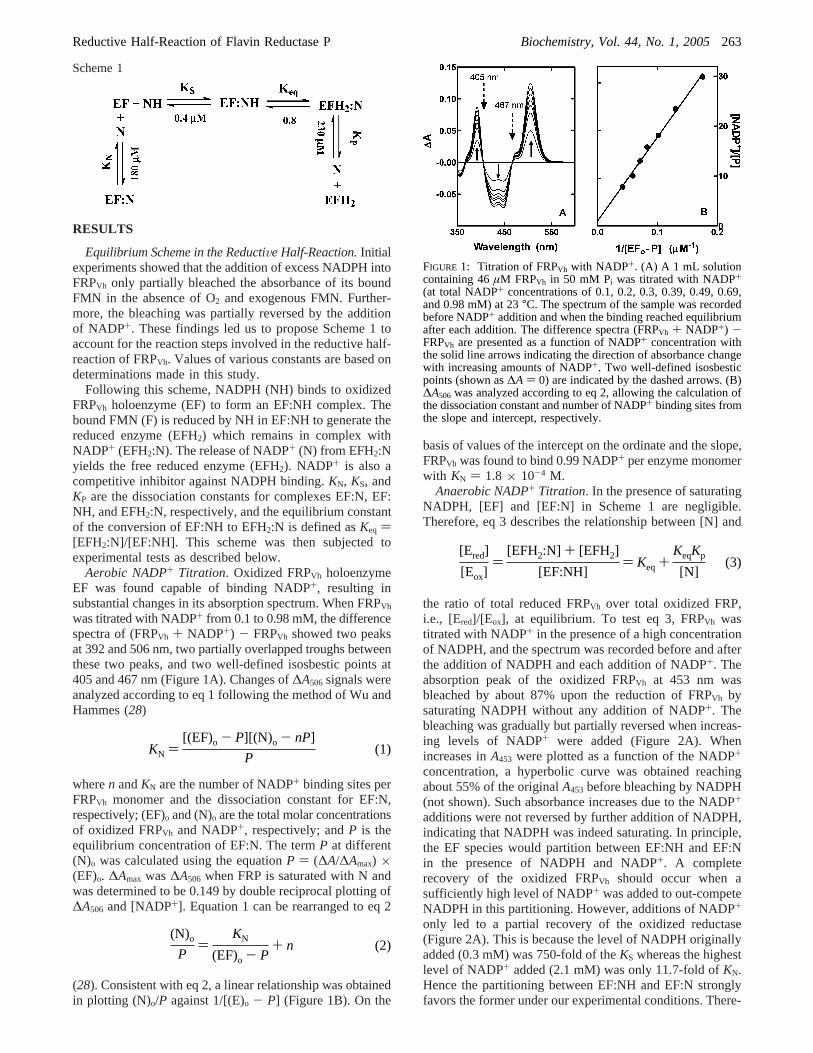
was titrated with NADP+ from 0.1 to 0.98 mM, the differencespectra of (FRPVh + NADP+) - FRPVh showed two peaksat 392 and 506 nm, two partially overlapped troughs betweenthese two peaks, and two well-defined isosbestic points at405 and 467 nm (Figure 1A). Changes of∆A506 signals wereanalyzed according to eq 1 following the method of Wu andHammes (28)
wheren andKN are the number of NADP+ binding sites perFRPVh monomer and the dissociation constant for EF:N,respectively; (EF)o and (N)o are the total molar concentrationsof oxidized FRPVh and NADP+, respectively; andP is theequilibrium concentration of EF:N. The termP at different(N)o was calculated using the equationP ) (∆A/∆Amax) ×(EF)o. ∆Amax was∆A506 when FRP is saturated with N andwas determined to be 0.149 by double reciprocal plotting of∆A506 and [NADP+]. Equation 1 can be rearranged to eq 2
(28). Consistent with eq 2, a linear relationship was obtainedin plotting (N)o/P against 1/[(E)o - P] (Figure 1B). On the
basis of values of the intercept on the ordinate and the slope,FRPVh was found to bind 0.99 NADP+ per enzyme monomerwith KN ) 1.8 × 10⁻⁴ M.
Anaerobic NADP+ Titration. In the presence of saturatingNADPH, [EF] and [EF:N] in Scheme 1 are negligible.Therefore, eq 3 describes the relationship between [N] and
the ratio of total reduced FRPVh over total oxidized FRP,i.e., [Ered]/[Eox], at equilibrium. To test eq 3, FRPVh wastitrated with NADP+ in the presence of a high concentrationof NADPH, and the spectrum was recorded before and afterthe addition of NADPH and each addition of NADP+. Theabsorption peak of the oxidized FRPVh at 453 nm wasbleached by about 87% upon the reduction of FRPVh bysaturating NADPH without any addition of NADP+. Thebleaching was gradually but partially reversed when increas-ing levels of NADP+ were added (Figure 2A). Whenincreases inA453 were plotted as a function of the NADP+
concentration, a hyperbolic curve was obtained reachingabout 55% of the originalA453 before bleaching by NADPH(not shown). Such absorbance increases due to the NADP+
additions were not reversed by further addition of NADPH,indicating that NADPH was indeed saturating. In principle,the EF species would partition between EF:NH and EF:Nin the presence of NADPH and NADP+. A completerecovery of the oxidized FRPVh should occur when asufficiently high level of NADP+ was added to out-competeNADPH in this partitioning. However, additions of NADP+
only led to a partial recovery of the oxidized reductase(Figure 2A). This is because the level of NADPH originallyadded (0.3 mM) was 750-fold of theKS whereas the highestlevel of NADP+ added (2.1 mM) was only 11.7-fold ofKN.Hence the partitioning between EF:NH and EF:N stronglyfavors the former under our experimental conditions. There-
Scheme 1
FIGURE 1: Titration of FRPVh with NADP+. (A) A 1 mL solutioncontaining 46µM FRPVh in 50 mM Pi was titrated with NADP+(at total NADP+ concentrations of 0.1, 0.2, 0.3, 0.39, 0.49, 0.69,and 0.98 mM) at 23°C. The spectrum of the sample was recordedbefore NADP+ addition and when the binding reached equilibriumafter each addition. The difference spectra (FRPVh + NADP+) -FRPVh are presented as a function of NADP+ concentration withthe solid line arrows indicating the direction of absorbance changewith increasing amounts of NADP+. Two well-defined isosbesticpoints (shown as∆A ) 0) are indicated by the dashed arrows. (B)∆A506 was analyzed according to eq 2, allowing the calculation ofthe dissociation constant and number of NADP+ binding sites fromthe slope and intercept, respectively.
[Ered]
[Eox])
[EFH2:N] + [EFH2]
[EF:NH]) Keq +
KeqKp
[N](3)
KN )[(EF)o - P][(N)o - nP]
P(1)
(N)o
P)
KN
(EF)o - P+ n (2)
Reductive Half-Reaction of Flavin Reductase P Biochemistry, Vol. 44, No. 1, 2005263
fore, the partial recoveries of oxidized FRPVh upon NADP+
additions were primarily a consequence of the reversion ofEFH2 to EFH2:N and the subsequent equilibrium betweenEFH2:N and EF:NH.
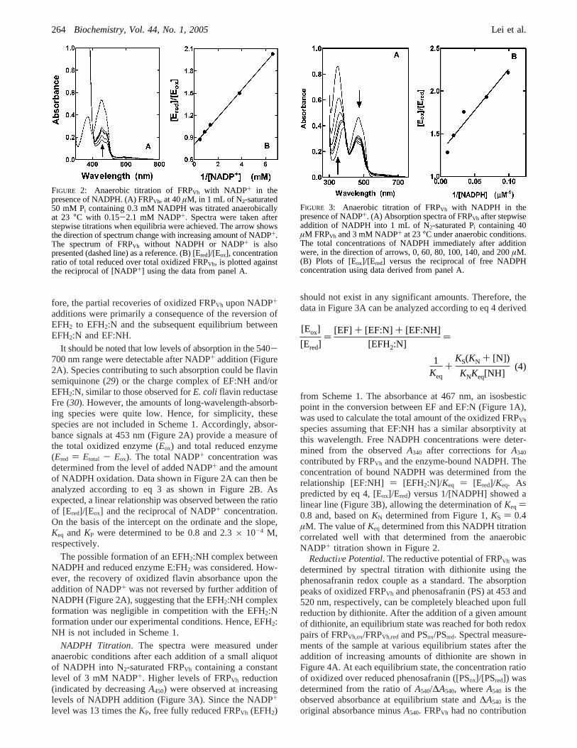
It should be noted that low levels of absorption in the 540-700 nm range were detectable after NADP+ addition (Figure2A). Species contributing to such absorption could be flavinsemiquinone (29) or the charge complex of EF:NH and/orEFH2:N, similar to those observed forE. coli flavin reductaseFre (30). However, the amounts of long-wavelength-absorb-ing species were quite low. Hence, for simplicity, thesespecies are not included in Scheme 1. Accordingly, absor-bance signals at 453 nm (Figure 2A) provide a measure ofthe total oxidized enzyme (Eox) and total reduced enzyme(Ered ) Etotal - Eox). The total NADP+ concentration wasdetermined from the level of added NADP+ and the amountof NADPH oxidation. Data shown in Figure 2A can then beanalyzed according to eq 3 as shown in Figure 2B. Asexpected, a linear relationship was observed between the ratioof [Ered]/[Eox] and the reciprocal of NADP+ concentration.On the basis of the intercept on the ordinate and the slope,Keq and KP were determined to be 0.8 and 2.3× 10⁻⁴ M,respectively.
The possible formation of an EFH2:NH complex betweenNADPH and reduced enzyme E:FH2 was considered. How-ever, the recovery of oxidized flavin absorbance upon theaddition of NADP+ was not reversed by further addition ofNADPH (Figure 2A), suggesting that the EFH2:NH complexformation was negligible in competition with the EFH2:Nformation under our experimental conditions. Hence, EFH2:NH is not included in Scheme 1.
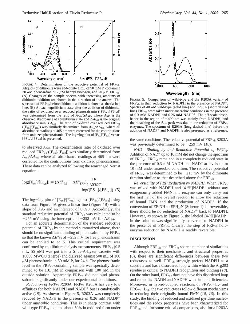
NADPH Titration. The spectra were measured underanaerobic conditions after each addition of a small aliquotof NADPH into N2-saturated FRPVh containing a constantlevel of 3 mM NADP+. Higher levels of FRPVh reduction(indicated by decreasingA450) were observed at increasinglevels of NADPH addition (Figure 3A). Since the NADP+
level was 13 times theKP, free fully reduced FRPVh (EFH2)
should not exist in any significant amounts. Therefore, thedata in Figure 3A can be analyzed according to eq 4 derived
from Scheme 1. The absorbance at 467 nm, an isosbesticpoint in the conversion between EF and EF:N (Figure 1A),was used to calculate the total amount of the oxidized FRPVh
species assuming that EF:NH has a similar absorptivity atthis wavelength. Free NADPH concentrations were deter-mined from the observedA340 after corrections forA340
contributed by FRPVh and the enzyme-bound NADPH. Theconcentration of bound NADPH was determined from therelationship [EF:NH] ) [EFH2:N]/Keq ) [Ered]/Keq. Aspredicted by eq 4, [Eox]/Ered) versus 1/[NADPH] showed alinear line (Figure 3B), allowing the determination ofKeq )0.8 and, based onKN determined from Figure 1,KS ) 0.4µM. The value ofKeq determined from this NADPH titrationcorrelated well with that determined from the anaerobicNADP+ titration shown in Figure 2.
ReductiVe Potential. The reductive potential of FRPVh wasdetermined by spectral titration with dithionite using thephenosafranin redox couple as a standard. The absorptionpeaks of oxidized FRPVh and phenosafranin (PS) at 453 and520 nm, respectively, can be completely bleached upon fullreduction by dithionite. After the addition of a given amountof dithionite, an equilibrium state was reached for both redoxpairs of FRPVh,ox/FRPVh,red and PSox/PSred. Spectral measure-ments of the sample at various equilibrium states after theaddition of increasing amounts of dithionite are shown inFigure 4A. At each equilibrium state, the concentration ratioof oxidized over reduced phenosafranin ([PSox]/[PSred]) wasdetermined from the ratio ofA540/∆A540, whereA540 is theobserved absorbance at equilibrium state and∆A540 is theoriginal absorbance minusA540. FRPVh had no contribution
FIGURE 2: Anaerobic titration of FRPVh with NADP+ in thepresence of NADPH. (A) FRPVh, at 40µM, in 1 mL of N2-saturated50 mM Pi containing 0.3 mM NADPH was titrated anaerobicallyat 23 °C with 0.15-2.1 mM NADP+. Spectra were taken afterstepwise titrations when equilibria were achieved. The arrow showsthe direction of spectrum change with increasing amount of NADP+.The spectrum of FRPVh without NADPH or NADP+ is alsopresented (dashed line) as a reference. (B) [Ered]/[Eox], concentrationratio of total reduced over total oxidized FRPVh, is plotted againstthe reciprocal of [NADP+] using the data from panel A.
FIGURE 3: Anaerobic titration of FRPVh with NADPH in thepresence of NADP+. (A) Absorption spectra of FRPVh after stepwiseaddition of NADPH into 1 mL of N2-saturated Pi containing 40µM FRPVh and 3 mM NADP+ at 23°C under anaerobic conditions.The total concentrations of NADPH immediately after additionwere, in the direction of arrows, 0, 60, 80, 100, 140, and 200µM.(B) Plots of [Eox]/[Ered] versus the reciprocal of free NADPHconcentration using data derived from panel A.
[Eox]
[Ered])
[EF] + [EF:N] + [EF:NH]
[EFH2:N])
1Keq
+KS(KN + [N])
KNKeq[NH](4)
264 Biochemistry, Vol. 44, No. 1, 2005 Lei et al.
to observedA540. The concentration ratio of oxidized overreduced FRPVh ([Eox]/[Ered]) was similarly determined fromA465/∆A465 where all absorbance readings at 465 nm werecorrected for the contributions from oxidized phenosafranin.These data can be analyzed following the rearranged Nernstequation:
The log-log plot of [Eox]/[Ered] against [PSox]/[PSred] usingdata from Figure 4A gives a linear line (Figure 4B) with aslope of 0.95 and an intercept of 0.098. Accordingly, thestandard reductive potential of FRPVh was calculated to be-255 mV using the intercept and-252 mV for ∆E°PS.
For an accurate determination of the standard reductivepotential of FRPVh by the method summarized above, thereshould be no significant binding of phenosafranin by FRPVh
so that the known∆E°PSof -252 mV for free phenosafranincan be applied to eq 5. This critical requirement wasconfirmed by equilibrium dialysis measurements. FRPVh (0.5mL, 55 µM) was put into a Slide-A-Lyzer cassette with10000 MWCO (Pierce) and dialyzed against 500 mL of 100µM phenosafranin in 50 mM Pi for 24 h. The phenosafraninlevel in the FRPVh-containing sample was spectrally deter-mined to be 101µM in comparison with 100µM in theoutside solution. Apparently FRPVh did not bind pheno-safranin significantly under our experimental conditions.
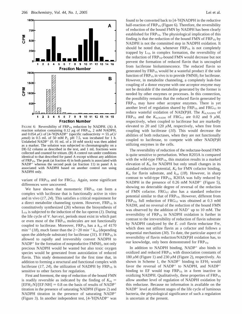
Reduction of FRPVh R203A. FRPVh R203A has very lowaffinities for both NADPH and NADP+ but is catalyticallyactive (18). As shown in Figure 5, R203A was completelyreduced by NADPH in the presence of 0.26 mM NADP+
under anaerobic conditions. This is in sharp contrast withwild-type FRPVh that had about 50% in oxidized form under
the same conditions. The reductive potential of FRPVh R203Awas previously determined to be-259 mV (18).
NAD+ Binding by and ReductiVe Potential of FRGVf.Addition of NAD+ up to 10 mM did not change the spectrumof FRGVf. FRGVf remained in a completely reduced state inthe presence of 0.3 mM NADH and NAD+ at levels up to10 mM under anaerobic condition. The reductive potentialof FRGVf was determined to be-215 mV by the dithionitetitration similar to that described above for FRPVh.
ReVersibility of FRP Reduction by NADPH. When FRPVh
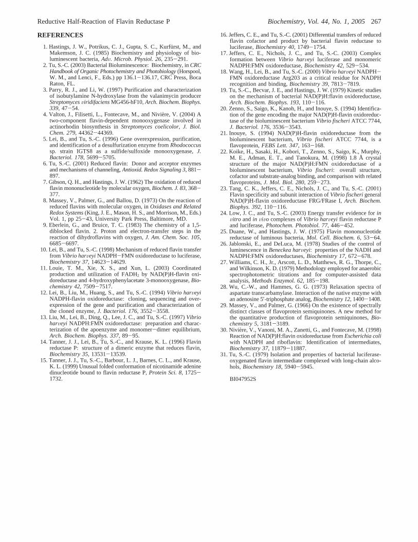
was mixed with NADPH and [4-3H]NADP+ without anyexogenously added FMN, the enzyme can only carry outthe first half of the overall reaction to allow the reductionof bound FMN and the production of NADP+. If theconversion of EF:NH to EFH2:N (Scheme 1) is irreversible,there should be no reduction of NADP+ back to NADPH.However, as shown in Figure 6, the labeled [4-3H]NADP+
in the solution was significantly converted to NADPH inthe presence of FRPVh. Clearly, the step of FRPVh holo-enzyme reduction by NADPH is readily reversible.
DISCUSSION
Although FRPVh and FRGVf share a number of similaritieswith respect to their mechanistic and structural properties(6), there are significant differences between these tworeductases as well. FRPVh strongly prefers NADPH as asubstrate and has a disordered loop within which the Arg203residue is critical to NADPH recognition and binding (18).On the other hand, FRGVh does not have this disordered loopand can utilize NADH and NADPH with similar efficiencies.Moreover, in hybrid-coupled reactions of FRPVh-LVf andFRGVf-LVh, the two reductases follow different mechanismsin reducing their originally bound FMN (10, 16). In thisstudy, the binding of reduced and oxidized pyridine nucleo-tides and the redox properties have been characterized forFRPVh and, for some critical comparisons, also for a R203A
FIGURE 4: Determination of the reductive potential of FRPVh.Aliquots of dithionite were added into 1 mL of 50 mM Pi containing20 µM phenosafranin, 2µM benzyl viologen, and 20µM FRPVh.(A) Changes of the sample spectra with increasing amounts ofdithionite addition are shown in the direction of the arrows. Thespectrum of FRPVh before dithionite addition is shown as the dashedline. (B) At each equilibrium state after the addition of dithionite,the ratio of oxidized over reduced phenosafranin ([PSox]/[PSred])was determined from the ratio ofA540/∆A540, whereA540 is theobserved absorbance at equilibrium state and∆A540 is the originalabsorbance minusA540. The ratio of oxidized over reduced FRPVh([Eox]/[Ered]) was similarly determined fromA465/∆A465, where allabsorbance readings at 465 nm were corrected for the contributionsfrom oxidized phenosafranin. The log-log plot of [Eox]/[Ered] versus[PSox]/[PSred] is presented.
log([Eox]/[Ered]) ) (∆E°PS- ∆E°FRP)2F
2.303RT+
log([PSox]/[PSred]) (5)
FIGURE 5: Comparison of wild-type and the R203A variant ofFRPVh in their reduction by NADPH in the presence of NADP+.Spectra of 40µM wild-type (solid line) and R203A (short dashedline) FRPVh were taken under anaerobic conditions in the presenceof 0.3 mM NADPH and 0.26 mM NADP+. The off-scale absor-bance in the region of<400 nm was mainly from NADPH, andthe bleaching of theA450 peak was due to the reduction of FRPVhenzymes. The spectrum of R203A (long dashed line) before theaddition of NADP+ and NADPH is also presented as a reference.
Reductive Half-Reaction of Flavin Reductase P Biochemistry, Vol. 44, No. 1, 2005265
variant of FRPVh and for FRGVf. Again, some significantdifferences were uncovered.
We have shown that monomeric FRPVh can form acomplex with luciferase that is functionally active in vitroand in vivo (17, 24). This satisfies a critical requirement fora direct metabolite channeling system. However, FRPVh isconstitutively synthesized (26) whereas the biosynthesis ofLVh is subjected to the induction of thelux operon (1). Duringthe life cycle ofV. harVeyi, periods must exist in which partor even most of the FRPVh molecules are not functionallycoupled to luciferase. Moreover, FRPVh has akcat of 4170min-1 (18), much faster than the 2-20 min-1 kcat (dependingupon the aldehyde substrate) for luciferase (31). If FRPVh isallowed to rapidly and irreversibly convert NADPH toNADP+ for the formation of nonproductive FMNH2, not onlyprecious NADPH would be wasted but also toxic oxygenspecies would be generated from autoxidation of reducedflavin. This study demonstrated for the first time that, inaddition to forming a structural and functional complex withluciferase (17, 24), the utilization of NADPH by FRPVh issensitive to other factors for regulation.
First and foremost, the step of reduction of the bound FMNis readily reversible as indicated by the finding ofKeq )[EFH2:N]/[EF:NH] ) 0.8 on the basis of results of NADP+
titration in the presence of saturating NADPH (Figure 2) andNADPH titration in the presence of saturating NADP+
(Figure 3). In another independent test, [4-3H]NADP+ was
found to be converted back to [4-3H]NADPH in the reductivehalf-reaction of FRPVh (Figure 6). Therefore, the reversibilityof reduction of the bound FMN by NADPH has been clearlyestablished for FRPVh. The physiological implication of thisfinding is that the reduction of the bound FMN of FRPVh byNADPH is not the committed step in NADPH oxidation. Itshould be noted that, whenever FRPVh is not completelytrapped by LVh in complex formation, the reversibility ofthe reduction of FRPVh-bound FMN would decrease but notprevent the formation of reduced flavin that is uncoupledfrom luciferase bioluminescence. The reduced flavin sogenerated by FRPVh would be a wasteful product if the solefunction of FRPVh in vivo is to provide FMNH2 for luciferase.However, in metabolite channeling, a completely leak-freecoupling of a donor enzyme with one acceptor enzyme maynot be desirable if the metabolite generated by the former isneeded by other enzymes or processes. In this connection,the possibility remains that the reduced flavin generated byFRPVh may have other acceptor enzymes. There is yetanother level of regulation shared by FRPVh and FRGVf toreduce wasteful oxidation of NAD(P)H. TheKm,NADPH ofFRPVh and the Km,NADH of FRGVf are 0.02 and 9µM,respectively, when coupled to luciferase but are markedlyelevated to 20 and 120µM, respectively, when free fromcoupling with luciferase (10). This would decrease theabilities of both reductases, when they are not functionallycoupled to luciferase, to compete with other NAD(P)Hutilizing enzymes in the cells.
The reversibility of reduction of the reductase-bound FMNis quite sensitive to perturbation. When R203A is comparedwith the wild-type FRPVh, this mutation results in a markedelevation ofKm for NADPH but only small changes in itsstandard reductive potential,Kd for FMN cofactor binding,Km for flavin substrate, andkcat (18). However, in sharpcontrast to wild-type FRPVh, R203A was fully reduced byNADPH in the presence of 0.26 mM NADP+ (Figure 5),showing no detectable degree of reversal of the reductionof FMN cofactor. FRGVf also has a standard reductivepotential similar to that of FRPVh. However, different fromFRPVh, full reduction of FRGVf was obtained at 0.3 mMNADH, and no reversal of the reduction of the bound FMNwas observed by the addition of NAD+ up to 10 mM. Thereversibility of FRPVh in NADPH oxidation is further incontrast to the irreversibility of reduction of flavin substrateby NADPH catalyzed by theE. coli flavin reductase Fre,which does not utilize flavin as a cofactor and follows asequential mechanism (30). To date, the particular aspect ofreversibility of flavin reduction/NAD(P)H oxidation has, toour knowledge, only been demonstrated for FRPVh.
In addition to NADPH binding, NADP+ also binds tooxidized and reduced FRPVh with dissociation constants of180µM (Figure 1) and 230µM (Figure 2), respectively. Asshown in Scheme 1, the NADP+ binding to EFH2 wouldfavor the reversal of NADP+ to NADPH, and NADP+
binding to EF would trap FRPVh in a form inactive inoxidizing NADPH. Qualitatively, these properties of FRPVh
allow another level of regulation of NADPH oxidation bythis reductase. Because no information is available on theNADP+ level at different stages of the life cycle of luminousbacteria, the physiological significance of such a regulationis uncertain at the present.
FIGURE 6: Reversibility of FRPVh reduction by NADPH. (A) Areaction solution containing 0.12µg of FRPVh, 2 mM NADPH,and 0.054µCi of [4-3H]NADP+ (specific radioactivity) 55 µCi/µmol) in 0.5 mL of 50 mM Pi, pH 7.5, was incubated for 1 h.Unlabeled NADP+ (0. 01 mL of a 10 mM stock) was then addedas a marker. The solution was subjected to chromatography on aDE-52 column as described in the text, and 1 mL fractions werecollected and counted for tritium. (B) A control run under conditionsidentical to that described for panel A except without any additionof FRPVh. The peak (at fraction 4) in both panels is associated withNADP+ whereas the second peak (at fraction 11) in panel A isassociated with NADPH based on another control run usingNADPH only.
266 Biochemistry, Vol. 44, No. 1, 2005 Lei et al.
REFERENCES
1. Hastings, J. W., Potrikus, C. J., Gupta, S. C., Kurfu¨rst, M., andMakemson, J. C. (1985) Biochemistry and physiology of bio-luminescent bacteria,AdV. Microb. Physiol. 26, 235-291.
2. Tu, S.-C. (2003) Bacterial Bioluminescence: Biochemistry, inCRCHandbook of Organic Photochemistry and Photobiology(Horspool,W. M., and Lenci, F., Eds.) pp 136.1-136.17, CRC Press, BocaRaton, FL.
3. Parry, R. J., and Li, W. (1997) Purification and characterizationof isobutylamine N-hydroxylase from the valanimycin producerStreptomycesViridifaciensMG456-hF10,Arch. Biochem. Biophys.339, 47-54.
4. Valton, J., Filisetti, L., Fontecave, M., and Nivie`re, V. (2004) Atwo-component flavin-dependent monooxygenase involved inactinorhodin biosynthesis inStreptomyces coelicolor, J. Biol.Chem. 279, 44362-44369.
5. Lei, B., and Tu, S.-C. (1996) Gene overexpression, purification,and identification of a desulfurization enzyme fromRhodococcussp. strain IGTS8 as a sulfide/sulfoxide monooxygenase,J.Bacteriol. 178, 5699-5705.
6. Tu, S.-C. (2001) Reduced flavin: Donor and acceptor enzymesand mechanisms of channeling,Antioxid. Redox Signaling 3, 881-897.
7. Gibson, Q. H., and Hastings, J. W. (1962) The oxidation of reducedflavin mononucleotide by molecular oxygen,Biochem. J. 83, 368-377.
8. Massey, V., Palmer, G., and Ballou, D. (1973) On the reaction ofreduced flavins with molecular oxygen, inOxidases and RelatedRedox Systems(King, J. E., Mason, H. S., and Morrison, M., Eds.)Vol. 1, pp 25-43, University Park Press, Baltimore, MD.
9. Eberlein, G., and Bruice, T. C. (1983) The chemistry of a 1,5-diblocked flavin. 2. Proton and electron-transfer steps in thereaction of dihydroflavins with oxygen,J. Am. Chem. Soc. 105,6685-6697.
10. Lei, B., and Tu, S.-C. (1998) Mechanism of reduced flavin transferfrom Vibrio harVeyiNADPH-FMN oxidoreductase to luciferase,Biochemistry 37, 14623-14629.
11. Louie, T. M., Xie, X. S., and Xun, L. (2003) Coordinatedproduction and utilization of FADH2 by NAD(P)H-flavin oxi-doreductase and 4-hydroxyphenylacetate 3-monooxygenase,Bio-chemistry 42, 7509-7517.
12. Lei, B., Liu, M., Huang, S., and Tu, S.-C. (1994)Vibrio harVeyiNADPH-flavin oxidoreductase: cloning, sequencing and over-expression of the gene and purification and characterization ofthe cloned enzyme,J. Bacteriol. 176, 3552-3558.
13. Liu, M., Lei, B., Ding, Q., Lee, J. C., and Tu, S.-C. (1997)VibrioharVeyi NADPH:FMN oxidoreductase: preparation and charac-terization of the apoenzyme and monomer-dimer equilibrium,Arch. Biochem. Biophys. 337, 89-95.
14. Tanner, J. J., Lei, B., Tu, S.-C., and Krause, K. L. (1996) Flavinreductase P: structure of a dimeric enzyme that reduces flavin,Biochemistry 35, 13531-13539.
15. Tanner, J. J., Tu, S.-C., Barbour, L. J., Barnes, C. L., and Krause,K. L. (1999) Unusual folded conformation of nicotinamide adeninedinucleotide bound to flavin reductase P,Protein Sci. 8, 1725-1732.
16. Jeffers, C. E., and Tu, S.-C. (2001) Differential transfers of reducedflavin cofactor and product by bacterial flavin reductase toluciferase,Biochemistry 40, 1749-1754.
17. Jeffers, C. E., Nichols, J. C., and Tu, S.-C. (2003) Complexformation betweenVibrio harVeyi luciferase and monomericNADPH:FMN oxidoreductase,Biochemistry 42, 529-534.
18. Wang, H., Lei, B., and Tu, S.-C. (2000)Vibrio harVeyiNADPH-FMN oxidoreductase Arg203 as a critical residue for NADPHrecognition and binding,Biochemistry 39, 7813-7819.
19. Tu, S.-C., Becvar, J. E., and Hastings, J. W. (1979) Kinetic studieson the mechanism of bacterial NAD(P)H:flavin oxidoreductase,Arch. Biochem. Biophys. 193, 110-116.
20. Zenno, S., Saigo, K., Kanoh, H., and Inouye, S. (1994) Identifica-tion of the gene encoding the major NAD(P)H-flavin oxidoreduc-tase of the bioluminescent bacteriumVibrio fischeriATCC 7744,J. Bacteriol. 176, 3536-3543.
21. Inouye, S. (1994) NAD(P)H-flavin oxidoreductase from thebioluminescent bacterium,Vibrio fischeri ATCC 7744, is aflavoprotein,FEBS Lett. 347, 163-168.
22. Koike, H., Sasaki, H., Kobori, T., Zenno, S., Saigo, K., Murphy,M. E., Adman, E. T., and Tanokura, M. (1998) 1.8 Å crystalstructure of the major NAD(P)H:FMN oxidoreductase of abioluminescent bacterium,Vibrio fischeri: overall structure,cofactor and substrate-analog binding, and comparison with relatedflavoproteins,J. Mol. Biol. 280, 259-273.
23. Tang, C. K., Jeffers, C. E., Nichols, J. C., and Tu, S.-C. (2001)Flavin specificity and subunit interaction ofVibrio fischerigeneralNAD(P)H-flavin oxidoreductase FRG/FRase I,Arch. Biochem.Biophys. 392, 110-116.
24. Low, J. C., and Tu, S.-C. (2003) Energy transfer evidence forinVitro andin ViVo complexes ofVibrio harVeyi flavin reductase Pand luciferase,Photochem. Photobiol. 77, 446-452.
25. Duane, W., and Hastings, J. W. (1975) Flavin mononucleotidereductase of luminous bacteria,Mol. Cell. Biochem. 6, 53-64.
26. Jablonski, E., and DeLuca, M. (1978) Studies of the control ofluminescence inBeneckea harVeyi: properties of the NADH andNADPH:FMN oxidoreductases,Biochemistry 17, 672-678.
27. Williams, C. H., Jr., Arscott, L. D., Matthews, R. G., Thorpe, C.,and Wilkinson, K. D. (1979) Methodology employed for anaerobicspectrophotometric titrations and for computer-assisted dataanalysis,Methods Enzymol. 62, 185-198.
28. Wu, C.-W., and Hammes, G. G. (1973) Relaxation spectra ofaspartate transcarbamylase. Interaction of the native enzyme withan adenosine 5′-triphosphate analog,Biochemistry 12, 1400-1408.
29. Massey, V., and Palmer, G. (1966) On the existence of spectrallydistinct classes of flavoprotein semiquinones. A new method forthe quantitative production of flavoprotein semiquinones,Bio-chemistry 5, 3181-3189.
30. Niviere, V., Vanoni, M. A., Zanetti, G., and Fontecave, M. (1998)Reaction of NAD(P)H:flavin oxidoreductase fromEscherichia coliwith NADPH and riboflavin: Identification of intermediates,Biochemistry 37, 11879-11887.
31. Tu, S.-C. (1979) Isolation and properties of bacterial luciferase-oxygenated flavin intermediate complexed with long-chain alco-hols,Biochemistry 18, 5940-5945.
BI047952S
Reductive Half-Reaction of Flavin Reductase P Biochemistry, Vol. 44, No. 1, 2005267


![706889 FMN CSR Booklet_v4[1]](https://img.pdfslide.net/doc/110x75/588587ab1a28ab84668b53ed/706889-fmn-csr-bookletv41.jpg)