Embed Size (px)
Citation preview
Dwayne Williams PA-C
North Carolina Academy of Physician Assistants
Recertification Exam Review 2017
RENAL DISEASES REVIEW

DISCLOSURES
• NONE
LEARNING OBJECTIVES
• Acute Kidney Injury (Acute Renal Failure) • Chronic Kidney disease • Glomerulonephritis • Nephrotic Syndrome • Polycystic Kidney Disease • Renal Vascular Disease • Fluid & Electrolyte Disorders • Hypervolemia/Hypovolemia • Acid Base Disorders
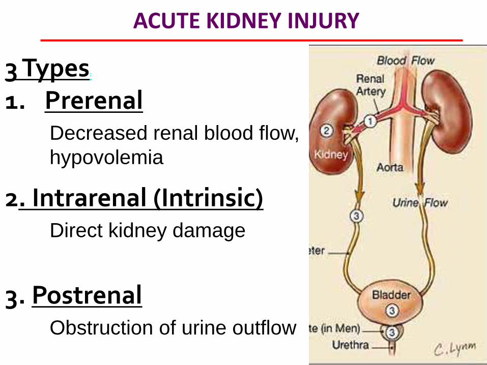
ACUTE KIDNEY INJURY
3 Types:
1. Prerenal 2. Intrarenal (Intrinsic) 3. Postrenal
Decreased renal blood flow,
hypovolemia
Direct kidney damage
Obstruction of urine outflow

ACUTE KIDNEY INJURY - PRERENAL
REDUCED RENAL PERFUSION* - GI losses: vomiting, diarrhea - Renal losses: diuretics, polyuria - Blood/fluid loss: hemorrhage, burns, pancreatitis MC type. May lead to ATN if not corrected. Hallmarks: FeNa <1%*, BUN/Cr >20:1*, ↑urine spp gravity.
Mgmt: volume repletion (rapid response).
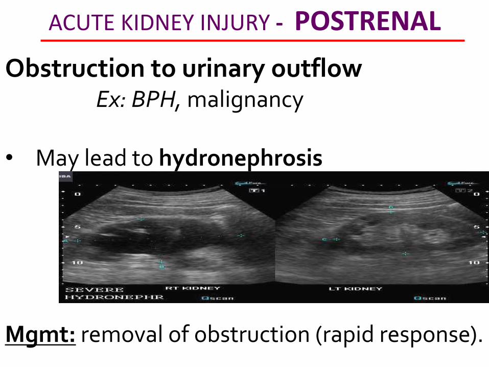
ACUTE KIDNEY INJURY - POSTRENAL
Obstruction to urinary outflow Ex: BPH, malignancy • May lead to hydronephrosis Mgmt: removal of obstruction (rapid response).
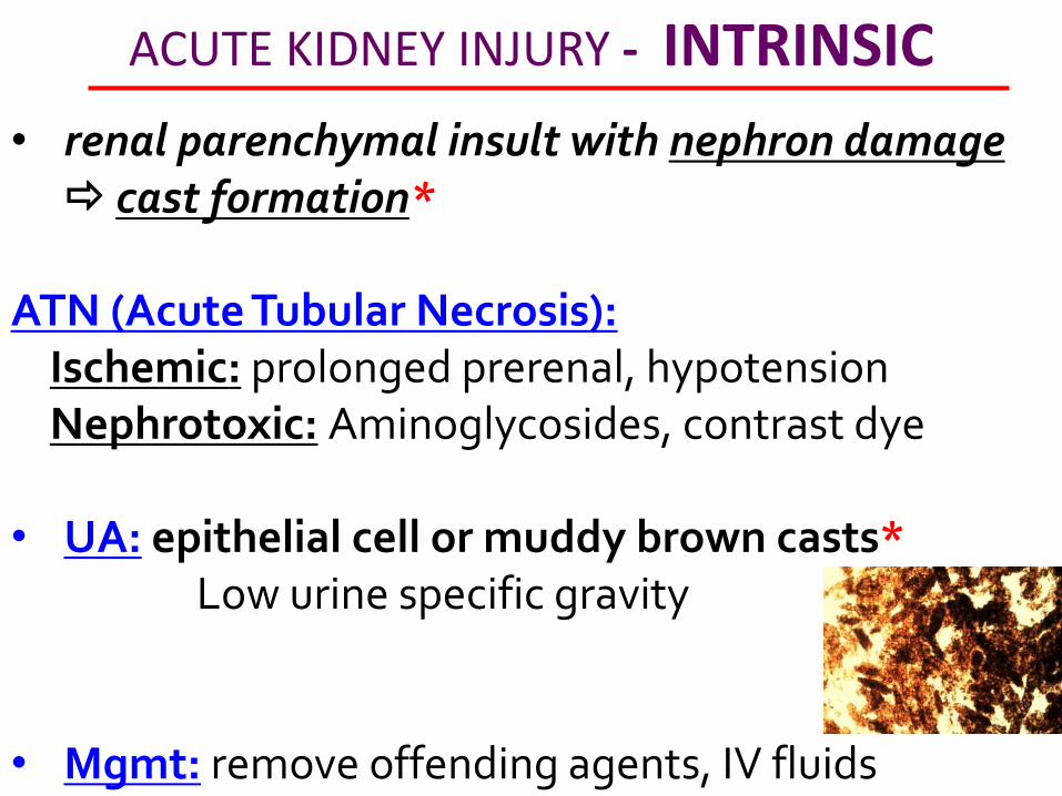
ACUTE KIDNEY INJURY - INTRINSIC
• renal parenchymal insult with nephron damage cast formation*
ATN (Acute Tubular Necrosis): Ischemic: prolonged prerenal, hypotension Nephrotoxic: Aminoglycosides, contrast dye • UA: epithelial cell or muddy brown casts* Low urine specific gravity • Mgmt: remove offending agents, IV fluids

ACUTE KIDNEY INJURY - INTRINSIC
• renal parenchymal insult with nephron damage cast formation*
AIN (Acute Interstitial Nephritis) Drug HSN: Penicillin, sulfa drugs, autoimmune Triad: rash, eosinophilia, fever • UA: white blood cell casts* • Mgmt: remove offending agent(s).

ACUTE KIDNEY INJURY - INTRINSIC
• renal parenchymal insult with nephron damage cast formation*
AGN (Acute Glomerulonephritis): • UA: red blood cell casts* • Mgmt: supportive

CHRONIC KIDNEY DISEASE
INSERT PICTURE OF STAGES
Renal transplant for end stage disease (stage 5).
Dialysis: GFR ≤ 10mL/min &/or serum creatinine ≥8mg/dL
Dialysis in diabetics:
GFR ≤ 15ml/min &/or serum creatinine of ≥6.
CHRONIC KIDNEY DISEASE
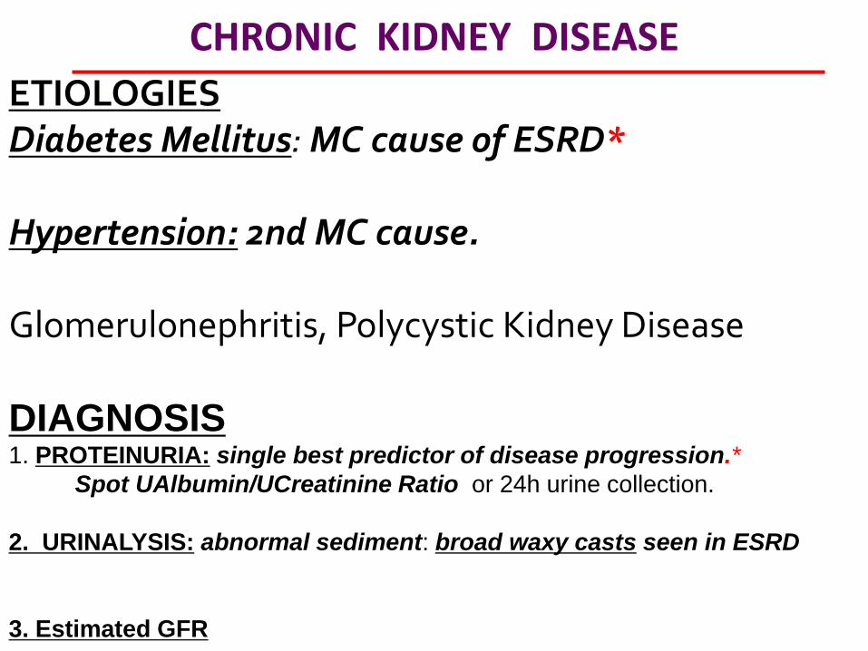
ETIOLOGIES Diabetes Mellitus: MC cause of ESRD* Hypertension: 2nd MC cause. Glomerulonephritis, Polycystic Kidney Disease DIAGNOSIS 1. PROTEINURIA: single best predictor of disease progression.*
Spot UAlbumin/UCreatinine Ratio or 24h urine collection.
2. URINALYSIS: abnormal sediment: broad waxy casts seen in ESRD
3. Estimated GFR
CHRONIC KIDNEY DISEASE
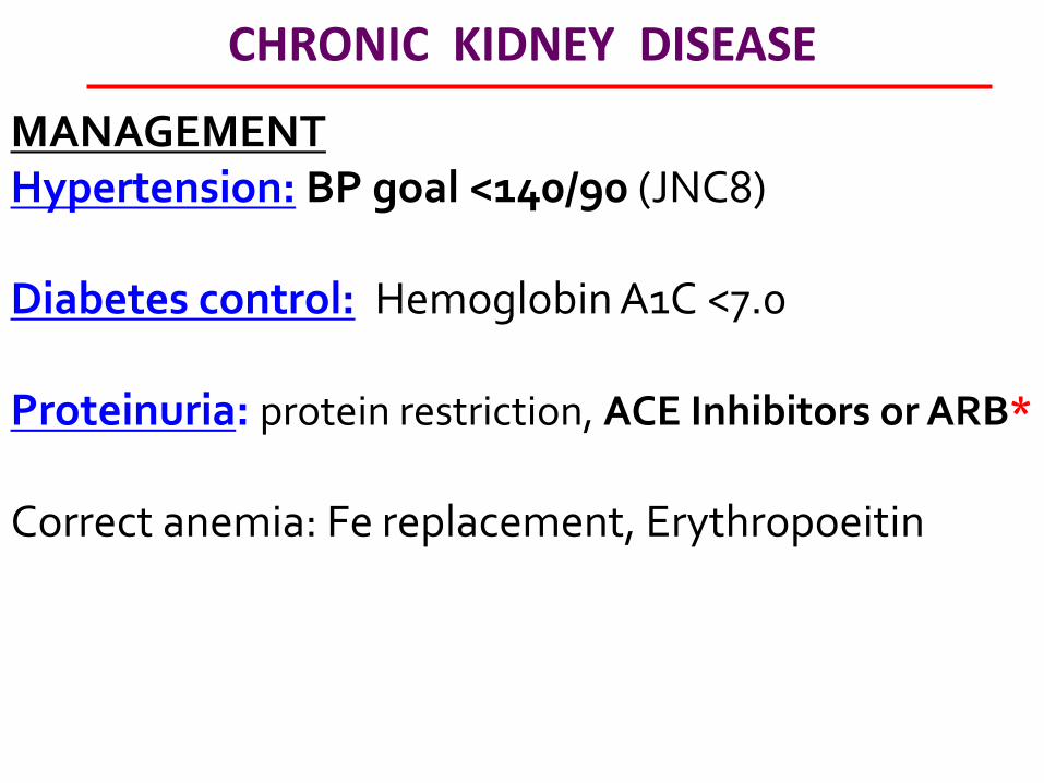
MANAGEMENT Hypertension: BP goal <140/90 (JNC8) Diabetes control: Hemoglobin A1C <7.0 Proteinuria: protein restriction, ACE Inhibitors or ARB*
Correct anemia: Fe replacement, Erythropoeitin

GLOMERULONEPHRITIS
Inflammation of the glomeruli RBC & protein leakage
•IgA Nephropathy (Berger’s disease) •Post infectious •Membranoproliferative/mesangiocapillary RAPIDLY PROGESSING GLOMERULONEPHRITIS • GoodPasture Syndrome •Vasculitis
GLOMERULONEPHRITIS
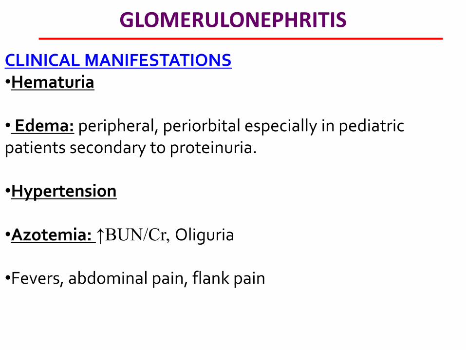
CLINICAL MANIFESTATIONS •Hematuria
• Edema: peripheral, periorbital especially in pediatric patients secondary to proteinuria. •Hypertension
•Azotemia: ↑BUN/Cr, Oliguria
•Fevers, abdominal pain, flank pain

GLOMERULONEPHRITIS - IgA Nephropathy
Aka Berger disease •MC cause of AGN in adults worldwide. •MC young males within days (24-48h) after URI or GI infection. Diagnosis: IgA deposition* on renal biopsy. Mgmt: ACE inhibitors ± Corticosteroids

GLOMERULONEPHRITIS - Post Infectious
• MC after GABHS but can occur after any infection). • Classically: 2-14y boy with puffiness of eyelids, facial
edema up to 3 weeks after strep infection with scanty cola-colored (dark) urine.
Diagnosis: Clinical, Renal biopsy: immune humps IgM, IgG.* Mgmt: ACE inhibitors ± Corticosteroids

GLOMERULONEPHRITIS - Goodpastures
• anti-GBM antibodies against the kidney and Type IV collagen antibodies of the lungs (develop glomerulonephritis and hemoptysis*
•Diagnosis: linear IgG deposits*
•Management: high dose steroid immunosuppression + cyclophosphamide* plus plasmapharesis

NEPHROTIC SYNDROME
HALLMARKS • Proteinuria • Hypoalbuminemia • Edema & hyperlipidemia
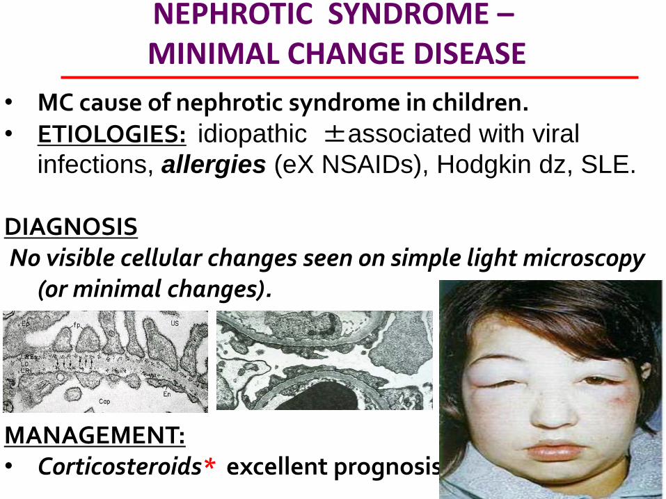
NEPHROTIC SYNDROME – MINIMAL CHANGE DISEASE
• MC cause of nephrotic syndrome in children. • ETIOLOGIES: idiopathic ±associated with viral
infections, allergies (eX NSAIDs), Hodgkin dz, SLE. DIAGNOSIS No visible cellular changes seen on simple light microscopy
(or minimal changes).
MANAGEMENT: • Corticosteroids* excellent prognosis • Cyclosporine if refractory
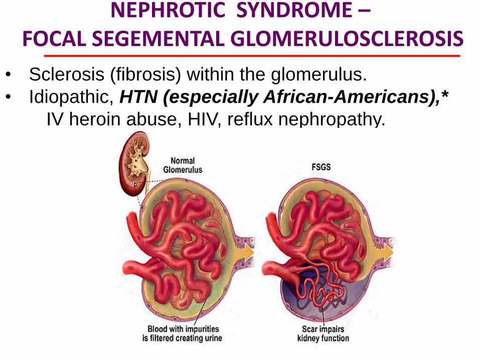
NEPHROTIC SYNDROME – FOCAL SEGEMENTAL GLOMERULOSCLEROSIS
• Sclerosis (fibrosis) within the glomerulus.
• Idiopathic, HTN (especially African-Americans),*
IV heroin abuse, HIV, reflux nephropathy.
NEPHROTIC SYNDROME – MEMBRANOUS
• Membranous: due to SLE, viral hepatitis, malaria, drugs (pencillamine) hypocomplementemia. Usually present with nephritic-nephrotic picture.
• Hallmark: Thickened basement membrane
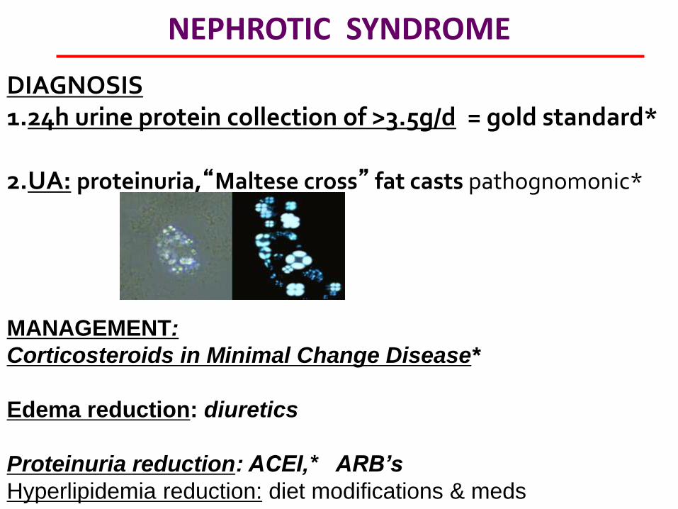
NEPHROTIC SYNDROME
DIAGNOSIS 1.24h urine protein collection of >3.5g/d = gold standard*
2.UA: proteinuria,“Maltese cross” fat casts pathognomonic*
MANAGEMENT:
Corticosteroids in Minimal Change Disease*
Edema reduction: diuretics
Proteinuria reduction: ACEI,* ARB’s
Hyperlipidemia reduction: diet modifications & meds
POLYCYSTIC KIDNEY DISEASE
•Autosomal dominant disorder of genes PKD1 or PKD2 •Kidney cysts & other organs (liver, spleen, pancreas).
CLINICAL MANIFESTATIONS •Pain: flank or abdominal pain, hepatomegaly Hematuria: cystic rupture into the renal pelvis results in gross hematuria. Nephrolithiasis, decreased urine concentrating ability, microalbuminuria.
•Patients also develop hepatic cysts, cerebral aneurysms,* mitral valve prolapse.

POLYCYSTIC KIDNEY DISEASE
DIAGNOSIS •Renal Ultrasound: best initial test*
MANAGEMENT • Lifestyle changes: protein restriction, lowered salt intake,
decreased caffeine intake, increased daily water intake. (to suppress)
• Tight Blood pressure control: to decrease activation of RAAS. (ACEI/ARBs). Goal 140/90
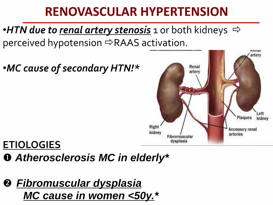
RENOVASCULAR HYPERTENSION
•HTN due to renal artery stenosis 1 or both kidneys perceived hypotension RAAS activation.
•MC cause of secondary HTN!* ETIOLOGIES Atherosclerosis MC in elderly*
Fibromuscular dysplasia
MC cause in women <50y.*

RENOVASCULAR HYPERTENSION
CLINICAL MANIFESTATIONS: severe/refractory HTN, Headache Abdominal (renal) bruit DIAGNOSIS • CT or MR angiography, Captopril renography.
• Renal arteriography: gold standard.*
RENOVASCULAR HYPERTENSION
MANAGEMENT Surgical: revascularization
•Angioplasty with stent – definitive.*
Medical
•ACE inhibitors*/ARBs (inhibits aldosterone &
angiotensin II-mediated vasoconstriction.
•However, ACEI/ARB contraindicated if bilateral
stenosis or solitary kidney* can markedly reduce
renal blood flow & GFR.
HYPERKALEMIA
ETIOLOGIES ↓Renal excretion: acute or chronic renal failure,*
Hypoaldosteronism, adrenal insufficiency Meds: K+ supplements, K+ sparing diuretics, ACEI/ARB’s, β-
blockers, digoxin, NSAID's, cyclosporine Cell lysis: rhabdomyolysis, burns, hypovolemia, thrombocytosis,
leukocytosis (intracellular release of K from cell lysis. K+ Redistribution: metabolic acidosis* (DKA), catabolic states
• •Pseudohyperkalemia: venipuncture MC, lab error.

HYPERKALEMIA
MANIFESTATIONS Neuromuscular: weakness (progressive ascending), fatigue, paresthesias, paralysis
Cardiovascular: palpitations, cardiac arrhythmias
HYPERKALEMIA
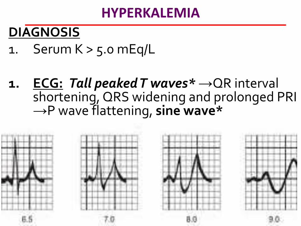
DIAGNOSIS 1. Serum K > 5.0 mEq/L 1. ECG: Tall peaked T waves* →QR interval
shortening, QRS widening and prolonged PRI →P wave flattening, sine wave*
HYPERKALEMIA
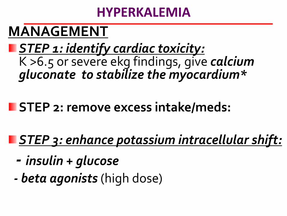
MANAGEMENT STEP 1: identify cardiac toxicity: K >6.5 or severe ekg findings, give calcium gluconate to stabilize the myocardium*
STEP 2: remove excess intake/meds:
STEP 3: enhance potassium intracellular shift:
- insulin + glucose
- beta agonists (high dose)
HYPOKALEMIA
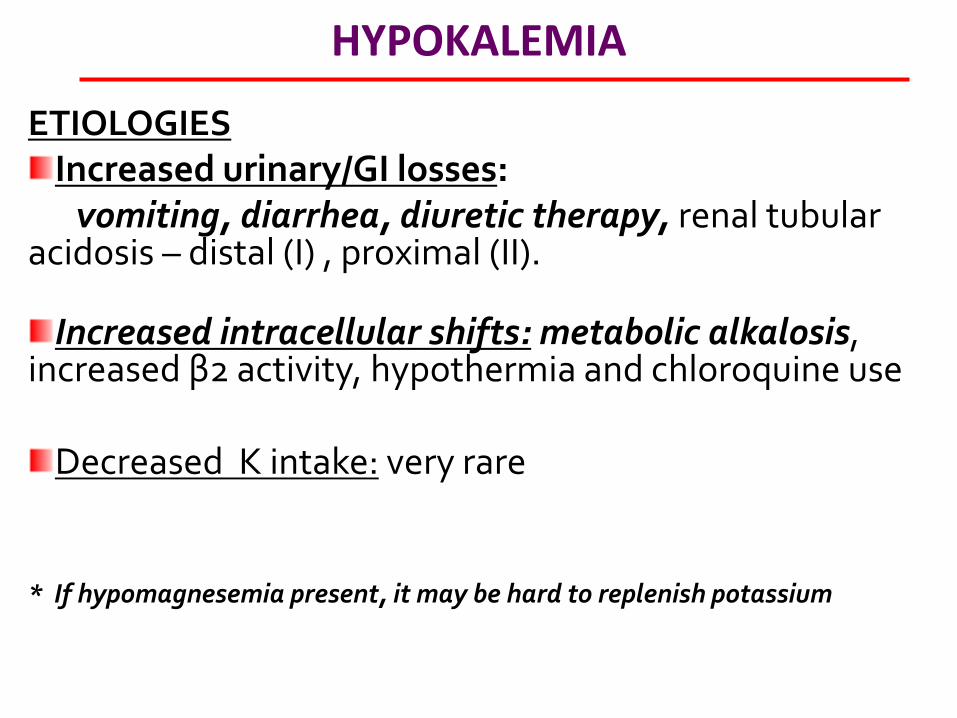
ETIOLOGIES Increased urinary/GI losses:
vomiting, diarrhea, diuretic therapy, renal tubular acidosis – distal (I) , proximal (II).
Increased intracellular shifts: metabolic alkalosis, increased β2 activity, hypothermia and chloroquine use
Decreased K intake: very rare * If hypomagnesemia present, it may be hard to replenish potassium
HYPOKALEMIA
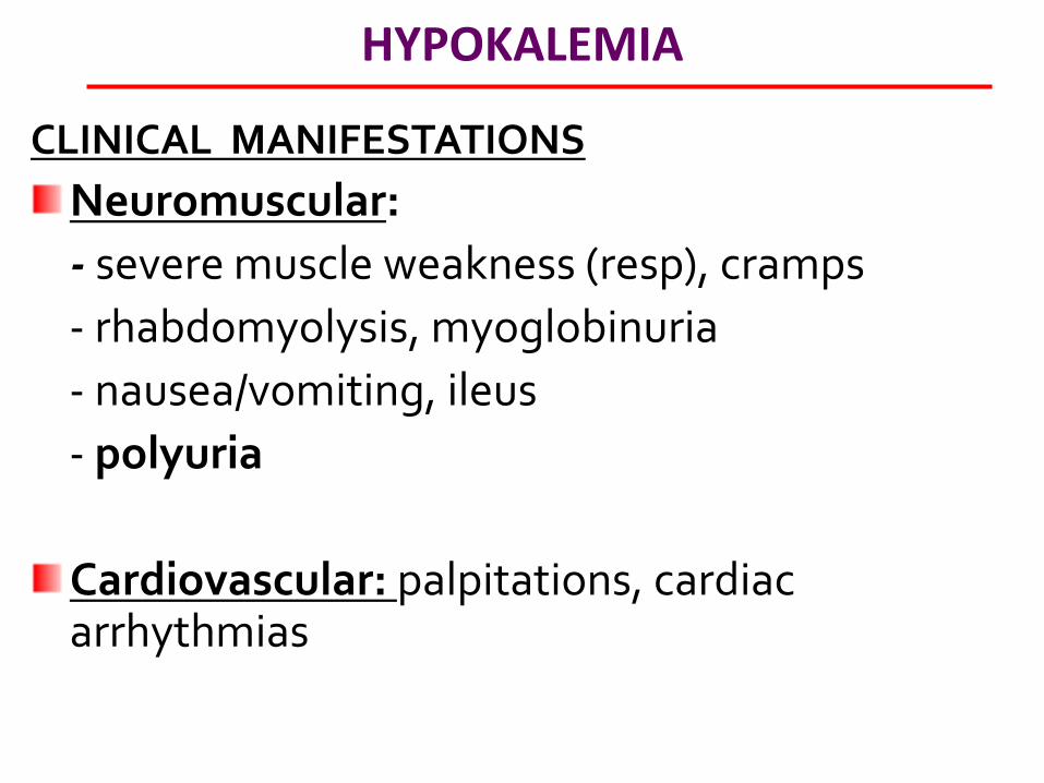
CLINICAL MANIFESTATIONS
Neuromuscular:
- severe muscle weakness (resp), cramps
- rhabdomyolysis, myoglobinuria
- nausea/vomiting, ileus
- polyuria
Cardiovascular: palpitations, cardiac arrhythmias
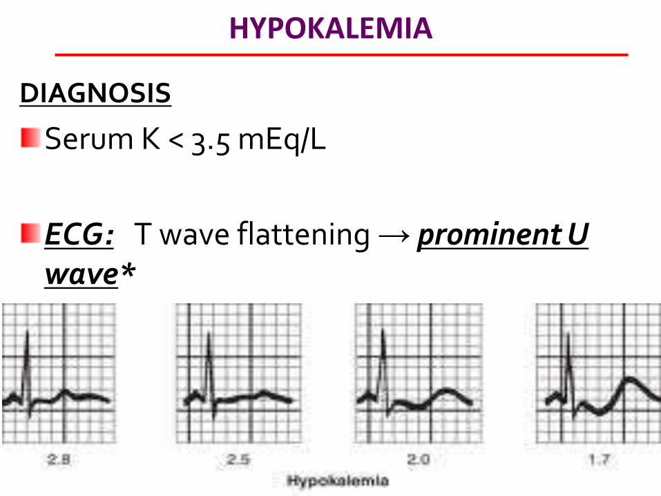
HYPOKALEMIA
DIAGNOSIS
Serum K < 3.5 mEq/L
ECG: T wave flattening → prominent U wave*
HYPOKALEMIA
MANAGEMENT
Potassium replacement:
KCl oral if possible
IV KCl given for rapid treatment/severe sx)
Replenish Magnesium if hypomagnesemic
HYPERNATREMIA
[Na] > 145 meq/L. Not as common as hyponatremia.
Cause is dehydration. Simply, the inability to drink free water (water loss)
Seen in patients who don’t have access to free water (infants, elderly, debilitated patients, nursing home)
.
HYPERNATREMIA
CNS dysfunction →water shifts out of cells →shrinkage of brain cells
(increased risk of SAH, intracranial hemorrhage). Confusion, lethargy, sz (not as common as hyponatremia), coma, muscle weakness. Lethargy
Symptoms Vary c degree and rapidity of hypernatremia. Chronic hypernatremia generally less symptomatic as a result of adaptive mechanisms.
.
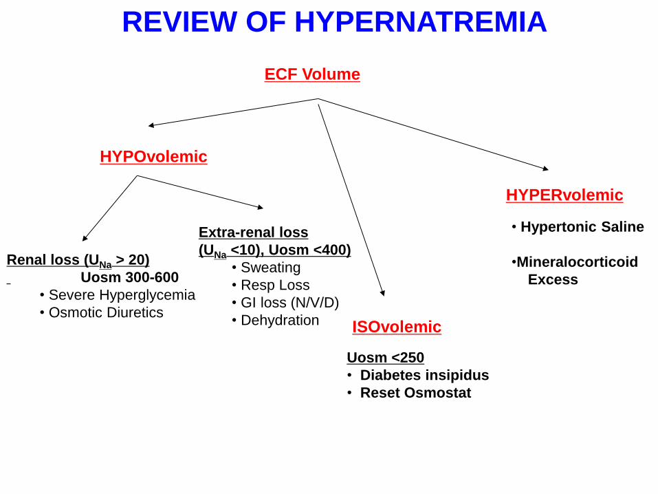
REVIEW OF HYPERNATREMIA
ECF Volume
HYPOvolemic
ISOvolemic
HYPERvolemic
• Hypertonic Saline
•Mineralocorticoid
Excess
Uosm <250
• Diabetes insipidus
• Reset Osmostat
Renal loss (UNa > 20)
Uosm 300-600
• Severe Hyperglycemia
• Osmotic Diuretics
Extra-renal loss
(UNa <10), Uosm <400)
• Sweating
• Resp Loss
• GI loss (N/V/D)
• Dehydration

HYPERNATREMIA
MANAGEMENT
Only hypotonic fluids are appropriate (ex D5W, pure water, 0.45% NS, 0.2% saline)
Preferred route is PO
Correction should be ≤0.5mEq/L/h
(to prevent cerebral edema.
except in cases of frank circulatory compromise 0.9% NS is unsuitable for managing hypernatremia.
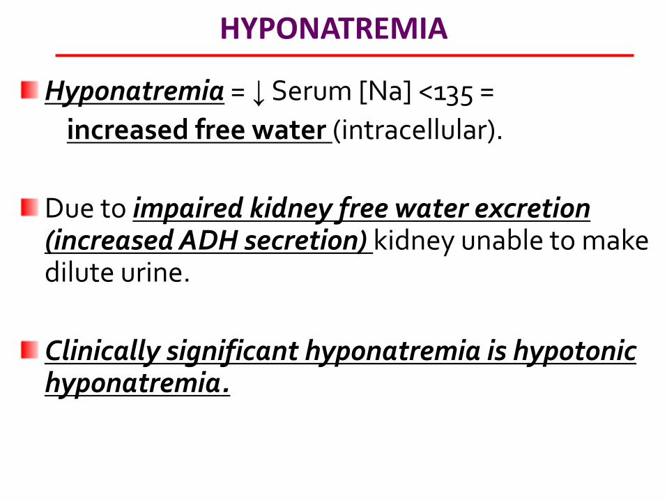
HYPONATREMIA
Hyponatremia = ↓ Serum [Na] <135 =
increased free water (intracellular).
Due to impaired kidney free water excretion (increased ADH secretion) kidney unable to make dilute urine.
Clinically significant hyponatremia is hypotonic hyponatremia.

REVIEW OF HYPONATREMIA
Serum OSM
Low Normal High
Hypotonic
Hyponatremia
(TRUE HYPONATREMIA)
Hyperglycemia
Mannitol
Lab Error
(Protein, TG)
*Note: all have ↑ADH
•SIADH: inappropriate
•Rest: appropriate
If pt critical,
Hypertonic saline
+ Loop diuretic


REVIEW OF HYPONATREMIA
Serum OSM
Low Normal High
Hypotonic
Hyponatremia
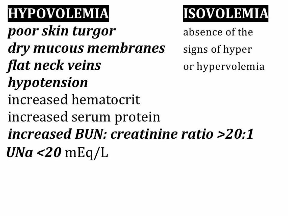
ECF Volume
Low Normal
High
Hyperglycemia
Mannitol
Lab Error
(Protein, TG)
•CHF
•Cirrhosis
•Nephrosis
•Mgmt: H2O/salt
restriction
•Hypothyroidism
•AI
•SIADH, post op
•Reset Osmostat
•Water Intoxication
1° Polydipsia
Mgmt: Water
Restriction
Renal loss (UNa > 20)
•Diuretics
•Thiazide
•K-sparing
•ACE-I, ARB
•IV RTA, Hypoaldo
Mgmt: Normal Saline
Extra-renal loss (UNa
<10), FeNa <1)
•Bleeding
•Burns
•GI (N/V, diarrhea)
•Pancreatitis
*Note: all have ↑ADH
•SIADH: inappropriate
•Rest: appropriate
If pt critical,
Hypertonic saline
+ Loop diuretic

HYPONATREMIA
MANAGEMENT
Acute hyponatremia (<48h) can be safely corrected more rapidly than chronic hyponatremia.
Rapid correction of hyponatremia can cause osmotic demyelination (rapid shrinking of brain cells leading to quadriplegia and other neurologic sequelae).
Correction should be ≤0.5mEq/L/h
(1-2mEq/L/h in severe/symptomatic patients)
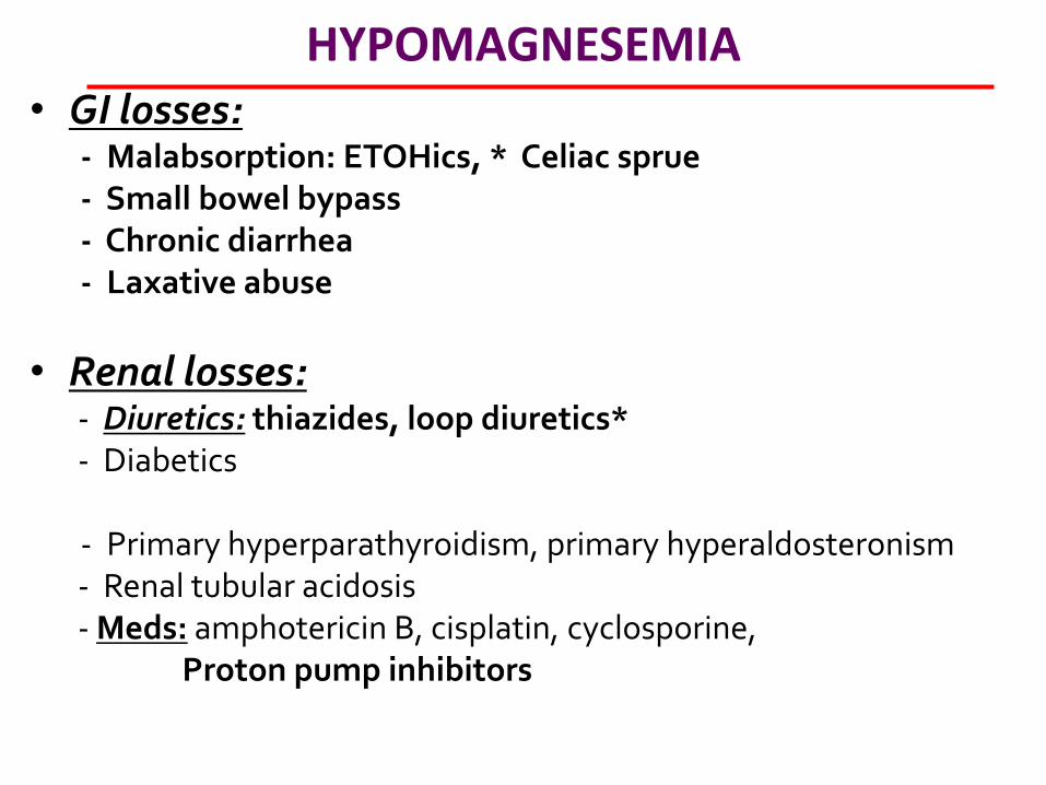
HYPOMAGNESEMIA • GI losses: - Malabsorption: ETOHics, * Celiac sprue - Small bowel bypass - Chronic diarrhea - Laxative abuse
• Renal losses: - Diuretics: thiazides, loop diuretics* - Diabetics - Primary hyperparathyroidism, primary hyperaldosteronism - Renal tubular acidosis - Meds: amphotericin B, cisplatin, cyclosporine, Proton pump inhibitors
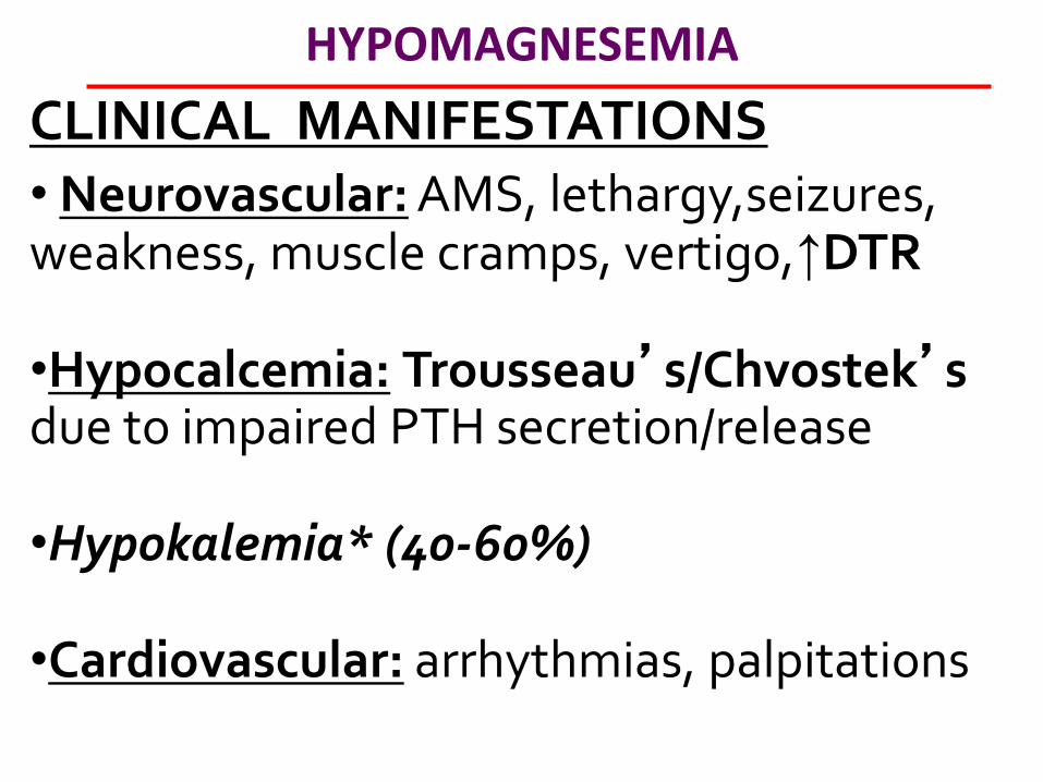
HYPOMAGNESEMIA
CLINICAL MANIFESTATIONS • Neurovascular: AMS, lethargy,seizures, weakness, muscle cramps, vertigo,↑DTR
•Hypocalcemia: Trousseau’s/Chvostek’s due to impaired PTH secretion/release
•Hypokalemia* (40-60%)
•Cardiovascular: arrhythmias, palpitations
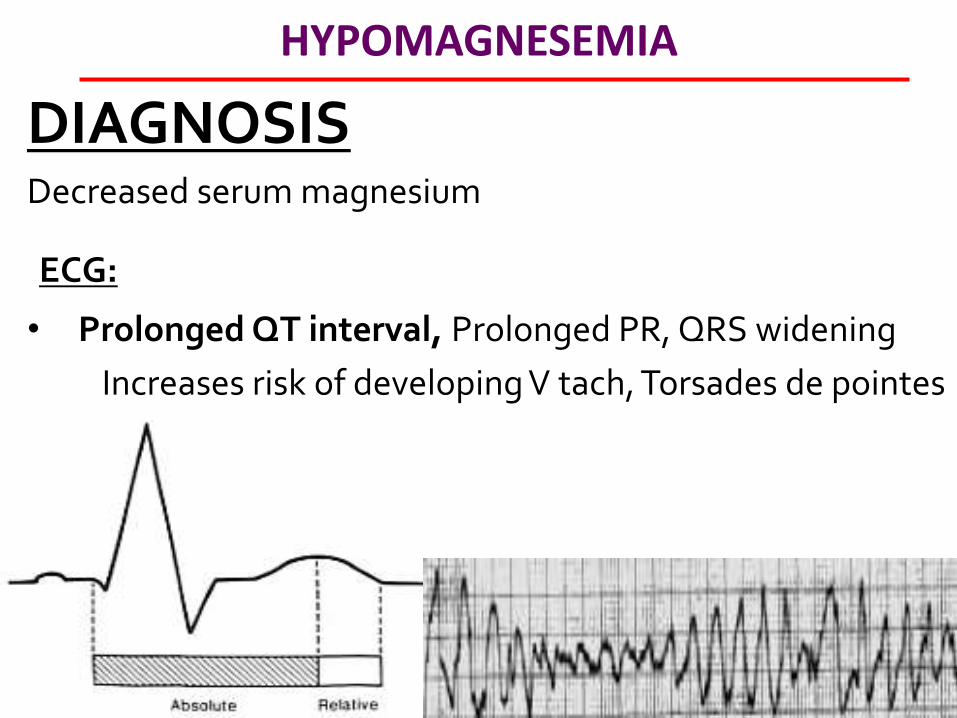
HYPOMAGNESEMIA
DIAGNOSIS Decreased serum magnesium
ECG:
• Prolonged QT interval, Prolonged PR, QRS widening
Increases risk of developing V tach, Torsades de pointes
HYPOMAGNESEMIA
MANAGEMENT
• Oral Magnesium:
• IV Magnesium Sulfate: drug of choice for torsades de pointe* or severe
HYPERMAGNESEMIA RARE. MC cause is renal insufficiency or overcorrection
of hypomagnesemia • Acute renal failure • Ingestion of Magnesium substances (ex. Vitamins,
antacids)
• Excess IV magnesium administration (ex asthma, eclampsia, Torsades de pointes, arrhythmias
• Adrenal sufficiency, milk alkali syndrome, Lithium
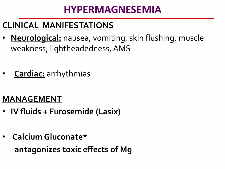
HYPERMAGNESEMIA
CLINICAL MANIFESTATIONS
• Neurological: nausea, vomiting, skin flushing, muscle weakness, lightheadedness, AMS
• Cardiac: arrhythmias
MANAGEMENT
• IV fluids + Furosemide (Lasix)
• Calcium Gluconate*
antagonizes toxic effects of Mg
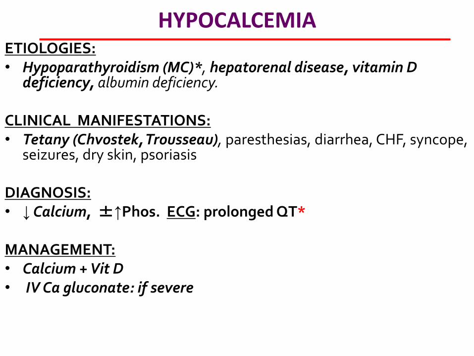
HYPOCALCEMIA ETIOLOGIES: • Hypoparathyroidism (MC)*, hepatorenal disease, vitamin D
deficiency, albumin deficiency. CLINICAL MANIFESTATIONS: • Tetany (Chvostek, Trousseau), paresthesias, diarrhea, CHF, syncope,
seizures, dry skin, psoriasis DIAGNOSIS: • ↓ Calcium, ±↑Phos. ECG: prolonged QT* MANAGEMENT: • Calcium + Vit D • IV Ca gluconate: if severe
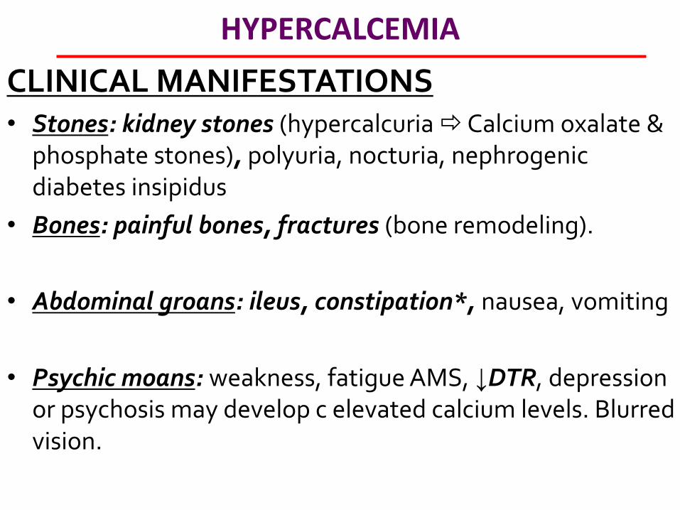
HYPERCALCEMIA
CLINICAL MANIFESTATIONS • Stones: kidney stones (hypercalcuria Calcium oxalate &
phosphate stones), polyuria, nocturia, nephrogenic diabetes insipidus
• Bones: painful bones, fractures (bone remodeling).
• Abdominal groans: ileus, constipation*, nausea, vomiting
• Psychic moans: weakness, fatigue AMS, ↓DTR, depression or psychosis may develop c elevated calcium levels. Blurred vision.
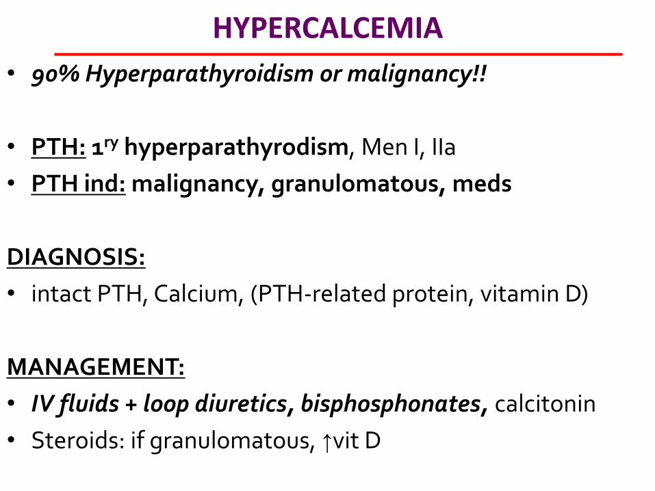
HYPERCALCEMIA
• 90% Hyperparathyroidism or malignancy!!
• PTH: 1ry hyperparathyrodism, Men I, IIa
• PTH ind: malignancy, granulomatous, meds
DIAGNOSIS:
• intact PTH, Calcium, (PTH-related protein, vitamin D)
MANAGEMENT:
• IV fluids + loop diuretics, bisphosphonates, calcitonin
• Steroids: if granulomatous, ↑vit D
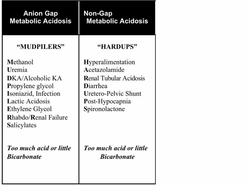
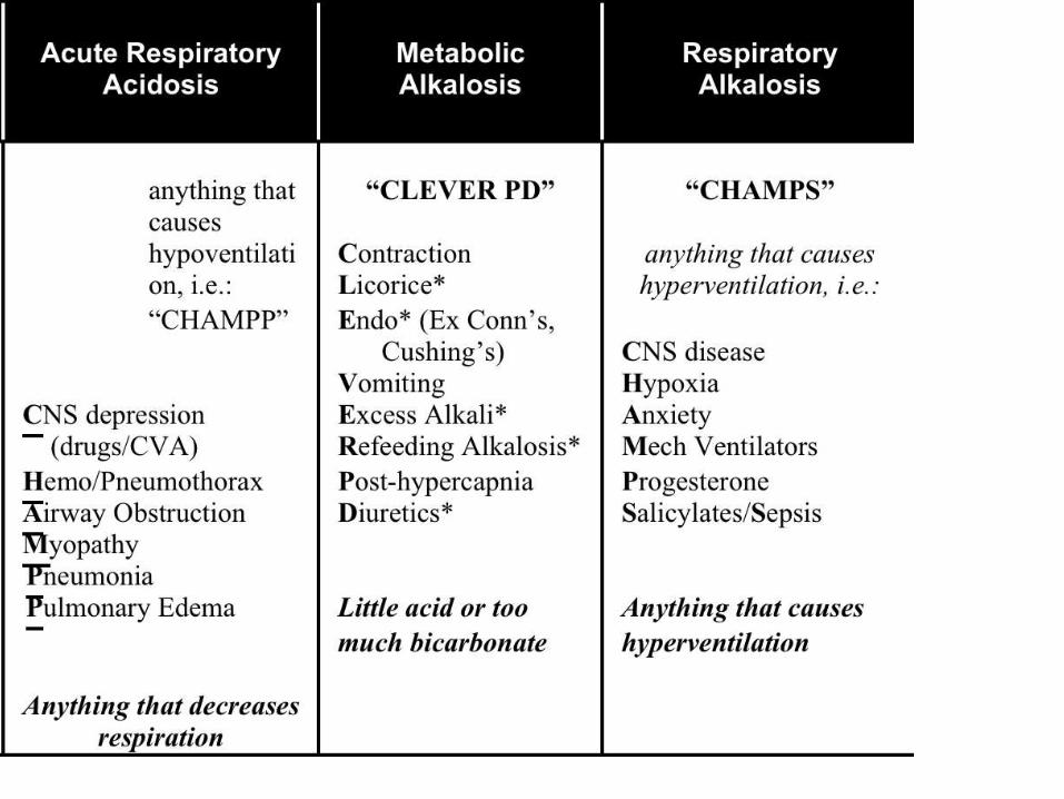
ABG STEP 1:
• LOOK AT PH
STEP 2:
• LOOK AT PCO2
STEP 3: • LOOK AT ANION GAP

ABG’S