Embed Size (px)
Citation preview
Full Terms & Conditions of access and use can be found athttp://www.tandfonline.com/action/journalInformation?journalCode=tjas20
Download by: [Western Sydney University] Date: 09 May 2017, At: 14:48
Italian Journal of Animal Science
ISSN: (Print) 1828-051X (Online) Journal homepage: http://www.tandfonline.com/loi/tjas20
Influences of the stress endocrine system on thereproductive endocrine axis in sheep (Ovis aries)
Edward Narayan & Simone Parisella
To cite this article: Edward Narayan & Simone Parisella (2017): Influences of the stress endocrinesystem on the reproductive endocrine axis in sheep (Ovis aries), Italian Journal of Animal Science,DOI: 10.1080/1828051X.2017.1321972
To link to this article: http://dx.doi.org/10.1080/1828051X.2017.1321972
© 2017 The Author(s). Published by InformaUK Limited, trading as Taylor & FrancisGroup.
Published online: 09 May 2017.
Submit your article to this journal
View related articles
View Crossmark data
REVIEW ARTICLE
Influences of the stress endocrine system on the reproductive endocrine axisin sheep (Ovis aries)
Edward Narayana,b and Simone Parisellaa
aSchool of Animal and Veterinary Sciences, Charles Sturt University, Wagga Wagga, Australia; bSchool of Science and Health, WesternSydney University, Penrith, Australia
ABSTRACTThe hypothalamic–pituitary–adrenal (HPA) axis and the hypothalamic–pituitary–gonadal (HPG)axis systems are inversely related in humans and animals. Although livestock animals, such assheep (Ovis aries), tend to be well adapted to their environment, it is known that the livestockproduction processes subject animals to a multitude of physical and psychological stressful stim-uli that have the potential to elevate the HPA axis activity. Chronic stress is one of the majorchallenges in sheep production, as it is difficult to detect and can result in prolonged dysfunc-tion of the HPA axis, causing downstream negative physiological effects such as immunosup-pression, increased susceptibility to disease and reproductive dysfunction. The elevation of HPAaxis activity during chronic stress has been suggested as the primary neuroendocrine mechanismunderlying the aetiology of reproductive dysfunction in sheep. Research in sheep has demon-strated that glucocorticoids act on the HPG axis at the level of the hypothalamus and hypophys-eal portal system to decrease gonadotrophin secretion and at the level of the pituitary gland toreduce responsiveness and sensitivity of gonadotroph cells and their receptors to GnRH. Sheepfarming enterprises rely on the breeding efficacy of ewes to optimise lambing percentage andreproductive success in order to ensure a profitable business. This review discusses the influen-ces of the HPA axis on the HPG axis and defines any significant reproductive function conse-quences caused by stress in ewes and places them into perspective for sheep management andproductivity.
ARTICLE HISTORYReceived 25 October 2016Revised 21 February 2017Accepted 24 March 2017
KEYWORDSSheep; stress; reproduction;hypothalamic–pituitary–adrenal axis; hypothala-mic–pituitary–gonadal axis;reproductive wastage
Introduction
It is already known that the livestock production pro-cess can produce a stressful environment for the ani-mals (Dantzer & Morm�ede 1983), whereby animals aresubjected to a multitude of stressful stimuli (Aggarwal& Upadhyay 2013). Although production animals (e.g.sheep) tend to be well adapted to aspects such asenvironmental conditions, feed availability and disease(Qureshi 2012), studies have demonstrated that stres-sors associated with animal production negativelyaffect reproductive physiology (Dobson et al. 2012).Sheep are prey animals and their natural response tostress is to mask any obvious behavioural signs toavoid predation (Stubsjøen et al. 2015). This makesprolonged or chronic stress difficult to detect in sheepfarming practices (Aggarwal & Upadhyay 2013).
Stress in livestock animals is defined as the inabilityof the animal to cope with stressful stimuli or situa-tions within its environment, whereby the animal fails
to meet its genetic potential in fitness parameterssuch as growth rate, milk production and fertility(Dobson & Smith 2000). A stressor is a stimulus thatdisrupts homeostasis (Smith & Dobson 2002) and acti-vates a variety of chemical and specific physiologicalprocesses and behavioural coping mechanisms torestore homeostasis and promote survival (Cizauskaset al. 2015). Acute stress is caused by a short-termnegative situation, whereby the individual is able toquickly and completely recover (Trevisi & Bertoni 2009;Papargiris et al. 2011). In contrast, chronic stress isdefined as a long lasting condition in response to along-term stressor, whereby the individual may neverrecover (Trevisi & Bertoni 2009). Stress-induced repro-ductive dysfunction is more likely attributed to chronicstress rather than acute stress and involves neuroen-docrine action at the level of the hypothalamus, pituit-ary glands and gonads. Chronic stress is one of themajor challenges in livestock production, as it can
CONTACT Dr. Edward Narayan [email protected] School of Science and Health, Western Sydney University, Locked Bag 1797,Penrith, NSW 2751, Australia� 2017 The Author(s). Published by Informa UK Limited, trading as Taylor & Francis Group.This is an Open Access article distributed under the terms of the Creative Commons Attribution-NonCommercial License (http://creativecommons.org/licenses/by-nc/4.0/), whichpermits unrestricted non-commercial use, distribution, and reproduction in any medium, provided the original work is properly cited.
ITALIAN JOURNAL OF ANIMAL SCIENCE, 2017https://doi.org/10.1080/1828051X.2017.1321972
result in immune suppression, increased susceptibilityto disease and reproductive dysfunction, potentiallyleading to undesired fitness consequences and failureto meet full genetic potential (Wagenmaker et al.2010). Macfarlane et al. (2000) suggested that stress-like concentrations of cortisol may actually arrest fol-licular development prior to maturation, which couldlead to stress related infertility in ewes. This informa-tion suggests that the effects of stress on reproductiveperformance in ewes may have severe consequenceson fecundity and fertility and that the actual impacton lambing percentage and reproductive wastage.
This review aims to discuss the proposed mecha-nisms of stress-induced reproductive effects in ewesinvolving the hypothalamo–pituitary–adrenal and thehypothalamo–pituitary–gonadal (HPA–HPG) axes inter-play. Furthermore, this review will define any signifi-cant reproductive fitness consequences caused byacute and chronic stress in ewes and place theminto perspective for farm management andproductivity.
Stress in the context of sheep reproduction:the HPA and HPG axis and their basicresponses to stress, interactions and feedbackmechanisms
Survival and growth remain the leading priority overproductivity and reproduction (Qureshi 2012).Reproduction has been suggested as the last priorityfor nutritional partitioning; therefore, it is usually thefirst physiological function to be negatively affectedby stress (Qureshi 2012). Exposure to stressful environ-mental stimuli activates and increases the secretion ofglucocorticoid hormones such as cortisol within theHPA axis, which are the downstream effectors thatregulate the physiological changes associated with thestress response (Adams et al. 1999). It has been postu-lated that glucocorticoids act on the HPG axis toinduce reproductive suppression in two definitiveways (Smith & Dobson 2002): first, at the level of thehypothalamus and hypophyseal portal system todecrease gonadotrophin secretion, and second, at thelevel of the pituitary gland to reduce responsivenessand sensitivity of gonadotroph cells and their recep-tors to gonadotropin releasing hormone (GnRH)(Adams et al. 1999). In a study that examined theeffects of inflammatory stress via the administration ofa bacterial endotoxin lipopolysaccharide (LPS), on thereproductive system at the level of the pituitary inanoestrous ewes, it was found that inflammatory stressinhibited luteinising hormone (LH) release by reducingLHb subunit transcription, resulting in decreased LH
secretion from the anterior pituitary (Herman et al.2010). The authors of this study suggested that theseresults were attributed to inflammatory stress affectingthe HPG axis firstly at the level of the hypothalamusthrough the modulation of GnRH production andrelease, by decreasing GnRH secretion to the anteriorpituitary and subsequently altering the expression ofGnRH receptor genes and LHb subunit transcription inthe anterior pituitary, thus inhibiting LH release(Herman et al. 2010). Although it has not been fullyelucidated, this insinuates that the influence of stres-sors on animals begins at the level of the hypothal-amus and precedes a cascade of physiological changesthat ultimately lead to reproductive inhibition insheep. This issue will be discussed later on in thisreview.
Functional interactions between the HPA andHPG axes
Understanding how both the HPA axis and the HPGaxis function individually is imperative in order to beable to understand and identify potential points ofcross-talks and possible feedback mechanismsbetween the two neuroendocrine pathways. The HPAaxis consists of three endocrine glands: the hypothal-amus, the anterior pituitary and the adrenal glands(Chrousos 2000). The feedback interactions betweenthese organs form the HPA axis, which is part of aneuroendocrine system that controls the physiologicaland behavioural responses to stress and can regulateother bodily processes such as glucose storage andrelease, immune function, digestion and reproduction(Chrousos 2000). Corticotrophin releasing factor (CRF)is secreted from the paraventricular nucleus (PVN) ofthe hypothalamus in response to stress (Tsigos &Chrousos 2002). Corticotrophin releasing factor is apeptide hormone and neurotransmitter, and functionsto stimulate adrenocorticotrophic hormone (ACTH)synthesis and release from the anterior pituitary(Tsigos & Chrousos 2002). Adrenocorticotrophic hor-mone is a polypeptide trophic hormone that functionsto stimulate the secretion of glucocorticoids fromadrenal cortex cells in the adrenal glands (Chrousos2000). Cortisol is a glucocorticoid steroid hormone andis produced in the adrenal glands in response to stressand low plasma glucose levels (Tsigos & Chrousos2002). When cortisol is released during the stressresponse, it functions to increase plasma glucose viagluconeogenesis, suppress the immune system andincrease fat, protein and carbohydrate metabolism toaid in the stress response (Chrousos 2000; Smith &Vale 2006).
2 E. NARAYAN AND S. PARISELLA
The HPG axis governs reproductive function in ani-mals and also plays a crucial role in development andageing (Viau 2002). The three structures that comprisethe HPG axis are the hypothalamus, the anterior pituit-ary and the gonads (Dismukes 2013). The most import-ant function of the HPG axis is to regulatereproduction in females by controlling the ovarian oroestrous cycle to prepare the follicle in the ovary forovulation (Dobson et al. 2012). Gonadotrophin releas-ing hormone is a decapeptide synthesised in special-ised neurons within the hypothalamus and isresponsible for initiating the synthesis and secretion ofgonadotrophins, LH and follicle stimulating hormone(FSH), from the anterior pituitary (Nestor 2012).Follicle-stimulating hormone and LH synthesis andsecretion are mediated by the amplitude and fre-quency of GnRH pulses and feedback interactions fromgonadal hormones (Dobson et al. 2012). In female ani-mals, gonadotrophin action is dependent upon thetarget structure on the ovary during the reproductivecycle, being either the corpus luteum or the dominant
follicle (Nestor 2012). During the follicular phase of thefemale reproductive cycle, FSH is responsible for ini-tiating follicular growth and maturation and producingoestrogen from the ovary (Breen & Karsch 2006).During the luteal phase of the female reproductivecycle, LH triggers ovulation and the development ofthe corpus luteum and stimulates progesterone pro-duction from the corpus luteum (Nestor 2012).Oestrogen is the primary female sex steroid hormone,which functions to promote sexual behaviour duringoestrus and prepare the female reproductive tract tofacilitate pregnancy and ovulation (Papargiris et al.2011). Progesterone, another important sex steroidhormone, is responsible for maintaining pregnancyand the corpus luteum to prevent future ovulations (Liet al. 2010; Papargiris et al. 2011).
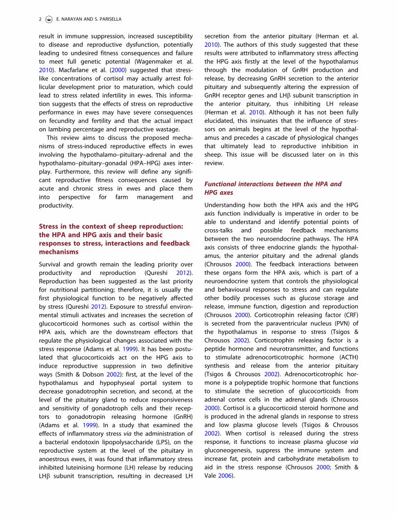
The HPA and HPG axes are parallel structures(Figure 1) that undergo a biochemical cascade ofevents that begin and end at the hypothalamus of thebrain (Dismukes 2013). The end products of both axes,such as cortisol, oestrogen and progesterone, regulate
Figure 1. Diagram of the hypothalamic–pituitary–adrenal axis (HPA-axis) and hypothalamic–pituitary–gonadal axis (HPG-axis) loopin the female animal, detailing feedback and interactions between the hormones and structures involved in both pathways.Oestrogen and progesterone confer positive/negative feedback onto the hypothalamus to regulate the HPG-axis. Cortisol confersnegative feedback onto the hypothalamus to regulate the HPA-axis. Cortisol also acts at the level of the anterior pituitary(P¼ pituitary) and reproductive organs to impede reproductive function. Hypothalamic kisspeptin (KNDy cells) are believed tomodulate the synthesis of gonadotropin releasing hormone (GnRH) within the hypothalamus. Gonadotrophin inhibitory hormone(GnIH) neurons are assumed to act both directly on GnRH neurons within the hypothalamus and project to the median eminenceto mediate pituitary function via G-protein coupled receptors on gonadotroph cells.
ITALIAN JOURNAL OF ANIMAL SCIENCE 3

these biochemical processes by conferring negativefeedback on the hypothalamus and the anterior pituit-ary, which are the same structures in the brain respon-sible for initiation of the cascade (Pierce et al. 2008).The anatomical structures that constitute both path-ways are found in the central nervous system and per-ipheral tissues and include the hypothalamus, anteriorpituitary gland, the gonads and the adrenal glands(Pierce et al. 2008).
There are many notable similarities between theHPA axis and HPG axis (Figure 1), the most significantbeing that the hypothalamus projects to and stimu-lates the anterior pituitary via hypophyseal portal ves-sels (Dismukes 2013). Interestingly, both CRF andGnRH are synthesised and secreted from neuronslocated in the preoptic area (POA) of the hypothal-amus (Dismukes 2013). Studies have suggested thatCRF receptors may be involved in regulating GnRHgene expression in the hypothalamus by regulatingGnRH mRNA levels and thus inhibiting GnRH pulsegeneration (Smith & Vale 2006).
Due to the parallel structure of both HPA–HPG axes(Figure 1), it is highly probable that there is a great dealof communication across and within the common struc-tures of the HPA and HPG axes (Viau 2002). The primaryinteractions between the HPA and HPG axes occur viathe negative feedback interactions of the end productsproduced by both axis, such as cortisol and oestrogen,the regulatory actions of the intermediaries CRF, ACTH,GnRH, LH and FSH and the involvement of other mole-cules such as gonadotrophin-inhibitory hormone (GnIH),inhibin and kisspeptin (Smith & Vale 2006).
HPA–HPG axes interplay on GnRH
In ewes, the cells that synthesise GnRH are located inthe POA and the arcuate nucleus of the hypothalamus(Dobson et al. 2012). Gonadotrophin-releasing
hormone axons project from the arcuate nucleus andPOA to the median eminence, which constitutes a dif-fuse neural network that forms the GnRH pulse gener-ator (Li et al. 2010). The pulsatile neuropeptidesecretion from GnRH neurons is dependent on intra-cellular signalling mechanisms that lead to the pulsa-tile release of GnRH from the hypothalamus (Dobsonet al. 2012). The GnRH pulse generator also constitutesthe pulsatile release of LH from the anterior pituitary(Li et al. 2010). Gonadotrophin-releasing hormone syn-thesis and secretion is usually assessed by monitoringpulsatile LH secretion, as the frequency of LH pulses iscorrelated to the activity of the GnRH pulse generator,with the amplitude of LH pulses reflecting pituitaryresponsiveness to GnRH (Clarke 2014). It is most likelythat stress induced suppression of the HPG axis beginsthrough the inhibition of GnRH secretion, which subse-quently alters the downstream effects of GnRH on LHand FSH release from the pituitary (Clarke 2014).
Glucocorticoids are assumed to suppress GnRH byacting at a central level (Figure 2). Stress-like levels ofcortisol could either reduce pituitary responsiveness toGnRH or inhibit hypothalamic synthesis and secretionof GnRH itself. Breen and Karsch (2006) found thatstress-like levels of cortisol directly reduced reproduct-ive neuroendocrine function in ovariectomised ewesby reducing pituitary responsiveness to both endogen-ous and exogenous GnRH pulses, instead of inhibitingGnRH itself. Furthermore, the suppressive action ofcortisol on pituitary responsiveness to GnRH is sup-ported during the presence of oestradiol (Oakley et al.2009). For example, Pierce et al. (2009b) set out todetermine if cortisol acts directly at the pituitary orindirectly via the hypothalamus in the presence of oes-tradiol to inhibit pituitary responsiveness to GnRH inovariectomised ewes that had undergone surgicalhypothalamo–pituitary disconnection. The resultsdetermined that oestradiol influences the suppressive
Figure 2. Diagram summarising the main neuroendocrine changes occurring in the animal (sheep model) due to direct exposureto chronic stressor, and the culminating effects on reproductive fitness of the animal (this summary diagram is based on the pub-lished literature).
4 E. NARAYAN AND S. PARISELLA
effect of cortisol on pituitary responsiveness to GnRH inewes, where LH pulses were reduced during cortisolinfusion in the presence of oestradiol (Pierce et al.2009b). Hypothalamic regulation of the anterior pituit-ary was eliminated in this experimental model, whichsuggests that cortisol is able to act directly on the pitu-itary to suppress LH secretion and hypothalamic GnRHdoes not influence the ability of cortisol to reduce pitu-itary responsiveness to GnRH. This suggests that the fre-quency and amplitude of GnRH pulses do not appearto influence the efficacy of cortisol to reduce pituitaryresponsiveness to GnRH. On the contrary, in anotherstudy that assessed the effects of psychosocial stress onewe reproduction by isolation, blindfold and predatorstress, it was found that psychosocial stress inhibitedpulsatile LH secretion in ovariectomised ewes by bothdecreasing GnRH pulse amplitude and pituitary respon-siveness to GnRH (Pierce et al. 2008).
Glucocorticoids exert their physiological actions bybinding to specific receptors on target cells (Ralph &Tilbrook 2016a,b). Glucocorticoid binding sites occurmainly in pituitary cytosols in both neonatal and adultsheep (Rose et al. 1985). Type I glucocorticoid recep-tors regulate the basal activity of the HPA axis and arelocated in the hypothalamus and other structures ofthe brain (Ralph & Tilbrook 2016b). Type II glucocortic-oid receptors are utilised during the stress responsewhen glucocorticoid concentration exceeds basal lev-els and localise in the hypothalamus and on cortico-troph cells in the pituitary (Ralph & Tilbrook 2016b). Ithas been postulated that pituitary responsiveness toGnRH is mediated via type II glucocorticoid receptors,as it was found that type II glucocorticoid receptorsare present on gonadotroph cells in the pituitary, sug-gesting that type II receptors within the pituitary maybe responsible for reducing pituitary responsiveness toGnRH via the direct actions of cortisol (Dobson et al.2012). The short-term effects of type II glucocorticoidreceptors on gonadotrophs within the pituitary aresuspected to inhibit GnRH-receptor affinity and disruptGnRH stimulation of LH release and long term by sup-pressing GnRH receptor synthesis (Dobson et al. 2012).On the contrary, type II glucocorticoid receptor immu-noreactivity determined that there was no colocalisa-tion between GnRH neurons and glucocorticoidreceptors in the hypothalamus of ewes, suggestingthat type II glucocorticoid receptors do not act directlyon GnRH neurons in the hypothalamus, but rather viaan indirect neuronal pathway on GnRH cells in theewe (Dufourny & Skinner 2002).
There are cells within the hypothalamus that syn-thesise the neuropeptide kisspeptin, called KNDy cells,that are named after the three neuropeptides that
they express, kisspeptin, neurokinin B and dynorphin(Dobson et al. 2012), and are implicated in the controlof GnRH secretion in the ewe (Ralph et al. 2016).Kisspeptin from KNDy cells stimulates the release ofGnRH from hypothalamic GnRH neurons, and oestro-gen regulates GnRH synthesis by acting on kisspeptinneurons (Kageyama 2013). Kisspeptin is believed toplay a crucial role in the transduction of stress-inducedreproductive dysfunction, as the expression of kisspep-tin is downregulated by stress (Kageyama 2013).Hypothalamic KNDy cells have oestrogen, progester-one and type II glucocorticoid receptors (Dobson et al.2012), suggesting that they are a target for cortisol aswell as oestrogen and progesterone. In addition, KNDycells have axons that enable direct projections withGnRH cell bodies and terminals within the hypothal-amus and are believed to be positioned in mannerthat enables them to control GnRH and LH release indifferent situations (Lehman et al. 2010; Dobson et al.2012). As previously mentioned, kisspeptin from KNDycells is believed to modulate the synthesis of GnRH,where the action of kisspeptin is regulated by oestra-diol. It is postulated that stress suppresses GnRH in asimilar manner, whereby glucocorticoids act on type IIglucocorticoid receptors on KNDy cells, suppressingthe expression of kisspeptin and, therefore, the synthe-sis of GnRH (Nestor 2012; Ralph et al. 2016).
Gonadotrophin-inhibitory hormone is a hypothalamicpeptide that inhibits gonadotrophin synthesis andsecretion in a variety of animal species including sheep(Kirby et al. 2009). G-protein coupled receptors are thereceptors for GnIH (Ubuka et al. 2013). Gonadotrophin-inhibitory hormone neurons act by projecting to themedian eminence, where they can mediate pituitaryfunction via G-protein coupled receptors expressed ingonadotroph cells (Ubuka et al. 2013). HypothalamicGnRH neurons also express G-protein coupled recep-tors, in which GnIH is able to bind to and inhibitgonadotrophin synthesis and secretion by decreasingthe activity of GnRH neurons and inhibit the effects ofGnRH on gonadotrophs (Ubuka et al. 2013).Hypothalamic GnIH neurons are believed to act directlywith GnRH neurons to suppress HPG axis function.Most recent research in sheep has demonstrated thatboth acute and chronic stressors increased the GnIHcell function in the hypothalamus and their input toGnRH cells in ewes (Clarke et al. 2016).
HPA–HPG axes interplay on LH and FSH
The inhibition of GnRH production in the hypothal-amus and reduced pituitary responsiveness to GnRHdisrupts the synthesis and secretion of LH and FSH in
ITALIAN JOURNAL OF ANIMAL SCIENCE 5
the anterior pituitary (Dismukes 2013). However, theexact mechanisms and potential actions of the HPAaxis intermediaries and secretory products have notbeen defined yet, specifically not in sheep. It has beenshown in ewes that psychosocial (Pierce et al. 2008),transportation (Dobson et al. 1999) and inflammatory(Herman et al. 2010) stressors disrupt pulsatile LHsecretion and delay the preovulatory LH surge. Theblocking or blunting of the preovulatory LH surgeresults in the delay or cessation of ovulation, whichcan impair fertilisation. Stress during the late follicularphase of the oestrous cycle is most likely to impactthe LH surge and thus affect ovulation, compared withstress during the early follicular phase or luteal phase(Ralph et al. 2016). One study found that the adminis-tration of stress-like concentrations of cortisol duringthe luteal and follicular phase suppressed the preovu-latory LH surge in ewes (Breen & Karsch 2004).Inflammatory stress caused by bacterial endotoxin inewes, delayed and blocked the LH surge during thefollicular phase. The timing of a stressor relative to theonset of the LH surge appears to be important in howa stressor affects the LH surge (Dobson et al. 1999).
Information detailing the effects of stress andglucocorticoid hormones on LH receptors in the repro-ductive organs of sheep is scarce. A study in cattlethat examined the direct effects of cortisol on LHreceptor numbers in cultured bovine granulosa cells,found that the addition of cortisol significantlydecreased the number of LH receptors on granulosacells (Kawate et al. 1993), demonstrating the directinhibitory effects of cortisol on LH receptor content.This data insinuate that glucocorticoids from stressmay be able to act directly on the reproductive organsto reduce LH receptor content and further altergonadal hormone synthesis and secretion.
The actions of oestrogen within the HPG axis arecritical for the generation of the LH surge (Nestor2012). In a study that examined the effects of cortisolon the action of oestrogen to induce the preovulatoryLH surge in ovariectomised ewes, it was determinedthat cortisol was able to disrupt the oestrogen-inducedLH surge, by either blocking the LH surge or suppress-ing LH surge amplitude (Pierce et al. 2009b). Thisstudy also found that cortisol caused an increase inthe latent period between the oestrogen stimulus andthe LH surge in the ewes (Pierce et al. 2009b). Insheep, oestrogen stimulates the hypothalamus to initi-ate GnRH secretion and stimulates the pituitary toenhance pituitary responsiveness to GnRH (Pierce et al.2009a,b). The effect of cortisol on the LH surge mayinvolve action by attenuating GnRH production or by
disrupting the actions of oestrogen at the level of thepituitary or hypothalamus.
Hypothalamic GnRH stimulates the synthesis of FSH,but secretion does not require the input of GnRH togonadotroph cells in the pituitary (Clarke 2014). In astudy where ewes underwent hypothalamo–pituitarydisconnection to eliminate the influence of endogen-ous GnRH on the pituitary, the cessation of pulsatileGnRH leads to the inhibition of pulsatile LH secretion,but had no effect on FSH secretion (Clarke 2014). Inanother study, cyclic ewes that were treated with2mg/d of dexamethasone had no change in plasmaFSH concentration, but plasma FSH response toexogenous GnRH was significantly reduced (Phillips &Clarke 1990). Sex steroids may play a significant role inhow stress impacts LH and FSH secretion. For example,a study found that prolonged cortisol infusion did notaffect LH pulse frequency in ovariectomised ewesdeprived of gonadal hormones, but lowered LH pulsefrequency in ewes that were supplied with oestrogenand progesterone (Li et al. 2010). There are very fewstudies that describe how CRF and ACTH affect LH andFSH secretion in sheep. In the ewe, the administrationof CRF either dramatically increased or had no effecton LH pulse frequency (Li et al. 2010). In contrast, inanother study, a CRF antagonist was unable to preventhypoglycaemic stress from inhibiting LH synthesis andsecretion in sheep (Li et al. 2010). In earlier studies,exogenous ACTH was found to suppress plasma LHconcentration and reduce LH response to exogenousGnRH in wethers and rams (Mohamed & Cox 1988;Mohamed et al. 1988). Van Lier et al. (1999) found thatexogenous ACTH suppressed LH pulse frequency inmale castrated rams, which was correlated with anincrease in plasma cortisol, but had no effect onplasma LH concentration.
Influences of acute and chronic stress onHPA–HPG axes interplay
Acute stress
Acute stress requires a short-term physiologicalresponse to avoid a negative outcome and the effectsof acute stress on reproductive function tend to be rela-tively short lived (Coburn et al. 2010). Transportation isan efficient way to induce acute stress in ruminantsin an experimental setting (Maziero et al. 2011). In astudy conducted on sheep that examined the effectsof transport stress on LH surge and secretion, bothovariectomised and intact ewes demonstrated a delayin the LH surge and a reduced LH concentration(Dobson et al. 1999). The resumption of normal LH
6 E. NARAYAN AND S. PARISELLA
profile in the next follicular phase of the ewes indi-cated that acute stress from transport leads to onlytemporary disruption of the HPG axis (Dobson et al.1999). In another study, there was no significant dif-ference observed in oestrus expression betweentransported ewes at different stages of oestrus andnon-transported ewes (Orihuela et al. 2002).
In terms of reproductive behaviour, the acute 5-hinfusion of cortisol reduced sexual receptive behaviourin ewes, where cortisol acted by inhibiting the actionof oestrogen to induce sexual receptive behaviour(Papargiris et al. 2011). The action of acute stress toinhibit sexual receptive behaviour could have relevantimplications for reproductive success as it may reducethe chance of the ewe being mounted by the ram infield conditions (Papargiris et al. 2011). However, bothacute and chronic stress-like levels of cortisol had noeffect on sexual attractivity and proceptivity behav-iours in this study (Papargiris et al. 2011). On the con-trary, another study found that the repeated exposureof ewes to acute psychosocial stressors over two oes-trous cycles did not alter the incidence, timing, ampli-tude or duration of the preovulatory LH surge(Wagenmaker et al. 2010). This study also found thatewes subjected to a variety of acute stressors duringthe follicular phase, such as food denial, unfamiliarnoises, exercise and transportation, had no effect onthe LH surge (Wagenmaker et al. 2010). There was nodifference in the mean latent period to the LH surgein the stressed ewes compared with the control ewesof this study (Wagenmaker et al. 2010). This suggeststhat the type or intensity of the acute stressor maydetermine the effect of reproductive dysfunction insheep; however, it contradicts the effects of transpor-tation stress on the preovulatory LH surge observed inother research.
Chronic stress
Research has shown that peripheral administration ofCRF has no effect on LH secretion, but centralinjection of CRF inhibits LH secretion, suggesting thata central mechanism is involved in regulating GnRHneuronal activity in the hypothalamus and down-stream LH release from the pituitary (Kageyama 2013).Furthermore, CRF inhibits the release of GnRH into thehypophyseal portal circulation both in vitro and invivo, confirming that CRF acts via a central mechanismwithin the hypothalamus (Polkowska & Przekop 1997;see Figure 2). In ewes, stress-induced suppression ofGnRH neuronal activity can be initiated by CRF releaseafter stressful stimulation (Polkowska & Przekop 1997).Corticotrophin-releasing factor has direct anatomical
connections between CRF axon terminals and the den-drites of GnRH secreting neurons (Rivier & Rivest1991). It has been suggested that CRF inhibits therelease of GnRH at the level of neurosecretory termi-nals in the median eminence of the hypothalamus,where CRF acts directly on GnRH nerve terminals(Rivier & Rivest 1991). A study that examined theeffects of CRF infusions in ewes during the late follicu-lar phase of the oestrous cycle determined that CRFinduced a decrease in GnRH stores in the nerve termi-nals of the median eminence, which was believed tobe responsible for blocking the GnRH/LH surge(Polkowska & Przekop 1997). In this study, immunohis-tochemistry of the hypothalamus found that GnRHneurons were most concentrated in the medial POA ofthe hypothalamus and GnRH neurons and cell bodieswere concentrated near the median eminence(Polkowska & Przekop 1997). The blockade of the preo-vulatory LH surge in CRF treated ewes in this studycould be attributed to diminished GnRH stores inGnRH nerve terminals of the median eminence(Polkowska & Przekop 1997). The decrease in hormonalstores in GnRH nerve terminals in the median emi-nence (Polkowska & Przekop 1997) suggests that CRFmay not block the biosynthesis of GnRH, but mayinstead inhibit GnRH release and transportation fromGnRH cell bodies to GnRH nerve terminals via theGnRH pulse generator. The suppression of GnRHrelease via CRF infusion did not occur after acutestress when CRF concentration is highest, but did soafter prolonged stimulation with CRF when HPA axisactivity was at normal levels (Polkowska & Przekop1997), suggesting that chronic stimulation of the hypo-thalamus with CRF is needed to modulate GnRHrelease.
In ewes, the predominant effect of chronic stress onthe HPG axis is the suppression of pituitary responsive-ness to GnRH (Pierce et al. 2008). In ovariectomisedewes, treatment with bacterial endotoxin marginallysuppressed GnRH secretion, but significantly disruptedLH pulsatility, demonstrating that immune stress isable to block LH pulses without inhibiting GnRH,therefore uncoupling GnRH and LH (Williams et al.2001). In this study, bacterial endotoxin also sup-pressed LH amplitude in the presence of exogenousGnRH (Williams et al. 2001), further confirming thatstress acts at the level of the pituitary by inhibitingpituitary responsiveness to GnRH. The mechanismsthat govern stress-induced reduction in pituitaryresponsiveness to GnRH have not been fully eluci-dated, but it is believed that it involves modulation ofGnRH receptors and the colocalisation of CRF
ITALIAN JOURNAL OF ANIMAL SCIENCE 7
receptors with gonadotroph cells within the anteriorpituitary.
Tissue concentration of GnRH receptors in theanterior pituitary is considered to be a determinant ofpituitary/gonadotroph responsiveness to GnRH (Daleyet al. 1999b). Stress-like concentrations of cortisol(intravenous cannula delivery into left jugular vein at3.6mg/50 kg per hour continuous infusion for a periodof 7) did not affect gonadotroph responsiveness orGnRH receptor and GnRH receptor mRNA in the pituit-ary of ovidectomised sheep, which was correlatedwith no significant change in GnRH secretion from thehypothalamus (Daley et al. 1999b). Endotoxin injectionsignificantly decreased the level of GnRH receptormRNA by 80% in the anterior pituitary of anoestrousewes, which was correlated with a decrease in LHsecretion (Herman & Tomaszewska-Zaremba 2010). Inanother study, GnRH receptor mRNA was significantlyreduced in the anterior pituitary of follicular phaseewes that underwent foot shock stimulation, whichwas associated with a decrease in plasma LH concen-tration (Ciechanowaska et al. 2007). On the contrary, asimilar experiment found that both short and pro-longed foot shock stimulation caused an increase inGnRH receptor mRNA in the pituitary of anoestrousewes (Łapot et al. 2007). Interestingly, GnRH infusionsignificantly increased GnRH receptor mRNA in thepituitary and plasma LH concentration of anoestrousewes (Parhar et al. 2012), suggesting that increasedGnRH concentration can augment the biosynthesisof GnRH receptors in the anterior pituitary.Hypothalamic GnRH is a crucial regulator of GnRHreceptor biosynthesis (Herman & Tomaszewska-Zaremba 2010). Decreased GnRH receptor geneexpression results in a reduction in GnRH receptors inthe membrane of gonadotroph cells (Herman &Tomaszewska-Zaremba 2010), which could very wellexplain stress-induced suppression of LH secretionfrom the anterior pituitary.
Fitness consequences of stress: folliculardevelopment and ovarian function
Cortisol is believed to affect follicular developmentand ovarian function by either acting at the level ofthe hypothalamus and pituitary to limit the amountof LH and FSH to support folliculogenesis and ovula-tion, or by acting directly at ovarian loci to impairfollicular maturation, ovulation and oestrogen secre-tion (Daley et al. 1999a). Serum oestrogen concentra-tion is used to estimate follicular maturation (Carsonet al. 1981). This is because large preovulatory fol-licles synthesise and secrete oestrogen and little
oestrogen is produced by small atretic follicles(Daley et al. 1999a). An early study found that ovinefollicles larger than 3.5mm in diameter are able tosynthesise and secrete oestradiol (Huet et al. 1997),which suggests that an alteration in oestrogen secre-tion could indicate that follicular development maybe impaired in the presence of significantly elevatedcortisol. In a study that examined follicular matur-ation and oestrogen production in sheep receivingstress-like levels of cortisol (intravenous delivery inright and left jugular veins at 0.1mg/kg per hour5 d before and 5 d after vaginal pessary removal)during the late luteal and early follicular phase ofthe oestrous cycle, oestrogen secretion was sup-pressed (Macfarlane et al. 2000). The suppression ofoestrogen during cortisol infusion demonstrated thatstress-like levels of cortisol arrest follicular develop-ment prior to follicular maturation (Macfarlane et al.2000). In a similar study in ewes that underwentvaginal pessary synchronisation of the oestrous cycle,it was found that serum oestrogen increased in con-trol ewes that were treated with saline solution andoestrogen levels were either blocked or attenuatedin ewes that received stress-like levels of cortisol(Daley et al. 1999a). In this study, oestrogen secre-tion was suppressed in all ewes that received0.1mg/kg per hour of exogenous cortisol and inonly 50% of ewes that received 0.08mg/kg per hourof exogenous cortisol, which suggests that cortisolconcentration or duration may be a factor thatdetermines if follicular development will be adverselyaffected (Daley et al. 1999a). In a second experimentby Daley et al. (1999a), ewes that were administeredcortisol and exogenous episodic GnRH had a signifi-cant increase in oestrogen secretion, the same ascontrol ewes in the previous experiment. In ewesthat received cortisol without exogenous episodicGnRH, oestrogen secretion was attenuated (Daleyet al. 1999a), suggesting that stress-like concentra-tions of cortisol can potentially arrest or delay fol-licular development and that episodic GnRH is ableto override the cortisol-induced delay in follicularmaturation and ovulation.
There is little information that defines the mecha-nisms that govern the effects of stress-like concentra-tions of cortisol on follicular maturation and ovulationin sheep; however, it may also involve action viaglucocorticoid or LH receptors in the ovary.Glucocorticoid receptors have been localised in theovarian follicles of humans, rats and cattle (Tetsuka2007), indicating that glucocorticoids can act directlyat the level of the reproductive organs. The expressionof LH receptors in granulosa and theca cells in the
8 E. NARAYAN AND S. PARISELLA
ovary is essential for follicular maturation (Tetsuka2007). A study in cattle that examined the directeffects of cortisol on oestradiol secretion and LHreceptor numbers in cultured bovine granulosa cellsfound that the addition of cortisol caused a significantdecrease in the amount of oestradiol secreted by gran-ulosa cells and a significant decrease in the number ofLH receptors on granulosa cells (Kawate et al. 1993).This demonstrates the direct inhibitory effects of corti-sol on LH receptor content and oestradiol secretion,indicating that cortisol from stress may be able toinhibit follicular maturation and ovulation at the levelof the reproductive organs. However, if cortisol actsvia glucocorticoid receptors on ovarian cells to inhibitovarian function or by reducing LH receptors in theovary, remains undetermined.
Embryo and foetal development, fecundity andoffspring birth weight
Livestock animals experience stressful situations fre-quently throughout pregnancy. Little is known aboutthe adverse effects of stress during gestation in farmanimals; however, other research confirmed that stressfollowing conception in sheep can result in earlyembryonic loss and reduced implantation rate (Edey1966; Dixon et al. 2007). Embryonic loss was signifi-cantly higher in ewes subjected to environmentalstress after mating (Doney et al. 1976). Similarly, heatstress during pregnancy in mice disrupted meioticmaturation of ova, increased preimplantation lossesand significantly increased early embryonic mortality(Baumgartner & Chrisman 1987).
There are very few research works that describe theimpact of stress on foetal development, fecundity andbirth weight in livestock animals. In a study that exam-ined the effects of repeated stress throughout preg-nancy on lamb birth weight and physiology, ewes thatwere exposed to isolation and dogs birthed lambs thathad heavier birth weights and higher basal cortisollevels than lambs born to non-stressed mothers(Roussel et al. 2004). In another study, nutritionallystressed ewes gave birth to lambs with significantlylower birth weights than lambs born to non-nutrition-ally stressed mothers (Meza-Herrera et al. 2015). Thisstudy also found that nutritionally stressed ewes hadfewer placental cotyledons, significantly smaller cotyle-don diameter and produced smaller litter sizes thannon-nutritionally stressed ewes (Meza-Herrera et al.2015). Another study in sheep found that chronic heatstress during gestation lowered placental weight by54%, which was correlated with low foetal weight (Bellet al. 1989), suggesting that heat stress adversely
affects foetal growth. Similarly, in cattle, calves born toheat stressed mothers had lower birth weights andweaning weights compared with calves born to non-heat-stressed mothers (Tao et al. 2012). Furthermore,embryonic and foetal mortality from stress has thepotential to reduce lambing rate and litter size, whichcould contribute to economic loss in the sheep farm-ing industry.
Conclusions
This review set out to investigate reproductive-stressendocrine axis interplay and stress-induced reproduct-ive dysfunction in sheep and the potential mecha-nisms and consequences for sheep production.Elevated HPA axis activity due to stress can causereproductive dysfunction and lead to reproductive fit-ness consequences in sheep, by interacting with andmodulating molecular and cellular aspects of the HPGaxis (Figure 2). The impact of HPA–HPG axes interplayon the reproductive fitness potential of sheep canimpede fertility, fecundity, ovulation rate, ova quality,offspring birth weight and increase embryonic and off-spring mortality. This has major implications for eco-nomic loss in the sheep farming industry, wherefarmers rely on the reproductive success of ewes inorder to ensure a profitable enterprise. Stress actsfirstly at the level of the hypothalamus to decreaseGnRH synthesis and secretion, and secondly at thelevel of the anterior pituitary gland to reduce pituitaryresponsiveness to GnRH and modulate gonadotrophinsecretion via a number of potential mechanisms. Themechanisms of reproductive-stress endocrine axisinterplay appear to involve more than just the actionof glucocorticoids to modulate HPG axis function, butinstead a series of complex interactions with hormonessuch as GnIH and CRF, colocalisation of CRF receptorswith gonadotroph cells and GnRH, and GnRH receptorgene expression modulation. Future areas of potentialresearch on reproductive-stress endocrine axis inter-play that could provide a valuable insight into sheepreproductive efficacy, may involve examiningGnIH–GnRH interaction in sheep and examining theeffects of chronic stress and elevated glucocorticoidson lambing percentage and fecundity.
Acknowledgements
This review paper forms the literature review for Masterof Animal Science thesis submitted by SP and supervisedby EN. The authors would like to thank the two anonym-ous reviewers for their comments on earlier versions ofthis article.
ITALIAN JOURNAL OF ANIMAL SCIENCE 9
Disclosure statement
No potential conflict of interest was reported by the authors.
Funding
The authors would also like to thank Australian WoolEducation Trust and Graham Centre for AgriculturalInnovation, Charles Sturt University for funding support.
ORCID
Edward Narayan http://orcid.org/0000-0003-2719-0900
References
Adams TE, Sakurai H, Adams BM. 1999. Effect of stress-likeconcentrations of cortisol on estradiol-dependent expres-sion of gonadotropin-releasing hormone receptor inorchidectomized sheep. Biol Reprod. 60:164–168.
Aggarwal A, Upadhyay R. 2013. Heat stress and animal prod-uctivity. New Delhi, India: Springer.
Baumgartner AP, Chrisman CL. 1987. Embryonic mortalitycaused by maternal heat stress during mouse oocyte mat-uration. Anim Reprod Sci. 14:309–316.
Bell AW, McBride BW, Slepetis R, Early RJ, Currie WB. 1989.Chronic heat stress and prenatal development in sheep: I.Conceptus growth and maternal plasma hormones andmetabolites. J Anim Sci. 67:3289–3299.
Breen KM, Karsch FJ. 2004. Does cortisol inhibit pulsatileluteinizing hormone secretion at the hypothalamic or pitu-itary level? Endocrinology. 145:692–698.
Breen KM, Karsch FJ. 2006. New insights regarding glucocor-ticoids, stress and gonadotropin suppression. FrontNeuroendocrinol. 27:233–245.
Carson RS, Findlay JK, Clarke IJ, Burger HG. 1981. Estradiol,testosterone, and androstenedione in ovine follicular fluidduring growth and atresia of ovarian follicles. Biol Reprod.24:105–113.
Chrousos GP. 2000. The HPA axis and the stress response.Endocr Res. 26:513–514.
Ciechanowaska M, Łapot M, Malewiski T, Misztal T, MateusiakK, Przekop F. 2007. Effect of stress on the expression ofGnRH and GnRH receptor (GnRH-R) genes in the preopticarea-hypothalamus and GnRH-R gene in the stalk/medianeminence and anterior pituitary gland in ewes during fol-licular phase of the estrous cycle. Acta Neurobiol Exp.67:1–12.
Cizauskas CA, Turner WC, Pitts N, Getz WM. 2015. Seasonalpatterns of hormones, macroparasites, and microparasitesin wild African ungulates: the interplay among stress,reproduction, and disease. PLoS ONE. 10:e0120800.
Clarke IJ, Bartolini D, Conductier G, Henry BA. 2016. Stressincreases gonadotropin inhibitory hormone cell activityand input to GnRH cells in ewes. Endocrinology.157:4339–4350.
Clarke IJ. 2014. Interface between metabolic balance andreproduction in ruminants: focus on the hypothalamusand pituitary. Horm Behav. 66:15–40.
Coburn S, Salman M, Rhyan J, Keefe T, McCollum M, Aune K,Spraker T, Miller L. 2010. Comparison of endocrineresponse to stress between captive-raised and wild-caughtbighorn sheep. J Wildlife Manage. 74:532–538.
Daley CA, Macfarlane MS, Sakurai H, Adams TE. 1999a. Effectof stress-like concentrations of cortisol on follicular devel-opment and the preovulatory surge of LH in sheep.Reproduction. 117:11–16.
Daley CA, Sakurai H, Adams BM, Adams TE. 1999b. Effect ofstress-like concentrations of cortisol on gonadotroph func-tion in orchidectomized sheep. Biol Reprod. 60:158–163.
Dantzer R, Morm�ede P. 1983. Stress in farm animals: a needfor reevaluation. J Anim Sci. 57:6–18.
Dismukes A. 2013. Coupling of the HPA and HPG axes [MSdissertation]. New Orleans (LA): University of New Orleans.
Dixon AB, Knights M, Winkler JL, Marsh DJ, Pate JL, WilsonME, Dailey RA, Seidel G, Inskeep EK. 2007. Patterns of lateembryonic and fetal mortality and association with severalfactors in sheep. J Anim Sci. 85:1274–1284.
Dobson H, Fergani C, Routly JE, Smith RF. 2012. Effects ofstress on reproduction in ewes. Anim Reprod Sci. 130:135–140.
Dobson H, Smith RF. 2000. What is stress, and how does itaffect reproduction? Anim Reprod Sci. 60–61:743–752.
Dobson H, Tebble JE, Phogat JB, Smith RF. 1999. Effect oftransport on pulsatile and surge secretion of LH in ewesin the breeding season. J Reprod Fertil. 116:1–8.
Doney JM, Smith WF, Gunn RG. 1976. Effects of post-matingenvironmental stress or administration of ACTH on earlyembryonic loss in sheep. J Agric Sci. 87:133–136.
Dufourny L, Skinner DC. 2002. Type II glucocorticoid recep-tors in the ovine hypothalamus: distribution, influence ofestrogen and absence of co-localization with GnRH. BrainRes. 946:79–86.
Edey TN. 1966. Nutritional stress and pre-implantationembryonic mortality in Merino sheep. J Agric Sci.74:187–192.
Herman AP, Romanowicz K, Tomaszewska-Zaremba D. 2010.Effect of LPS on reproductive system at the level of thepituitary of anestrous ewes. Reprod Domest Anim.45:e351–e359.
Herman AP, Tomaszewska-Zaremba D. 2010. Effect of endo-toxin on the expression of GnRH and GnRHR genes in thehypothalamus and anterior pituitary gland of anestrousewes. Anim Reprod Sci. 120:105–111.
Huet C, Monget P, Pisselet C, Monniaux D. 1997. Changes inextracellular matrix components and steroidogenicenzymes during growth and atresia of antral ovarian fol-licles in the sheep. Biol Reprod. 56:1025–1034.
Kageyama K. 2013. Regulation of gonadotropins by cortico-tropin-releasing factor and urocortin. Front Endocrinol(Lausanne). 4:12.
Kawate N, Inaba T, Mori J. 1993. Effects of cortisol on theamounts of estradiol-17b and progesterone secreted andthe number of luteinizing hormone receptors in culturedbovine granulosa cells. Anim Reprod Sci. 32:15–25.
Kirby ED, Geraghty AC, Ubuka T, Bentley GE, Kaufer D. 2009.Stress increases putative gonadotropin inhibitory hormoneand decreases luteinizing hormone in male rats. Proc NatlAcad Sci USA. 106:11324–11329.
Łapot M, Ciechanowaska M, Melewski T, Misztal T, MateusiakK, Przekop F. 2007. The effect of stress on the expression
10 E. NARAYAN AND S. PARISELLA
of GnRH and GnRH receptor genes in the discrete regionsof the hypothalamus and pituitary of anestrous ewes.Reprod Biol. 7:55–71.
Lehman MH, Coolen LM, Goodman RL. 2010. Minireview:Kisspeptin/neurokinin B/dynorphin (KNDy) cells of thearcuate nucleus: a central node in the control of gonado-tropin-releasing hormone secretion. Endocrinology.151:3479–3489.
Li XF, Knox AM, O’Byrne KT. 2010. Corticotrophin-releasingfactor and stress-induced inhibition of the gonadotrophin-releasing hormone pulse generator in the female. BrainRes. 1364:153–163.
Macfarlane MS, Breen KM, Sakurai H, Adams BM, Adams TE.2000. Effect of duration of infusion of stress-like concen-trations of cortisol on follicular development and the pre-ovulatory surge of LH in sheep. Anim Reprod Sci.63:167–175.
Maziero RRD, Martins AC, Mollo MR, Martin I, Bastos MR,Ferreira JCP, Rumpf R, Sartori R. 2011. Ovarian function incows submitted to acute stress during proestrus. LivestSci. 138:105–108.
Meza-Herrera CA, Vicente-P�erez A, Osorio-Mar�ın Y, Gir�on-G�omez BS, Beltran-Calderon E, Avenda~no-Reyes L, Correa-Calderon A, Mac�ıas-Cruz U. 2015. Heat stress, divergentnutrition level, and late pregnancy in hair sheep: effectsupon cotyledon development and litter weight at birth.Trop Anim Health Prod. 47:819–824.
Mohamed FHA, Cox JE, Moonan V. 1988. Studies of pituitary-adrenal-testis interaction in sheep. I. The effects ofrepeated injections of adrenocorticotrophic hormone dur-ing the breeding season. Theriogenology. 29:849–857.
Mohamed FHA, Cox JE. 1988. Studies of pituitary–adrenal–-testis interaction in sheep. II. The effects of repeated injec-tions of adrenocorticotrophic hormone outside thebreeding season. Theriogenology. 29:859–865.
Nestor CC. 2012. Evidence of a role for three neuropeptidesthat mediate steroid negative feedback on gonadotropinreleasing hormone/luteinizing hormone secretion in theewe: Kisspeptin, neurokinin B and orphanin FQ [PhD dis-sertation]. Morgantown, WV: West Virginia University.
Oakley AE, Breen KM, Clarke IJ, Karsch FJ, Wagenmaker ER,Tilbrook AJ. 2009. Cortisol reduces gonadotropin-releasinghormone pulse frequency in follicular phase ewes: influ-ence of ovarian steroids. Endocrinology. 150:341–349.
Orihuela A, S�anchez-Mejorada H, Toledo M. 2002. Effect ofshort transport during di-oestrus and pro-oestrus on corti-sol levels and oestrous behaviour of sheep. J Agric Sci.138:93–96.
Papargiris MM, Rivalland ETA, Hemsworth PH, Morrissey AD,Tilbrook AJ. 2011. Acute and chronic stress-like levels ofcortisol inhibit the oestradiol stimulus to induce sexualreceptivity but have no effect on sexual attractivity or pro-ceptivity in female sheep. Horm Behav. 60:336–345.
Parhar I, Ogawa S, Kitahashi T. 2012. RFamide peptides asmediators in environmental control of GnRH neurons.Prog Neurobiol. 98:176–196.
Phillips DJ, Clarke IJ. 1990. Effects of the synthetic gluco-corticoid dexamethasone on reproductive function in theewe. J Endocrinol. 126:289–295.
Pierce BN, Clarke IJ, Turner AI, Rivalland ETA, Tilbrook AJA.2009a. Cortisol disrupts the ability of estradiol-17beta to
induce the LH surge in ovariectomized ewes. DomestAnim Endocrinol. 36:202–208.
Pierce BN, Hemsworth PH, Rivalland ETA, Wagenmaker ER,Morrissey AD, Papargiris MM, Clarke IJ, Karsch FJ, TurnerAI, Tilbrook AJ. 2008. Psychosocial stress suppresses attrac-tivity, proceptivity and pulsatile LH secretion in the ewe.Horm Behav. 54:424–434.
Pierce BN, Stackpole CA, Breen KM, Clarke IJ, Karsch FJ,Rivalland ETA, Turner AI, Caddy DJ, Wagenmaker ER,Oakley AE, Tilbrook AJ. 2009b. Estradiol enables cortisol toact directly upon the pituitary to suppress pituitaryresponsiveness to GnRH in sheep. Neuroendocrinology.89:86–97.
Polkowska J, Przekop F. 1997. The effect of corticotropin-releasing factor (CRF) on the gonadotropin hormonereleasing hormone (GnRH) hypothalamic neuronal systemduring preovulatory period in the ewe. Acta NeurobiolExp. 57:91–99.
Qureshi M. 2012. Stress impedes reproductive physiology ofdairy animals under subtropical conditions – a review. JAnim Plant Sci. 22:75–78.
Ralph CR, Lehman MN, Goodman RL, Tilbrook AJ. 2016.Impact of psychosocial stress on gonadotrophins and sex-ual behaviour in females: role for cortisol? Reproduction.152:R1–R14.
Ralph CR, Tilbrook AJ. 2016a. The hypothalamo-pituitary-adrenal (HPA) axis in sheep is attenuated during lactationin response to psychosocial and predator stress. DomestAnim Endocrinol. 55:66–73.
Ralph CR, Tilbrook AJ. 2016b. Invited review: the usefulnessof measuring glucocorticoids for assessing animal welfare.J Anim Sci. 94:457–470.
Rivier C, Rivest S. 1991. Effect of stress on the activity of thehypothalamic–pituitary–gonadal axis: peripheral and cen-tral mechanisms. Biol Reprod. 45:523–532.
Rose JC, Kute TE, Winkler L. 1985. Glucocorticoid receptors insheep brain tissues during development. Am J Physiol.249:E345–E349.
Roussel S, Hemsworth PH, Boissy A, Duvaux-Ponter C. 2004.Effects of repeated stress during pregnancy in ewes onthe behavioural and physiological responses to stressfulevents and birth weight of their offspring. Appl AnimBehav Sci. 85:259–276.
Smith RF, Dobson H. 2002. Hormonal interactions within thehypothalamus and pituitary with respect to stress andreproduction in sheep. Domest Anim Endocrinol.23:75–85.
Smith SM, Vale WW. 2006. The role of the hypothalamic–pi-tuitary–adrenal axis in neuroendocrine responses to stress.Dialog Clin Neurosci. 8:383–395.
Stubsjøen SM, Bohlin J, Dahl E, Knappe-Poindecker M,Fjeldaas T, Lepschy M, Palme R, Langbein J, Ropstad E.2015. Assessment of chronic stress in sheep (part I): theuse of cortisol and cortisone in hair as non-invasive bio-logical markers. Small Ruminant Res. 132:25–31.
Tao S, Monteiro AP, Thompson IM, Hayen MJ, Dahl GE. 2012.Effect of late-gestation maternal heat stress on growthand immune function of dairy calves. J Dairy Sci.95:7128–7136.
Tetsuka M. 2007. Actions of glucocorticoid and their regula-tory mechanisms in the ovary. Anim Sci J. 78:112–120.
ITALIAN JOURNAL OF ANIMAL SCIENCE 11

Trevisi E, Bertoni G. 2009. Some physiological and biochem-ical methods for acute and chronic stress evaluation indairy cows. Ital J Anim Sci. 8:265–286.
Tsigos C, Chrousos GP. 2002. Hypothalamic–pituitary–adrenalaxis, neuroendocrine factors and stress. J Psychosom Res.53:865–871.
Ubuka T, Son YL, Bentley GE, Millar RP, Tsutsui K.2013. Gonadotropin-inhibitory hormone (GnIH), GnIHreceptor and cell signaling. Gen Comp Endocrinol.190:10–17.
Van Lier E, Regueiro M, P�erez-Clariget R, Andersson H,Kindahl H, Forsberg M. 1999. Effects of adrenocortico-trophin (ACTH) and progesterone on luteinising hormone
(LH) secretion in recently castrated rams. Anim Reprod Sci.55:115–126.
Viau V. 2002. Functional cross-talk between the hypothala-mic–pituitary–gonadal and -adrenal axes. J Neuroendocrinol.14:506–513.
Wagenmaker ER, Breen KM, Oakley AE, Tilbrook AJ, KarschFJ. 2010. The estrous cycle of the ewe is resistant to dis-ruption by repeated, acute psychosocial stress. BiolReprod. 82:1206–1215.
Williams CY, Harris TG, Battaglia DF, Vigui�e C, Karsch FJ.2001. Endotoxin inhibits pituitary responsiveness togonadotropin-releasing hormone 1. Endocrinology.142:1915–1922.
12 E. NARAYAN AND S. PARISELLA


![[XLS] · Web view1336 gs Caenorhabditis briggsae Echinococcus granulosus Galaxea fascicularis Ovis aries Rhodopirellula baltica Suberites domuncula Schistosoma mansoni Steinernema](https://img.pdfslide.net/doc/110x75/5b075a377f8b9a58148e2bed/xls-view1336-gs-caenorhabditis-briggsae-echinococcus-granulosus-galaxea-fascicularis.jpg)