Embed Size (px)
Citation preview
Cutuli et al. Alzheimer's Research & Therapy 2013, 5:50http://alzres.com/content/5/5/50
RESEARCH Open Access
Neuroprotective effects of donepezil againstcholinergic depletionDebora Cutuli1,2*, Paola De Bartolo1,2, Paola Caporali1,2, Anna Maria Tartaglione1,2, Diego Oddi1,3,Francesca Romana D’Amato1,3, Annalisa Nobili1, Marcello D’Amelio1,4† and Laura Petrosini1,2†
Abstract
Introduction: Intraparenchymal injections of the immunotoxin 192-IgG-saporin into medial septum and nucleusbasalis magnocellularis causes a selective depletion of basal forebrain cholinergic neurons. Thus, it represents a validmodel to mimic a key component of the cognitive deficits associated with aging and dementia. Here we administereddonepezil, a potent acetylcholinesterase inhibitor developed for treating Alzheimer’s disease, 15 days before192-IgG-saporin injection, and thus we examined donepezil effects on neurodegeneration and cognitive deficits.
Methods: Caspase-3 activity and cognitive performances of lesioned rats pre-treated with donepezil or salinewere analyzed and compared to the outcomes obtained in pre-treated sham-lesioned rats.
Results: Cholinergic depletion increased hippocampal and neocortical caspase-3 activity and impaired workingmemory, spatial discrimination, social novelty preference, and ultrasonic vocalizations, without affecting anxietylevels and fear conditioning. In lesioned animals, donepezil pre-treatment reduced hippocampal and neocorticalcaspase-3 activity and improved working memory and spatial discrimination performances and partially rescuedultrasonic vocalizations, without preventing social novelty alterations.
Conclusions: Present data indicate that donepezil pre-treatment exerts beneficial effects on behavioral deficitsinduced by cholinergic depletion, attenuating the concomitant hippocampal and neocortical neurodegeneration.
IntroductionExperimental and clinical evidence supports the hypoth-esis that the loss of basal forebrain (BF) cholinergic neu-rons and the consequent reduction of acetylcholine (ACh)synthesis and release significantly contribute to the cogni-tive impairment of aging disorders, such as mild cognitiveimpairment (MCI) and Alzheimer’s disease (AD) [1-3].Acetylcholinesterase inhibitors (AChE-Is) such as donepe-zil prevent the hydrolysis of the residual ACh in the brainand represent the best pharmacological tool to attenuatecognitive disturbances in patients with mild to moderateAD [4,5]. AChE-Is are currently used as a symptomatictreatment to improve or at least maintain central choliner-gic function [6,7].
* Correspondence: [email protected]†Equal contributors1IRCCS Fondazione Santa Lucia, Via del Fosso di Fiorano 64, Rome 00143, Italy2Department of Psychology, University of Rome “Sapienza”, Via dei Marsi 78,Rome 00185, ItalyFull list of author information is available at the end of the article
© 2013 Cutuli et al.; licensee BioMed Central LCommons Attribution License (http://creativecreproduction in any medium, provided the or
To date, besides the research of new drugs able to com-bat age-related cognitive decline, the protection of neuronsfrom damage and death associated with neurodegenerativedisorders is a major challenge in neuroscience. The conceptof neuroprotection has found increasing acceptance inneurology during the past decade and includes interven-tions aimed to slow or even halt the progress of neuronaldegeneration. Interestingly, there is growing evidence that,beyond allowing alleviation of cognitive symptoms, AChE-Is produce effective neuroprotection [7]. In fact, it has beenshown that AChE-Is protect against glutamate excitotoxi-city, neuronal damage and amyloid β (Aβ) neurotoxicity.Furthermore, many studies have shown that they induceupregulation of nicotinic ACh receptors (nAChRs) [8-18].Importantly, α4 and α7 nAChRs play a crucial role inAChE-I-mediated neuroprotection, mainly through theinvolvement of the phosphatidylinositol 3-kinase (PI3K)pathway [8,17]. Unfortunately, few in vivo studies haveexamined AChE-I neuroprotective action [7]. Althougha lot of studies have demonstrated the symptomatic effectsof donepezil in models of aging and dementia [19-25], a
td. This is an open access article distributed under the terms of the Creativeommons.org/licenses/by/2.0), which permits unrestricted use, distribution, andiginal work is properly cited.
Cutuli et al. Alzheimer's Research & Therapy 2013, 5:50 Page 2 of 18http://alzres.com/content/5/5/50
handful of studies have distinguished symptomatic fromneuroprotective effects by administering donepezil only be-fore (not during) behavioral testing [12-14,26,27]. Namely,injecting donepezil before a hypoxic insult has been shownto alleviate hypoxia-induced neurodegeneration and behav-ioral impairment [12], and, similarly, administrating done-pezil before Aβ injection was demonstrated to block lipidperoxidation and learning deficits [13]. In both studies,donepezil neuroprotective effects appeared to be medi-ated by the activation of the σ1 receptor, a protein involvedin modulation of intracellular Ca2+ mobilization, oxida-tive stress and neurotransmitter response. Furthermore,donepezil pre-treatment significantly prevented isoflurane-induced cholinergic degeneration and spatial memoryimpairment in aged mice [27] and attenuated okadaicacid–induced memory impairment, mitochondrial dys-function and apoptotic cell death [26]. Donepezil pre-treatment also prevented streptozotocin (STZ)-inducedmemory deficits, oxidative stress, mitochondrial dys-function and caspase-3 activity through the specific in-volvement of nAChRs [14].In the light of these studies, it seemed interesting to as-
sess the neuroprotective properties of long-term pre-le-sion donepezil treatment in a rat model of BF cholinergicdepletion induced by 192-immunoglobulin G (IgG)-saporin (Sap) injection. The resulting permanent and se-lective Sap-dependent loss of cholinergic BF neuronsmimics neuropathological features and cognitive impair-ments associated with MCI and AD. In fact, Sap selectivelycauses death of cholinergic cells by inhibiting ribosomalprotein synthesis when it is taken up into cells expressingthe low-affinity neurotrophin receptor p75 [28]. In the pasttwo decades, the availability of Sap allowed studying therole of BF cholinergic system in several cognitive functionsand its implications in aging and dementia [29].In the present study, we focused on the neuroprotec-
tive action of donepezil by investigating the influence ofdonepezil pre-treatment on cognitive deficits and neur-onal impairment induced by intraparenchymal Sap injec-tions into the medial septum (MS) and nucleus basalismagnocellularis (NBM). To achieve this aim, cognitiveperformance and caspase-3 activity levels of cholinergi-cally depleted rats pre-treated with donepezil or salinewere compared with those of pre-treated sham-lesionedanimals. Cognitive function was analyzed by means of awide battery of behavioral tests, including elevated plusmaze (EPM), open field with objects (OF), radial armmaze (RAM), sociability and preference for social noveltytest (PSNT) and fear conditioning (FC) with ultrasonicvocalization (USV) recording. The low-frequency USVsreflect a negative affective state [30,31] and are positivelycorrelated with the aversiveness of the situation [32]. Afterbehavioral testing, neurodegeneration was analyzed bymeasuring caspase-3 activity in the hippocampus and
neocortex, projection areas of the lesion sites. In fact,caspase-3 is the main effector caspase, whose localizedactivation can trigger synaptic loss, causing cognitiveand behavioral deficits [33], whereas strong activationleads to switching on of the apoptotic cascade. Becausethe increase in caspase-3 activity has been proposed as anearly neurodegenerative event in AD progression [34-36],its quantification might be useful in evaluating the efficacyof neuroprotective pharmacological treatment.
MethodsStudy animalsMale Wistar rats (about 300 g and 2.5 months of age)kept in standard laboratory conditions (08.00:20.00 light,food and water ad libitum and controlled humidity andtemperature) were used in the experiments. The animalswere maintained according to the guidelines for ethicalconduct developed by the European Union directive of22 September 2010 (2010/63/EU). The study was approvedby the local ethics committee of the IRCCS FondazioneSanta Lucia. Rats subjected to behavioral testing wererandomly assigned to the following experimental groups:(1) donepezil-treated sham-lesioned (Don-Sham) rats(n = 7), which were treated with donepezil andthen sham-lesioned; (2) donepezil-treated Sap-lesioned(Don-Sap) rats (n = 8), which were treated with donepeziland then Sap-lesioned; (3) saline-treated Sap-lesioned(Sal-Sap) rats (n = 8), which were treated with saline(NaCl 0.9%) and then Sap-lesioned; and (4) saline-treatedsham-lesioned (Sal-Sham) rats (n = 12), which weretreated with saline and then sham-lesioned. This groupencompassed six intact rats treated with saline and sixrats pre-treated with saline and then sham-lesioned.The behavioral performance of the two groups was notstatistically different in all the following behavioralparameters (multivariate analysis of variance (group ×parameter): group: F1,10 = 0.01, P = n.s.; parameter:F37,370 = 120.79, P < 0.000001; and group × parameter:F37,370 = 1.25, P = n.s.; Additional file 1). These animalswere pooled in the Sal-Sham group. At the end of behav-ioral testing, all rats were killed to verify the lesion bycholine acetyltransferase (ChAT) immunohistochemis-try on the lesion sites (MS and NBM). Furthermore, anadditional three rats per group were prepared to verifyChAT levels in target areas of cholinergic projections(hippocampus and neocortex) and synaptic impairment.In these rats, ChAT densitometry and caspase-3activity were measured 2.5 weeks after Sap or shamsurgery.
DrugDonepezil hydrochloride, (RS)-2-[(1-benzyl-4-piperidyl)methyl]-5,6-dimethoxy-2,3-dihydroinden-1-one (Aricept;Pfizer Inc, New York, NY, USA), was intraperitoneally
Cutuli et al. Alzheimer's Research & Therapy 2013, 5:50 Page 3 of 18http://alzres.com/content/5/5/50
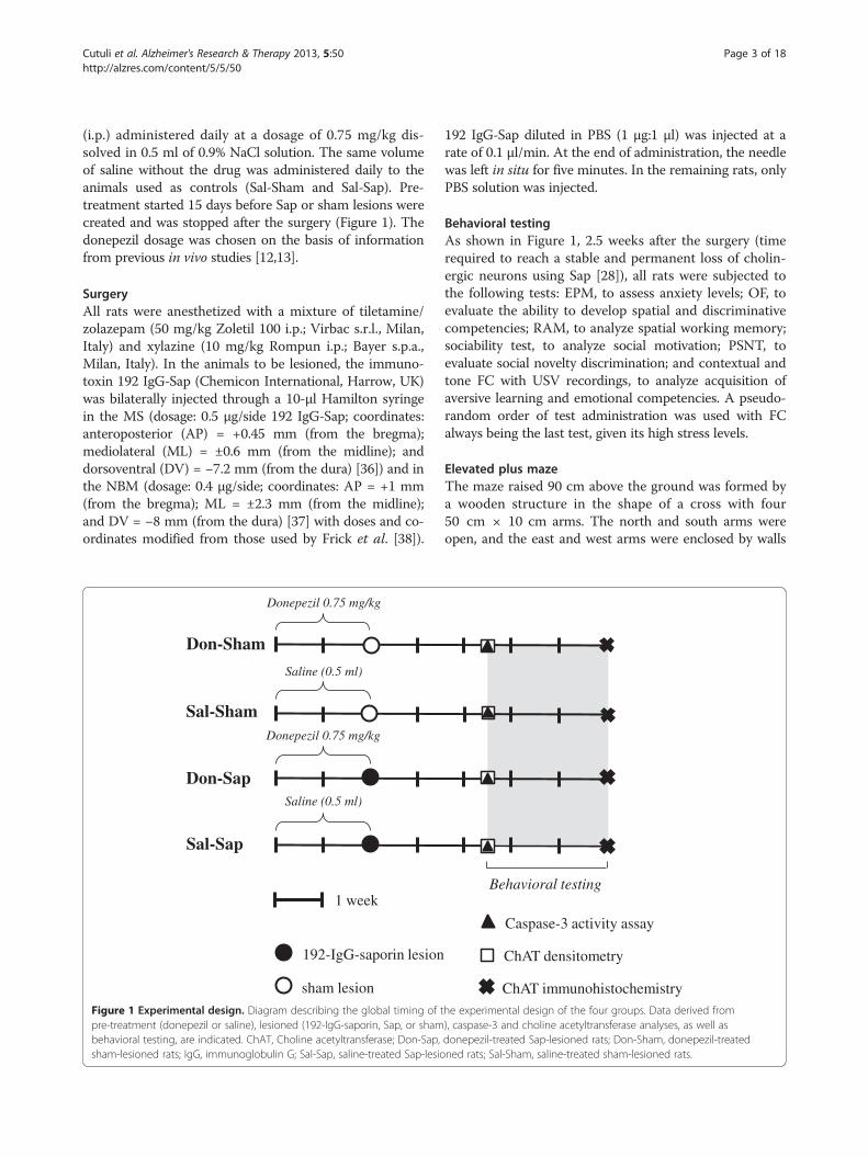
(i.p.) administered daily at a dosage of 0.75 mg/kg dis-solved in 0.5 ml of 0.9% NaCl solution. The same volumeof saline without the drug was administered daily to theanimals used as controls (Sal-Sham and Sal-Sap). Pre-treatment started 15 days before Sap or sham lesions werecreated and was stopped after the surgery (Figure 1). Thedonepezil dosage was chosen on the basis of informationfrom previous in vivo studies [12,13].
SurgeryAll rats were anesthetized with a mixture of tiletamine/zolazepam (50 mg/kg Zoletil 100 i.p.; Virbac s.r.l., Milan,Italy) and xylazine (10 mg/kg Rompun i.p.; Bayer s.p.a.,Milan, Italy). In the animals to be lesioned, the immuno-toxin 192 IgG-Sap (Chemicon International, Harrow, UK)was bilaterally injected through a 10-μl Hamilton syringein the MS (dosage: 0.5 μg/side 192 IgG-Sap; coordinates:anteroposterior (AP) = +0.45 mm (from the bregma);mediolateral (ML) = ±0.6 mm (from the midline); anddorsoventral (DV) = −7.2 mm (from the dura) [36]) and inthe NBM (dosage: 0.4 μg/side; coordinates: AP = +1 mm(from the bregma); ML = ±2.3 mm (from the midline);and DV = −8 mm (from the dura) [37] with doses and co-ordinates modified from those used by Frick et al. [38]).
Sal-Sap
Saline (0.5 ml)
1 week
Don-Sap
Donepezil 0.75 mg/kg
Donepezil 0.75 mg/kg
Don-Sham
Saline (0.5 ml)
Sal-Sham
192-IgG-saporin lesion
sham lesion
Figure 1 Experimental design. Diagram describing the global timing of tpre-treatment (donepezil or saline), lesioned (192-IgG-saporin, Sap, or shambehavioral testing, are indicated. ChAT, Choline acetyltransferase; Don-Sap,sham-lesioned rats; IgG, immunoglobulin G; Sal-Sap, saline-treated Sap-lesio
192 IgG-Sap diluted in PBS (1 μg:1 μl) was injected at arate of 0.1 μl/min. At the end of administration, the needlewas left in situ for five minutes. In the remaining rats, onlyPBS solution was injected.
Behavioral testingAs shown in Figure 1, 2.5 weeks after the surgery (timerequired to reach a stable and permanent loss of cholin-ergic neurons using Sap [28]), all rats were subjected tothe following tests: EPM, to assess anxiety levels; OF, toevaluate the ability to develop spatial and discriminativecompetencies; RAM, to analyze spatial working memory;sociability test, to analyze social motivation; PSNT, toevaluate social novelty discrimination; and contextual andtone FC with USV recordings, to analyze acquisition ofaversive learning and emotional competencies. A pseudo-random order of test administration was used with FCalways being the last test, given its high stress levels.
Elevated plus mazeThe maze raised 90 cm above the ground was formed bya wooden structure in the shape of a cross with four50 cm × 10 cm arms. The north and south arms wereopen, and the east and west arms were enclosed by walls
Behavioral testing
Caspase-3 activity assay
ChAT densitometry
ChAT immunohistochemistryhe experimental design of the four groups. Data derived from), caspase-3 and choline acetyltransferase analyses, as well asdonepezil-treated Sap-lesioned rats; Don-Sham, donepezil-treatedned rats; Sal-Sham, saline-treated sham-lesioned rats.
Cutuli et al. Alzheimer's Research & Therapy 2013, 5:50 Page 4 of 18http://alzres.com/content/5/5/50
36 cm high. The parameters taken into account were fre-quency of entries into the open and closed arms, totaltime spent in the open and closed arms and number ofdefecations [20].The parameters taken into account were: frequency of
entries in the open and closed arms; total time spent inthe open and closed arms; number of defecations [20].
Open field with objectsThe apparatus consisted of a circular arena (diameter140 cm) delimited by a 30-cm-high wall. During session1 (S1), each rat was allowed to move freely in the emptyopen field and the baseline activity level was measured.During S2 to S4 (habituation phase), four objects wereplaced in a square arrangement in the middle annulusof the arena and a fifth one was placed in the centralarea. In S5 and S6 (spatial change), the spatial configur-ation was changed by moving objects 2 and 5 so that theinitial square arrangement was changed to a polygon-shaped configuration without any central object. During S7(novelty), the configuration was modified by substitut-ing one object with another new one. Sessions lasted sixminutes, and intersession intervals were three minuteslong. All testing was recorded by a video camera whosesignal was relayed to a monitor and to an image analyzer(EthoVision, Noldus Information Technology, Wageningen,The Netherlands). The parameters taken into account weretotal and peripheral distances traveled in S1, time spentcontacting the objects, frequency of rearing, motionlesstime and number of defecations [20,39].
Radial arm mazeThe apparatus consisted of a central platform (30-cmdiameter) from which eight arms (12.5 cm wide × 60 cmlong) radiated like the spokes of a wheel. A food well(5-cm diameter, 2-cm depth) was located at the end ofeach arm [20]. A 40-W red light bulb provided theonly source of illumination in the testing room. Testingwas performed between 09:00 and 17:00 hours. After ahabituation phase, all rats (whose food was restricted todecrease their weight by approximately 15%) were sub-jected to the RAM full-baited procedure in which allarms were baited with a piece of Purina chow (PurinaMills, Gray Summit, MO, USA) with the goal of havingthe rats collect the eight rewards in a maximum of 16entries. The animals were subjected to two trials dailyfor five consecutive days. The intersession interval wasfour hours. The parameters taken into account werepercentage of total errors (number of revisited armsdivided by total number of visits × 100), mean spatialspan (longest sequence of correctly visited arms in eachsession) and perseverations (sum of consecutive entriesin the same arm or in a fixed sequence of arms).
Sociability and preference for social novelty testThe apparatus consisted of a white rectangular three-chamber wooden box (150 × 40 × 40 cm). The centralchamber was 30 cm long, and the two lateral chamberswere 60 cm long. The three chambers were divided bytwo transparent Plexiglas walls with an open middle door(height 10 cm, width 8 cm), which allowed free accessto each lateral chamber. Each lateral chamber con-tained a small plastic cage (18-cm diameter) with mesh-like holes in which stranger rats were confined for socialinteraction.The test comprised 3 sessions: Habituation, Sociability
and PSNT. During the habituation, each rat was allowedto freely move in the apparatus for 10 min. DuringSociability, an unfamiliar juvenile (35 to 45 days old)male conspecific (Stranger 1) was placed inside the smallcage in one of the lateral chambers (randomly selectedand counterbalanced for each group). The experimentalrat was placed in the apparatus and it was allowed tofreely explore the three chambers and contact the smallcages for ten minutes.During PSNT, another unfamiliar rat (stranger 2) was
placed inside the plastic cage of the opposite lateralchamber that had been empty during the Sociabilitysession. The experimental rat was allowed to move freelyand contact the plastic cages housing the strangers forten minutes. Inter-session intervals lasted three mi-nutes. Rats’ behavior was recorded by a video cameramounted on the ceiling. The resulting video signal wasrelayed to a monitor and to an EthoVision imageanalyzer. The parameters analyzed in each lateralchamber were frequency of entries, total duration (inseconds) and total distance traveled (in centimeters).
Context and tone fear conditioningThe apparatus consisted of a 21 × 21 × 49 cm condition-ing chamber (model 7532; Ugo Basile, Comerio, Italy).Chamber walls were made of gray Plexiglas, and theceiling was made of transparent Plexiglas to allow videorecording. The grid floor (steel pieces spaced by 1.5 cm)was connected to a shock generator scrambler (condi-tioner 7531; Ugo Basile).The FC test encompassed three sessions: training,
context and tone. During training session, following a120 seconds of acclimation to the conditioning appar-atus (baseline), three trials consisting of the 30-secondtone exposure (2 kHz, 90 dB) were carried out. Thelast 2 seconds of each tone were paired with a 1-mAfoot shock. Tone- and shock-free 60-second inter-trialintervals were used.After 24 hours, rats were placed for 240 seconds in
the training chamber (context test). After 4 hours, therats were submitted to a tone test in a white Plexiglasbox (21 × 18 × 45 cm) with black stripes applied on the
Cutuli et al. Alzheimer's Research & Therapy 2013, 5:50 Page 5 of 18http://alzres.com/content/5/5/50
walls. After 120 seconds of acclimation, a 120-secondtone identical to that used in the training session wassounded without any shock (tone test).During training, context and tone tests, the rats’ be-
havior was recorded by a video camera mounted on theceiling. The resulting video signal was relayed to a moni-tor and to an EthoVision image analyzer. In addition,22-kHz USVs were recorded [38,40].Behavioral parameters taken into account were frequency
and duration of freezing (behavioral immobility, except forrespiration movements) and number of defecations.USVs were collected via an ultrasound microphone
(UltraSoundGate CM16; Avisoft Bioacoustics, Berlin,Germany) placed through a hole in the middle of thetest chamber roof approximately 21 cm above the shockfloor. The microphone was sensitive to 15 to 180 kHzfrequencies with a flat frequency response (±6 dB) be-tween 25 and 140 kHz. It was connected by an Ultra-SoundGate USB audio device to a personal computer,which recorded data at 250,000 Hz in 16-bit format andstored as .wav files for subsequent analysis. Sound fileswere transferred to SASLab Pro (version 5.2; AvisoftBioacoustics) for sonographic analysis and a fast Fouriertransform (FFT) was performed (512 FFT length, 100%frame, Hamming window and 75% time window over-lap). USV parameters taken into account were numberof calls emitted, peak amplitude and frequency, frequencymodulation and call duration.
Histological analysesAt the end of behavioral testing, the animals weredeeply anesthetized and transcardially perfused with sa-line, followed by 4% paraformaldehyde and 0.1% glutar-aldehyde in PBS (4°C, pH 7.5). Brains were removed andcryoprotected in 30% buffered sucrose and cut on afreezing microtome. The anterior part of the brain wascut into coronal sections of 40 μm and stored for ChATimmunohistochemistry.
Choline acetyltransferase immunohistochemical stainingSections (40 μm) immunostained for ChAT were prein-cubated in PBS at 4°C, then in 0.4% Triton X-100 in PBSand finally in 0.1% Triton X-100 plus 1% bovine serumalbumin (Sigma-Aldrich, St Louis, MO, USA) plus nor-mal goat serum (NGS; Vector Laboratories, Burlingame,CA, USA) in PBS. Sections were incubated for 16 hoursat 4°C with 0.1% Triton X-100 and NGS in PBS with theprimary antibody for ChAT diluted 1:1,000 (ChemiconInternational). Subsequently, sections were incubatedwith biotinylated secondary antibody (goat anti-rabbit IgG-biotin conjugate) and 3% NGS (Kit Elite PK-6101; VectorLaboratories) in PBS for 10 minutes at room temperature.Staining was visualized with 0.05% diaminobenzidineand ammonium nickel(II) sulfate (Sigma-Aldrich Chemie,
Steinheim, Germany) after incubation with avidin andbiotinylated peroxidase (Kit Elite PK-6101). The sec-tions were then rinsed in PBS. Stained sections weremounted on slides, dehydrated and coverslipped. To ex-clude artefacts, in each case some random sections wereprocessed as previously described. The only differencewas the absence of the primary antibody.
Biochemical analysesTotal homogenate preparation from hippocampal andneocortical tissuesAfter the animals were decapitated, hippocampal and neo-cortical tissues were dissected and homogenized in lysisbuffer (320 mM sucrose, 50 mM NaCl, 50 mM Tris-HCl(pH 7.5), 1% Triton X-100, 0.5 mM sodium orthovanadate,5 mM β-glycerophosphate, proteases inhibitors), then incu-bated on ice for 30 minutes and centrifuged at 13,000 gfor 10 minutes. The total protein content of the resultingsupernatant was determined by the Bradford assay method.
Immunoblot analysis and antibodiesProteins were subjected to SDS-PAGE and electroblottedonto a polyvinylidene fluoride membrane. Immunoblotanalysis was performed using a chemiluminescence de-tection kit. The relative levels of immunoreactivity weredetermined by densitometry using ImageQuant 5.0 software.Antibodies to anti-ChAT were purchased from ChemiconInternational (AB143), and anti-actin clone EP184E rabbitmonoclonal antibody was obtained from EMD Millipore(04-1040; Billerica, MA, USA).
Fluorometric assay of caspase-3 activityTotal hippocampal and neocortical tissue was homoge-nized in lysis assay buffer (100 mM 2-[4-(2-hydroxyethyl)piperazin-1-yl]ethanesulfonic acid (pH 7.4), 0.1% 3-[(3-cholamidopropyl)dimethylammonio]-1-propanesulfonate(wt/vol), 1 mM ethylenediaminetetraacetic acid, 10 mMdithiothreitol, 1 mM phenylmethylsulfonyl fluoride) andlysed by freezing in liquid N2 and thawing at 37°C threetimes. After centrifugation at 11,500 g for 5 minutes, theprotein concentration of resulting supernatant was deter-mined and the same amount of protein was incubated at37°C in lysis assay buffer containing 50 μM caspase-3substrate II, fluorogenic (Ac-DEVD-AMC; Calbiochem,San Diego, CA, USA). The fluorescence was measuredwith 380-nm excitation wavelength and 460-nm emissionwavelength.
Statistical analysisAll data were tested for normality (Shapiro-Wilk test) andhomoscedasticity (Levene’s test). Behavioral data wereanalyzed using two-way analysis of variance (ANOVA;F-statistic) (with drug and lesion as between-animal fac-tors) or a mixed model of three-way ANOVA (with drug

Cutuli et al. Alzheimer's Research & Therapy 2013, 5:50 Page 6 of 18http://alzres.com/content/5/5/50
and lesion as between-animal factors and arm/session/object/day/chamber as within-animal factors). Post hoccomparisons were performed by means of Tukey’s honestlysignificant difference test. When parametric assumptionswere not fully met, data transformations (angular trans-formation for percentages) or nonparametric ANOVAs(Kruskal-Wallis test (H-statistic) and Mann-WhitneyU test; Z-statistic) were used. Biochemical data of ChATlevels were analyzed by Student’s t-test, and data regardingcaspase-3 activity were analyzed using the Bonferronimultiple comparisons test. Differences were consideredsignificant at the P ≤ 0.05 level.
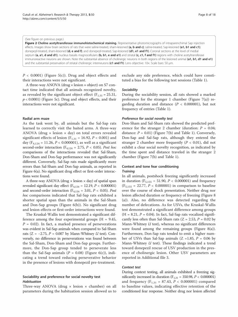
ResultsLesion verification by choline acetyltransferaseimmunohistochemical stainingThe presence of ChAT immunoreactive (ChAT-IR) neu-rons in the BF projection areas was assessed by inspection(Figures 2(a), 2(a1), 2(b), 2(b1), 2(d), 2(d1), 2(e) and 2(e1)).Brain sections were visualized with the light microscope-interfaced software Neurolucida (MBF Bioscience, Williston,VT, USA). Using a 10× lens objective, ChAT-IR neuronswere assessed in the two main regions of the BF: the MS,taking into account five 40-μm sections between 1.20 and0.20 mm anterior to bregma, and the NBM, taking intoaccount eight 40-μm sections between 0.80 and 2.30 mmposterior to the bregma [37]. Additional visual inspectionwas carried out to exclude eventual degeneration ofstriatal cholinergic interneurons after i.p. Sap injections inthe NBM (Figures 2(c), 2(c1), 2(f) and 2(f1)).
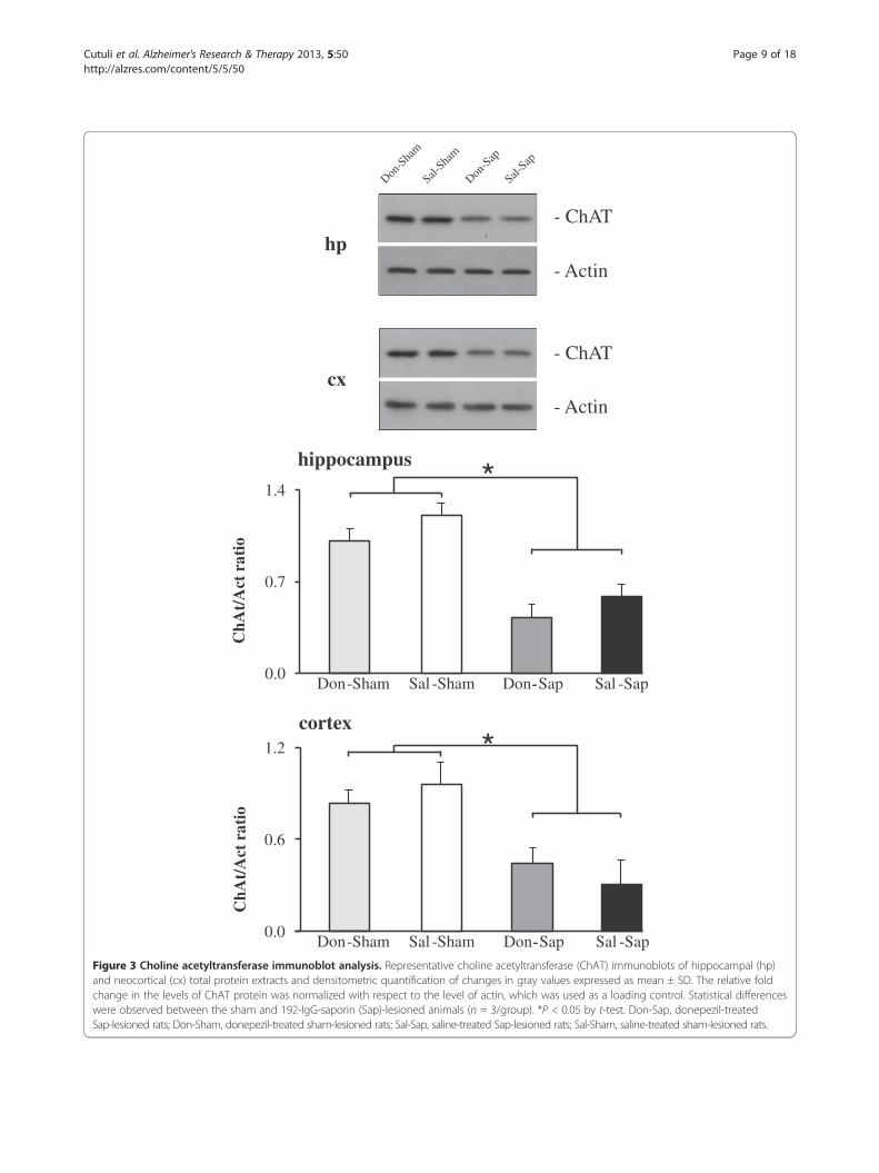
Lesion verification by choline acetyltransferaseimmunoblot analysisIntraparenchymal Sap injections in the NBM and MSinduced an extensive loss of ChAT-IR in the synapticboutons of the neocortex and hippocampus, as demon-strated by a strong reduction in ChAT expression (Figure 3).A comparable reduction of ChAT expression was de-tected in the hippocampi and neocortices from bothlesioned groups (Don-Sap and Sal-Sap). Conversely, ChATexpression was not significantly different in the sham-lesioned groups (Sal-Sham and Don-Sham).
Hippocampal and neocortical caspase-3 activityA significant increase in caspase-3 activity was evident inthe Sal-Sap group; however, a partial but significant rescuewas found in the Don-Sap group in both hippocampal andneocortical extracts. Both sham-lesioned groups (Don-Shamand Sal-Sham) exhibited similar levels of caspase-3 activityin both hippocampal and neocortical extracts (Figure 4).
Elevated plus mazeThe animals of all groups entered more frequently (F1,31 =47.94; P < 0.000001) and spent more time (F1,31 = 200.66,
P < 0.000001) in the closed arms than in the open arms.No difference was detected in the total number of defe-cations. Thus, neither drug nor lesion affected anxiety-related behavior in the EPM (Additional file 2).
Open field with objectsTwo-way ANOVA on total or peripheral distances trav-eled in S1 did not reveal any significant effect of drugor lesion. A three-way ANOVA (drug × lesion × session)on motionless time failed to reveal any significant effectof drug or lesion. Session effect (F6,186 = 5.01, P < 0.0001)was significant, because motionless time increased through-out the task in all groups. Lesion × session interactionwas also significant (F6,186 = 2.42, P = 0.03). Post hoccomparisons revealed that both lesioned groups (Don-Sapand Sal-Sap) tended to exhibit motionless time lowerthan that of sham animals (Don-Sham and Sal-Sham) inthe final sessions of the test (S6: P = 0.02; S7: P = 0.07).No other significant interactions were found. As ses-sions went by, rearing (F6,186 = 28.52, P = 0.000001) anddefecation (F6,186 = 14.88, P = 0.000001) decreased with-out any significant differences among groups. For bothparameters, no significant drug and lesion effects wereobserved.In the presence of the objects, all animals showed
habituation, as revealed by the significant session effect(F2,62 = 42.26, P < 0.000001) of three-way ANOVA (drug ×lesion × session) on contact times. Drug and lesion effectsand their interaction were not significant.A three-way ANOVA (drug × lesion × object) on S5 con-
tact time revealed a significant object effect (F1,31 = 23.28,P < 0.0001), as well as significant interactions of drug × object(F1,31 = 14.22, P < 0.001), lesion × object (F1,31 = 8.11,P < 0.01) and drug × lesion × object (F1,31 = 7.15, P = 0.01).Post hoc comparisons performed on the second-order inter-action revealed that, in S5, both sham groups (Don-Shamand Sal-Sham), as well as the Don-Sap group, recognizedthe spatial change (P < 0.01), whereas the Sal-Sap rats failedto detect the new spatial configuration (Figure 5(a)). Drugand lesion effects and their interaction were not significant.A three-way ANOVA (drug × lesion × object) on S6 con-
tact time revealed a significant lesion effect (F1,31 = 24.64,P < 0.0001), as well as significant interactions of drug × ob-ject (F1,31 = 13.18, P = 0.001), lesion × object (F1,31 = 14.05,P = 0.001) and drug × lesion × object (F1,31 = 9.26, P =0.005). Post hoc comparisons on the second-order inter-action revealed that, though Don-Sham, Sal-Sham and Don-Sap groups did not show any preference towards displacedobjects, Sal-Sap rats continued to explore nondisplaced ob-jects longer (P < 0.001) (Figure 5(b)). Drug and object effectsand drug × lesion interactions were not significant.A three-way ANOVA (drug × lesion × object) on S7
contact time indicated that all animals recognized novelty,as revealed by the significant object effect (F1,31 = 25.31,
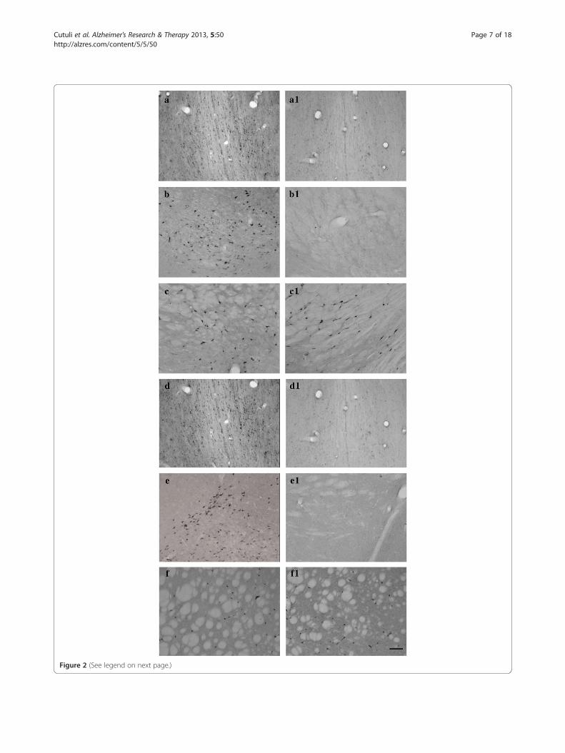
Figure 2 (See legend on next page.)
Cutuli et al. Alzheimer's Research & Therapy 2013, 5:50 Page 7 of 18http://alzres.com/content/5/5/50
(See figure on previous page.)Figure 2 Choline acetyltransferase immunohistochemical staining. Representative photomicrographs of intraparenchimal Sap injectioneffects. Images show brain sections of rats that were saline-treated, sham-lesioned (a, b and c); saline-treated, Sap-lesioned (a1, b1 and c1);donepezil-treated, sham-lesioned (d, e and f); and donepezil-treated, Sap-lesioned (d1, e1 and f1). Coronal sections at the level of medialseptum (a, a1, d and d1), nucleus basalis magnocellularis (b, b1, e and e1) and striatal (c, c1, f and f1) regions with choline acetyltransferaseimmunoreactive neurons are shown. Note the substantial absence of cholinergic neurons in both regions of the lesioned animal (a1, b1, d1 and e1)and the substantial preservation of striatal cholinergic interneurons (c1 and f1). Lens objective: 10×. Scale bars: 50 μm.
Cutuli et al. Alzheimer's Research & Therapy 2013, 5:50 Page 8 of 18http://alzres.com/content/5/5/50
P < 0.0001) (Figure 5(c)). Drug and object effects andtheir interactions were not significant.A three-way ANOVA (drug × lesion × object) on S7 con-
tact time indicated that all animals recognized novelty,as revealed by the significant object effect (F1,31 = 25.31,p < 0.0001) (Figure 5c). Drug and object effects, and theirinteractions were not significant.
Radial arm mazeAs the task went by, all animals but the Sal-Sap ratslearned to correctly visit the baited arms. A three-wayANOVA (drug × lesion × day) on total errors revealedsignificant effects of lesion (F1,31 = 16.92, P < 0.001) andday (F4,124 = 11.26, P < 0.000001), as well as a significantsecond-order interaction (F4,124 = 2.75, P < 0.05). Post hoccomparisons of the interactions revealed that Sal-Sham,Don-Sham and Don-Sap performance was not significantlydifferent. Conversely, Sal-Sap rats made significantly moreerrors than Sal-Sham and Don-Sap animals, as reported inFigure 6(a). No significant drug effect or first-order interac-tions were found.A three-way ANOVA (drug × lesion × day) of spatial span
revealed significant day effect (F4,124 = 12.19, P < 0.000001)and second-order interaction (F4,124 = 3.01, P < 0.05). Posthoc comparisons indicated that Sal-Sap rats exhibited ashorter spatial span than the animals in the Sal-Shamand Don-Sap groups (Figure 6(b)). No significant drugand lesion effects or first-order interactions were found.The Kruskal-Wallis test demonstrated a significant dif-
ference among the four experimental groups (H = 9.45,P = 0.02). In fact, a marked presence of perseverationswas evident in Sal-Sap animals when compared to Sal-Shamrats (Z = −2.75, P = 0.007 by Mann-Whitney U test). Con-versely, no difference in perseverations was found betweenthe Sal-Sham, Don-Sham and Don-Sap groups. Further-more, the Don-Sap group tended to perseverate lessthan the Sal-Sap animals (P = 0.08) (Figure 6(c)), indi-cating a trend toward reducing perseverative behaviorin the presence of lesions with donepezil pre-treatment.
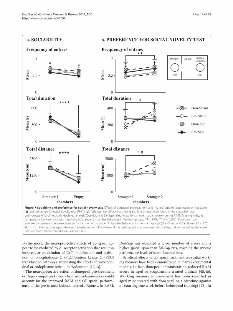
Sociability and preference for social novelty testHabituationThree-way ANOVA (drug × lesion × chamber) on allparameters during the habituation session allowed us to
exclude any side preference, which could have consti-tuted a bias for the following test sessions (Table 1).
SociabilityDuring the sociability session, all rats showed a markedpreference for the stranger 1 chamber (Figure 7(a)) re-garding duration and distance (P < 0.000001), but notfrequency of entries (Table 1).
Preference for social novelty testDon-Sham and Sal-Sham rats showed the predicted pref-erence for the stranger 2 chamber (duration: P = 0.04;distance: P = 0.01) (Figure 7(b) and Table 1). Conversely,Don-Sap and Sal-Sap rats, although they entered thestranger 2 chamber more frequently (P < 0.01), did notexhibit a clear social novelty recognition, as indicated bythe time spent and distance traveled in the stranger 2chamber (Figure 7(b) and Table 1).
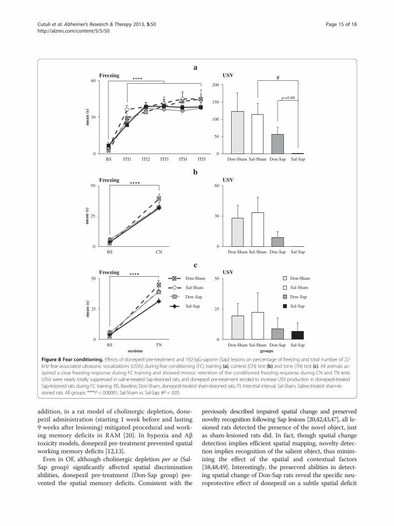
Context and tone fear conditioningTrainingIn all animals, postshock freezing significantly increasedin duration (F5,155 = 51.90, P < 0.000001) and frequency(F5,155 = 22.77, P < 0.000001) in comparison to baselineover the course of shock presentation. Neither drug norlesion affected duration or frequency of freezing (Figure 8(a)). Also, no difference was detected regarding thenumber of defecations. As for USVs, the Kruskal-Wallistest demonstrated a significant difference among groups(H = 8.21, P = 0.04). In fact, Sal-Sap rats vocalized signifi-cantly less often than Sal-Sham rats (Z = 2.53, P = 0.02 byMann-Whitney U test), whereas no significant differenceswere found among the remaining groups (Figure 8(a)).Furthermore, Don-Sap rats tended to emit a higher num-ber of USVs than Sal-Sap animals (Z =1.85, P = 0.06 byMann-Whitney U test). These findings indicated a trendtoward donepezil rescue of USV production in the pres-ence of cholinergic lesion. Other USV parameters arereported in Additional file 3.
Context testDuring context testing, all animals exhibited a freezing sig-nificantly increased in duration (F1,31 = 350.98, P < 0.000001)and frequency (F1,31 = 87.43, P < 0.000001) comparedto baseline values, indicating effective retention of theconditioned fear response. Neither drug nor lesion affected
ChA
t/A
ct r
atio
0.0
0.7
1.4 *
Don-Sham Sal -Sham -Don Sap SapSal -
hippocampus
ChA
t/A
ctra
tio
0.0
0.6
1.2
cortex
Don-Sham Sal -Sham -Don Sap Sal -Sap
*
- ChAT
- Actincx
- ChAT
- Actin
Sal-Sap
Don-S
ap
Don-S
ham
Sal-Sha
m
hp
Figure 3 Choline acetyltransferase immunoblot analysis. Representative choline acetyltransferase (ChAT) immunoblots of hippocampal (hp)and neocortical (cx) total protein extracts and densitometric quantification of changes in gray values expressed as mean ± SD. The relative foldchange in the levels of ChAT protein was normalized with respect to the level of actin, which was used as a loading control. Statistical differenceswere observed between the sham and 192-IgG-saporin (Sap)-lesioned animals (n = 3/group). *P < 0.05 by t-test. Don-Sap, donepezil-treatedSap-lesioned rats; Don-Sham, donepezil-treated sham-lesioned rats; Sal-Sap, saline-treated Sap-lesioned rats; Sal-Sham, saline-treated sham-lesioned rats.
Cutuli et al. Alzheimer's Research & Therapy 2013, 5:50 Page 9 of 18http://alzres.com/content/5/5/50

Figure 4 Hippocampal and neocortical caspase-3 activity.Caspase-3 activity was revealed by a fluorometric assay in totalhippocampal and neocortical homogenates from sham and lesionedrats (n = 3/group). The fluorometric data are expressed as mean ± SEM.*P < 0.05, ***P ≤ 0.001 (Bonferroni multiple comparisons test).Don-Sap, donepezil-treated Sap-lesioned rats; Don-Sham,donepezil-treated sham-lesioned rats; Sal-Sap, saline-treatedSap-lesioned rats; Sal-Sham, saline-treated sham-lesioned rats.
Cutuli et al. Alzheimer's Research & Therapy 2013, 5:50 Page 10 of 18http://alzres.com/content/5/5/50
duration or frequency of freezing (Figure 8(b)). No diffe-rences were observed among groups in number of defeca-tions. As for USVs, the experimental groups just tended todiffer (H = 6.71, P = 0.08 by Kruskal-Wallis test), withDon-Sap and Sal-Sap lesioned rats emitting fewer USVsthan sham rats. Further USV parameters are reported inAdditional file 4.
Tone testAll rats showed a significant increase in duration (F1,31 =113.44, P < 0.000001) and frequency (F1,31 = 52.61,P < 0.000001) of freezing during tone reexposure incomparison to baseline values, indicating once again an ef-fective retention of the conditioned fear response. Neitherdrug nor lesion affected duration or frequency of freezing(Figure 8(c)). No differences were observed among groupsin number of defecations. USVs were scarcely emitted byall rats, regardless of drug and lesion conditions (H = 2.53,P = 0.47 by Kruskal-Wallis test) (Figure 8(c)). Further USVparameters are reported in Additional file 5.
DiscussionAs ACh reduction is a morphofunctional hallmark in AD,AChE-Is, such as donepezil, are used to improve AD-related cognitive deterioration [7]. Besides symptomaticeffects, many in vitro studies have shown that AChE-Ismight exert neuroprotective action against neurotoxicagents, such as glutamate and Aβ plaques [8-11,15-18].Unfortunately, only a few in vivo experiments haveconfirmed AChE-I pre-lesion neuroprotective action[12-14,26,27]. Interestingly, the present study demon-strates a marked neuroprotective effect of donepezilagainst neurodegeneration and cognitive impairmentinduced by cholinergic depletion in rats. Donepezil pre-treatment did not induce any biochemical or behavioralmodification in sham-lesioned rats, which is in accordwith the findings of Saxena et al. [14] and Su et al. [27].We previously demonstrated that healthy rats treatedwith donepezil during behavioral testing showed enhancedmemory and explorative functions in RAM and OF, as wellas reduced anxiety levels in EPM, but they did not exhibitany improvement in spatial span, motivational levelsand associative learning [41]. Thus, although donepeziltreatment during testing is able to improve cognitiveperformance even in unlesioned animals, donepezil treat-ment before testing (pre-treatment) exerts neuroprotectiveaction only in the presence of lesion or insult.Cholinergic depletion per se (Sal-Sap group) increased
hippocampal and neocortical caspase-3 activity and im-paired working memory performance in RAM and spatialchange detection in OF, altered social discrimination per-formance in PSNT and reduced USV production duringFC, but it did not affect anxiety levels in EPM and FC ac-quisition. The behavioral results in our study are in linewith those reported in previous studies of memory deficits[20,29,42,43] and impaired production of USVs [38,40,43]and no effects on anxiety levels following 192 IgG-Saplesion [20,44]. Our present is the first one that explainscaspase-3 activity in the target areas of cholinergicprojections. Our results demonstrate that donepezil pre-treatment (Don-Sap group) was able to counteract neu-rodegeneration associated with cholinergic depletion. Infact, hippocampal and neocortical caspase-3 levels ob-served in Don-Sap rats were significantly decreased incomparison to Sal-Sap. This result is in line with previ-ous studies using donepezil pre-treatment in differentexperimental paradigms. In a glutamate excitotoxicitystudy, donepezil pre-treatment significantly preventedcaspase-3 activation, contributing to internalization anddownregulation of N-methyl-D-aspartate (NMDA) re-ceptors through α7 nAChR stimulation [15]. Moreover,following STZ lesioning, donepezil pre-treatment signifi-cantly prevented hippocampal and neocortical caspase-3activity [14]. The reduction of hippocampal and neocorticalcaspase-3 activity by donepezil pre-treatment following

Figure 5 Open field with objects. Effects of donepezil pre-treatment and 192-IgG-saporin (Sap) lesions on open field with objects contacttime with objects during spatial change in session 5 (S5) (a), S6 (b) and novelty (S7) (c). Cholinergic depletion per se (Sal-Sap group)significantly affected spatial discrimination abilities in S5 and S6, and donepezil pre-treatment (Don-Sap group) prevented spatial memorydeficits. In S7, all rats detected the presence of the novel object. Behavioral data are expressed as means ± SEM, and asterisks indicate post hoccomparisons between groups. DO, Displaced object; Don-Sap, donepezil-treated Sap-lesioned rats; Don-Sham, donepezil-treated sham-lesioned rats;FO, familiar object; NDO, non-displaced objects; NO, novel object; Sal-Sap, saline-treated Sap-lesioned rats; Sal-Sham, saline-treated sham-lesioned rats.**P < 0.01, ***P < 0.001.
Cutuli et al. Alzheimer's Research & Therapy 2013, 5:50 Page 11 of 18http://alzres.com/content/5/5/50

Mea
n
0
1
2
3
Sal -Sham Sal -SapDon -Sham Don-Sap
##
Sal-Sap
Sal-Sham
Don-Sham
Don-Sap
RADIAL ARM MAZE
days
###
a
b
c
Per
cent
age
0
25
50
75
100
##*
I II III IV V
* Sal-Sap
Sal-Sham
Don-Sap
Don-Sham
Mea
n
#
0
2
4
6
8
I II III IV V
days
*
Figure 6 Radial arm maze. Effects of donepezil pre-treatment and 192-IgG-saporin (Sap) lesions on radial arm maze total errors (a), spatialspans (b) and perseverations (c). Donepezil pre-treatment reduced the number of errors and increased spatial span in cholinergicallydepleted rats. Sal-Sham vs. Sal-Sap: #p < 0.05; ##p < 0.01; ###p < 0.001; Don-Sap vs. Sal-Sap: *p = 0.05, **p = 0.01. Don-Sap, donepezil-treatedSap-lesioned rats; Don-Sham, donepezil-treated sham-lesioned rats; Sal-Sap, saline-treated Sap-lesioned rats; Sal-Sham, saline-treatedsham-lesioned rats.
Cutuli et al. Alzheimer's Research & Therapy 2013, 5:50 Page 12 of 18http://alzres.com/content/5/5/50
cholinergic depletion adds further evidence to donepezilneuroprotective action. This effect could be ascribed tothe up-regulation of nAChRs and to the consequentactivation of the anti-apoptotic nAChR/PI3K/Akt/Bcl2pathway, as previously hypothesized [8,17]. Namely,several studies have demonstrated the involvement ofnAChRs in donepezil-induced neuroprotection. In fact, in
contrast to the AChE-Is galantamine and tacrine, pro-longed donepezil administration induces not only a func-tional up-regulation but also an increased expression ofnAChRs [9,17]. Importantly, nAChR up-regulation bydonepezil is able to activate intracellular anti-apoptoticsecondary messenger systems, such as the nAChR/PI3Kpathway, that protect neurons against neuron death [17].
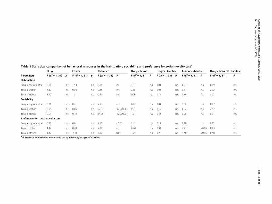
Table 1 Statistical comparison of behavioral responses in the habituation, sociability and preference for social novelty testa
Drug Lesion Chamber Drug × lesion Drug × chamber Lesion × chamber Drug × lesion × chamber
Parameters F (df = 1, 31) p F (df = 1, 31) p F (df = 1, 31) P F (df = 1, 31) P F (df = 1, 31) P F (df = 1, 31) P F (df = 1, 31) P
Habituation
Frequency of entries 0.01 n.s. 1.54 n.s. 3.11 n.s. 4.01 n.s. 3.01 n.s. 0.81 n.s. 0.89 n.s.
Total duration 3.62 n.s. 0.30 n.s. 0.38 n.s. 2.68 n.s. 0.01 n.s. 3.61 n.s. 2.92 n.s.
Total distance 1.90 n.s. 1.31 n.s. 0.25 n.s. 0.06 n.s. 0.15 n.s. 3.84 n.s. 3.67 n.s.
Sociability
Frequency of entries 0.01 n.s. 0.21 n.s. 2.95 n.s. 0.47 n.s. 0.01 n.s. 1.06 n.s. 0.47 n.s.
Total duration 0.04 n.s. 0.66 n.s. 57.87 <0.000001 0.04 n.s. 0.19 n.s. 0.02 n.s. 2.47 n.s.
Total distance 0.01 n.s. 0.18 n.s. 64.05 <0.000001 1.71 n.s. 0.43 n.s. 0.05 n.s. 0.91 n.s.
Preference for social novelty test
Frequency of entries 0.28 n.s. 0.01 n.s. 9.13 <0.01 2.41 n.s. 0.11 n.s. 0.18 n.s. 0.12 n.s.
Total duration 1.42 n.s. 0.20 n.s. 2.84 n.s. 0.76 n.s. 0.34 n.s. 4.21 <0.05 0.13 n.s.
Total distance 1.47 n.s. 2.18 n.s. 7.17 0.01 1.25 n.s. 0.27 n.s. 4.48 <0.05 0.48 n.s.aAll statistical comparisons were carried out by three-way analysis of variance.
Cutuliet
al.Alzheim
er'sResearch
&Therapy
2013,5:50Page
13of
18http://alzres.com
/content/5/5/50
Sal-Sap
Sal-Sham
Don-Sap
Don-Sham
a. SOCIABILITY
chambersStranger 1 Empty
chambersStranger 1 Stranger 2
b. PREFERENCE FOR SOCIAL NOVELTY TEST
** **Total distance
0
1250
2500
Mea
n(c
m)
** **Total duration
0
300
600
Mea
n(s
)
Frequency of entries
0
3.5
7
Mea
n
##Total distance
0
1000
2000
Mea
n(c
m)
#Total duration
0
200
400
Mea
n (s
)
**Frequency of entries
0
3.5
7
Mea
n
cagecage
Stranger 1 EmptyorStranger 2
Central
Figure 7 Sociability and preference for social novelty test. Effects of donepezil pre-treatment and 192-IgG-saporin (Sap) lesions on sociability(a) and preference for social novelty test (PSNT) (b). Although no differences among the four groups were found in the sociability test,both groups of cholinergically depleted animals (Don-Sap and Sal-Sap) failed to exhibit an overt social novelty during PSNT. Asterisks indicatecomparisons between stranger 1 and empty/stranger 2 chamber behaviors in the four groups. **P < 0.01, ****P < 0.0001. Pound symbolsindicate comparisons between stranger 1 chamber and stranger 2 chamber behaviors in the sham groups (Don-Sham and Sal-Sham). #P < 0.05,##P = 0.01. Don-Sap, donepezil-treated Sap-lesioned rats; Don-Sham, donepezil-treated sham-lesioned rats; Sal-Sap, saline-treated Sap-lesionedrats; Sal-Sham, saline-treated sham-lesioned rats.
Cutuli et al. Alzheimer's Research & Therapy 2013, 5:50 Page 14 of 18http://alzres.com/content/5/5/50
Furthermore, the neuroprotective effects of donepezil ap-pear to be mediated by σ1 receptor activation that result inintracellular modulation of Ca2+ mobilization and activa-tion of phospholipase C (PLC)/protein kinase C (PKC)transduction pathways, attenuating the effects of mitochon-drial or endoplasmic reticulum dysfunction [12,13].The neuroprotective action of donepezil pre-treatment
on hippocampal and neocortical neurodegeneration couldaccount for the improved RAM and OF spatial perform-ance of the pre-treated lesioned animals. Namely, in RAM,
Don-Sap rats exhibited a lower number of errors and ahigher spatial span than Sal-Sap rats, reaching the mnesicperformance levels of sham-lesioned rats.Beneficial effects of donepezil treatment on spatial work-
ing memory have been demonstrated in many experimentalmodels. In fact, donepezil administration reduced RAMerrors in aged or scopolamine-treated animals [45,46].Working memory improvement has been reported inaged mice treated with donepezil or a nicotinic agonistα7 (starting one week before behavioral training) [23]. In
Sal-Sap
Sal-Sham
Don-Sap
Don-Sham
b
0
25
50
BS CN
mea
n (s
)
***Freezing
0
30
60
Don-Sham Sal-Sham Don-Sap Sal-Sap
USV
a
0
30
60
BS ITI1 ITI2 ITI3 ITI4 ITI5
mea
n (s
)
Freezing
0
50
100
150
200
Don-Sham Sal-Sham Don-Sap Sal-Sap
p=0.06
#USV
c
0
25
50
BS TNsessions
mea
n (s
)
Freezing ***
0
25
50
Don-Sham Sal-Sham Don-Sap Sal-Sap
USV
groups
Sal-Sap
Sal-Sham
Don-Sham
Don-Sap
***
Figure 8 Fear conditioning. Effects of donepezil pre-treatment and 192-IgG-saporin (Sap) lesions on percentage of freezing and total number of 22-kHz fear-associated ultrasonic vocalizations (USVs) during fear conditioning (FC) training (a), context (CN) test (b) and tone (TN) test (c). All animals ac-quired a clear freezing response during FC training and showed mnesic retention of the conditioned freezing response during CN and TN tests.USVs were nearly totally suppressed in saline-treated Sap-lesioned rats, and donepezil pre-treatment tended to increase USV production in donepezil-treatedSap-lesioned rats during FC training. BS, Baseline; Don-Sham, donepezil-treated sham-lesioned rats; ITI, Inter-trial interval; Sal-Sham, Saline-treated sham-le-sioned rats. All groups: ****P < 0.00001; Sal-Sham vs. Sal-Sap: #P < 0.05.
Cutuli et al. Alzheimer's Research & Therapy 2013, 5:50 Page 15 of 18http://alzres.com/content/5/5/50
addition, in a rat model of cholinergic depletion, done-pezil administration (starting 1 week before and lasting9 weeks after lesioning) mitigated procedural and work-ing memory deficits in RAM [20]. In hypoxia and Aβtoxicity models, donepezil pre-treatment prevented spatialworking memory deficits [12,13].Even in OF, although cholinergic depletion per se (Sal-
Sap group) significantly affected spatial discriminationabilities, donepezil pre-treatment (Don-Sap group) pre-vented the spatial memory deficits. Consistent with the
previously described impaired spatial change and preservednovelty recognition following Sap lesions [20,42,43,47], all le-sioned rats detected the presence of the novel object, justas sham-lesioned rats did. In fact, though spatial changedetection implies efficient spatial mapping, novelty detec-tion implies recognition of the salient object, thus minim-izing the effect of the spatial and contextual factors[38,48,49]. Interestingly, the preserved abilities in detect-ing spatial change of Don-Sap rats reveal the specific neu-roprotective effect of donepezil on a subtle spatial deficit
Cutuli et al. Alzheimer's Research & Therapy 2013, 5:50 Page 16 of 18http://alzres.com/content/5/5/50
induced by the cholinergic lesion and are in agreementwith donepezil neuroprotective properties on spatialmnesic performances in different brain damage models[12-14,26,27].In all animals, the shock exposure induced a clear
freezing response during FC training, and the condi-tioned freezing response was retained during contextand tone tests. Although the lesion did not alter freezingresponse, it decreased the concomitant production of22-kHz USVs. Interestingly, donepezil pre-treatment dur-ing FC training just tended to rescue the defect of fear-associated USVs, which were nearly totally suppressed inSal-Sap rats. The 22-kHz USVs are part of the rodentdefensive repertoire and are emitted in response to nega-tive experiences, such as foot-shock, presence of predatorsor startling noise exposure [30-32]. These USVs are closelyassociated with freezing response to actual or potentialthreats and might serve as “alarm calls” for conspecificity.There is converging evidence that Sap lesions selectivelyimpair the functionality of neural systems regulating USVs[38,40,43]. Overall, our data are in agreement with thepreviously described disassociation between intact freezingresponse and impaired USV production in cholinergicallydepleted rats [38]. Intriguingly, donepezil pre-treatment(Don-Sap group) appeared to mitigate the USV suppres-sion of lesioned rats during FC training. Thus, our datacorroborate the added value of USV measurement, whichprovides additional information about the emotional stateof the animals in situations of coping with inescapableaversive stimuli.Also, social interactions and recognition of conspecifi-
city are fundamental and adaptive components of thebehavioral repertoire of numerous species. Similarly tosocial transmission of food preference, social recognitionmemory, which relies on the functional activation of hip-pocampal and prelimbic cortical circuitry [50], is impairedin the early phases of AD. Clinical trials have demon-strated that donepezil enhances AD patients’ social inter-action, engagement and interest [51]. On these bases, inthe present research, we investigated for the first timedonepezil pre-treatment effects on social skills using thethree-chamber test. During sociability tests, all animalsshowed a clear social motivation. In fact, all of them pre-ferred the conspecific stranger, as they spent more timeand traveled longer distances in the chamber containingthe social stimulus. These results fit with those obtainedin the sociability test described by Riedel et al. [50], butthey seem to be in disagreement with those obtained bySavage et al. [47]. On the basis of use of a social inter-action test, these authors reported a significant reductionin active interactions following selective cholinergic deple-tion of the neocortex in rats. Notably, at variance with thesocial interaction test, sociability does not allow any directcontact with the social stranger. Thus, in the present
study, the indirect social interactions and the scarce sa-lience of the empty compartment featuring sociabilityappear to have facilitated the exploration of the strangerby the lesioned rats.During PSNT, though sham rats showed a clear prefer-
ence for the novel stranger, the cholinergically depletedanimals did not exhibit overt social novelty. This resulthas to be interpreted as a specific social recognitionmemory deficit (as described by Riedel et al. [50]), be-cause the OF lesioned rats did recognize the novel ob-ject, which is in line with the findings reported bySavage et al. [47]. Although Riedel et al. [50] reportedthat donepezil administration succeeded in rescuing so-cial memory scopolamine-induced deficits, donepezilpre-treatment failed to prevent PSNT deficits in lesionedrats in our present study. The different results could beexplained by methodological differences, such as treat-ment time (acute vs. pre-treatment), behavioral protocols(short-term vs. long-term memory) and cholinergic ma-nipulations (scopolamine vs. Sap). Notably, hippocampaland neocortical cholinergic deafferentation by Sap re-sults in massive dysregulation of other neurotransmittersystems, such as dopaminergic and glutamatergic ones,which is known to contribute to social discrimination[46,52,53].Our current research reveals that donepezil pre-treatment
is able to reduce hippocampal and neocortical caspase-3activity, thus preventing neuron degeneration, and to exertbeneficial effects on specific behavioral deficits inducedby cholinergic depletion. As indicated in previous stud-ies, donepezil neuroprotective effects might be medi-ated by many protective mechanisms, such as nAChRupregulation and activation of the nAChR/PI3K path-way [8,15,17] and the σ1 receptor/PLC/PKC pathway[12,13]. Such effects result in a reduction of neurotox-icity linked to NMDA receptor–mediated Ca2+ influx,oxidative stress and caspase-3 activity [14,15]. Further-more, donepezil exerts a protective action against Aβtoxicity [7]. In fact, in AD patients, Aβ plaques colocalizewith nAChRs and α4 and α7 nAChR expression is reduced.In CA1, the α4-nAChR activation causes γ-aminobutyricacid (GABA) release from interneurons that inhibit pyr-amidal neurons, whereas the activation of α7 nAChRsin pyramidal neurons results in Ca2+ influx, presynapticneurotransmitter release and postsynaptic depolarization[7]. Thus, Aβ interferes with nAChR activity, and theconsequent increase of glutamate and decrease of GABAinduce glutamate excitotoxicity and excitation–inhibitionimbalance, altering the fine-tuning of hippocampal firing[6]. In AD patients, increase of caspase-3 activity has beenreported in the hippocampal and neocortical postsynapticdensity fractions [54]. Also, in the Tg2576 AD mousemodel, the increase of caspase-3 in hippocampal post-synaptic compartment leads to alteration of synaptic
Cutuli et al. Alzheimer's Research & Therapy 2013, 5:50 Page 17 of 18http://alzres.com/content/5/5/50
plasticity and dendritic spine loss. In parallel, the Sap-induced cholinergic depletion mimicking Aβ interferencemight cause an alteration of the excitation–inhibitionbalance and produce excitotoxic damage in hippocam-pal and neocortical neurons, which accumulate activecaspase-3. On these bases, the question is: How candonepezil neuroprotection be exerted? By inhibiting AChEactivity, the donepezil pre-treatment might reduce theGABAergic alterations and prevent glutamatergic exci-totoxicity and the excitation–inhibition imbalance. Inthis way, caspase-3 accumulation would be decreased,contributing to the maintenance of hippocampal andneocortical functioning.
ConclusionsThe present results show for the first time that donepezilpre-treatment is able to slow down the memory deficitsinduced by cholinergic depletion and to reduce caspase-3accumulation in hippocampal and neocortical areas.Although further studies deepening understanding of themolecular mechanisms of donepezil neuroprotective ac-tion are needed, the present results are promising andmay lead to the development of novel strategies for pre-vention and therapy of neurodegenerative diseases.
Additional files
Additional file 1: Figure representing sham rats’ data.
Additional file 2: Table of EPM data.
Additional file 3: Table of the main spectrographic parameters ofUSVs emitted during FC training.
Additional file 4: Table of the main spectrographic parameters ofUSVs emitted during the context test.
Additional file 5: Table of the main spectrographic parameters ofUSVs emitted during tone test.
AbbreviationsACh: Acetylcholine; AChE-I: Acetylcholinesterase inhibitor; AD: Alzheimer’sdisease; ANOVA: Analysis of variance; AP: Anteroposterior; Aβ: Amyloid β;ChAT: Choline acetyltransferase; Don-Sap: Donepezil-treated immunoglobulinG saporin-lesioned rats; Don-Sham: Donepezil-treated sham-lesioned rats;DV: Dorsoventral; EPM: Elevated plus maze; FC: Fear conditioning; MCI: Mildcognitive impairment; ML: Mediolateral; MS: Medial septum; nAChR: Nicotinicacetylcholine receptor; NBM: Nucleus basalis magnocellularis; OF: Open fieldwith objects; PBS: Phosphate-buffered saline; PI3K: Phosphatidylinositol 3-kinase; PKC: Protein kinase C; PLC: Phospholipase C; PSNT: Preference forsocial novelty test; RAM: Radial arm maze; Sal-Sap: Saline-treatedimmunoglobulin G saporin-lesioned rats; Sal-Sham: Saline-treated sham-lesioned rats; Sap: 192 immunoglobulin G saporin; STZ: Streptozotocin;USV: Ultrasonic vocalization.
Competing interestsThe authors have no conflicts of interest to declare.
Authors’ contributionsDC, LP and MDA made substantial contributions to the conception anddesign of the study. PDB performed immunotoxic lesioning and histologicalanalyses. DC, PC and AMT treated the animals and performed behavioralevaluations. DO contributed to acquisition and analysis of USV data. ANperformed biochemical analyses. DC and AN undertook the statistical
analyses. DC, LP, MDA and FDA were involved in drafting the manuscriptand revising it critically for important intellectual content. All authorscontributed to the interpretation of data and gave their final approval of themanuscript version to be published.
AcknowledgementsDonepezil was a generous gift from Pfizer, Inc. This study was supported byItalian Ministry of Education, Universities and Research grants (to LP andMDA). MDA is financially supported by a grant from the Alzheimer’sAssociation (NIRG-11-204588).
Author details1IRCCS Fondazione Santa Lucia, Via del Fosso di Fiorano 64, Rome 00143, Italy.2Department of Psychology, University of Rome “Sapienza”, Via dei Marsi 78,Rome 00185, Italy. 3Cell Biology and Neurobiology Institute, National ResearchCouncil, Via del Fosso di Fiorano 64, Rome 00143, Italy. 4Medical School,Campus Bio-Medico University Molecular Neuroscience Unit, Via Alvaro delPortillo 21, Rome 00128, Italy.
Received: 27 June 2013 Accepted: 16 October 2013Published: 24 October 2013
References1. Bartus RT, Dean RL 3rd, Beer B, Lippa AS: The cholinergic hypothesis of
geriatric memory dysfunction. Science 1982, 217:408–414.2. Bartus RT: On neurodegenerative diseases, models, and treatment
strategies: lessons learned and lessons forgotten a generation followingthe cholinergic hypothesis. Exp Neurol 2000, 163:495–529.
3. Sarter M, Bruno JP: Developmental origins of the age-related decline incortical cholinergic function and associated cognitive abilities. NeurobiolAging 2004, 25:1127–1139.
4. Galimberti D, Scarpini E: Treatment of Alzheimer’s disease: symptomaticand disease-modifying approaches. Curr Aging Sci 2010, 3:46–56.
5. Schwarz S, Froelich L, Burns A: Pharmacological treatment of dementia.Curr Opin Psychiatry 2012, 25:542–550.
6. D’Amelio M, Rossini PM: Brain excitability and connectivity of neuronalassemblies in Alzheimer’s disease: from animal models to humanfindings. Prog Neurobiol 2012, 99:42–60.
7. Pepeu G, Giovannini MG: Cholinesterase inhibitors and beyond. Curr AlzheimerRes 2009, 6:86–96.
8. Akaike A, Takada-Takatori Y, Kume T, Izumi Y: Mechanisms of neuroprotectiveeffects of nicotine and acetylcholinesterase inhibitors: role of α4 and α7receptors in neuroprotection. J Mol Neurosci 2010, 40:211–216.
9. Akasofu S, Kimura M, Kosasa T, Sawada K, Ogura H: Study of neuroprotectionof donepezil, a therapy for Alzheimer’s disease. Chem Biol Interact 2008,175:222–226.
10. Arias E, Alés E, Gabilan NH, Cano-Abad MF, Villarroya M, García AG, López MG:Galantamine prevents apoptosis induced by β-amyloid and thapsigargin:involvement of nicotinic acetylcholine receptors. Neuropharmacology 2004,46:103–114.
11. Geerts H: Indicators of neuroprotection with galantamine. Brain Res Bull2005, 64:519–524.
12. Meunier J, Ieni J, Maurice T: Antiamnesic and neuroprotective effects ofdonepezil against learning impairments induced in mice by exposure tocarbon monoxide gas. J Pharmacol Exp Ther 2006, 317:1307–1319.
13. Meunier J, Ieni J, Maurice T: The anti-amnesic and neuroprotective effects ofdonepezil against amyloid β25-35 peptide-induced toxicity in mice involvean interaction with the σ1 recepto. Br J Pharmacol 2006, 149:998–1012.
14. Saxena G, Patro IK, Nath C: ICV STZ induced impairment in memory andneuronal mitochondrial function: a protective role of nicotinic receptor.Behav Brain Res 2011, 224:50–57.
15. Shen H, Kihara T, Hongo H, Wu X, Kem WR, Shimohama S, Akaike A,Niidome T, Sugimoto H: Neuroprotection by donepezil against glutamateexcitotoxicity involves stimulation of α7 nicotinic receptors andinternalization of NMDA receptors. Br J Pharmacol 2010, 161:127–139.
16. Takada Y, Yonezawa A, Kume T, Katsuki H, Kaneko S, Sugimoto H, Akaike A:Nicotinic acetylcholine receptor-mediated neuroprotection by donepezilagainst glutamate neuroto xicity in rat cortical neurons. J Pharmacol ExpTher 2003, 306:772–777.
17. Takada-Takatori Y, Kume T, Izumi Y, Ohgi Y, Niidome T, Fujii T, Sugimoto H,Akaike A: Roles of nicotinic receptors in acetylcholinesterase inhibitor-
Cutuli et al. Alzheimer's Research & Therapy 2013, 5:50 Page 18 of 18http://alzres.com/content/5/5/50
induced neuroprotection and nicotinic receptor up-regulation. Biol PharmBull 2009, 32:318–324.
18. Wang R, Tang XC: Neuroprotective effects of huperzine A: a naturalcholinesterase inhibitor for the treatment of Alzheimer’s disease.Neurosignals 2005, 14:71–82.
19. Barnes CA, Meltzer J, Houston F, Orr G, McGann K, Wenk GL: Chronictreatment of old rats with donepezil or galantamine: effects on memory,hippocampal plasticity and nicotinic receptors. Neuroscience 2000, 99:17–23.
20. Cutuli D, Foti F, Mandolesi L, De Bartolo P, Gelfo F, Federico F, Petrosini L:Cognitive performances of cholinergically depleted rats followingchronic donepezil administration. J Alzheimers Dis 2009, 17:161–176.
21. Dong H, Csernansky CA, Martin MV, Bertchume A, Vallera D, Csernansky JG:Acetylcholinesterase inhibitors ameliorate behavioral deficits in theTg2576 mouse model of Alzheimer’s disease. Psychopharmacology (Berl)2005, 181:145–152.
22. Hernandez CM, Gearhart DA, Parikh V, Hohnadel EJ, Davis LW, Middlemore ML,Warsi SP, Waller JL, Terry AV Jr: Comparison of galantamine and donepezilfor effects on nerve growth factor, cholinergic markers, and memoryperformance in aged rats. J Pharmacol Exp Ther 2006, 316:679–694.
23. Marighetto A, Valerio S, Desmedt A, Philippin JN, Trocmé-Thibierge C,Morain P: Comparative effects of the α7 nicotinic partial agonist, S 24795,and the cholinesterase inhibitor, donepezil, against aging-related deficits indeclarative and working memory in mice. Psychopharmacol (Berl) 2008,197:499–508.
24. Sonkusare S, Srinivasan K, Kaul C, Ramarao P: Effect of donepezil andlercanidipine on memory impairment induced by intracerebroventricularstreptozotocin in rats. Life Sci 2005, 77:1–14.
25. Yamada K, Takayanagi M, Kamei H, Nagai T, Dohniwa M, Kobayashi K,Yoshida S, Ohhara T, Takuma K, Nabeshima T: Effects of memantine anddonepezil on amyloid β-induced memory impairment in a delayed-matching to position task in rats. Behav Brain Res 2005, 162:191–199.
26. Kamat PK, Tota S, Shukla R, Ali S, Najmi AK, Nath C: Mitochondrialdysfunction: a crucial event in okadaic acid (ICV) induced memoryimpairment and apoptotic cell death in rat brain. Pharmacol BiochemBehav 2011, 100:311–319.
27. Su D, Zhao Y, Wang B, Xu H, Li W, Chen J, Wang X: Isoflurane-inducedspatial memory impairment in mice is prevented by theacetylcholinesterase inhibitor donepezil. PLoS One 2011, 6:e27632.
28. Waite JJ, Chen AD: Differential changes in rat cholinergic parameterssubsequent to immunotoxic lesion of the basal forebrain nuclei.Brain Res 2001, 918:113–120.
29. Petrosini L, De Bartolo P, Cutuli D: Neurotoxic effects, mechanisms andoutcome of 192-IgG-saporin. In Handbook of Neurotoxicity. Edited by KostrzewaRM. New York: Springer-Verlag; 2013. in press. ISBN: 978-1-4614-5835-7.
30. Brudzynski SM: Communication of adult rats by ultrasonic vocalization:biological, sociobiological, and neuroscience approaches. ILAR J 2009,50:43–50.
31. Brudzynski SM, Holland G: Acoustic characteristics of air puff-induced 22-kHzalarm calls in direct recordings. Neurosci Biobehav Rev 2005, 29:1169–1180.
32. Wöhr M, Borta A, Schwarting RK: Overt behavior and ultrasonicvocalization in a fear conditioning paradigm: a dose–response study inthe rat. Neurobiol Learn Mem 2005, 84:228–240.
33. Cavallucci V, Berretta N, Nobili A, Nisticò R, Mercuri NB, D’Amelio M:Calcineurin inhibition rescues early synaptic plasticity deficits in a mousemodel of Alzheimer’s disease. Neuromolecular Med 2013, 15:541–548.
34. Coleman PD, Yao PJ: Synaptic slaughter in Alzheimer’s disease.Neurobiol Aging 2003, 24:1023–1027.
35. D’Amelio M, Cavallucci V, Middei S, Marchetti C, Pacioni S, Ferri A,Diamantini A, De Zio D, Carrara P, Battistini L, Moreno S, Bacci A,Ammassari-Teule M, Marie H, Cecconi F: Caspase-3 triggers early synapticdysfunction in a mouse model of Alzheimer’s disease. Nat Neurosci 2011,14:69–76.
36. D’Amelio M, Sheng M, Cecconi F: Caspase-3 in the central nervoussystem: beyond apoptosis. Trends Neurosci 2012, 35:700–709.
37. Paxinos G, Watson C: The Rat Brain in Stereotaxic Coordinates. 4th edition.San Diego, CA: Academic Press; 1998.
38. Frick KM, Kim JJ, Baxter MG: Effects of complete immunotoxin lesions ofthe cholinergic basal forebrain on fear conditioning and spatial learning.Hippocampus 2004, 14:244–254.
39. Save E, Poucet B, Foreman N, Buhot MC: Object exploration andreactions to spatial and nonspatial changes in hooded rats following
damage to parietal cortex or hippocampal formation. Behav Neurosci1992, 106:447–456.
40. Ricceri L, Cutuli D, Venerosi A, Scattoni ML, Calamandrei G: Neonatal basalforebrain cholinergic hypofunction affects ultrasonic vocalizations andfear conditioning responses in preweaning rats. Behav Brain Res 2007,183:111–117.
41. Cutuli D, Foti F, Mandolesi L, De Bartolo P, Gelfo F, Federico F, Petrosini L:Cognitive performance of healthy young rats following chronicdonepezil administration. Psychopharmacology (Berl) 2008, 197:661–673.
42. De Bartolo P, Cutuli D, Ricceri L, Gelfo F, Foti F, Laricchiuta D, Scattoni ML,Calamandrei G, Petrosini L: Does age matter? Behavioral and neuro-anatomical effects of neonatal and adult basal forebrain cholinergiclesions. J Alzheimers Dis 2010, 20:207–227.
43. Ricceri L: Behavioral patterns under cholinergic control duringdevelopment: lessons learned from the selective immunotoxin 192 IgGsaporin. Neurosci Biobehav Rev 2003, 27:377–384.
44. Knox D, Berntson GG: Effect of nucleus basalis magnocellularischolinergic lesions on fear-like and anxiety-like behavior. Behav Neurosci2006, 120:307–312.
45. Bontempi B, Whelan KT, Risbrough VB, Lloyd GK, Menzaghi F: Cognitiveenhancing properties and tolerability of cholinergic agents in mice:a comparative study of nicotine, donepezil, and SIB-1553A, asubtype-selective ligand for nicotinic acetylcholine receptors.Neuropsychopharmacology 2003, 28:1235–1246.
46. Ogura H, Kosasa T, Kuriya Y, Yamanishi Y: Donepezil, a centrally actingacetylcholinesterase inhibitor, alleviates learning deficits inhypocholinergic models in rats. Methods Find Exp Clin Pharmacol 2000,22:89–95.
47. Savage S, Kehr J, Olson L, Mattsson A: Impaired social interaction andenhanced sensitivity to phencyclidine-induced deficits in novel objectrecognition in rats with cortical cholinergic denervation. Neuroscience 2011,195:60–69.
48. Buhot MC, Rage P, Segu L: Changes in exploratory behaviour of hamstersfollowing treatment with 8-hydroxy-2-(di-n-propylamino)tetralin.Behav Brain Res 1989, 35:163–179.
49. Bussey TJ, Saksida LM: Object memory and perception in the medialtemporal lobe: an alternative approach. Curr Opin Neurobiol 2005,15:730–737.
50. Riedel G, Kang SH, Choi DY, Platt B: Scopolamine-induced deficits in socialmemory in mice: reversal by donepezil. Behav Brain Res 2009, 204:217–225.
51. Rockwood K, Graham JE, Fay S, ACADIE Investigators: Goal setting andattainment in Alzheimer’s disease patients treated with donepezil.J Neurol Neurosurg Psychiatry 2002, 73:500–507.
52. Mattsson A, Lindqvist E, Ogren SO, Olson L: Increased phencyclidine-induced hyperactivity following cortical cholinergic denervation.Neuroreport 2005, 16:1815–1819.
53. Mattsson A, Olson L, Svensson TH, Schilström B: Cortical cholinergicdeficiency enhances amphetamine-induced dopamine release in theaccumbens but not striatum. Exp Neurol 2007, 208:73–79.
54. Louneva N, Cohen JW, Han LY, Talbot K, Wilson RS, Bennett DA, Trojanowski JQ,Arnold SE: Caspase-3 is enriched in postsynaptic densities and increased inAlzheimer’s disease. Am J Pathol 2008, 173:1488–1495.
doi:10.1186/alzrt215Cite this article as: Cutuli et al.: Neuroprotective effects of donepezilagainst cholinergic depletion. Alzheimer's Research & Therapy 2013 5:50.