Embed Size (px)
Citation preview
Advanced Drug Delivery Reviews 64 (2012) 1590–1602
Contents lists available at SciVerse ScienceDirect
Advanced Drug Delivery Reviews
j ourna l homepage: www.e lsev ie r .com/ locate /addr
Reservoir-based drug delivery systems utilizing microtechnology☆
Cynthia L. Stevenson a, John T. Santini Jr. a,⁎, Robert Langer b
a On Demand Therapeutics, Inc., One Industrial Way, Unit 1A, Tyngsboro, MA 01879, USAb Department of Chemical Engineering, Massachusetts Institute of Technology, 500 Main Street, MIT Room 76-661, Cambridge, MA 02139, USA
☆ This review is part of the Advanced Drug Delivery RevMicro- and Nanotechnologies for the Development of NoSystems”.⁎ Corresponding author. Tel.: +1 978 649 2882x302;
E-mail addresses: [email protected] ([email protected] (J.T. Santini), [email protected]
0169-409X/$ – see front matter © 2012 Elsevier B.V. Aldoi:10.1016/j.addr.2012.02.005
a b s t r a c t
a r t i c l e i n f oArticle history:Received 1 September 2011Accepted 15 February 2012Available online 21 February 2012
Keywords:Controlled releaseImplantMEMSMicroneedleMicropumpOcularOn demandPulsatile
This review covers reservoir-based drug delivery systems that incorporate microtechnology, with an empha-sis on oral, dermal, and implantable systems. Key features of each technology are highlighted such as workingprinciples, fabrication methods, dimensional constraints, and performance criteria. Reservoir-based systemsinclude a subset of microfabricated drug delivery systems and provide unique advantages. Reservoirs, wheth-er external to the body or implanted, provide a well-controlled environment for a drug formulation, allowingincreased drug stability and prolonged delivery times. Reservoir systems have the flexibility to accommodatevarious delivery schemes, including zero order, pulsatile, and on demand dosing, as opposed to a standardsustained release profile. Furthermore, the development of reservoir-based systems for targeted deliveryfor difficult to treat applications (e.g., ocular) has resulted in potential platforms for patient therapy.
© 2012 Elsevier B.V. All rights reserved.
Contents
1. Introduction . . . . . . . . . . . . . . . . . . . . . . . . . . . . . . . . . . . . . . . . . . . . . . . . . . . . . . . . . . . . . 15902. Oral delivery . . . . . . . . . . . . . . . . . . . . . . . . . . . . . . . . . . . . . . . . . . . . . . . . . . . . . . . . . . . . . 15913. Dermal delivery . . . . . . . . . . . . . . . . . . . . . . . . . . . . . . . . . . . . . . . . . . . . . . . . . . . . . . . . . . . 1591
3.1. Microneedle syringes . . . . . . . . . . . . . . . . . . . . . . . . . . . . . . . . . . . . . . . . . . . . . . . . . . . . . 15913.2. Microneedle patches . . . . . . . . . . . . . . . . . . . . . . . . . . . . . . . . . . . . . . . . . . . . . . . . . . . . . . 1592
4. Implantable delivery . . . . . . . . . . . . . . . . . . . . . . . . . . . . . . . . . . . . . . . . . . . . . . . . . . . . . . . . . 15924.1. Passive delivery systems . . . . . . . . . . . . . . . . . . . . . . . . . . . . . . . . . . . . . . . . . . . . . . . . . . . . 1592
4.1.1. Membrane controlled diffusion systems . . . . . . . . . . . . . . . . . . . . . . . . . . . . . . . . . . . . . . . . 15924.1.2. Matrix controlled diffusion systems . . . . . . . . . . . . . . . . . . . . . . . . . . . . . . . . . . . . . . . . . . 15944.1.3. Microchannel implants . . . . . . . . . . . . . . . . . . . . . . . . . . . . . . . . . . . . . . . . . . . . . . . . 15964.1.4. Osmotic pumps . . . . . . . . . . . . . . . . . . . . . . . . . . . . . . . . . . . . . . . . . . . . . . . . . . . . 1597
4.2. Active delivery systems . . . . . . . . . . . . . . . . . . . . . . . . . . . . . . . . . . . . . . . . . . . . . . . . . . . . 15984.2.1. Single reservoir active systems . . . . . . . . . . . . . . . . . . . . . . . . . . . . . . . . . . . . . . . . . . . . . 15984.2.2. Multi-reservoir active systems . . . . . . . . . . . . . . . . . . . . . . . . . . . . . . . . . . . . . . . . . . . . . 1599
5. Outlook . . . . . . . . . . . . . . . . . . . . . . . . . . . . . . . . . . . . . . . . . . . . . . . . . . . . . . . . . . . . . . . 16006. Conclusions . . . . . . . . . . . . . . . . . . . . . . . . . . . . . . . . . . . . . . . . . . . . . . . . . . . . . . . . . . . . . 1601Acknowledgements . . . . . . . . . . . . . . . . . . . . . . . . . . . . . . . . . . . . . . . . . . . . . . . . . . . . . . . . . . . . 1601References . . . . . . . . . . . . . . . . . . . . . . . . . . . . . . . . . . . . . . . . . . . . . . . . . . . . . . . . . . . . . . . . 1601
iews theme issue on “Emergingvel Drug Delivery Devices and
fax: +1 978 926 0210.L. Stevenson),u (R. Langer).
l rights reserved.
1. Introduction
The sales for advanced drug delivery systems were $37.9B, $54.2B,and $64.1B in 2000, 2004, and 2005, respectively [1,2]. In 2005, thelargest sectors of the market consisted of sustained release implantsand transdermal drug delivery systems [1]. The global market for
1591C.L. Stevenson et al. / Advanced Drug Delivery Reviews 64 (2012) 1590–1602
drug delivery systems was $134.3B in 2008 and projected to increaseto $196.4B in 2014. This market potential has provided the drivingforce behind the development of micro and nanodelivery systems[3,4].
Many therapeutically active agents, especially biomolecules, haveshort plasma half-lives and require frequent dosing to maintainblood levels in the therapeutic window. A more controlled therapeu-tic blood level can result in fewer adverse reactions because a highCmax after injection is not required to keep the drug in the therapeuticwindow for the required time period. Drug delivery systems canimprove therapeutic response by providing more consistent bloodlevels than immediate release or sustained release depot parenterals.Therefore, an objective for delivery system development is a lessfrequent, more efficacious dosing regimen with increased patientcomfort, safety, and compliance. Such systems may also make it pos-sible to get improved efficacy with smaller quantities of drug.
Reservoir-based delivery systems that utilize microtechnology areresulting in a decrease in the overall size/profile of delivery systems.Smaller systems can incorporate features or mechanisms that allowmore precise control over the drug delivery rate, enable the patientor physician to actively start/modify/stop drug release in an interac-tive format, and provide ways to reach difficult to treat locations.These next generation targeted delivery systems offer the possibilitythat the drug can be delivered preferentially to a relatively inaccessi-ble site, a specific tissue type, or simply to limit side effects due tosystemic exposure.
Microtechnology for drug delivery can be defined as the construc-tion or assembly of components that result in delivery aspects in themicro scale, forming either a complete device or a critical componentof a larger delivery system. A more rigid definition may include onlythose devices where the entire unit has micro scale dimensions. Forthe purposes of this review, the former definition was selected andreservoir-based, microtechnology drug delivery systems includingoral, dermal, and several types of implantable delivery approachesfor both systemic and targeted delivery will be discussed in detail.
2. Oral delivery
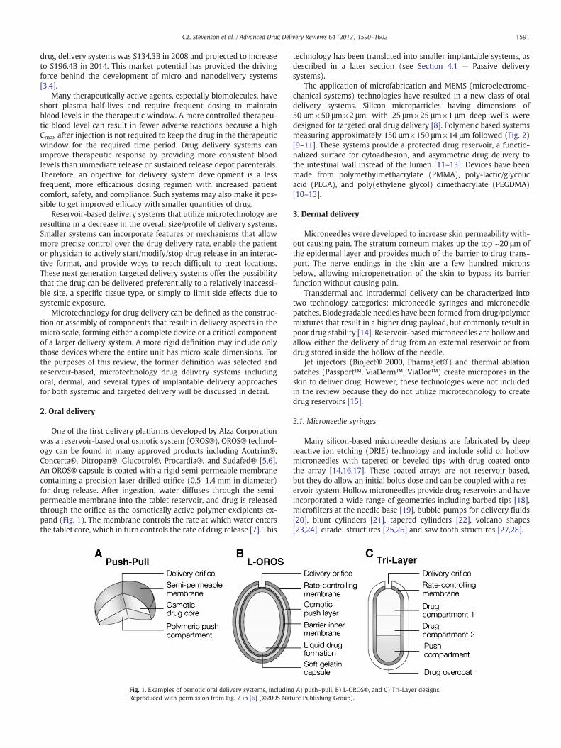
One of the first delivery platforms developed by Alza Corporationwas a reservoir-based oral osmotic system (OROS®). OROS® technol-ogy can be found in many approved products including Acutrim®,Concerta®, Ditropan®, Glucotrol®, Procardia®, and Sudafed® [5,6].An OROS® capsule is coated with a rigid semi-permeable membranecontaining a precision laser-drilled orifice (0.5–1.4 mm in diameter)for drug release. After ingestion, water diffuses through the semi-permeable membrane into the tablet reservoir, and drug is releasedthrough the orifice as the osmotically active polymer excipients ex-pand (Fig. 1). The membrane controls the rate at which water entersthe tablet core, which in turn controls the rate of drug release [7]. This
Fig. 1. Examples of osmotic oral delivery systems, includinReproduced with permission from Fig. 2 in [6] (©2005 Nat
technology has been translated into smaller implantable systems, asdescribed in a later section (see Section 4.1 — Passive deliverysystems).
The application of microfabrication and MEMS (microelectrome-chanical systems) technologies have resulted in a new class of oraldelivery systems. Silicon microparticles having dimensions of50 μm×50 μm×2 μm, with 25 μm×25 μm×1 μm deep wells weredesigned for targeted oral drug delivery [8]. Polymeric based systemsmeasuring approximately 150 μm×150 μm×14 μm followed (Fig. 2)[9–11]. These systems provide a protected drug reservoir, a functio-nalized surface for cytoadhesion, and asymmetric drug delivery tothe intestinal wall instead of the lumen [11–13]. Devices have beenmade from polymethylmethacrylate (PMMA), poly-lactic/glycolicacid (PLGA), and poly(ethylene glycol) dimethacrylate (PEGDMA)[10–13].
3. Dermal delivery
Microneedles were developed to increase skin permeability with-out causing pain. The stratum corneum makes up the top ~20 μm ofthe epidermal layer and provides much of the barrier to drug trans-port. The nerve endings in the skin are a few hundred micronsbelow, allowing micropenetration of the skin to bypass its barrierfunction without causing pain.
Transdermal and intradermal delivery can be characterized intotwo technology categories: microneedle syringes and microneedlepatches. Biodegradable needles have been formed from drug/polymermixtures that result in a higher drug payload, but commonly result inpoor drug stability [14]. Reservoir-based microneedles are hollow andallow either the delivery of drug from an external reservoir or fromdrug stored inside the hollow of the needle.
Jet injectors (BioJect® 2000, PharmaJet®) and thermal ablationpatches (Passport™, ViaDerm™, ViaDor™) create micropores in theskin to deliver drug. However, these technologies were not includedin the review because they do not utilize microtechnology to createdrug reservoirs [15].
3.1. Microneedle syringes
Many silicon-based microneedle designs are fabricated by deepreactive ion etching (DRIE) technology and include solid or hollowmicroneedles with tapered or beveled tips with drug coated ontothe array [14,16,17]. These coated arrays are not reservoir-based,but they do allow an initial bolus dose and can be coupled with a res-ervoir system. Hollowmicroneedles provide drug reservoirs and haveincorporated a wide range of geometries including barbed tips [18],microfilters at the needle base [19], bubble pumps for delivery fluids[20], blunt cylinders [21], tapered cylinders [22], volcano shapes[23,24], citadel structures [25,26] and saw tooth structures [27,28].
g A) push–pull, B) L-OROS®, and C) Tri-Layer designs.ure Publishing Group).
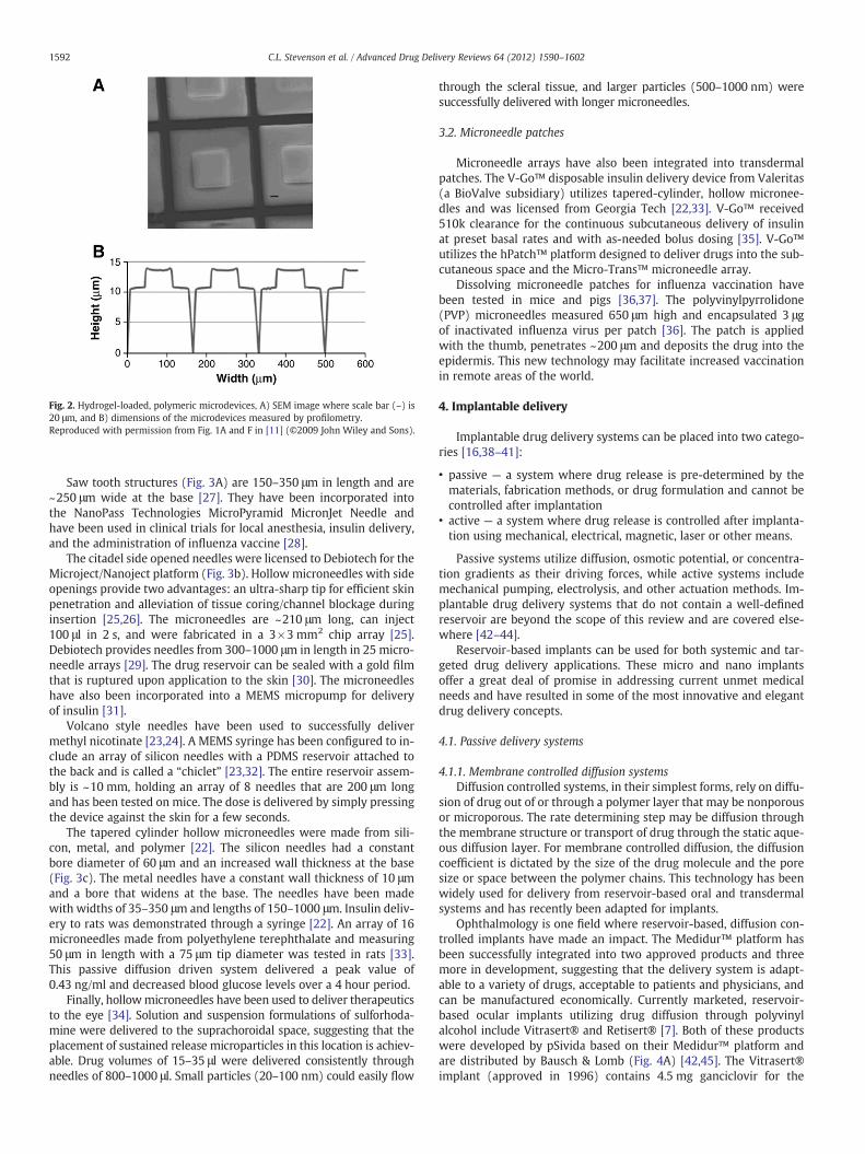
Fig. 2. Hydrogel-loaded, polymeric microdevices, A) SEM image where scale bar (–) is20 μm, and B) dimensions of the microdevices measured by profilometry.Reproduced with permission from Fig. 1A and F in [11] (©2009 John Wiley and Sons).
1592 C.L. Stevenson et al. / Advanced Drug Delivery Reviews 64 (2012) 1590–1602
Saw tooth structures (Fig. 3A) are 150–350 μm in length and are~250 μm wide at the base [27]. They have been incorporated intothe NanoPass Technologies MicroPyramid MicronJet Needle andhave been used in clinical trials for local anesthesia, insulin delivery,and the administration of influenza vaccine [28].
The citadel side opened needles were licensed to Debiotech for theMicroject/Nanoject platform (Fig. 3b). Hollow microneedles with sideopenings provide two advantages: an ultra-sharp tip for efficient skinpenetration and alleviation of tissue coring/channel blockage duringinsertion [25,26]. The microneedles are ~210 μm long, can inject100 μl in 2 s, and were fabricated in a 3×3 mm2 chip array [25].Debiotech provides needles from 300–1000 μm in length in 25 micro-needle arrays [29]. The drug reservoir can be sealed with a gold filmthat is ruptured upon application to the skin [30]. The microneedleshave also been incorporated into a MEMS micropump for deliveryof insulin [31].
Volcano style needles have been used to successfully delivermethyl nicotinate [23,24]. A MEMS syringe has been configured to in-clude an array of silicon needles with a PDMS reservoir attached tothe back and is called a “chiclet” [23,32]. The entire reservoir assem-bly is ~10 mm, holding an array of 8 needles that are 200 μm longand has been tested on mice. The dose is delivered by simply pressingthe device against the skin for a few seconds.
The tapered cylinder hollow microneedles were made from sili-con, metal, and polymer [22]. The silicon needles had a constantbore diameter of 60 μm and an increased wall thickness at the base(Fig. 3c). The metal needles have a constant wall thickness of 10 μmand a bore that widens at the base. The needles have been madewith widths of 35–350 μm and lengths of 150–1000 μm. Insulin deliv-ery to rats was demonstrated through a syringe [22]. An array of 16microneedles made from polyethylene terephthalate and measuring50 μm in length with a 75 μm tip diameter was tested in rats [33].This passive diffusion driven system delivered a peak value of0.43 ng/ml and decreased blood glucose levels over a 4 hour period.
Finally, hollowmicroneedles have been used to deliver therapeuticsto the eye [34]. Solution and suspension formulations of sulforhoda-mine were delivered to the suprachoroidal space, suggesting that theplacement of sustained release microparticles in this location is achiev-able. Drug volumes of 15–35 μl were delivered consistently throughneedles of 800–1000 μl. Small particles (20–100 nm) could easily flow
through the scleral tissue, and larger particles (500–1000 nm) weresuccessfully delivered with longer microneedles.
3.2. Microneedle patches
Microneedle arrays have also been integrated into transdermalpatches. The V-Go™ disposable insulin delivery device from Valeritas(a BioValve subsidiary) utilizes tapered-cylinder, hollow micronee-dles and was licensed from Georgia Tech [22,33]. V-Go™ received510k clearance for the continuous subcutaneous delivery of insulinat preset basal rates and with as-needed bolus dosing [35]. V-Go™utilizes the hPatch™ platform designed to deliver drugs into the sub-cutaneous space and the Micro-Trans™ microneedle array.
Dissolving microneedle patches for influenza vaccination havebeen tested in mice and pigs [36,37]. The polyvinylpyrrolidone(PVP) microneedles measured 650 μm high and encapsulated 3 μgof inactivated influenza virus per patch [36]. The patch is appliedwith the thumb, penetrates ~200 μm and deposits the drug into theepidermis. This new technology may facilitate increased vaccinationin remote areas of the world.
4. Implantable delivery
Implantable drug delivery systems can be placed into two catego-ries [16,38–41]:
• passive — a system where drug release is pre-determined by thematerials, fabrication methods, or drug formulation and cannot becontrolled after implantation
• active — a system where drug release is controlled after implanta-tion using mechanical, electrical, magnetic, laser or other means.
Passive systems utilize diffusion, osmotic potential, or concentra-tion gradients as their driving forces, while active systems includemechanical pumping, electrolysis, and other actuation methods. Im-plantable drug delivery systems that do not contain a well-definedreservoir are beyond the scope of this review and are covered else-where [42–44].
Reservoir-based implants can be used for both systemic and tar-geted drug delivery applications. These micro and nano implantsoffer a great deal of promise in addressing current unmet medicalneeds and have resulted in some of the most innovative and elegantdrug delivery concepts.
4.1. Passive delivery systems
4.1.1. Membrane controlled diffusion systemsDiffusion controlled systems, in their simplest forms, rely on diffu-
sion of drug out of or through a polymer layer that may be nonporousor microporous. The rate determining step may be diffusion throughthe membrane structure or transport of drug through the static aque-ous diffusion layer. For membrane controlled diffusion, the diffusioncoefficient is dictated by the size of the drug molecule and the poresize or space between the polymer chains. This technology has beenwidely used for delivery from reservoir-based oral and transdermalsystems and has recently been adapted for implants.
Ophthalmology is one field where reservoir-based, diffusion con-trolled implants have made an impact. The Medidur™ platform hasbeen successfully integrated into two approved products and threemore in development, suggesting that the delivery system is adapt-able to a variety of drugs, acceptable to patients and physicians, andcan be manufactured economically. Currently marketed, reservoir-based ocular implants utilizing drug diffusion through polyvinylalcohol include Vitrasert® and Retisert® [7]. Both of these productswere developed by pSivida based on their Medidur™ platform andare distributed by Bausch & Lomb (Fig. 4A) [42,45]. The Vitrasert®implant (approved in 1996) contains 4.5 mg ganciclovir for the
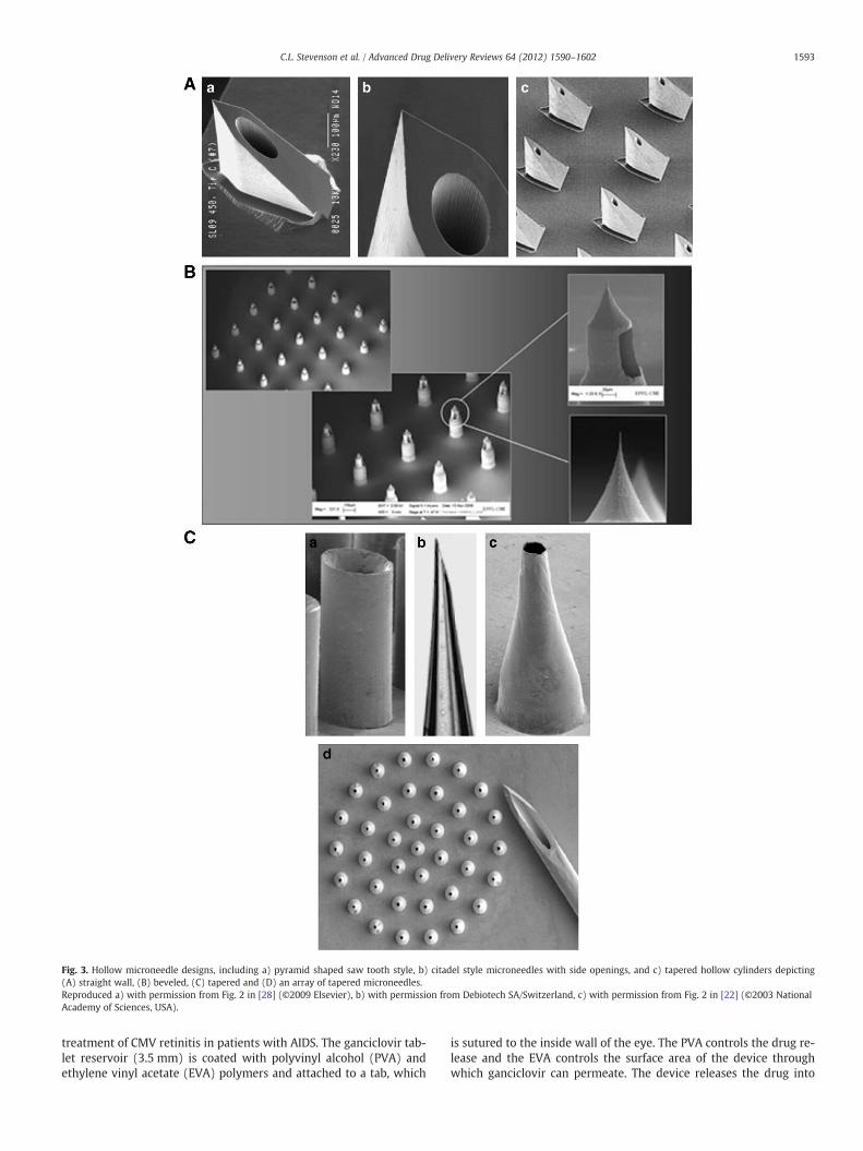
Fig. 3. Hollow microneedle designs, including a) pyramid shaped saw tooth style, b) citadel style microneedles with side openings, and c) tapered hollow cylinders depicting(A) straight wall, (B) beveled, (C) tapered and (D) an array of tapered microneedles.Reproduced a) with permission from Fig. 2 in [28] (©2009 Elsevier), b) with permission from Debiotech SA/Switzerland, c) with permission from Fig. 2 in [22] (©2003 NationalAcademy of Sciences, USA).
1593C.L. Stevenson et al. / Advanced Drug Delivery Reviews 64 (2012) 1590–1602
treatment of CMV retinitis in patients with AIDS. The ganciclovir tab-let reservoir (3.5 mm) is coated with polyvinyl alcohol (PVA) andethylene vinyl acetate (EVA) polymers and attached to a tab, which
is sutured to the inside wall of the eye. The PVA controls the drug re-lease and the EVA controls the surface area of the device throughwhich ganciclovir can permeate. The device releases the drug into
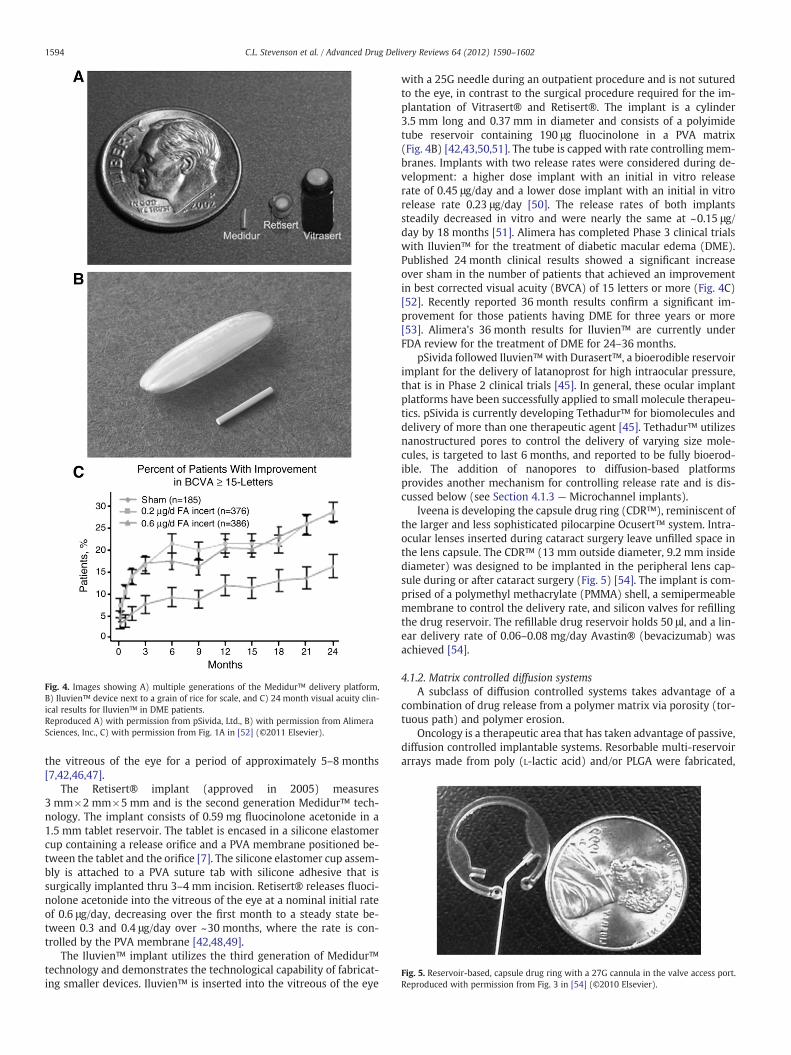
Fig. 4. Images showing A) multiple generations of the Medidur™ delivery platform,B) Iluvien™ device next to a grain of rice for scale, and C) 24 month visual acuity clin-ical results for Iluvien™ in DME patients.Reproduced A) with permission from pSivida, Ltd., B) with permission from AlimeraSciences, Inc., C) with permission from Fig. 1A in [52] (©2011 Elsevier).
Fig. 5. Reservoir-based, capsule drug ring with a 27G cannula in the valve access port.Reproduced with permission from Fig. 3 in [54] (©2010 Elsevier).
1594 C.L. Stevenson et al. / Advanced Drug Delivery Reviews 64 (2012) 1590–1602
the vitreous of the eye for a period of approximately 5–8 months[7,42,46,47].
The Retisert® implant (approved in 2005) measures3 mm×2 mm×5 mm and is the second generation Medidur™ tech-nology. The implant consists of 0.59 mg fluocinolone acetonide in a1.5 mm tablet reservoir. The tablet is encased in a silicone elastomercup containing a release orifice and a PVA membrane positioned be-tween the tablet and the orifice [7]. The silicone elastomer cup assem-bly is attached to a PVA suture tab with silicone adhesive that issurgically implanted thru 3–4 mm incision. Retisert® releases fluoci-nolone acetonide into the vitreous of the eye at a nominal initial rateof 0.6 μg/day, decreasing over the first month to a steady state be-tween 0.3 and 0.4 μg/day over ~30 months, where the rate is con-trolled by the PVA membrane [42,48,49].
The Iluvien™ implant utilizes the third generation of Medidur™technology and demonstrates the technological capability of fabricat-ing smaller devices. Iluvien™ is inserted into the vitreous of the eye
with a 25G needle during an outpatient procedure and is not suturedto the eye, in contrast to the surgical procedure required for the im-plantation of Vitrasert® and Retisert®. The implant is a cylinder3.5 mm long and 0.37 mm in diameter and consists of a polyimidetube reservoir containing 190 μg fluocinolone in a PVA matrix(Fig. 4B) [42,43,50,51]. The tube is capped with rate controlling mem-branes. Implants with two release rates were considered during de-velopment: a higher dose implant with an initial in vitro releaserate of 0.45 μg/day and a lower dose implant with an initial in vitrorelease rate 0.23 μg/day [50]. The release rates of both implantssteadily decreased in vitro and were nearly the same at ~0.15 μg/day by 18 months [51]. Alimera has completed Phase 3 clinical trialswith Iluvien™ for the treatment of diabetic macular edema (DME).Published 24 month clinical results showed a significant increaseover sham in the number of patients that achieved an improvementin best corrected visual acuity (BVCA) of 15 letters or more (Fig. 4C)[52]. Recently reported 36 month results confirm a significant im-provement for those patients having DME for three years or more[53]. Alimera's 36 month results for Iluvien™ are currently underFDA review for the treatment of DME for 24–36 months.
pSivida followed Iluvien™with Durasert™, a bioerodible reservoirimplant for the delivery of latanoprost for high intraocular pressure,that is in Phase 2 clinical trials [45]. In general, these ocular implantplatforms have been successfully applied to small molecule therapeu-tics. pSivida is currently developing Tethadur™ for biomolecules anddelivery of more than one therapeutic agent [45]. Tethadur™ utilizesnanostructured pores to control the delivery of varying size mole-cules, is targeted to last 6 months, and reported to be fully bioerod-ible. The addition of nanopores to diffusion-based platformsprovides another mechanism for controlling release rate and is dis-cussed below (see Section 4.1.3 — Microchannel implants).
Iveena is developing the capsule drug ring (CDR™), reminiscent ofthe larger and less sophisticated pilocarpine Ocusert™ system. Intra-ocular lenses inserted during cataract surgery leave unfilled space inthe lens capsule. The CDR™ (13 mm outside diameter, 9.2 mm insidediameter) was designed to be implanted in the peripheral lens cap-sule during or after cataract surgery (Fig. 5) [54]. The implant is com-prised of a polymethyl methacrylate (PMMA) shell, a semipermeablemembrane to control the delivery rate, and silicon valves for refillingthe drug reservoir. The refillable drug reservoir holds 50 μl, and a lin-ear delivery rate of 0.06–0.08 mg/day Avastin® (bevacizumab) wasachieved [54].
4.1.2. Matrix controlled diffusion systemsA subclass of diffusion controlled systems takes advantage of a
combination of drug release from a polymer matrix via porosity (tor-tuous path) and polymer erosion.
Oncology is a therapeutic area that has taken advantage of passive,diffusion controlled implantable systems. Resorbable multi-reservoirarrays made from poly (L-lactic acid) and/or PLGA were fabricated,
1595C.L. Stevenson et al. / Advanced Drug Delivery Reviews 64 (2012) 1590–1602
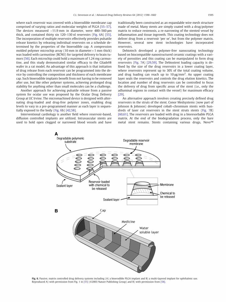
where each reservoir was covered with a bioerodible membrane capcomprised of varying ratios and molecular weights of PLGA [55–57].The devices measured ~11.9 mm in diameter, were 480–560 μmthick, and contained thirty six 120–130 nl reservoirs (Fig. 6A) [55].The incorporation of multiple reservoirs effectively provides pulsatilerelease kinetics by releasing individual reservoirs on a schedule de-termined by the properties of the bioerodible cap. A compressionmolded polymer microchip array (10 mm in diameter×1 mm thick)was loaded with carmustine (BCNU) for targeted delivery to brain tu-mors [56]. Each microchip could hold a maximum of 1.24 mg carmus-tine, and this study demonstrated similar efficacy to the Gliadel®wafer in a rat model. An advantage of this approach is that initiationof drug release from each reservoir can be programmed into the de-vice by controlling the composition and thickness of each membranecap. Such bioerodible implants benefit from not having to be removedafter use, but like other polymer systems, achieving prolonged drugstability for anything other than small molecules can be a challenge.
Another approach for achieving pulsatile release from a passivesystem for ocular use was proposed by the Ocular Drug DeliveryGroup at UC Irvine. The micromachined device is designed with alter-nating drug-loaded and drug-free polymer zones, enabling druglevels to vary in a pre-programmed manner as each layer is sequen-tially exposed to the body (Fig. 6b) [42,58].
Interventional cardiology is another field where reservoir-based,diffusion controlled implants are utilized. Intravascular stents areused to hold open clogged or narrowed blood vessels and have
Fig. 6. Passive, matrix controlled drug delivery systems including (A) a bioerReproduced A) with permission from Fig. 1 in [55] (©2003 Nature Publishing
traditionally been constructed as an expandable wire mesh structuremade of metal. Many stents are simply coated with a drug/polymermatrix to reduce restenosis, a re-narrowing of the stented vessel byinflammation and tissue ingrowth. This coating technology does notdeliver drug from a reservoir ‘per se’, but from the polymer matrix.However, several new stent technologies have incorporatedreservoirs.
Debiotech developed a polymer-free nanocoating technologybased on biocompatible nanostructured ceramic coatings with a vari-ety of porosities and this coating can be manipulated to form drugreservoirs (Fig. 7A) [29,59]. The Debiostent loading capacity is de-fined by the size of the drug reservoirs in a lower coating layer,where reservoirs represent up to 30% of the total coating volume,and drug loading can reach up to 10 μg/mm2. An upper coatinglayer seals the reservoirs and controls the drug elution kinetics. Thelocation and number of drug reservoirs can be controlled to focusthe delivery of drug from specific areas of the stent (i.e., only theadluminal regions in contact with the vessel) for maximum efficacy[29].
An alternative approach involves creating precisely defined drugreservoirs in the struts of the stent. Conor Medsystems (now part ofJohnson & Johnson) developed cobalt–chromium stents with hun-dreds of laser cut reservoirs in the stent struts stents (Fig. 7B)[60,61]. The reservoirs are loaded with drug in a bioresorbable PLGAmatrix. At the end of the biodegradation process, only the baremetal stent remains. Stents containing various drugs, Nevo™
odible PLGA implant and B) a multi-layered implant for ophthalmic use.Group) and B) with permission from [58].
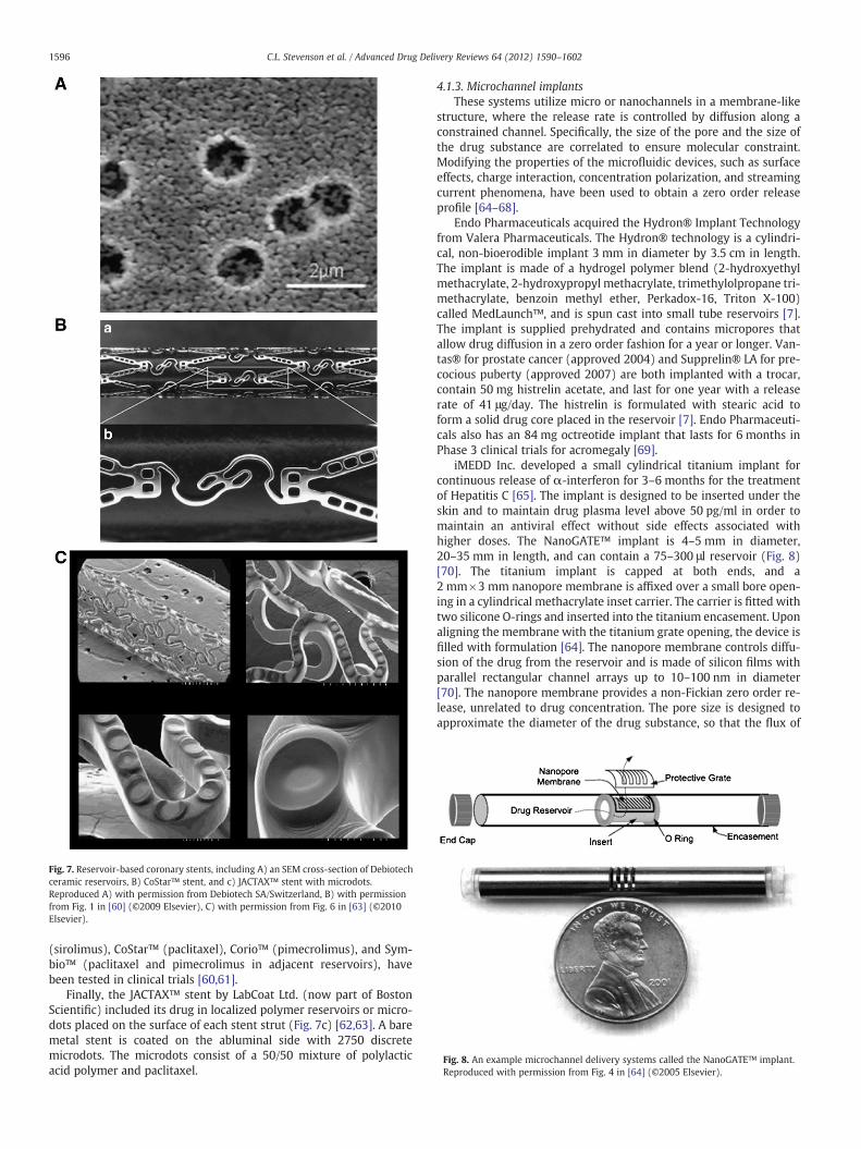
Fig. 7. Reservoir-based coronary stents, including A) an SEM cross-section of Debiotechceramic reservoirs, B) CoStar™ stent, and c) JACTAX™ stent with microdots.Reproduced A) with permission from Debiotech SA/Switzerland, B) with permissionfrom Fig. 1 in [60] (©2009 Elsevier), C) with permission from Fig. 6 in [63] (©2010Elsevier).
Fig. 8. An example microchannel delivery systems called the NanoGATE™ implant.Reproduced with permission from Fig. 4 in [64] (©2005 Elsevier).
1596 C.L. Stevenson et al. / Advanced Drug Delivery Reviews 64 (2012) 1590–1602
(sirolimus), CoStar™ (paclitaxel), Corio™ (pimecrolimus), and Sym-bio™ (paclitaxel and pimecrolimus in adjacent reservoirs), havebeen tested in clinical trials [60,61].
Finally, the JACTAX™ stent by LabCoat Ltd. (now part of BostonScientific) included its drug in localized polymer reservoirs or micro-dots placed on the surface of each stent strut (Fig. 7c) [62,63]. A baremetal stent is coated on the abluminal side with 2750 discretemicrodots. The microdots consist of a 50/50 mixture of polylacticacid polymer and paclitaxel.
4.1.3. Microchannel implantsThese systems utilize micro or nanochannels in a membrane-like
structure, where the release rate is controlled by diffusion along aconstrained channel. Specifically, the size of the pore and the size ofthe drug substance are correlated to ensure molecular constraint.Modifying the properties of the microfluidic devices, such as surfaceeffects, charge interaction, concentration polarization, and streamingcurrent phenomena, have been used to obtain a zero order releaseprofile [64–68].
Endo Pharmaceuticals acquired the Hydron® Implant Technologyfrom Valera Pharmaceuticals. The Hydron® technology is a cylindri-cal, non-bioerodible implant 3 mm in diameter by 3.5 cm in length.The implant is made of a hydrogel polymer blend (2-hydroxyethylmethacrylate, 2-hydroxypropyl methacrylate, trimethylolpropane tri-methacrylate, benzoin methyl ether, Perkadox-16, Triton X-100)called MedLaunch™, and is spun cast into small tube reservoirs [7].The implant is supplied prehydrated and contains micropores thatallow drug diffusion in a zero order fashion for a year or longer. Van-tas® for prostate cancer (approved 2004) and Supprelin® LA for pre-cocious puberty (approved 2007) are both implanted with a trocar,contain 50 mg histrelin acetate, and last for one year with a releaserate of 41 μg/day. The histrelin is formulated with stearic acid toform a solid drug core placed in the reservoir [7]. Endo Pharmaceuti-cals also has an 84 mg octreotide implant that lasts for 6 months inPhase 3 clinical trials for acromegaly [69].
iMEDD Inc. developed a small cylindrical titanium implant forcontinuous release of α-interferon for 3–6 months for the treatmentof Hepatitis C [65]. The implant is designed to be inserted under theskin and to maintain drug plasma level above 50 pg/ml in order tomaintain an antiviral effect without side effects associated withhigher doses. The NanoGATE™ implant is 4–5 mm in diameter,20–35 mm in length, and can contain a 75–300 μl reservoir (Fig. 8)[70]. The titanium implant is capped at both ends, and a2 mm×3 mm nanopore membrane is affixed over a small bore open-ing in a cylindrical methacrylate inset carrier. The carrier is fitted withtwo silicone O-rings and inserted into the titanium encasement. Uponaligning the membrane with the titanium grate opening, the device isfilled with formulation [64]. The nanopore membrane controls diffu-sion of the drug from the reservoir and is made of silicon films withparallel rectangular channel arrays up to 10–100 nm in diameter[70]. The nanopore membrane provides a non-Fickian zero order re-lease, unrelated to drug concentration. The pore size is designed toapproximate the diameter of the drug substance, so that the flux of
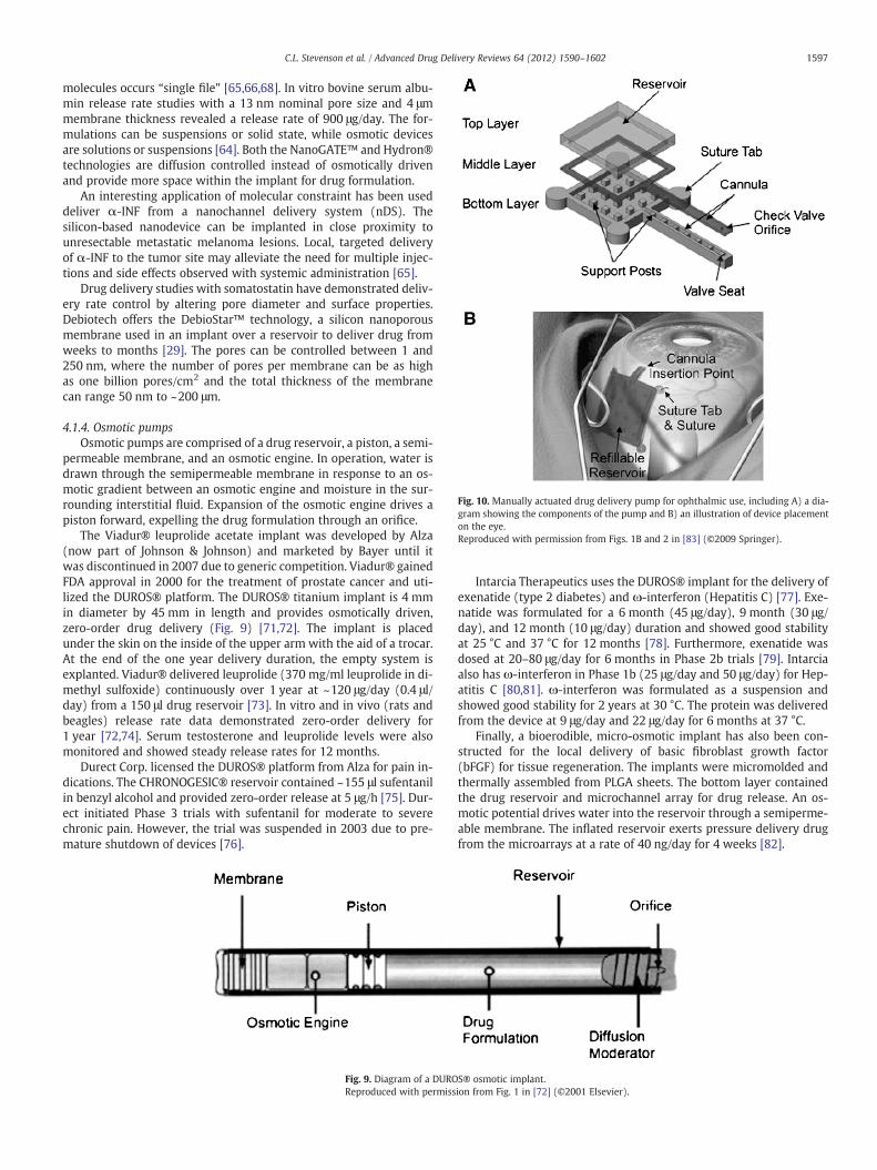
Fig. 10. Manually actuated drug delivery pump for ophthalmic use, including A) a dia-gram showing the components of the pump and B) an illustration of device placementon the eye.Reproduced with permission from Figs. 1B and 2 in [83] (©2009 Springer).
1597C.L. Stevenson et al. / Advanced Drug Delivery Reviews 64 (2012) 1590–1602
molecules occurs “single file” [65,66,68]. In vitro bovine serum albu-min release rate studies with a 13 nm nominal pore size and 4 μmmembrane thickness revealed a release rate of 900 μg/day. The for-mulations can be suspensions or solid state, while osmotic devicesare solutions or suspensions [64]. Both the NanoGATE™ and Hydron®technologies are diffusion controlled instead of osmotically drivenand provide more space within the implant for drug formulation.
An interesting application of molecular constraint has been useddeliver α-INF from a nanochannel delivery system (nDS). Thesilicon-based nanodevice can be implanted in close proximity tounresectable metastatic melanoma lesions. Local, targeted deliveryof α-INF to the tumor site may alleviate the need for multiple injec-tions and side effects observed with systemic administration [65].
Drug delivery studies with somatostatin have demonstrated deliv-ery rate control by altering pore diameter and surface properties.Debiotech offers the DebioStar™ technology, a silicon nanoporousmembrane used in an implant over a reservoir to deliver drug fromweeks to months [29]. The pores can be controlled between 1 and250 nm, where the number of pores per membrane can be as highas one billion pores/cm2 and the total thickness of the membranecan range 50 nm to ~200 μm.
4.1.4. Osmotic pumpsOsmotic pumps are comprised of a drug reservoir, a piston, a semi-
permeable membrane, and an osmotic engine. In operation, water isdrawn through the semipermeable membrane in response to an os-motic gradient between an osmotic engine and moisture in the sur-rounding interstitial fluid. Expansion of the osmotic engine drives apiston forward, expelling the drug formulation through an orifice.
The Viadur® leuprolide acetate implant was developed by Alza(now part of Johnson & Johnson) and marketed by Bayer until itwas discontinued in 2007 due to generic competition. Viadur® gainedFDA approval in 2000 for the treatment of prostate cancer and uti-lized the DUROS® platform. The DUROS® titanium implant is 4 mmin diameter by 45 mm in length and provides osmotically driven,zero-order drug delivery (Fig. 9) [71,72]. The implant is placedunder the skin on the inside of the upper arm with the aid of a trocar.At the end of the one year delivery duration, the empty system isexplanted. Viadur® delivered leuprolide (370 mg/ml leuprolide in di-methyl sulfoxide) continuously over 1 year at ~120 μg/day (0.4 μl/day) from a 150 μl drug reservoir [73]. In vitro and in vivo (rats andbeagles) release rate data demonstrated zero-order delivery for1 year [72,74]. Serum testosterone and leuprolide levels were alsomonitored and showed steady release rates for 12 months.
Durect Corp. licensed the DUROS® platform from Alza for pain in-dications. The CHRONOGESIC® reservoir contained ~155 μl sufentanilin benzyl alcohol and provided zero-order release at 5 μg/h [75]. Dur-ect initiated Phase 3 trials with sufentanil for moderate to severechronic pain. However, the trial was suspended in 2003 due to pre-mature shutdown of devices [76].
Fig. 9. Diagram of a DUROReproduced with permiss
Intarcia Therapeutics uses the DUROS® implant for the delivery ofexenatide (type 2 diabetes) and ω-interferon (Hepatitis C) [77]. Exe-natide was formulated for a 6 month (45 μg/day), 9 month (30 μg/day), and 12 month (10 μg/day) duration and showed good stabilityat 25 °C and 37 °C for 12 months [78]. Furthermore, exenatide wasdosed at 20–80 μg/day for 6 months in Phase 2b trials [79]. Intarciaalso has ω-interferon in Phase 1b (25 μg/day and 50 μg/day) for Hep-atitis C [80,81]. ω-interferon was formulated as a suspension andshowed good stability for 2 years at 30 °C. The protein was deliveredfrom the device at 9 μg/day and 22 μg/day for 6 months at 37 °C.
Finally, a bioerodible, micro-osmotic implant has also been con-structed for the local delivery of basic fibroblast growth factor(bFGF) for tissue regeneration. The implants were micromolded andthermally assembled from PLGA sheets. The bottom layer containedthe drug reservoir and microchannel array for drug release. An os-motic potential drives water into the reservoir through a semiperme-able membrane. The inflated reservoir exerts pressure delivery drugfrom the microarrays at a rate of 40 ng/day for 4 weeks [82].
S® osmotic implant.ion from Fig. 1 in [72] (©2001 Elsevier).
Fig. 11. Active delivery pump for ophthalmic use, including A) a diagram showing thecomponents of the pump and B) a cross-section of the pump showing electrolysis en-abled pumping of drug into the eye.Reproduced with permission from Figs. 1 and 2 in [84] (©2008 Elsevier).
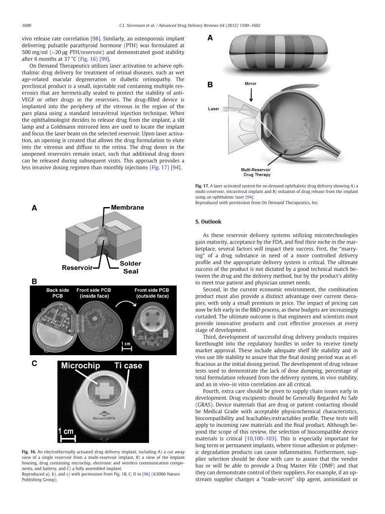
Fig. 13. An implantable active drug delivery pump, including A) a view of the piezo-electric membrane pumping mechanism and check valves and B) a view of the fluidicconnections to the pump.Reproduced with permission from Debiotech SA/Switzerland.
1598 C.L. Stevenson et al. / Advanced Drug Delivery Reviews 64 (2012) 1590–1602
4.2. Active delivery systems
4.2.1. Single reservoir active systemsPumps generally include a drug reservoir, an actuator, and one or
more valves in order to accurately control the delivery of small vol-umes of a drug in solution. They may operate by manual actuation,electrolysis, piezoelectric actuation, resistive heating, magnetic actua-tion, or by incorporation of reversible polymeric valves.
The simplest example of an active drug delivery pump is one thatis actuated manually by pressing on it with an instrument or a finger.Such a system was designed for the treatment of glaucoma, age-related macular degeneration, and diabetic retinopathy [44,83]. Thepump is fabricated from three layers of polymethylsiloxane (PDMS)using soft lithography. The device was sutured to the outside of theeye and contained a drug reservoir of approximately 200 μl, a checkvalve, and a cannula (10 mm×1 mm×1 mm) placed through thewall of the eye (Fig. 10). When the pump was manually actuated bypressing on the drug reservoir, the increase in pressure in the
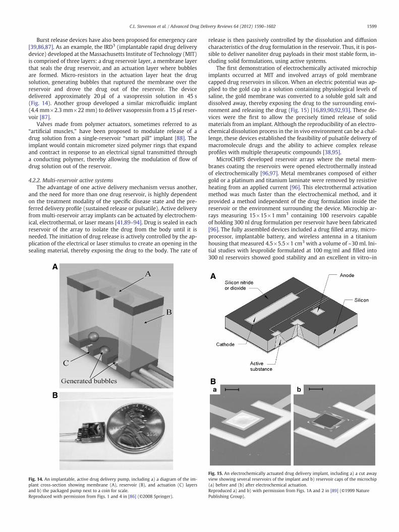
Fig. 12. Ocular drug delivery implant actuated by a magnetic field.Reproduced with permission from Fig. 1 in [85] (©2011 Royal Society ofChemistry).
reservoir caused the check valve in the cannula to open, dispensingthe drug.
Replenish has developed an electrochemically driven drug deliv-ery pump based on silicon MEMS technology that is designed toallow variable drug delivery rates. It contains a refillable reservoirfor a drug solution and a one way check valve made of parylene.The device is surgically implanted beneath the conjunctiva with aflexible parylene cannula inserted through the eye wall. An electriccurrent is passed between two electrodes located on the silicon incontact with the drug solution when a dose is required. The gas gen-erated by electrolysis of the water increases the pressure on the flex-ible membrane of the drug reservoir, pushing drug solution outthrough the cannula and into the eye (Fig. 11) [84].
A magnetically actuated ocular implant has been reported by theUniversity of British Columbia for the treatment of diabetic retinopa-thy [85]. The device is designed to be surgically implanted behind theeye. A prototype implant consisted of a reservoir (6 mm×550 μm)containing docetaxel, sealed with an elastic magnetic polydimethylsi-loxane (PDMS) membrane (6 mm×40 μm), and a laser drilled orifice(100×100 μm2) (Fig. 12). Upon application of a magnetic field(255 mT), the membrane deforms, causing expulsion of drug solution(~171 ng/actuation) from the implant.
The MIP implantable pump from Debiotech is a piezo-actuated sil-icon micropump for drug delivery. The pump consists of pair of checkvalves and a reciprocating pumping membrane to guide liquid flow inthe proper direction from a drug reservoir to the target location. Thepump is fabricated from multiple bonded layers of silicon and glass,with a piezoelectric ceramic disk and titanium fluid connectors andhas a typical flow rate of 1 ml/min (Fig. 13) [29].
1599C.L. Stevenson et al. / Advanced Drug Delivery Reviews 64 (2012) 1590–1602
Burst release devices have also been proposed for emergency care[39,86,87]. As an example, the IRD3 (implantable rapid drug deliverydevice) developed at the Massachusetts Institute of Technology (MIT)is comprised of three layers: a drug reservoir layer, a membrane layerthat seals the drug reservoir, and an actuation layer where bubblesare formed. Micro-resistors in the actuation layer heat the drugsolution, generating bubbles that ruptured the membrane over thereservoir and drove the drug out of the reservoir. The devicedelivered approximately 20 μl of a vasopressin solution in 45 s(Fig. 14). Another group developed a similar microfluidic implant(4.4 mm×2.3 mm×22 mm) to deliver vasopressin from a 15 μl reser-voir [87].
Valves made from polymer actuators, sometimes referred to as“artificial muscles,” have been proposed to modulate release of adrug solution from a single-reservoir “smart pill” implant [88]. Theimplant would contain micrometer sized polymer rings that expandand contract in response to an electrical signal transmitted througha conducting polymer, thereby allowing the modulation of flow ofdrug solution out of the reservoir.
4.2.2. Multi-reservoir active systemsThe advantage of one active delivery mechanism versus another,
and the need for more than one drug reservoir, is highly dependenton the treatment modality of the specific disease state and the pre-ferred delivery profile (sustained release or pulsatile). Active deliveryfrom multi-reservoir array implants can be actuated by electrochem-ical, electrothermal, or laser means [41,89–94]. Drug is sealed in eachreservoir of the array to isolate the drug from the body until it isneeded. The initiation of drug release is actively controlled by the ap-plication of the electrical or laser stimulus to create an opening in thesealing material, thereby exposing the drug to the body. The rate of
Fig. 14. An implantable, active drug delivery pump, including a) a diagram of the im-plant cross-section showing membrane (A), reservoir (B), and actuation (C) layersand b) the packaged pump next to a coin for scale.Reproduced with permission from Figs. 1 and 4 in [86] (©2008 Springer).
release is then passively controlled by the dissolution and diffusioncharacteristics of the drug formulation in the reservoir. Thus, it is pos-sible to deliver nanoliter drug payloads in their most stable form, in-cluding solid formulations, using active systems.
The first demonstration of electrochemically activated microchipimplants occurred at MIT and involved arrays of gold membranecapped drug reservoirs in silicon. When an electric potential was ap-plied to the gold cap in a solution containing physiological levels ofsaline, the gold membrane was converted to a soluble gold salt anddissolved away, thereby exposing the drug to the surrounding envi-ronment and releasing the drug (Fig. 15) [16,89,90,92,93]. These de-vices were the first to allow the precisely timed release of solidmaterials from an implant. Although the reproducibility of an electro-chemical dissolution process in the in vivo environment can be a chal-lenge, these devices established the feasibility of pulsatile delivery ofmacromolecule drugs and the ability to achieve complex releaseprofiles with multiple therapeutic compounds [38,95].
MicroCHIPS developed reservoir arrays where the metal mem-branes coating the reservoirs were opened electrothermally insteadof electrochemically [96,97]. Metal membranes composed of eithergold or a platinum and titanium laminate were removed by resistiveheating from an applied current [96]. This electrothermal activationmethod was much faster than the electrochemical method, and itprovided a method independent of the drug formulation inside thereservoir or the environment surrounding the device. Microchip ar-rays measuring 15×15×1 mm3 containing 100 reservoirs capableof holding 300 nl drug formulation per reservoir have been fabricated[96]. The fully assembled devices included a drug filled array, micro-processor, implantable battery, and wireless antenna in a titaniumhousing that measured 4.5×5.5×1 cm3 with a volume of ~30 ml. Ini-tial studies with leuprolide formulated at 100 mg/ml and filled into300 nl reservoirs showed good stability and an excellent in vitro–in
Fig. 15. An electrochemically actuated drug delivery implant, including a) a cut awayview showing several reservoirs of the implant and b) reservoir caps of the microchip(a) before and (b) after electrochemical actuation.Reproduced a) and b) with permission from Figs. 1A and 2 in [89] (©1999 NaturePublishing Group).
1600 C.L. Stevenson et al. / Advanced Drug Delivery Reviews 64 (2012) 1590–1602
vivo release rate correlation [98]. Similarly, an osteoporosis implantdelivering pulsatile parathyroid hormone (PTH) was formulated at500 mg/ml (~20 μg PTH/reservoir) and demonstrated good stabilityafter 6 months at 37 °C (Fig. 16) [99].
On Demand Therapeutics utilizes laser activation to achieve oph-thalmic drug delivery for treatment of retinal diseases, such as wetage-related macular degeneration or diabetic retinopathy. Thepreclinical product is a small, injectable rod containing multiple res-ervoirs that are hermetically sealed to protect the stability of anti-VEGF or other drugs in the reservoirs. The drug-filled device isimplanted into the periphery of the vitreous in the region of thepars plana using a standard intravitreal injection technique. Whenthe ophthalmologist decides to release drug from the implant, a slitlamp and a Goldmann mirrored lens are used to locate the implantand focus the laser beam on the selected reservoir. Upon laser activa-tion, an opening is created that allows the drug formulation to eluteinto the vitreous and diffuse to the retina. The drug doses in theunopened reservoirs remain intact, such that additional drug dosescan be released during subsequent visits. This approach provides aless invasive dosing regimen than monthly injections (Fig. 17) [94].
Fig. 16. An electrothermally actuated drug delivery implant, including A) a cut awayview of a single reservoir from a multi-reservoir implant, B) a view of the implanthousing, drug containing microchip, electronic and wireless communication compo-nents, and battery, and C) a fully assembled implant.Reproduced a), b), and c) with permission from Fig. 1B, C, D in [96] (©2006 NaturePublishing Group).
Fig. 17. A laser activated system for on demand ophthalmic drug delivery showing A) amulti-reservoir, intravitreal implant and B) initiation of drug release from the implantusing an ophthalmic laser [94].Reproduced with permission from On Demand Therapeutics, Inc.
5. Outlook
As these reservoir delivery systems utilizing microtechnologiesgain maturity, acceptance by the FDA, and find their niche in the mar-ketplace, several factors will impact their success. First, the “marry-ing” of a drug substance in need of a more controlled deliveryprofile and the appropriate delivery system is critical. The ultimatesuccess of the product is not dictated by a good technical match be-tween the drug and the delivery method, but by the product's abilityto meet true patient and physician unmet needs.
Second, in the current economic environment, the combinationproduct must also provide a distinct advantage over current thera-pies, with only a small premium in price. The impact of pricing cannow be felt early in the R&D process, as these budgets are increasinglycurtailed. The ultimate outcome is that engineers and scientists mustprovide innovative products and cost effective processes at everystage of development.
Third, development of successful drug delivery products requiresforethought into the regulatory hurdles in order to receive timelymarket approval. These include adequate shelf life stability and invivo use life stability to assure that the final dosing period was as ef-ficacious as the initial dosing period. The development of drug releasetests used to demonstrate the lack of dose dumping, percentage oftotal formulation released from the delivery system, in vivo stability,and an in vivo–in vitro correlation are all critical.
Fourth, extra care should be given to supply chain issues early indevelopment. Drug excipients should be Generally Regarded As Safe(GRAS). Device materials that are drug or patient contacting shouldbe Medical Grade with acceptable physicochemical characteristics,biocompatibility and leachables/extractables profile. These tests willapply to incoming raw materials and the final product. Although be-yond the scope of this review, the selection of biocompatible devicematerials is critical [10,100–103]. This is especially important forlong term or permanent implants, where tissue adhesion or polymer-ic degradation products can cause inflammation. Furthermore, sup-plier selection should be done with care to assure that the vendorhas or will be able to provide a Drug Master File (DMF) and thatthey can demonstrate control of their suppliers. For example, if an up-stream supplier changes a “trade-secret” slip agent, antioxidant or
1601C.L. Stevenson et al. / Advanced Drug Delivery Reviews 64 (2012) 1590–1602
dye package in a commodity material, the downstream drug productproducer must understand the impact on the product before it is soldto a patient (and prevent the need for a product recall).
6. Conclusions
The knowledge and tools enabling the development of reservoir-based drug delivery systems utilizing microtechnology have comefrom number of diverse fields of study including chemistry, materialsscience, mechanics, information technology, and microelectronics.The innovations at the core of such novel oral, dermal, and implant-able delivery systems come from the intersection of these disparatefields. Devices combining reservoirs and microtechnology driven bypassive or active mechanisms are enabling the delivery of bothsmall and macromolecule drugs with increased specificity andcontrol.
While tremendous progress continues on the development ofmicro and nanotechnology for reservoir-based drug delivery devices,much additional work will be required to translate the promisingtechnology into safe and effective, well-controlled, patient acceptableproducts.
Acknowledgements
Robert Langer would like to acknowledge grant support forreservoir-based, microchip drug delivery systems from the U.S. Na-tional Institutes of Health, grant #EB006365.
References
[1] D.C. Mandal, M. Mandal, Current status and future prospects of new drug deliv-ery system, Pharma Times 42 (Apr. 2010).
[2] C. Kaparissides, S. Alexandridou, K. Kotti, S. Chaididou, Advanced Drug DeliverySystems: New Developments, New Technologies, AzoM.com Pvt.L Ltd, October2006.
[3] R. Langer, N.A. Peppas, Advances in biomaterials, drug delivery and bionano-technology, AICHE J. 49 (2003) 2990–3006.
[4] J.Z. Hilt, N.A. Peppas, Microfabricated drug delivery devices, Int. J. Pharm. 306(2005) 15–23.
[5] R.K. Verma, S. Garg, Drug delivery technologies and future directions, Pharm.Technol. 25 (2001) 1–14.
[6] H. Rosen, T. Abribat, The rise and rise of drug delivery, Nat. Rev. Drug Discov. 4(2005) 381–385.
[7] Thomson Physicians' Desk Reference, 63th Edition Thomson Publishing, Mon-tvale, N.J, 2009.
[8] A. Ahmed, C. Bonner, T. Desai, Bioadhesive microdevices with multiple reser-voirs: a new platform for oral drug delivery, J. Control. Release 81 (2002)291–306.
[9] S.L. Tao, T.A. Desai, Micromachined devices: the impact of controlled geometryfor cell-targeting to bioavailability, J Control. Release 109 (2005) 127–138.
[10] K.M. Ainslie, T.A. Desai, Microfabricated implant for applications in therapeuticdelivery, tissue engineering and biosensing, Lab Chip 8 (2008) 1864–1878.
[11] K.M. Ainslie, R.D. Lowe, T.T. Beaudette, L. Petty, E.M. Bachelder, T.A. Desai, Micro-fabricated devices for enhanced bioadhesive drug delivery: attachment to andsmall-molecule release through a cell monolayer under flow, Small 5 (2009)2857–2863.
[12] S.L. Tao, T.A. Desai, Microfabrication of multilayer, asymetric, polymeric devicesfor drug delivery, Adv. Mater. 17 (2005) 1625–1630.
[13] S.L. Tao, T.A. Desai, Gastrointestinal patch systems for oral drug delivery, DrugDiscov. Today. 10 (2005) 909–915.
[14] E.E. Nuxoll, R.A. Siegel, BioMEMS devices for drug delivery, IEEE Eng. Med. Biol.(Jan/Feb 2009) 31–39.
[15] A. Aroro, M. Prausnitz, S. Mitragotri, Micro-scale devices for transdermal drugdelivery, Int. J. Pharm. 8 (2008) 227–236.
[16] M. Staples, K. Daniel, M.J. Cima, R. Langer, Application of micro- andnano-electromechanical devices to drug delivery, Pharm. Res. 23 (2006) 847–863.
[17] J. Matriano, M. Cormier, J. Johnson, W. Young, M. Buttery, K. Nyam, P. Daddona,Macroflux microprojection array patch technology: a new and efficient ap-proach for intracutaneous immunization, Pharm. Res. 19 (2002) 1653–1664.
[18] S. Chandrasekara, J. Brazzle, A. Frazier, Surface machined metallic microneedles,J. Microelectromech. Syst. 12 (2003) 281–288.
[19] J. Zahn, N. Talbot, D. Liepmann, A. Pisano, Microfabricated polysilicon micronee-dles for minimally invasive biomedical devices, Biomed. Microdevices 2 (2000)295–303.
[20] J. Zahn, A. Deshmekh, A. Pisano, D. Liepmann, Continuous on-chip micropump-ing from microneedled enhance drug delivery, Biomed. Microdevices 6 (2004)183–190.
[21] M.A.L. Teo, C. Shearwood, K.C. Ng, J. Lu, S. Moochhala, In vitro and in vivo char-acterization of MEMS microneedles, Biomed. Microdevices 7 (2005) 47–52.
[22] D.V. McAllister, P.M. Wang, S.P. Davis, J.H. Park, P.J. Canatella, M.G. Allen, M.R.Prausnitz, Microfabricated needles for transdermal delivery of macromoleculesand nanoparticles: fabrication methods and transport studies, Proc. Natl. Acad.Sci. U. S. A. 100 (2003) 13755–13760.
[23] B. Stoeber, D. Liepmann, Arrays of hollow out-of-plane microneedles for drugdelivery, J. Microelectromech. Syst. 14 (2005) 472–479.
[24] R. Sivamani, B. Stoeber, G. Wu, H. Zhai, D. Liepmann, H. Maibach, Clinical micro-needle injection of methyl nicotinate: stratum corneum penetration, Skin Res.Technol. 11 (2005) 152–156.
[25] P. Griss, G. Stemme, Side-opened out-of-plane microneedles for microfluidictransdermal liquid transfers, J. Microelectromech. Syst. 12 (2003) 296–301.
[26] N. Roxhed, C. Gasser, P. Griss, G. Holzapfel, G. Stemme, Penetration enhancedultrashorp microneedles and prediction in skin interaction for efficient trans-dermal drug delivery, J. Microelectromech. Syst. 16 (2007) 1429–1440.
[27] H. Gardeniers, R. Luttge, E. Bereschot, M. de Boer, S. Yeshurun, M. Hefetz, R. van'tOever, A. van den Berg, Silicon micromachines hollow microneedles for trans-dermal liquid transport, J. Microelectromech. Syst. 12 (2003) 855–862.
[28] P. Van Damme, F. Oosterhuis-Kafeja, M. Van der Wielen, Y. Almagor, O. Sharon,Y. Levin, Safety and efficacy of a novel microneedle device for dose sparing intra-dermal influenza vaccination in healthy adults, Vaccine 27 (2009) 454–459.
[29] http://www.debiotech.com/.[30] N. Roxhed, P. Griss, G. Stemme, Membrane-sealed hollow microneedles and re-
lated administration schemes for transdermal drug delivery, Biomed. Microde-vices 10 (2008) 271–279.
[31] L. Nordquist, N. Roxhed, P. Griss, G. Stemme, Novel microneedle patches for ac-tive insulin delivery are efficient in maintaining glycaemic control: an initialcomparison with subcutaneous administration, Pharm. Res. 24 (2007)1381–1387.
[32] U.O. Hafeli, A. Mikhtari, D. Liepmann, In vivo evaluation of a microneedle-basedminiature syringe for intradermal drug delivery, Biomed. Microdevices 11(2009) 943–950.
[33] S.P. Davis, W. Martanto, M.G. Allen, M.R. Prausnitz, Hollow metal microneedlesfor insulin delivery to diabetic rats, IEEE T. Bio-Med. Eng. 52 (2005) 909–915.
[34] S.R. Patel, A.S.P. Lin, H.F. Edelhauser, M.R. Prausnitz, Suprachoroidal drug deliv-ery to the back of the eye using hollow microneedles, Pharm. Res. 28 (2010)166–176.
[35] http://www.valeritas.com/.[36] S.P. Sullivan, D.G. Koutsonanos, M.D.P. Martin, J.W. Lee, V. Zarnitsyn, S.O. Choi, N.
Murthy, R.W. Compans, I. Skountzou, M.R. Prausnitz, Dissolving polymer micro-needle patches for influenza vaccination, Nat. Med. 16 (2010) 915–920.
[37] S. Kommareddy, B.C. Boudner, S. Oh, S.Y. Kwon, M. Singh, D.T. O'Hagan, Dissolv-able microneedle patches for the delivery of cell culture derived influenza vac-cine antigens, J. Pharm. Sci. 101 (2012) 1021–1027.
[38] A.C.R. Grayson, R.S. Shawgo, A.M. Johnson, N.T. Flynn, Y. Li, M.J. Cima, R. Langer,A BioMEMS review: MEMS technology for physiologically integrated devices,Proc. IEEE 92 (2009) 6–21.
[39] J.H. Sakamoto, A.L. van de Ven, B. Godin, E. Blanco, R.E. Serda, A. Grattoni, A.Ziemys, A. Bouramrani, T. Hu, S.I. Ranganathan, E. De Rosa, J.O. Martinez, C.A.Smid, R.M. Chchana, S.Y. Lee, S. Srinavasan, M. Landry, A. Meyn, E. Tasciotti, X.Liu, P. Decuzzi, M. Ferrari, Enabling individualized therapy through nanotech-nology, Pharmacol. Res. 62 (2010) 57–89.
[40] A. Nisar, N. Afzulpurkar, B. Mahaisavariya, A. Tuantranont, MEMS-based micro-pumps in drug delivery and biomedical applications, Sensor. Actuat. B-Chem.130 (2008) 917–942.
[41] R.A.M. Receveur, F.W. Linemans, N.F. de Rooij, Microsystem technologies for im-plantable applications, J. Micromech. Microeng. 17 (2007) R50–R80.
[42] Y.E. Choonara, V. Pillay, M.P. Danckwerts, T.R. Carmichael, L.C. Du Toit, A reviewof implantable intravitreal drug delivery technologies for the treatment of pos-terior segment eye diseases, J. Pharm. Sci. 99 (2010) 2219–2239.
[43] E. Eljarrat-Binstock, J. Pe'er, A.J. Domb, New techniques for drug delivery to theposterior eye segment, Pharm. Res. 27 (2010) 530–543.
[44] S. Mansoor, B.D. Kuppermann, M.C. Kenney, Intraocular sustained release deliv-ery systems for triamcinolone acetonide, Pharm. Res. 26 (2009) 770–784.
[45] http://www.psivida.com/products.html.[46] G.E. Sanborn, R. Anand, R.E. Torti, S.D. Nightingale, S.X. Cal, B. Yates, P. Aston, T.J.
Smith, Sustained release ganciclovir therapy for treatment of cytomegalovirusretinitis; uses of an intravitreal device, Arch. Ophthalmol. 110 (1992) 188–195.
[47] T.J. Smith, P.A. Pearson, D.L. Blandford, J.D. Brown, K.A. Goins, J.L. Hollins, E.T.Schmiesser, P. Glavinos, L.B. Baldwin, P. Ashton, Intravitreal sustained releaseganciclovir, Arch. Ophthalmol. 110 (1992) 255–258.
[48] P. Mruthyunjaya, D. Khalatbari, P. Yang, S. Stinnett, R. Tano, P. Ashton, H. Guo, M.Nazzar, G.J. Jaffe, Efficacy of low release rate fluocinolone acetonide intravitrealimplants to treat experiments uveitis, Arch. Ophthalmol. 124 (2006)1012–1018.
[49] D.G. Callanan, G.J. Jaffe, D.F. Martin, P.A. Pearson, T.L. Comstock, Treatment ofposterior uveitis with a flucinolone acetonide implant, Arch. Ophthalmol. 126(2008) 1191–1201.
[50] http://www.alimerasciences.com/Products/iluvien.aspx.[51] F.E. Kane, J. Burdan, A. Cutino, K.E. Green, Iluvien: a new sustained delivery tech-
nology for posterior eye disease, Expert Opin. Drug Del. 5 (2008) 1039–1046.[52] P.A. Campochiaro, D.M. Brown, A. Pearson, T. Ciulla, D. Boyer, F.G. Holz, M.
Tolentino, A. Gupta, L. Duarte, S. Madreperla, J. Gonder, B. Kapik, K. Billman, F.Kane, Long-term benefit of sustained-delivery fluocinolone acetonide vitreousinserts for diabetic macular edema, Ophthalmology 118 (2011) 626–635.
1602 C.L. Stevenson et al. / Advanced Drug Delivery Reviews 64 (2012) 1590–1602
[53] http://investor.alimerasciences.com/releasedetail.cfm?ReleaseID=574002.[54] S.A. Molokhia, H. Sant, J. Simonis, C.J. Bishop, R.M. Burr, B.K. Gale, B.K. Ambati,
The capsule drug device: novel approach for drug delivery to the eye, VisionRes. 50 (2010) 680–685.
[55] A.C.R. Grayson, I.S. Choi, B.M. Tyler, P.P. Wang, H. Brem, M.J. Cima, R. Langer,Multi-pulse drug delivery from a resorbable polymeric microchip device, Nat.Mater. 2 (2003) 767–772.
[56] G.Y. Kim, B.M. Tyler, M.M. Tupper, J.M. Karp, R.S. Langer, H. Brem, M.J. Cima,Resorbable polymer microchips releasing BCNU inhibit tumor growth in therat 9L flank model, J. Control. Release 123 (2007) 172–178.
[57] A.C.R. Grayson, M.J. Cima, R. Langer, Molecular release from a polymeric micro-reservoir device: influence of chemistry, polymer swelling and loading on de-vice performance, J. Biomed. Mater. Res. A 69A (2004) 502–512.
[58] B.D. Kuppermann, Implant delivery of corticosteroids and other pharmacologicagents, Retina 2006: Emerging New Concepts. American Academy of Ophthal-mology Annual Meeting, Las Vegas, NV, Nov 10–11 2006.
[59] 59.L.D. Piveteau, H. Hofmann, F. Neftel, Anisotropic nanoporous coatings formedical implants. EP Patent 1,891,988 (2008)
[60] S. Verhey, P. Adostoni, K.D. Dawkins, J. Dens, W. Rutsch, D. Carrie, J. Schofer, C.Lotan, C.L. Dubois, S.A. Cohen, P.J. Fitzgerald, A.J. Lansky, The GENESIS (random-ized, multicenter study of the pimecrolimus-eluting and pimecrolimus/paclitax-el-eluting coronary stent system in patients with de novo lesions of the nativecoronary arties) Trial, J. Am. Coll. Cardiol. Intv. 2 (2009) 205–214.
[61] D. Capodanno, F. Dipasqua, C. Tamburino, Novel drug-eluting stents in the treat-ment of de novo coronary lesions, Vasc. Health Risk Manag. 7 (2011) 103–118.
[62] E. Grube, J. Schofer, K.E. Hauptmann, G. Nickenig, N. Curzen, D.J. Allocco, K.D.Dawkins, A novel paclitaxel-eluting stent with an ultrathin abluminal biode-gradable polymer, J. Am. Coll. Cardiol. Intv. 3 (2010) 431–438.
[63] S. Garg, P.W. Serruys, Coronary stents: looking forward, J. Am. Coll. Cardiol. 56(2010) S43–S78.
[64] F. Martin, R. Walczak, A. Boiarski, M. Cohen, T. West, C. Casentino, M. Ferrari, Tai-loring width of microfabricated nanochannels to solute size can be used to con-trol diffusion kinetics, J. Control. Release 102 (2005) 123–133.
[65] G. Lesinski, S. Sharma, K. Varker, P. Sinha, M. Ferrari, W. Carson, Release of bio-logically functional interferon-alpha from a nanochannel delivery system,Biomed. Microdevices 7 (2005) 71–79.
[66] S. Pricl, M. Ferrone, M. Fermeglia, F. Amato, C. Cosentino, M.M. Cheng, R.Walczak, M. Ferrari, Multiscale modeling of protein transport in silicon mem-brane nanochannels. Part 1. Derivation of molecular parameters from computersimulations, Biomed. Microdevices 8 (2006) 277–290.
[67] J.C.T. Eijkel, A. van den Berg, Nanofluidics: what is it and what can we expectfrom it? Microfluid. Nanofluid. 5 (2005) 249–267.
[68] P.M. Sinha, G. Valco, S. Sharma, X. Liu, M. Ferarri, Nanoengineered device fordrug delivery application, Nanotechnology 15 (2004) S585–S589.
[69] A.N. Paisley, P.J. Trainer, Recent advances in the treatment of acromegaly, RecentAdv. Investig. Drugs 15 (2006) 251–256.
[70] P. Gardner, Use of a nanopore membrane in novel a drug delivery device, FutureDrug Delivery June (2006) 59–60.
[71] C.L. Stevenson, F. Theeuwes, J.C. Wright, Osmotic implantable delivery systems,in: D. Wise (Ed.), Handbook of Pharmaceutical Controlled Release Technology,Marcel Dekker, Inc., New York, 2000, pp. 225–253.
[72] J.C. Wright, S.T. Leonard, C.L. Stevenson, J.C. Beck, G. Chen, R.M. Jao, P.A. Johnson,J. Leonard, R.J. Skowronski, An in vivo/in vitro comparison with a leuprolide os-motic implant for the treatment of prostate cancer, J. Control. Release 75 (2001)1–10.
[73] C.L. Stevenson, Formulation of leuprolide at high concentration for delivery froma one year implant, in: E.J. McNally, J.E. Hastedt (Eds.), Protein Formulation andDelivery, 2nd Edition, Marcel Dekker, Inc., NY., 2007, pp. 153–175.
[74] M.J. Cukierski, P.A. Johnson, J.C. Beck, Chronic (60 week) toxicity study of DUROSleuprolide implants in dogs, Int. J. Toxicol. 20 (2001) 369–381.
[75] D.M. Fisher, N. Kellett, R. Lenhardt, Pharmacokinetics of an implanted osmoticpump delivering sufentanil for the treatment of chronic pain, Anesthesiology99 (2003) 929–937.
[76] D. Corp, Press Release from, http://phx.corporate-ir.net/phoenix.zhtml?c=121590&p=irol-newsArticle&ID=459693&highlight16-Oct-2003.
[77] www.intarcia.com.[78] B. Yang, C. Negulescu, R. D'vas, C. Eftimie, J. Carr, S. Lautenbach, K. Horwege, C.
Mercer, D. Ford, T. Alessi, Stability of ICTA 560 for continuous subcutaneous de-livery of Exenatide at body temperature for 12 months, 2009 Diabetes Technol-ogy Meeting, Long Beach, CA, April 3, 2009.
[79] R.R. Henry, R. Cuddig, J. Rosenstock, T. Alessi, K. Luskey, Comparing ITCA 650,continuous subcutaneous delivery of exenatide via DUROS® device, vs. twice
daily exenatide injections in metformin-treated type 2 diabetes, European Asso-ciation for the Study of Diabetes (EASD) 46th Annual Meeting: Abstract 78, Sep-tember 22, 2010.
[80] B. Yang, T. Alessi, C. Rohloff, R. Mercer, C. Negulescu, S. Lautenbach, J. Gumucio,M. Guo, E. Weeks, J. Carr, D. Ford, Continuous delivery of proteins and peptidesat consistent rates for at least 3 months form the DUROS® device, AAPS 2008Meeting, Atlanta Georgia, Nov 18, 2008, Poster T3150.
[81] C.M. Rohloff, T.R. Alessi, B. Yang, J. Dahms, J.P. Carr, S.D. Lautenbach, DUROS®technology delivers peptides and proteins at consistent rate continuously for 3to 12 months, J. Diabetes Sci. Technol. 2 (2008) 461–467.
[82] W.H. Ryu, Z. Huang, F.B. Prinz, S.B. Goodman, R. Fasching, Biodegradable micro-osmotic pump for long-term and controller lease of basic fibroblast growth fac-tor, J. Control. Release 124 (2007) 98–105.
[83] R. Lo, K. Kuwahar, P.Y. Li, S. Saati, R.N. Agrawal, M.S. Humayun, E. Meng, A pas-sive MEMS drug delivery pump for treatment of ocular diseases, Biomed. Micro-devices 11 (2009) 959–970.
[84] P.Y. Li, J. Shih, R. Lo, S. Saati, R. Agrawal, M.S. Humayun, Y.C. Tai, E. Meng, An elec-trochemical intraocular drug delivery device, Sensor. Actuat. A-Phys. 143 (2008)41–48.
[85] F.N. Pirmoradi, J.K. Jackson, H.M. Burt, M. Chaio, On-demand controlled releaseof docetaxel from a battery-less MEMS drug delivery device, Lab Chip 11(2011) 2744–2752.
[86] N.M. Elman, H.L. Ho Duc, M.J. Cima, An implantable MEMS drug delivery devicefor rapid delivery in ambulatory emergency care, Biomed. Microdevices 11(2009) 625–631.
[87] A.J. Chung, Y.S. Huh, D. Erickson, A robust, electrochemically driven microwelldrug delivery system for controlled vasopressin release, Biomed. Microdevices11 (2009) 861–867.
[88] L.M. Low, S. Seetharaman, K.Q. He, M.J. Madou, Microactuators toward micro-valves for responsive controlled drug delivery, Sensor. Actuat. B-Chem. 1(2000) 149–160.
[89] J.T. Santini Jr., M.J. Cima, R. Langer, A controlled-release microchip, Nature 397(1999) 335–338.
[90] J.T. Santini Jr., A.C. Richards, R.A. Scheidt, M.J. Cima, R. Langer, Microchips as im-plantable drug delivery devices, Agnew. Chem. Int. Edit. 39 (2000) 2396–2407.
[91] R. Shawgo, A.C.R. Grayson, Y. Li, M.J. Cima, BioMEMS for drug delivery, Curr.Opin. Solid State Mater. Sci. 6 (2002) 329–334.
[92] Y. Li, H.L. Ho Duc, B. Tyler, T. Williams, M. Tupper, R. Langer, H. Brem, M.J. Cima,In vivo delivery of BCNU from a MEMS device to a tumor model, J. Control. Re-lease 106 (2005) 138–145.
[93] Y. Li, R. Shawgo, B. Tyler, P. Henderson, J. Gobel, A. Rosenberg, P.B. Storm, R.Langer, H. Brem, M.J. Cima, In vivo release from a drug delivery MEMS device,J. Control. Release 100 (2004) 211–291.
[94] www.ondemandtx.com.[95] A.C.R. Grayson, R.S. Shawgo, Y. Li, M.J. Cima, Electronic MEMS for triggered deliv-
ery, Adv. Drug Delivery Rev. 56 (2004) 173–184.[96] J.H. Prescott, S. Lipka, S. Baldwin, N.F. Sheppard Jr., J.M. Maloney, J. Coppeta, B.
Yomtov, M.A. Stables, J.T. Santini Jr., Chronic, programmed polypeptide deliveryfrom an implanted, multireservoir microchip device, Nat. Biotechnol. 24 (2006)437–438.
[97] J.M. Maloney, S.A. Uhland, B.F. Polito, N.F. Sheppard Jr., C.M. Pelta, J.T. Santini Jr.,Electrothermally activated microchips for implantable drug delivery and bio-sensing, J. Control. Release 109 (2005) 244–255.
[98] J.H. Prescott, T.J. Krieger, S. Lipka, M.A. Staples, Dosage form development, invitro release kinetics, and in vitro–in vivo correlation for leuprolide releasedfrom an implantable multi-reservoir array, Pharm. Res. 24 (2007) 1252–1261.
[99] E.R. Proos, J.H. Prescott, M.A. Staples, Long-term stability and in vitro release ofhPTH(1–34) from a multi-reservoir array, Pharm. Res. 25 (2008) 1387–1395.
[100] K.M. Ainslie, R.G. Thakar, D.A. Bernards, T.A. Desai, Inflammatory response toimplanted nanostructured materials, in: D.A. Puleo, R. Bizios (Eds.), BiologicalInteractions on Material Surfaces, Springer, New York, 2009, pp. 355–371.
[101] K.M. Ainslie, S.L. Tao, K.C. Popat, T.A. Desai, In vitro immunogenicity of siliconbased micro and nanostructured surfaces, ACS Nano 2 (2008) 1076–1084.
[102] L. Muthusubramaniam, R. Lowe, W.H. Fissell, L. Li, R.E. Marchant, T.A. Desai, S.Roy, Hemocompatibility of silicon based substrates for biomedical implant ap-plications, Ann. Biomed. Eng. 39 (2011) 1296–1305.
[103] G. Voskerician, M.S. Shive, R.S. Shawgo, H. von Recum, J.M. Anderson, M.J. Cima,R. Langer, Biocompatibility and biofouling of MEMS drug delivery devices, Bio-materials 24 (2003) 1959–1967.