Embed Size (px)
Citation preview

REVIEW
Resolution of inflammation pathways in preeclampsia—anarrative review
Luiza Oliveira Perucci1,2 & Mário Dias Corrêa3 & Luci Maria Dusse1,2 &
Karina Braga Gomes1,2 & Lirlândia Pires Sousa1,2
Published online: 8 April 2017# Springer Science+Business Media New York 2017
Abstract Preeclampsia (PE) is one of the leading causes ofmaternal morbidity and mortality worldwide. This disease isbelieved to occur in two stages with placental dysfunction inearly pregnancy leading to maternal clinical findings after20 weeks of gestation, as consequence of systemic inflamma-tion, oxidative stress, and endothelial dysfunction. Much evi-dence suggests that PE women display an overshooting in-flammatory response throughout pregnancy due to an unbal-anced regulation of innate and adaptive immune responses.Recently, it has been suggested that dysregulation of endoge-nous protective pathways might be associated with PEetiopathogenesis. Resolution of inflammation is an active pro-cess coordinated by mediators from diverse nature that regu-late key cellular events to restore tissue homeostasis.Inadequate or insufficient resolution of inflammation is be-lieved to play an important role in the development of chronicinflammatory diseases, like PE. In this narrative review, wediscuss possible pro-resolution pathways that might be com-promised in PE women, which could be targets to novel ther-apeutic strategies in this disease.
Keywords Preeclampsia . Inflammation . Resolution .
Pro-resolvingmediators
Introduction
Preeclampsia (PE) has been defined as a new onset of hyper-tension and either proteinuria or end-organ dysfunction at ges-tational age ≥20 weeks as consequence of systemic inflamma-tion, endothelial dysfunction, and oxidative stress [1, 2].Because PE is a heterogeneous disease, different classifica-tions based on severity (mild PE/severe PE) and onset of clin-ical symptoms (early PE <34 weeks/late PE ≥34 weeks; pre-term PE <37 weeks/term PE ≥37 weeks) have been proposed[3, 4]. It is widely accepted that early PE and late PE havedifferent clinical features, prognosis, and probably distinctetiopathogenesis [5, 6].
Traditionally, a Btwo-stage^ theory of PE etiopathogenesishas been considered. According to this theory, an abnormalspiral artery remodeling in early pregnancy causes placentalhypoxia (stage 1) and the ischemic placenta releases largeamounts of soluble factors, such as reactive oxygen species,pro-inflammatory cytokines, and anti-angiogenic factors, intothe maternal circulation, which lead to the clinical manifesta-tions and complications of the disease (stage 2) [7, 8]. Anotherparadigm has been recently proposed by Ahmed and Ramma[9], in which they use a metaphor to compare normotensivepregnancy as a car with accelerators and functioning brakes.The Baccelerators^ represent inflammation, oxidative stress,and an anti-angiogenic state, while the Bbrakes^ are the en-dogenous protective pathways. According to this theory, PEmanifests when the braking systems fail and the acceleratorscannot be stopped in early pregnancy. In their review, Ashmedand Ramma focused on the carbon monoxide, hydrogen sul-fide, and nitric oxide pathways. These gases have been
* Lirlândia Pires [email protected]
1 Departamento de Análises Clínicas e Toxicológicas, Faculdade deFarmácia, Universidade Federal de Minas Gerais, Avenida AntônioCarlos, 6627, Pampulha, Belo Horizonte, Minas Gerais 31270-901,Brazil
2 Programa de Pós-Graduação em Análises Clínicas e Toxicológicas,Universidade Federal de Minas Gerais, Avenida Antônio Carlos,6627, Pampulha, Belo Horizonte, Minas Gerais 31270-901, Brazil
3 Departamento de Ginecologia e Obstetrícia, Faculdade de Medicina,Universidade Federal de Minas Gerais, Avenida Professor AlfredoBalena, 190, Santa Efigênia, Belo Horizonte, MinasGerais 30130-100, Brazil
Immunol Res (2017) 65:774–789DOI 10.1007/s12026-017-8921-3

associated with protective roles, such as regulation ofuteroplacental perfusion and inhibition of oxidative stressand inflammation [10–12]. The new paradigm of dysregulatedendogenous protective pathways can be combined with thetraditional two-stage theory of PE pathogenesis (Fig. 1).Here, we raise the hypothesis of another protective pathwaythat may be compromised in PE women, the resolution ofinflammation pathway.
Resolution of inflammation
Acute inflammation is usually a self-limited response that canbe triggered by infectious or sterile injury and has the physi-ological purpose to restore tissue homeostasis [13]. Successfulresolution of inflammation is an active and highly regulatedprocess that evolves several cellular and biochemical events[14, 15]. During this process, the production of anti-inflam-matory/pro-resolving factors prevails over the production ofpro-inflammatory mediators. However, inflammation and res-olution are not isolated events. In fact, they continuously over-lap because pro-inflammatory signals can induce anti-inflammatory and pro-resolving signals aiming to temper in-flammation [16, 17].
In recent years, endogenous pro-resolution mediators fromdiverse nature have been identified, including proteins/
peptides, specialized pro-resolving lipid mediators, gaseousmediators, protease inhibitors, and neuromodulators [16, 18,19]. They inhibit further leukocyte recruitment, induce neu-trophil apoptosis, and enhance the efferocytosis of apoptoticneutrophils by macrophages, thus acting as brakes for theinflammatory response. They are also able to switch macro-phages from pro-inflammatory (M1) to anti-inflammatory andpro-resolving phenotypes (M2 andMres), drain non-apoptoticleukocytes to lymph nodes, and participate in tissue repair/healing mechanisms [14–16, 20]. Figure 2 shows the keysteps of successful resolution of an inflammatory process.
Inflammation may become chronic and lead to further tis-sue damage if resolution process fails. Dysfunctional resolu-tion of inflammation can occur due to decreased synthesis ofpro-resolving mediators and receptors, altered receptors con-formation, and increased inactivation of pro-resolving media-tors. Inadequate amount or action of pro-resolving mediatorscan lead, for example, to persistent recruitment and survival ofneutrophils, failure to reprogram macrophage phenotype, andineffective clearance of apoptotic neutrophils. In this sense, ifthe inflammatory stimulus is too high, it would be necessary ahigher production of anti-inflammatory/pro-resolving mole-cules to neutralize the overshooting inflammation.
In a recent review, Fullerton and Gilroy proposed that res-olution could be a bridge between innate and adaptive
Oxidative/nitrosativestress
Inflammation Endothelial dysfunction
Maternal clinical symptoms and complications
Dysregulation of endogenous protective pathways
Environmental factors
Placental hypoxia/ischemia
Genetic factors
Abnormal placentation
ROS
O2
RNS
LEGEND
ROS Reactive oxygen species RNS Reactive nitrogen species
Hypertension Renal dysfunction Liver dysfunction Cerebral edema
Fig. 1 The combination of Bthetwo-stage^ and Bthe acceleratorand brake^ theories might explainPE etiopathogenesis. Thisschematic diagram illustrates thesequential events involved in PEetiopathogenesis. Genetic andenvironmental factors disruptendogenous protective pathways,leading to inadequate invasion ofuterine spiral arteries by placentaltrophoblasts and a failure ofphysiological transformation ofuterine spiral arteries. This resultsin placental hypoxia/ischemia.The dysfunctional placentareleases large amounts of solublefactors into the maternalcirculation, which lead togeneralized inflammation,oxidative stress/nitrosative stress,and endothelial dysfunction,events that are interconnected andprecede PE clinical symptomsand complications. Alternatively,dysregulation of endogenousprotective pathways can directlycause inflammation, oxidativestress, and endothelialdysfunction
Immunol Res (2017) 65:774–789 775
immunities. Therefore, unresolved inflammation could lead tomaladaptive immune responses, which are commonly associ-ated with chronic inflammatory diseases [21].
Hypothesis
Embryo implantation, trophoblast invasion of uterine spiralarteries, and labor are inflammatory events. Therefore, in-flammation is necessary to successful reproduction [22].Normotensive pregnancy is characterized by a state ofmild/low-grade inflammation, as demonstrated, for exam-ple, by increased levels of pro-inflammatory cytokineswhen compared to the non-pregnant state [23]. Innate im-mune responses are upregulated in normotensive pregnantwomen, while adaptive immune responses are modulatedin order to maintain maternal immune tolerance to the
fetal allograft. By contrast, innate immune responses areeven more activated and adaptive immune responses aredysregulated in PE [24]. Indeed, there is a shift from Thelper (Th)2/regulatory T cell responses in normotensivepregnant women to a predominant Th1/Th17 immunityin PE women [25]. Furthermore, there are evidences ofplacental M2 macrophage polarization in normotensivepregnancy and a predominant M1 phenotype in PE [26,27]. Consequently, PE women display an overshootinginflammatory response throughout pregnancy [23].
Most studies of the literature have focused on the acceler-ators of the inflammatory response in PE. Here, we proposethat the exaggerated inflammatory response seen in this dis-ease may result from failures in a Bbraking system^ calledresolution of inflammation. If so, unresolved inflammationmay account for maladaptive immune responses in PEwomen.
TIME
ESN
OPSERYR
OTAM
MALFNI
EHTF
OYTIS
NETNI
Non-resolving inflamma�on
Successful resolu�on ofinflamma�on
M1 M2
Mres
Mres
M1
M1
M1
Monocytes and M1 macrophages
Pro-inflammatory mediators• Pro-inflammatory cytokines (e.g.: IL-6, TNF-α),
chemokines, prostaglandins, etc.
Anti-inflammatory/pro-resolving mediators• Anti-inflammatory cytokines (e.g.: IL-10, TGF-β), AnxA1,
SPMs, etc.
PMNs
Lymphocytes
LEGEND
M2 macrophages
Mres macrophages
Apoptotic PMNs
Transition phase Resolution phaseProductive phase
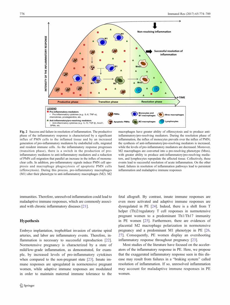
Fig. 2 Success and failure in resolution of inflammation. The productivephase of the inflammatory response is characterized by a significantinflux of PMN cells to the inflamed tissue and by an increasedgeneration of pro-inflammatory mediators by endothelial cells, migratedand resident immune cells. As the inflammatory response progresses(transition phase), there is a switch in the production of pro-inflammatory mediators to anti-inflammatory mediators and a reductionof PMN cell migration that parallel an increase in the influx of mononu-clear cells. In addition, pro-inflammatory signals induce PMN cell apo-ptosis and macrophage phagocytosis of apoptotic PMN cells(efferocytosis). During this process, pro-inflammatory macrophages(M1) alter their phenotype to anti-inflammatory macrophages (M2). M2
macrophages have greater ability of efferocytosis and to produce anti-inflammatory/pro-resolving mediators. During the resolution phase ofinflammation, the influx of monocytes prevails over the influx of PMN;the synthesis of anti-inflammatory/pro-resolving mediators is increased,while the levels of pro-inflammatory mediators are decreased. Moreover,M2 macrophages are converted into a pro-resolving phenotype (Mres),with greater ability to produce anti-inflammatory/pro-resolving media-tors, and lymphocytes repopulate the affected tissue. Collectively, theseevents lead to successful resolution of acute inflammation. On the otherhand, failures in resolution of inflammation pathways lead to persistentinflammation and maladaptive immune responses
776 Immunol Res (2017) 65:774–789

Methods
First, we performed a screening on PubMed database resultsthrough reading of titles and abstracts about pro-resolvingmediators previously described in general works [16, 18,19]. We used as key terms the specific pro-resolving mediatorand preeclampsia. The pro-resolving mediators that had beenstudied in the context of PE according to this research wereannexin A1, galectins, chemerin, lipoxin A4, nitric oxide, hy-drogen sulfide, carbon monoxide, acetylcholine, netrin-1, andprotease inhibitors. The final selection was based on full read-ing of each preselected article. Original research articles wereincluded if they addressed these pro-resolvingmediators in thecontext of PE (human disease and animal models). Originalarticles about pro-resolving mediators in other inflammatorydiseases and general review articles were also included toprovide a background on the role of these mediators.Articles that had not focused on these issues were excluded.
Results
This review included a total of 225 articles published between1985 and 2017. The main conclusions obtained from them aredescribed in the next subsections.
Annexin A1
Annexin A1 (AnxA1) is a 37-kDa glucocorticoid-regulatedprotein that elicits anti-inflammatory/pro-resolving effectsthrough binding to formyl peptide receptor type 2/lipoxinA4 receptor (FPR2/ALXR). These effects lie within AnxA1N-terminal domain and include inhibition of neutrophil migra-tion to inflamed tissues, induction of neutrophil apoptosis,stimulation of macrophage efferocytosis of apoptotic neutro-phils, and induction of macrophage reprogramming to a pro-resolving phenotype [28, 29]. AnxA1 was first recognized byits capacity to inhibit phospholipase A2 activity and the gen-eration of eicosanoids, but subsequent studies revealed thatthis protein exerts a wider range of actions. It has been sug-gested that AnxA1mediates part of neuroendocrine responsesof the glucocorticoids, particularly in the hypothalamic-pituitary-adrenocortical axis. In addition, experimental dataindicate that AnxA1 may be implicated in processes regulat-ing pregnancy, lactation, and fetal development [30, 31].
Altered AnxA1 synthesis might be involved in the patho-genesis of chronic inflammatory diseases, like asthma [32]. Ofimportance, intact AnxA1 (37 kDa) can be cleaved in its N-terminal domain by proteases, such as neutrophil elastase,generating various fragments that are believed to be inactiveor pro-inflammatory [33]. Indeed, increased levels of AnxA1cleavage products (e.g., 33 kDa) have been reported in inflam-matory samples [34]. Moreover, FPR2/ALXR decreased
expression or altered receptor conformation can impairAnxA1 to regulate inflammation [35, 36].
Previously, Perucci et al. investigated AnxA1 in PE andfound increased plasma levels when compared to normoten-sive pregnancy [37]. The increased concentration of AnxA1combined with an overwhelming inflammatory response sug-gests a failure in this resolution pathway in PE, which could bea consequence of decreased expression of FPR2/ALXR [38,39] or presence of anti-AnxA1 auto-antibodies [40].Considering that neutrophilia is a common feature in PEwomen and that neutrophil elastase is increased in their plas-ma and placenta [41–43], it is also plausible to hypothesizethat AnxA1 cleavage could interfere with its actions.Although AnxA1 expression has been studied in placentaltissues [44], the differential expression of its intact andcleaved forms in preeclamptic and normotensive pregnancieshas not been determined, a matter under investigation in ourgroup.
Galectins
Galectins are β-galactoside-binding proteins that were ini-tially known to mediate developmental processes, includ-ing tissue organization and embryo implantation [45].Further research indicated that galectins are secreted inresponse to inflammatory signals and cellular damage,acting as pattern recognition receptors, immunomodula-tors, or damage-associated molecular patterns in innateand adaptive immune responses [46, 47]. Galectins arethought to modulate intracellular signaling pathways inimmune cells due to their ability to induce the aggregationof specific cell-surface glycoreceptors [48, 49]. Thereby,galectins may elicit pro-resolving effects, as described be-low. Emerging evidences also suggest that galectins arecapable of triggering platelet activation and inducing an-giogenesis [50, 51]. Here, we give a general overview onthe role of galectin-1 (Gal-1) and galectin-13 (Gal-13), themost studied galectins in PE.
Galectin-1
It has been suggested that Gal-1 plays a role in maternal-fetal tolerance, which is thought to be impaired in PEwomen. Blois et al. reported that Gal-1 deficient(LGALS1−/−) mice had increased fetal loss when comparedto wild-type mice, an effect that was prevented by thetreatment with recombinant Gal-1. According to their re-sults, Gal-1 restored maternal immune tolerance by pro-moting the expansion of IL-10-secreting regulatory T cells[52]. This data was corroborated by the study of van derLeij et al. [53]. Gal-1 might also improve maternal-fetaltolerance by inducing the apoptosis of activated CD8+ Tcells, Th1 cells, and Th17 CD4+ cells [54]. Other
Immunol Res (2017) 65:774–789 777
immunomodulatory actions of Gal-1 have been proposed.For instance, Rostoker et al. showed that Gal-1 induced 12/15-lipoxygenase expression (lipoxin A4 synthetizing en-zyme; see the BLipoxin A4^ section) in murine macro-phages and promoted their conversion into a pro-resolving phenotype [55].
Some works have demonstrated that the gene expression ofGal-1 was upregulated in placentas from PEwomen comparedwith normotensive pregnant women [56–58]. Interestingly,LGALS1-knockout dams develop PE symptoms. However,when stratifying PE women according to the onset of clinicalsymptoms, early PE women showed lower placental expres-sion of Gal-1 than pregnant controls, while an opposite find-ing was reported for late PE women [58]. It has been proposedthat the decreased expression of Gal-1 in early PE could beassociated with placental dysfunction, whereas its overexpres-sion might be a compensatory mechanism to attenuate inflam-mation in late PE [59]. In addition, the circulating levels ofGal-1 may reflect its placental expression in late PE, but not inearly PE. Accordingly, Freitag et al. reported increased serumlevels of Gal-1 in late PE when compared with early PE andnormotensive pregnancies, but no difference was found be-tween early PE women and normotensive women [58].However, when both clinical forms were included in the samecohort, the serum levels of Gal-1 seemed to be similar be-tween patients and controls [60]. Pregnant women in the sec-ond trimester of pregnancy who developed PE also showedlower levels of Gal-1 than healthy pregnant women, indicatingthat Gal-1 might be an early predictor of PE [58].
Gal-1 seems to be differentially expressed in cells/tissuesfromwomen with PE. Gal-1 is downregulated in Tand naturalkiller cells in PE when compared with these cells from nor-motensive pregnancy, while no difference was detected inGal-1 messenger RNA (mRNA) expression in decidual sam-ples between these pregnant groups [58, 60]. The decreasedexpression of Gal-1 expression in these immune cells can beassociated with maternal-fetal intolerance and exacerbated in-flammatory response in PE women, as discussed above.
Galectin-13
Gal-13 is a galectin uniquely expressed in the placenta, mainlyin the syncytiotrophoblast, and it is released from the placentainto the maternal circulation [61]. In vitro studies suggest thatGal-13 participates in the morphological differentiation of thecytotrophoblast into the syncytiotrophoblast [62, 63]. In addi-tion, it has been demonstrated that Gal-13 is able to induce theapoptosis of activated human CD3+ T cells [64]. Interestingly,phagocytosed Gal-13 immunopositive deposits in immunecells coincided with zones of apoptotic and necrotic immunecells in Kliman et al. study [65]. These data indicate that Gal-13 might participate in placentation and in maternal adaptiveimmune responses at the maternal-fetal interface. Considering
that these processes are impaired in PE women, it can behypothesized that Gal-13 is involved in the pathogenesis ofthe disease.
Gal-13 placental-specific expression makes it a promisingbiomarker for PE early prediction. Indeed, Gal-13 protein andmRNA content in blood and placenta are decreased in the firsttrimester of gestation in women who developed PE, especiallyin the early clinical form [66–71], and this could be associatedwith single-nucleotide polymorphisms in the LGALS13 gene[72]. Moreover, combining Gal-13 with background risk fac-tors, other serum biomarkers and physical parameters increasethe accuracy of predicting PE [73]. Low serum levels of Gal-13 in early gestation may lead to impaired placentation andmaternal immune intolerance to the fetus [65, 74].
It has been demonstrated that the serum levels of Gal-13increase throughout normotensive pregnancy and that pretermPE women have higher serum levels of Gal-13 than pretermcontrols [66]. It was proposed that the increase in maternalserum concentration of Gal-13 during the third trimester ofgestation in PE women is a consequence of augmented pla-cental shedding of microvesicles containing Gal-13, and thiscould be a compensatory mechanism aiming to restore ho-meostasis [66]. Nevertheless, both decreased and increasedexpressions of placental Gal-13 have been reported in PEwomen [66, 75].
Chemerin
Chemerin is an adipocyte-secreted protein originally identi-fied as the natural ligand of chemR23 receptor, which is im-plicated in several biological processes, such as adipogenesis,glucose homeostasis, and immune cell migration [76]. It hasbeen suggested that chemerin is abundantly expressed in stro-mal cells and in extravillous trophoblast cells, but not in de-cidual endothelial cells in early pregnancy [77]. Moreover,chemerin may stimulate angiogenesis and the accumulationof natural killer cells at maternal-fetal interface, and theseimmune cells have been implicated in uterine spiral arteryremodeling [77–79]. Thus, chemerin can be involved in pla-cental development, which is impaired in PE women.Chemerin also acts as a chemoattractant for dendritic cells[80]. Several lines of evidence indicate crucial roles for bothnatural killer and dendritic cells in the modulation of adaptiveimmune responses [81, 82]. Based on these data, it can beadmitted that chemerin may contribute to maternal-fetal toler-ance, but more studies are necessary to clarify this issue.
Fragments with distinct inflammatory actions can be gen-erated after chemerin C-terminal proteolytic processing, de-pending on the types of proteases predominating in the micro-environment [80, 83]. Some chemerin fragments can inducethe chemotaxis of immune cells, in particular dendritic cells,macrophages, and natural killer cells, toward inflammatorysites, thus contributing to the onset of inflammation. By
778 Immunol Res (2017) 65:774–789

contrast, other fragments can inhibit the synthesis of pro-inflammatory mediators. In addition, the activation of thechemerin/chemR23 axis may increase the non-phlogisticphagocytosis of apoptotic cells by macrophages, and inhibitneutrophil activation and influx to inflammatory sites, thuspromoting the resolution of inflammation. Therefore,chemerin-derived peptides may play a role both in initiationand in resolution of the inflammatory response [80].
It has been shown that the serum levels of chemerin in-crease throughout normotensive pregnancy [84, 85]. Somestudies have reported increased circulating levels of chemerin,as well as increased mRNA and protein expressions in pla-centas from PEwomenwhen compared to normotensive preg-nant women [86–88]. Higher levels of chemerin were detectedin the first trimester of gestation in women who developed PE,were associated with disease severity, and remained signifi-cantly higher 6 months after delivery in former PE womencompared with controls [87–89]. Moreover, there is a positivecorrelation among chemerin levels, pro-inflammatory media-tors, and blood pressure [86–89]. However, these studies didnot specify the types of chemerin-derived peptides quantified.Hence, their role in PE pathogenesis remains unclear.
Specialized pro-resolving lipid mediators
The polyunsaturated fatty acids omega-6 and -3 are substratesfor the biosynthesis of lipoxins (LXs), maresins, resolvins,and protectins, which are collectively called specialized pro-resolving lipid mediators (SPMs). Prostaglandins and leuko-trienes are lipid mediators that play pivotal roles in the initia-tion of the inflammatory response, while SPMs attenuate in-flammation and contribute to its timely resolution [90].Curiously, aspirin induces the endogenous synthesis of LX15-epimers [91]. Endogenous LXs and their epimers havebeen shown to counter-regulate inflammation in a variety ofexperimental models of inflammatory diseases. They down-regulate pro-inflammatory mediators’ synthesis (includingprostaglandins and leukotrienes), inhibit neutrophil infiltra-tion, induce macrophage efferocytosis of apoptotic neutro-phils, and stimulate interleukin (IL)-10 production [90, 92].Furthermore, LXs may modulate other biological actions,such as angiogenesis, airway smooth muscle function, andactivity of neuronal ion channels that convey nociceptive sig-nals [93–95]. In this sense, LXs and other SPMs may contrib-ute to resolution of both inflammation and pain [94].
Lipoxin A4
Lipoxin A4 (LXA4) is an eicosanoid synthesized from arachi-donic acid, an omega-6 derivate, through the metabolism oflipoxygenase enzymes [96]. LXA4 interacts with FPR2/ALXR receptor, which also binds to AnxA1 [97]. Anin vitro study showed that LXA4 inhibited the production of
IL-1β by monocytes from severe PE women in a dose-dependent manner [98]. In another experiment, 15-epi-LXA4 reduced neutrophil-endothelium cell adhesion trig-gered by PE plasma [99]. Lin et al. administrated an LXA4analogue in low-dose-endotoxin-treated pregnant rats andfound that it attenuated inflammation and PE symptoms[100]. These experimental data suggest protective roles forLXA4 and its analogues in the disease.
Three works showed higher circulating levels of LXA4 inPE women compared to normotensive pregnant women [39,101, 102]. However, an opposite finding has also been report-ed [38]. Different studied populations or methodologies toquantify LXA4 might have contributed to these divergent re-sults. Interestingly, LXA4 plasma levels correlated with ma-ternal blood pressure, white blood cell count, and C-reactiveprotein levels in Perucci et al. study [102]. Similar to AnxA1discussion, LXA4 inefficiency to resolve inflammation couldbe a consequence, for example, of the decreased expression ofFPR2/ALXR and/or increased LXA4 inactivation. However,these hypotheses remain to be investigated.
Gaseous mediators
Nitric oxide, hydrogen sulfide, and carbon monoxide are themost studied gaseous mediators, and, for many years, onlytheir toxicity was known [103]. Recently, they have been im-plicated in key physiological functions, such as angiogenesis,inflammation, and vascular tone regulation [104, 105]. Theyalso participate in trophoblast invasion and in spiral arteryremodeling [106]. Experimental studies have demonstratedthat these gases act as anti-inflammatory mediators at lowconcentrations, promoting resolution of inflammation, but ex-ert pro-inflammatory and damaging effects at high concentra-tions [12]. In line with this data, altered production or signal-ing of gaseous mediators has been reported in inflammatorydiseases, like atherosclerosis and arthritis [107, 108].
Nitric oxide
Nitric oxide (NO) is synthesized by the conversion of L-arginine to L-citrulline by one of the following three isoformsof nitric oxide synthase (NOS): neuronal, endothelial (eNOS),or inducible (iNOS). NO acts as a vasodilatory molecule byinducing cyclic guanosine monophosphate (cGMP) synthesis[109]. However, NO can act by cGMP-independent pathwaysto regulate other mechanisms, such as leukocyte apoptosis[110, 111]. NO may have pro- or anti-inflammatory actionsdepending on the concentration used in the experiment, thedelivery method, and the system/disease model studied [112].Low amounts of NO inhibit the synthesis of pro-inflammatorycytokines and reduce leukocyte-endothelium adhesion andtransmigration to inflamed tissues, while high levels of NOincrease vascular permeability and leukocyte migration [113,
Immunol Res (2017) 65:774–789 779

114]. NO might also play a role in resolution of inflammationsince it induces neutrophil, but not macrophage, apoptosis[115].
In normal pregnancy, NO and cGMP biosynthesis are in-creased due to eNOS upregulation. In addition, the biosynthe-sis of asymmetrical dimethylarginine (ADMA), a competitiveinhibitor of NOS, is reduced. These events are important toregulate peripheral and placental bed vascular resistances, an-giogenesis, platelet adhesion/aggregation, and trophoblast in-vasion [116]. On the other hand, most studies have reporteddecreased activity of placental iNOS and eNOS and increasedlevels of ADMA in PE, but data on NO levels are inconsistent[117–119]. Moreover, increased levels of ADMA in the firsttrimester of pregnancy may predict PE [120]. Additionally,ADMA increase seems to be more prominent in early severePE than in late severe PE and eNOS polymorphisms mayinfluence the onset time of the disease [121, 122].
Other mechanisms may interfere with NO signaling in PE.It has been demonstrated that polymorphisms in the transcrip-tion factor STOX1 gene are associated with maternal suscep-tibility to PE and that the overexpression of placental STOX1induces a PE-like syndrome in mice [123, 124]. According tothe study of Doridot et al., placentas overexpressing STOX1showed high concentration of reactive nitrogen species(RNS). They proposed that RNS could be rapidly generatedin the placenta through the association of NO with reactiveoxygen species (ROS) [125]. Therefore, the overexpression ofSTOX1 could decreaseNO bioavailability in endothelial cells,preventing the protective actions of this gaseous mediator invascular tone and inflammation and also contributing to oxi-dative and nitrosative stress in PE women [11]. Further evi-dence suggested a risk allele (Y153H) in STOX1 gene thatmight be associated with a less invasive trophoblast pheno-type [126]. Accordingly, a previous study showed that theability of trophoblasts to remodel uteroplacental arteriesdepended on NO produced by extravillous trophoblasts inguinea pig pregnancy [127].
Considering that NO interferes with several pathways thatare known to be compromised in PE women, such as vasculartone, inflammation, and oxidative/anti-oxidative status, im-paired bioavailability and/or action of this gaseous mediatormay be associated with the pathogenesis of the disease. In thissense, the therapeutical potential of drugs that enhance NOavailability, inhibit cGMP degradation, or reduce ADMAlevels has been investigated in vitro and in experimentalmodels of PE. PE was mimicked by inducing reduced uterineperfusion pressure (RUPP) model and the overexpression ofthe anti-angiogenic molecule soluble fms-like tyrosine kinase(sFlt1) and by administrating the NOS inhibitor NG-nitro-L-arginine methyl ester (L-NAME) in rodents [128]. Althoughsome of these findings seem promising, there is insufficientclinical evidence to use these drugs for PE treatment or pre-vention [128, 129].
Hydrogen sulfide
Endogenous hydrogen sulfide (H2S) is primarily synthetized bythe conversion of L-cysteine or homocysteine by two enzymes,which are cystathionine β-synthase (CBS) and cystathionine γ-lyase (CSE) [130]. H2S is a vasodilatory mediator, an effect thatcan be mediated through eNOS activation and NO production[131]. H2S exert anti-oxidant effects on cells and it seems to becytoprotective at low concentrations, while higher H2S exposurefavors oxidative stress and cell apoptosis [132]. As for NO, H2Srole in inflammation is complex and not fully elucidated. H2Smight act as a pro-inflammatory mediator, as demonstrated inexperimental sepsis [133, 134], or as an anti-inflammatory/pro-resolvingmolecule, for example, in gastrointestinal inflammation[135]. Evidences suggest that H2S promotes resolution of inflam-mation by inducing neutrophil apoptosis, M2macrophage polar-ization, and clearance of apoptotic neutrophils by macrophages[136–138]. Recently, it has been suggested that part of H2S anti-inflammatory/pro-resolving effects are mediated by AnxA1[139].
H2S has been implicated in placental vascular developmentand function due to its pro-angiogenic and vasodilatory activ-ities [131, 140]. Both H2S-synthetizing enzymes CBS andCSE are expressed in human placenta during normal pregnan-cies [141], but the studies on their expression in PE haveconflicting results. Wang et al. reported a downregulation ofCSE (mRNA and protein) in placentas from PEwomen [142].Moreover, placentas with abnormal Doppler have increasedexpression of microRNA-21, which negatively regulates CSEexpression [143]. By contrast, in Holwerda et al. study,mRNA levels of CSE in the placenta were unchanged, whilemRNA levels of CBSwere decreased in early PE [144]. Thesedivergent results might be attributed to differences in the stud-ied clinical forms of PE [128]. Furthermore, Wang et al. re-ported decreased plasma levels of H2S in PE women [142].Abnormal synthesis of H2S could contribute to endothelialdysfunction, oxidative stress, and overwhelming inflamma-tion observed in PE women [128].
H2S-based therapies have been studied in animal modelsof PE and in human disease. The administration of a slow-releasing H2S-generating compound (GYY4137) ameliorat-ed PE-like symptoms induced by the treatment with aninhibitor of H2S synthesis (DL-propargylglycine) [142].However, oral administration of an H2S donor (N-acetylcysteine) to severe early PE women did not improvematernal outcomes [145]. More studies should be conduct-ed in order to evaluate the therapeutical potential of H2S-releasing compounds in PE.
Carbon monoxide
Heme oxygenase (HO) enzymes convert heme to biliverdin,free iron, and carbon monoxide (CO) in the endoplasmatic
780 Immunol Res (2017) 65:774–789

reticulum. HO enzymes exist as inducible (HO-1) and consti-tutive (HO-2) isoforms [146]. HO-CO system regulatesmany biological processes, such as vascular tone, oxidant-anti-oxidant status, and platelet aggregation. Further, CO actsas a signaling molecule in the neuronal system, where it reg-ulates the release of neurotransmitters. Like NO and H2S, COis toxic at high concentrations but has cytoprotective actions atlow concentrations [147, 148]. It has been suggested that partof the protective and deleterious effects of CO are due to itsability to regulate different types of ion channels [149]. Moststudies have reported counter-regulatory actions for CO ininflammatory responses. Low CO exposure inhibitsneutrophil-endothelial adhesion and transmigration to in-flamed tissues, suppresses the production of pro-inflammatory cytokines, promotes neutrophil apoptosis, andenhances macrophage efferocytosis of apoptotic neutrophils[150–153]. Moreover, CO accelerates resolution of inflamma-tion by shifting the lipid profile in the inflammatory milieu[153].
Both HO-1 and HO-2 are expressed in human pla-centa [154]. During pregnancy, CO regulates perfusionand oxidant-anti-oxidant status within placental tissues,as well as spiral artery transformation [154–156].Adequate expression of HO might also be important tomaintain maternal-fetal tolerance [157]. Considering theimportance of CO in regulating multiple processes dur-ing pregnancy, alterations in the HO-CO system in PEwould be expected. Indeed, PE women seem to havelower CO breath levels and carboxyhemoglobin concen-tration in the umbilical cord blood than normotensivepregnant women [158, 159]. These data are in line withthe observation that CO exposure in cigarette smokedecreases the risk of developing PE [160, 161].However, HO placental expression during PE is notclear. Either decreased, increased, or unchanged, placen-tal expressions of HO-1 and HO-2 have been reportedin PE women [162–166].
Compounds that induce HO expression have beenstudied in experimental and in human PE [128]. Arecent work showed that pravastatin treatment stabi-lized blood pressure, proteinuria, and serum uric acidlevels in severe PE women [167]. These effects seemto be partially mediated by upregulating HO-1 placen-ta l express ion [168] . McCar thy et a l . s tudiedrosiglitazone effects using the RUPP rat model of PEand found that this drug prevented the development ofdisease-like symptoms via HO-dependent pathway[169]. Moreover, CO applicat ion at low dosesprevented hypertension and proteinuria in adenovirussFlt1 PE-like mouse model [170]. In conclusion, theadministration of the CO or HO-inducing agents mightbe beneficial for treating or preventing PE, but furtherinvestigation is necessary.
Neuromodulators
Acetylcholine
Cholinergic neurons release acetylcholine (ACh), a neu-rotransmitter known to regulate skeletal, smooth, andcardiac muscle contract ions. ACh also acts asneuromodulator in the central nervous system, where italters neural excitability, synaptic transmission, andplasticity, thus interfering with learning, memory, andmood [171, 172]. ACh can induce endothelium vasodi-latation through NO-dependent and independent mecha-nisms, for example, by inducing the production of pros-taglandins [173]. Indeed, it has been demonstrated thatpharmacological administration of ACh reduces bloodpressure in rats [174]. These data suggest that Ach-deficient synthesis or action may be associated withPE pathogenesis.
Studies on the role of neural reflexes in inflammationand immunity are recent. It has been demonstrated thatACh binding to α7-nicotinic receptors in macrophages in-hibits the synthesis and the release of pro-inflammatorycytokines [175–178]. Alternative anti-inflammatory cholin-ergic mechanisms have been proposed. For instance, nico-tine (a cholinergic agonist drug) attenuates inflammationby upregulating the expression of HO-1 in macrophages[179, 180]. Other anti-inflammatory and pro-resolving ef-fects of nicotine include inhibition of neutrophil migrationand stimulation of its apoptosis [181, 182]. Moreover,ACh receptor activation by nicotine enhances macrophagephagocytosis and protects M2 macrophages from apoptosis[183, 184]. The role of this neural pathway in controllinginflammatory responses was further confirmed by studiesshowing that vagus nerve lesions enhance pro-inflammatory cytokines’ production and are associatedwith non-resolving inflammation [176, 185, 186].Accordingly, chronic inflammatory conditions, such as in-flammatory bowel disease, have decreased vagus nervefunction [176].
Yang et al. reported reduced vagus nerve function in PEwomen [187]. Thus, Ach-reduced synthesis in these womenmight contribute to excessive inflammation and hypertension.Accordingly, nicotine binding to ACh receptor supressesex vivo placental cytokines’ production [188]. In a recentstudy, nicotine was able to reduce systolic blood pressure inLPS-induced PE rat model [189]. This effect might be associ-ated with nicotine protective effects on the endothelium, aspreviously demonstrated by Mimura et al. [190]. These datacorroborate with the theory that nicotine, and also CO, incigarette smoke might protect from PE [160, 161]. Some stud-ies have also shown that nicotinic ACh receptors are upregu-lated in PE women [191, 192], which could be a compensa-tory mechanism to decreased ACh levels.
Immunol Res (2017) 65:774–789 781

Netrin-1
Netrin-1 was originally described as a laminin-related proteinthat guides axonal trajectories during the development of cen-tral nervous system, by repulsing/abolishing the attraction ofneuronal cells expressing the UNC5b receptor [193].Subsequently, it was implicated in the regulation of variousbiological processes, including angiogenesis and, recently, in-flammation. Netrin-1 suppresses neutrophil trafficking, prob-ably as consequence of the strong expression of UNC5b re-ceptor in these cells [194, 195]. It also inhibits prostaglandinE2 synthesis, regulates Th1/Th2/Th17 cytokines’ production,induces M2 polarization, increases apoptotic polymorphonu-clear (PMN) cell efferocytosis, and stimulates the endogenousbiosynthesis of SPMs [196–199]. In accordance, in vivo stud-ies have reported protective functions of netrin-1 in inflamma-tory conditions [199–202]. Interestingly, Mirakaj et al. foundthat netrin-1 stimulated the resolution of peritoneal inflamma-tion induced by zymosan via resolvin D1, a pro-resolving lipidmediator [186]. Yang et al. investigated the placental expres-sion of netrin-1 and found that it was downregulated in severePE women [203]. More studies are needed to understand theassociation between netrin-1 and inflammation in PE.
Protease inhibitors
Proteases are enzymes that hydrolyze peptide bonds of pro-teins, releasing polypeptides or free amino acids. They regu-late the activity and the localization of several proteins, mod-ulate the interactions among them, and participate in cellular
signaling events. Currently, proteases are classified based ontheir mechanisms of catalysis into the following four classes:serine proteases, metalloproteases, aspartic proteases, and cys-teine proteases. Their activities are tempered by protease in-hibitors or anti-proteases [204, 205]. Proteases are usuallyupregulated in inflammatory conditions, and defective anti-proteolytic control mechanisms may participate in the patho-genesis of chronic inflammatory diseases, like cystic fibrosis[206, 207]. Thus, protease inhibitors have the potential to bedeveloped as new therapeutic agents for these diseases.
Metalloproteinase inhibitors
Metalloproteinases are proteolytic enzymes that hydrolyze ex-tracellular matrix components, playing important roles on tis-sue repair. They participate in extracellular matrix remodelingduring trophoblast invasion and in uterine spiral artery trans-formation. This family of enzymes comprises, among othermembers, matrix metalloproteinases (MMPs) and membrane-anchored disintegrin metalloproteinases (ADAMs) [208,209]. Activated metalloproteinases can be regulated by gen-eral or specific protease inhibitors (tissue inhibitors of metal-loproteinases (TIMPs)) [210].
Several non-matrix substrates for metalloproteinases havebeen identified, including cytokines, chemokines, and theirreceptors. Metalloproteinases cleave these substrates in shortfragments, altering their bioactions, and, in the case of recep-tors, interfering with their responsiveness and downstreamsignaling. Metalloproteinases modulate additional aspects ofinflammation, such as integrity of physical barriers,
Non-pregnant women
Normotensive pregnant women
PE women
Intensity of the inflammatory response
Anti-inflammatory/proresolving mediators*
Pro-inflammatory mediators*
Resolution of inflammation mechanisms
Legend
Failure(s)
* This is a general overview. The levels of inflammatorymediators might be increased, decreased or unchangeddepending on their nature and physiological factors (e.g.maternal gestational age).
Fig. 3 Schematic representation of inflammatory and counter-regulatorymechanisms in non-pregnant women, normotensive pregnant women,and PE women. Healthy non-pregnant women have basal levels of anti-inflammatory/pro-resolving mediators and pro-inflammatory mediators,which are in a state of equilibrium due to functioning resolution of in-flammation mechanisms. Normotensive pregnant women show higherlevels of pro-inflammatory mediators than non-pregnant women, but the
inflammatory response is mild and controlled, because resolution of in-flammation mechanisms are able to adjust properly to this physiologicalstate (increased gear symbol). By contrast, failures in pro-resolvingmech-anisms probably lead to an exacerbated inflammatory response in PEwomen, despite the upregulation of some anti-inflammatory/pro-resolv-ing mediators
782 Immunol Res (2017) 65:774–789

leukocytes’ transmigration, and survival [211, 212].Metalloproteinases may have pro-inflammatory or anti-in-flammatory/pro-resolving actions. For instance, ADAM17,also known as tumor necrosis factor alpha (TNF-α)converting enzyme, releases the membrane-bound TNF-α,increasing the bioavailability of this pro-inflammatory cyto-kine. By contrast, ADAM17 sheds TNF-α soluble receptors(sTNFRs) in the circulation, which sequester TNF-α, neutral-izing its systemic effects [213]. ADAM17 also prevents neu-trophil transmigration trough the endothelium by shedding L-selectin from them, without altering monocyte recruitment. Inaddition, ADAM17 induces neutrophil apoptosis [214, 215].
There is increasing evidence of metalloproteinase/TIMPimbalance in inflammatory diseases, like inflammatory boweldiseases [216]. Metalloproteinases and their inhibitors mayalso participate in PE pathogenesis. Ma et al. reported thatADAM17 was upregulated in placentas from PE womenand induced TNF-α production by placental trophoblasts[217]. Later, they showed that the placental levels of TIMP3(ADAM17 inhibitor) were decreased in PE women and thatTIMP3 downregulation increased TNF-α production by pla-cental trophoblasts [218]. Further reports on increased circu-lating levels of TNF-α and sTNFRs in PE women corroborat-ed with these findings [219–221], since increased ADAM 17levels and decreased TIMP3 levels may induce TNF-α releaseand the consequent shedding of neutralizing sTNFR receptorsin the circulation of PE women. Decreased, increased, or sim-ilar levels of other metalloproteinases and TIMPs have beendescribed in PE [222]. These discrepancies are probably dueto differences in the types of specimens analyzed, gestationalage of specimen collection, and quantification methodologies.Therefore, the role of metalloproteinases and their inhibitorsin PE pathogenesis requires further investigation.
Concluding remarks
Several evidences support that there is a balance of pro-inflammatory and anti-inflammatory/pro-resolving pathways innormotensive pregnant women as consequence of functioningmechanisms of resolution of inflammation, leading to a state ofcontrolled inflammatory response in these women. On the otherhand, inflammation is overwhelming in PE women, probablybecause of dysregulated resolution of inflammation mechanisms(Fig. 3). Moreover, pro-inflammatory and anti-inflammatory/pro-resolving mediators from diverse nature might be at higher,lower, or similar levels in PE women compared with normoten-sive pregnant women, reinforcing the complex regulation of res-olution pathways.
The apparently contradictory findings regarding the mea-surement of pro-resolving mediators in PE can be a mirror ofthe biological sample tested (serum/plasma—systemic vs. pla-centa— local) and moment (onset vs. established
inflammation), in which such mediators were measured. It isknown that some of the pro-resolving mediators may havedual activities during the inflammatory response; i.e., theycan be pro-inflammatory at the beginning of inflammation toassure proper activation of the immunologic system and, asinflammation progresses, they can be pro-resolving, acting asbrakes for the inflammatory response. In addition, the activityof some mediators may be influenced by several factors, suchas molecule structure (e.g., AnxA1 cleavage generates shortpeptides believed to have pro-inflammatory activities), the celltype in which they act (e.g., LXA4 induces apoptosis of neu-trophils while rescue macrophage from death), or concentra-tion (e.g., NO and H2S have anti-inflammatory actions at lowconcentrations but pro-inflammatory actions at high concen-trations) [33, 113, 114, 133–135, 223, 224]. However, wheth-er these activities may be applied in the context of PE remainsto be determined.
There are few mechanistic studies in the literature for mostof the pro-resolving mediators described in this work in thecontext of PE. Further investigation about the role of pro-resolving mediators in PE pathogenesis is warranted, for ex-ample, using knockout animals and therapeutic strategies inanimal models. However, none of the available animal modelsof PE can mimic the full spectrum of the human disease [225].Prospective studies with standardized methodologies wouldalso be valuable to assess whether altered levels and/or actionsof pro-resolving mediators in PE women are causes or conse-quences of the disease. The knowledge acquired from thesestudies will provide a basis for future clinical trials about noveltherapies targeting pro-resolving mechanisms in PE.
Acknowledgments The authors are grateful to Fundação de Amparo àPesquisa do Estado de Minas Gerais (FAPEMIG), Conselho Nacional deDesenvolvimento Científico e Tecnológico (CNPq), and Coordenação deAperfeiçoamento de Pessoal de Nível Superior (CAPES) ResearchFellowship.
Compliance with ethical standards
Funding This study was funded by Fundação deAmparo à Pesquisa doEstado de Minas Gerais (FAPEMIG; APQ-03318-15), ConselhoNacional de Desenvolvimento Científico e Tecnológico (CNPq;Research Fellowship), and Coordenação de Aperfeiçoamento dePessoal de Nível Superior (CAPES; Ph.D. scholarship).
Conflict of interest The authors declare that they have no conflict ofinterest.
References
1. American College of Obstetricians and Gynecologists.Hypertension in pregnancy. Report of the American College ofObstetricians and Gynecologists’ Task Force on Hypertension inPregnancy. Obstet Gynecol. 2013;122(5):1122–31.
Immunol Res (2017) 65:774–789 783

2. Chaiworapongsa T, Chaemsaithong P, Yeo L, Romero R. Pre-eclampsia part 1: current understanding of its pathophysiology.Nat Rev Nephrol. 2014;10(8):466–80.
3. American College of Obstetricians and Gynecologists. ACOGpractice bulletin. Diagnosis and management of preeclampsiaand eclampsia. Number 33, January 2002. Obstet Gynecol.2002;99(1):159–67.
4. von Dadelszen P, Magee LA, Roberts JM. Subclassification ofpreeclampsia. Hypertens Pregnancy. 2003;22(2):143–8.
5. Steegers EA, von Dadelszen P, Duvekot JJ, Pijnenborg R. Pre-eclampsia. Lancet. 2010;376(9741):631–44.
6. Raymond D, Peterson E. A critical review of early-onset and late-onset preeclampsia. Obstet Gynecol Surv. 2011;66(8):497–506.
7. Roberts JM, Hubel CA. The two stage model of preeclampsia:variations on the theme. Placenta. 2009;30(Suppl A):S32–7.
8. Lam C, Lim KH, Karumanchi SA. Circulating angiogenic factorsin the pathogenesis and prediction of preeclampsia. Hypertension.2005;46(5):1077–85.
9. Ahmed A, Ramma W. Unravelling the theories of pre-eclampsia:are the protective pathways the new paradigm? Br J Pharmacol.2015;172(6):1574–86.
10. Cotechini T, Komisarenko M, Sperou A, Macdonald-GoodfellowS, Adams MA, Graham CH. Inflammation in rat pregnancy in-hibits spiral artery remodeling leading to fetal growth restrictionand features of preeclampsia. J Exp Med. 2014;211(1):165–79.
11. Matsubara K, Higaki T, Matsubara Y, Nawa A. Nitric oxide andreactive oxygen species in the pathogenesis of preeclampsia. Int JMol Sci. 2015;16(3):4600–14.
12. Wallace JL, Ianaro A, Flannigan KL, Cirino G. Gaseous mediatorsin resolution of inflammation. Semin Immunol. 2015;27(3):227–33.
13. Chovatiya R, Medzhitov R. Stress, inflammation, and defense ofhomeostasis. Mol Cell. 2014;54(2):281–8.
14. Serhan CN, Savill J. Resolution of inflammation: the beginningprograms the end. Nat Immunol. 2005;6(12):1191–7.
15. Serhan CN, Brain SD, Buckley CD, Gilroy DW, Haslett C,O’Neill LA, et al. Resolution of inflammation: state of the art,definitions and terms. FASEB J. 2007;21(2):325–32.
16. Headland SE, Norling LV. The resolution of inflammation: prin-ciples and challenges. Semin Immunol. 2015;27(3):149–60.
17. SugimotoM, Sousa L, PinhoV, Perretti M, TeixeiraM. Resolutionof inflammation: what controls its onset? Front Immunol.2016;160(7):1–18.
18. Vago JP, Tavares LP, Sugimoto MA, Lima GL, Galvão I, de CauxTR, et al. Proresolving actions of synthetic and natural proteaseinhibitors are mediated by Annexin A1. J Immunol. 2016;196(4):1922–32.
19. Odaka C, Mizuochi T, Yang J, Ding A. Murine macrophagesproduce secretory leukocyte protease inhibitor during clearanceof apoptotic cells: implications for resolution of the inflammatoryresponse. J Immunol. 2003;171(3):1507–14.
20. Titos E, Rius B, González-Périz A, López-Vicario C, Morán-Salvador E, Martínez-Clemente M, et al. Resolvin D1 and itsprecursor docosahexaenoic acid promote resolution of adiposetissue inflammation by eliciting macrophage polarization towardan M2-like phenotype. J Immunol. 2011;187(10):5408–18.
21. Fullerton JN, Gilroy DW. Resolution of inflammation: a new ther-apeutic frontier. Nat Rev Drug Discov. 2016;15(8):551–67.
22. Orsi NM. Cytokine networks in the establishment and mainte-nance of pregnancy. Hum Fertil (Camb). 2008;11(4):222–30.
23. Redman CW, Sacks GP, Sargent IL. Preeclampsia: an excessivematernal inflammatory response to pregnancy. Am J ObstetGynecol. 1999;180(2 Pt 1):499–506.
24. Schminkey DL, Groer M. Imitating a stress response: a new hy-pothesis about the innate immune system’s role in pregnancy.MedHypotheses. 2014;82(6):721–9.
25. Saito S, Nakashima A, Shima T, Ito M. Th1/Th2/Th17 and regu-latory T-cell paradigm in pregnancy. Am J Reprod Immunol.2010;63(6):601–10.
26. Li M, Piao L, Chen CP, Wu X, Yeh CC, Masch R, et al.Modulation of decidual macrophage polarization by macrophagecolony-stimulating factor derived from first-trimester decidualcells: implication in preeclampsia. Am J Pathol. 2016;186(5):1258–66.
27. Lee CL, Guo Y, So KH, Vijayan M, Wong VH, Yao Y, et al.Soluble human leukocyte antigen G5 polarizes differentiation ofmacrophages toward a decidual macrophage-like phenotype. HumReprod. 2015;30(10):2263–74.
28. Perretti M, D’Acquisto F. Annexin A1 and glucocorticoids aseffectors of the resolution of inflammation. Nat Rev Immunol.2009;9(1):62–70.
29. SugimotoMA, Vago JP, Teixeira MM, Sousa LP. Annexin A1 andthe resolution of inflammation: modulation of neutrophil recruit-ment, apoptosis, and clearance. J Immunol Res. 2016;2016:8239258.
30. John CD, Gavins FN, Buss NA, Cover PO, Buckingham JC.Annexin A1 and the formyl peptide receptor family: neuroendo-crine and metabolic aspects. Curr Opin Pharmacol. 2008;8(6):765–76.
31. John CD, Christian HC, Morris JF, Flower RJ, Solito E,Buckingham JC. Annexin 1 and the regulation of endocrine func-tion. Trends Endocrinol Metab. 2004;15(3):103–9.
32. Eke Gungor H, Tahan F, Gokahmetoglu S, Saraymen B.Decreased levels of lipoxin A4 and annexin A1 in wheezy infants.Int Arch Allergy Immunol. 2014;163(3):193–7.
33. Williams SL, Milne IR, Bagley CJ, Gamble JR, Vadas MA, PitsonSM, et al. A proinflammatory role for proteolytically cleavedannexin A1 in neutrophil transendothelial migration. J Immunol.2010;185(5):3057–63.
34. Tsao FH,Meyer KC, Chen X, Rosenthal NS, Hu J. Degradation ofannexin I in bronchoalveolar lavage fluid from patients with cysticfibrosis. Am J Respir Cell Mol Biol. 1998;18(1):120–8.
35. Cooray SN, Gobbetti T, Montero-Melendez T, McArthur S,Thompson D, Clark AJ, et al. Ligand-specific conformationalchange of the G-protein-coupled receptor ALX/FPR2 determinesproresolving functional responses. Proc Natl Acad Sci U S A.2013;110(45):18232–7.
36. Planagumà A, Kazani S, Marigowda G, Haworth O, Mariani TJ,Israel E, et al. Airway lipoxin A4 generation and lipoxin A4 re-ceptor expression are decreased in severe asthma. Am J RespirCrit Care Med. 2008;178(6):574–82.
37. Perucci LO, Carneiro FS, Ferreira CN, Sugimoto MA, SorianiFM, Martins GG, et al. Annexin A1 is increased in the plasmaof preeclamptic women. PLoS One. 2015;10(9):e0138475.
38. Xu Z, Zhao F, Lin F, Xiang H, Wang N, Ye D, et al. Preeclampsiais associated with a deficiency of lipoxin A4, an endogenous anti-inflammatory mediator. Fertil Steril. 2014;102(1):282–90.e4.
39. Dong W, Yin L. Expression of lipoxin A4, TNFα and IL-1β inmaternal peripheral blood, umbilical cord blood and placenta, andtheir significance in pre-eclampsia. Hypertens Pregnancy.2014;33(4):449–56.
40. Behrouz GF, Farzaneh GS, Leila J, Jaleh Z, Eskandar KS.Presence of auto-antibody against two placental proteins, annexinA1 and vitamin D binding protein, in sera of women with pre-eclampsia. J Reprod Immunol. 2013;99(1–2):10–6.
41. Canzoneri BJ, Lewis DF, Groome L, Wang Y. Increased neutro-phil numbers account for leukocytosis in women with preeclamp-sia. Am J Perinatol. 2009;26(10):729–32.
42. Gupta AK, Gebhardt S, Hillermann R, Holzgreve W, Hahn S.Analysis of plasma elastase levels in early and late onset pre-eclampsia. Arch Gynecol Obstet. 2006;273(4):239–42.
784 Immunol Res (2017) 65:774–789

43. Salama RH, Fathalla MM, Mekki AR, Elsadek B-K. Implicationof umbilical cord in preeclampsia. Med Princ Pract. 2011;20(2):124–8.
44. Sun M, Liu Y, Gibb W. Distribution of annexin I and II in termhuman fetal membranes, decidua and placenta. Placenta.1996;17(2–3):181–4.
45. Leffler H, Carlsson S, Hedlund M, Qian Y, Poirier F. Introductionto galectins. Glycoconj J. 2004;19(7–9):433–40.
46. Sato S, St-Pierre C, Bhaumik P, Nieminen J. Galectins in innateimmunity: dual functions of host soluble beta-galactoside-bindinglectins as damage-associated molecular patterns (DAMPs) and asreceptors for pathogen-associated molecular patterns (PAMPs).Immunol Rev. 2009;230(1):172–87.
47. Rubinstein N, Ilarregui JM, Toscano MA, Rabinovich GA. Therole of galectins in the initiation, amplification and resolution ofthe inflammatory response. Tissue Antigens. 2004;64(1):1–12.
48. Arikawa T, Simamura E, Shimada H, Nakamura T, Hatta T, ShojiH. Significance of sugar chain recognition by galectins and itsinvolvement in disease-associated glycosylation. CongenitAnom (Kyoto). 2014;54(2):77–81.
49. Vasta GR. Galectins as pattern recognition receptors: structure,function, and evolution. Adv Exp Med Biol. 2012;946:21–36.
50. Romaniuk MA, Negrotto S, Campetella O, Rabinovich GA,Schattner M. Identification of galectins as novel regulators ofplatelet signaling and function. IUBMB Life. 2011;63(7):521–7.
51. Blois SM, Conrad ML, Freitag N, Barrientos G. Galectins in an-giogenesis: consequences for gestation. J Reprod Immunol.2015;108:33–41.
52. Blois SM, Ilarregui JM, Tometten M, Garcia M, Orsal AS, Cordo-Russo R, et al. A pivotal role for galectin-1 in fetomaternal toler-ance. Nat Med. 2007;13(12):1450–7.
53. van der Leij J, van den Berg A, Blokzijl T, Harms G, van Goor H,Zwiers P, et al. Dimeric galectin-1 induces IL-10 production in T-lymphocytes: an important tool in the regulation of the immuneresponse. J Pathol. 2004;204(5):511–8.
54. KopcowHD, Rosetti F, Leung Y, Allan DS, Kutok JL, StromingerJL. T cell apoptosis at the maternal-fetal interface in early humanpregnancy, involvement of galectin-1. Proc Natl Acad Sci U S A.2008;105(47):18472–7.
55. Rostoker R, Yaseen H, Schif-Zuck S, Lichtenstein RG,Rabinovich GA, Ariel A. Galectin-1 induces 12/15-lipoxygenase expression in murine macrophages and favors theirconversion toward a pro-resolving phenotype. ProstaglandinsOther Lipid Mediat. 2013;107:85–94.
56. Jeschke U, Mayr D, Schiessl B, Mylonas I, Schulze S, Kuhn C,et al. Expression of galectin-1, −3 (gal-1, gal-3) and the Thomsen-Friedenreich (TF) antigen in normal, IUGR, preeclamptic andHELLP placentas. Placenta. 2007;28(11–12):1165–73.
57. Than NG, Erez O, Wildman DE, Tarca AL, Edwin SS, Abbas A,et al. Severe preeclampsia is characterized by increased placentalexpression of galectin-1. J Matern Fetal Neonatal Med.2008;21(7):429–42.
58. Freitag N, Tirado-González I, Barrientos G, Herse F, Thijssen VL,Weedon-Fekjær SM, et al. Interfering with Gal-1-mediated angio-genesis contributes to the pathogenesis of preeclampsia. Proc NatlAcad Sci U S A. 2013;110(28):11451–6.
59. Blois SM, Dechend R, Barrientos G, Staff AC. A potential path-ophysiological role for galectins and the renin-angiotensin systemin preeclampsia. Cell Mol Life Sci. 2015;72(1):39–50.
60. Molvarec A, Blois SM, Stenczer B, Toldi G, Tirado-Gonzalez I,Ito M, et al. Peripheral blood galectin-1-expressing T and naturalkiller cells in normal pregnancy and preeclampsia. Clin Immunol.2011;139(1):48–56.
61. Than NG, Pick E, Bellyei S, Szigeti A, Burger O, Berente Z, et al.Functional analyses of placental protein 13/galectin-13. Eur JBiochem. 2004;271(6):1065–78.
62. Orendi K, Gauster M, Moser G, Meiri H, Huppertz B. The cho-riocarcinoma cell line BeWo: syncytial fusion and expression ofsyncytium-specific proteins. Reproduction. 2010;140(5):759–66.
63. Than NG, Romero R, Xu Y, Erez O, Xu Z, Bhatti G, et al.Evolutionary origins of the placental expression of chromosome19 cluster galectins and their complex dysregulation in preeclamp-sia. Placenta. 2014;35(11):855–65.
64. Than NG, Romero R, Goodman M, Weckle A, Xing J, Dong Z,et al. A primate subfamily of galectins expressed at the maternal-fetal interface that promote immune cell death. Proc Natl Acad SciU S A. 2009;106(24):9731–6.
65. Kliman HJ, SammarM, Grimpel YI, Lynch SK,Milano KM, PickE, et al. Placental protein 13 and decidual zones of necrosis: animmunologic diversion that may be linked to preeclampsia.Reprod Sci. 2012;19(1):16–30.
66. Than NG, Abdul Rahman O, Magenheim R, Nagy B, Fule T,Hargitai B, et al. Placental protein 13 (galectin-13) has decreasedplacental expression but increased shedding and maternal serumconcentrations in patients presenting with preterm pre-eclampsiaand HELLP syndrome. Virchows Arch. 2008;453(4):387–400.
67. Huppertz B, Sammar M, Chefetz I, Neumaier-Wagner P, Bartz C,Meiri H. Longitudinal determination of serum placental protein 13during development of preeclampsia. Fetal Diagn Ther.2008;24(3):230–6.
68. Gonen R, Shahar R, Grimpel YI, Chefetz I, Sammar M, Meiri H,et al. Placental protein 13 as an early marker for pre-eclampsia: aprospective longitudinal study. BJOG. 2008;115(12):1465–72.
69. Sekizawa A, Purwosunu Y, Yoshimura S, Nakamura M, ShimizuH, Okai T, et al. PP13 mRNA expression in trophoblasts frompreeclamptic placentas. Reprod Sci. 2009;16(4):408–13.
70. Shimizu H, Sekizawa A, Purwosunu Y, Nakamura M, Farina A,Rizzo N, et al. PP13 mRNA expression in the cellular componentof maternal blood as a marker for preeclampsia. Prenat Diagn.2009;29(13):1231–6.
71. Farina A, Zucchini C, Sekizawa A, Purwosunu Y, de Sanctis P,Santarsiero G, et al. Performance of messenger RNAs circulatingin maternal blood in the prediction of preeclampsia at 10–14 weeks. Am J Obstet Gynecol. 2010;203(6):575.e1–7.
72. Gebhardt S, Bruiners N, Hillermann R. A novel exonic variant(221delT) in the LGALS13 gene encoding placental protein 13(PP13) is associatedwith preterm labour in a low risk population. JReprod Immunol. 2009;82(2):166–73.
73. Than NG, Balogh A, Romero R, Kárpáti E, Erez O, Szilágyi A,et al. Placental protein 13 (PP13)—a placental Immunoregulatorygalectin protecting pregnancy. Front Immunol. 2014;5:348.
74. Huppertz B,Meiri H, Gizurarson S, Osol G, Sammar M. Placentalprotein 13 (PP13): a new biological target shifting individualizedrisk assessment to personalized drug design combating pre-eclampsia. Hum Reprod Update. 2013;19(4):391–405.
75. Sammar M, Nisemblat S, Fleischfarb Z, Golan A, Sadan O, MeiriH, et al. Placenta-bound and body fluid PP13 and its mRNA innormal pregnancy compared to preeclampsia, HELLP and pre-term delivery. Placenta. 2011;32(Suppl):S30–6.
76. Zabel BA, Kwitniewski M, Banas M, Zabieglo K, Murzyn K,Cichy J. Chemerin regulation and role in host defense. Am JClin Exp Immunol. 2014;3(1):1–19.
77. Carlino C, Trotta E, Stabile H, Morrone S, Bulla R, Soriani A,et al. Chemerin regulates NK cell accumulation and endothelialcell morphogenesis in the decidua during early pregnancy. J ClinEndocrinol Metab. 2012;97(10):3603–12.
78. Tessier DR, Yockell-Lelievre J, Gruslin A. Uterine spiral arteryremodeling: the role of uterine natural killer cells and extravilloustrophoblasts in normal and high-risk human pregnancies. Am JReprod Immunol. 2015;74(1):1–11.
79. Kaur J, Adya R, Tan BK, Chen J, Randeva HS. Identification ofchemerin receptor (ChemR23) in human endothelial cells:
Immunol Res (2017) 65:774–789 785

chemerin-induced endothelial angiogenesis. Biochem BiophysRes Commun. 2010;391(4):1762–8.
80. Mariani F, Roncucci L. Chemerin/chemR23 axis in inflammationonset and resolution. Inflamm Res. 2015;64(2):85–95.
81. Moretta A, Marcenaro E, Parolini S, Ferlazzo G, Moretta L. NKcells at the interface between innate and adaptive immunity. CellDeath Differ. 2008;15(2):226–33.
82. Hespel C,MoserM. Role of inflammatory dendritic cells in innateand adaptive immunity. Eur J Immunol. 2012;42(10):2535–43.
83. Zabel BA, Allen SJ, Kulig P, Allen JA, Cichy J, Handel TM, et al.Chemerin activation by serine proteases of the coagulation, fibri-nolytic, and inflammatory cascades. J Biol Chem. 2005;280(41):34661–6.
84. Garces MF, Sanchez E, Ruiz-Parra AI, Rubio-Romero JA, Angel-Muller E, Suarez MA, et al. Serum chemerin levels during normalhuman pregnancy. Peptides. 2013;42:138–43.
85. Kasher-MeronM,Mazaki-Tovi S, Barhod E, Hemi R, Haas J, GatI, et al. Chemerin concentrations in maternal and fetal compart-ments: implications for metabolic adaptations to normal humanpregnancy. J Perinat Med. 2014;42(3):371–8.
86. Wang L, Yang T, Ding Y, ZhongY, Yu L, PengM. Chemerin playsa protective role by regulating human umbilical vein endothelialcell-induced nitric oxide signaling in preeclampsia. Endocrine.2015;48(1):299–308.
87. Duan DM, Niu JM, Lei Q, Lin XH, Chen X. Serum levels of theadipokine chemerin in preeclampsia. J Perinat Med. 2012;40(2):121–7.
88. Stepan H, PhilippA, Roth I, Kralisch S, JankA, SchaarschmidtW,et al. Serum levels of the adipokine chemerin are increased inpreeclampsia during and 6 months after pregnancy. Regul Pept.2011;168(1–3):69–72.
89. Xu QL, Zhu M, Jin Y, Wang N, Xu HX, Quan LM, et al. Thepredictive value of the first-trimester maternal serum chemerinlevel for pre-eclampsia. Peptides. 2014;62:150–4.
90. Serhan CN. Pro-resolving lipid mediators are leads for resolutionphysiology. Nature. 2014;510(7503):92–101.
91 Claria J, Serhan CN. Aspirin triggers previously undescribed bio-active eicosanoids by human endothelial cell-leukocyte interac-tions. Proc Natl Acad Sci U S A. 1995;92(21):9475–9.
92. Serhan CN. Lipoxins and aspirin-triggered 15-epi-lipoxins are thefirst lipid mediators of endogenous anti-inflammation and resolu-tion. Prostaglandins Leukot Essent Fatty Acids. 2005;73(3–4):141–62.
93. Fierro IM. Angiogenesis and lipoxins. Prostaglandins LeukotEssent Fatty Acids. 2005;73(3–4):271–5.
94. Choi G, Hwang SW. Modulation of the activities of neuronal ionchannels by fatty acid-derived pro-resolvents. Front Physiol.2016;7:523.
95. Levy BD, Serhan CN. Exploring new approaches to the treatmentof asthma: potential roles for lipoxins and aspirin-triggered lipidmediators. Drugs Today (Barc). 2003;39(5):373–84.
96. Lee TH. Lipoxin A4: a novel anti-inflammatory molecule?Thorax. 1995;50(2):111–2.
97. Gavins FN, Sawmynaden P, Chatterjee BE, Perretti M. A twist inanti-inflammation: annexin 1 acts via the lipoxin A4 receptor.Prostaglandins Leukot Essent Fatty Acids. 2005;73(3–4):211–9.
98. Wang J, Huang Y, Zhou J, Liu X. Effect of lipoxin A4 on IL-1βproduction of monocytes and its possible mechanism in severepreeclampsia. J Huazhong Univ Sci Technolog Med Sci.2010;30(6):767–70.
99. Gil-Villa AM, Norling LV, Serhan CN, Cordero D, Rojas M,Cadavid A. Aspirin triggered-lipoxin A4 reduces the adhesion ofhuman polymorphonuclear neutrophils to endothelial cells initiat-ed by preeclamptic plasma. Prostaglandins Leukot Essent FattyAcids. 2012;87(4–5):127–34.
100. Lin F, Zeng P, Xu Z, Ye D, Yu X, Wang N, et al. Treatment oflipoxin a(4) and its analogue on low-dose endotoxin induced pre-eclampsia in rat and possible mechanisms. Reprod Toxicol.2012;34(4):677–85.
101. Huang LL, Su S, Awale R, Zhang XY, Zhong LL, Tang H.Expression of anti-inflammatory mediator lipoxin A4 and inflam-mation responsive transcriptive factors NF-kappa B in patientswith preeclampsia. Clin Exp Obstet Gynecol. 2014;41(5):561–6.
102. Perucci LO, Santos PC, Ribeiro LS, Souza DG, GomesKB, DusseLM, et al. Lipoxin A4 is increased in the plasma of preeclampticwomen. Am J Hypertens. 2016;29(10):1179–85.
103. Cooper CE, Brown GC. The inhibition of mitochondrial cyto-chrome oxidase by the gases carbon monoxide, nitric oxide, hy-drogen cyanide and hydrogen sulfide: chemical mechanism andphysiological significance. J Bioenerg Biomembr. 2008;40(5):533–9.
104. Wang R. Two’s company, three’s a crowd: can H2S be the thirdendogenous gaseous transmitter? FASEB J. 2002;16(13):1792–8.
105. KajimuraM, Fukuda R, BatemanRM, Yamamoto T, SuematsuM.Interactions of multiple gas-transducing systems: hallmarks anduncertainties of CO, NO, and H2S gas biology. Antioxid RedoxSignal. 2010;13(2):157–92.
106. Lyall F. Development of the utero-placental circulation: the role ofcarbon monoxide and nitric oxide in trophoblast invasion andspiral artery transformation. Microsc Res Tech. 2003;60(4):402–11.
107. Li H, Horke S, Förstermann U. Vascular oxidative stress, nitricoxide and atherosclerosis. Atherosclerosis. 2014;237(1):208–19.
108. Nagy G, Koncz A, Telarico T, Fernandez D, Ersek B, Buzás E,et al. Central role of nitric oxide in the pathogenesis of rheumatoidarthritis and systemic lupus erythematosus. Arthritis Res Ther.2010;12(3):210.
109. Ignarro LJ, Buga GM, Wood KS, Byrns RE, Chaudhuri G.Endothelium-derived relaxing factor produced and released fromartery and vein is nitric oxide. Proc Natl Acad Sci U S A.1987;84(24):9265–9.
110. SogoN,Magid KS, ShawCA,Webb DJ,Megson IL. Inhibition ofhuman platelet aggregation by nitric oxide donor drugs: relativecontribution of cGMP-independent mechanisms. BiochemBiophys Res Commun. 2000;279(2):412–9.
111. Ward C, Wong TH, Murray J, Rahman I, Haslett C, Chilvers ER,et al. Induction of human neutrophil apoptosis by nitric oxidedonors: evidence for a caspase-dependent, cyclic-GMP-indepen-dent, mechanism. Biochem Pharmacol. 2000;59(3):305–14.
112. Lo Faro ML, Fox B, Whatmore JL, Winyard PG, Whiteman M.Hydrogen sulfide and nitric oxide interactions in inflammation.Nitric Oxide. 2014;41:38–47.
113. Laroux FS, Lefer DJ, Kawachi S, Scalia R, Cockrell AS, Gray L,et al. Role of nitric oxide in the regulation of acute and chronicinflammation. Antioxid Redox Signal. 2000;2(3):391–6.
114. Guzik TJ, Korbut R, Adamek-Guzik T. Nitric oxide and superox-ide in inflammation and immune regulation. J Physiol Pharmacol.2003;54(4):469–87.
115. Shaw CA, Taylor EL, Fox S, Megson IL, Rossi AG. Differentialsusceptibility to nitric oxide-evoked apoptosis in human inflam-matory cells. Free Radic Biol Med. 2011;50(1):93–101.
116. Huang LT, Hsieh CS, Chang KA, Tain YL. Roles of nitric oxideand asymmetric dimethylarginine in pregnancy and fetal program-ming. Int J Mol Sci. 2012;13(11):14606–22.
117. Morris NH, Sooranna SR, Learmont JG, Poston L, Ramsey B,Pearson JD, et al. Nitric oxide synthase activities in placental tissuefrom normotensive, pre-eclamptic and growth retarded pregnan-cies. Br J Obstet Gynaecol. 1995;102(9):711–4.
118. Böger RH, Diemert A, Schwedhelm E, Lüneburg N, Maas R,Hecher K. The role of nitric oxide synthase inhibition by
786 Immunol Res (2017) 65:774–789

asymmetric dimethylarginine in the pathophysiology of pre-eclampsia. Gynecol Obstet Investig. 2010;69(1):1–13.
119. López-Jaramillo P, Arenas WD, García RG, Rincon MY, LópezM. The role of the L-arginine-nitric oxide pathway in preeclamp-sia. Ther Adv Cardiovasc Dis. 2008;2(4):261–75.
120. Bian Z, Shixia C, Duan T. First-trimester maternal serum levels ofsFLT1, PGF and ADMA predict preeclampsia. PLoS One.2015;10(4):e0124684.
121. Alpoim PN, Godoi LC, Freitas LG, Gomes KB, Dusse LM.Assessment of L-arginine asymmetric 1 dimethyl (ADMA) inearly-onset and late-onset (severe) preeclampsia. Nitric Oxide.2013;33:81–2.
122. Alpoim PN, Gomes KB, Pinheiro MB, Godoi LC, Jardim LL,Muniz LG, et al. Polymorphisms in endothelial nitric oxide syn-thase gene in early and late severe preeclampsia. Nitric Oxide.2014;42:19–23.
123. Doridot L, Passet B, Mehats C, Rigourd V, Barbaux S, Ducat A,et al. Preeclampsia-like symptoms induced in mice byfetoplacental expression of STOX1 are reversed by aspirin treat-ment. Hypertension. 2013;61(3):662–8.
124. van Dijk M, Mulders J, Poutsma A, Konst AA, Lachmeijer AM,Dekker GA, et al. Maternal segregation of the Dutch preeclampsialocus at 10q22 with a new member of the winged helix genefamily. Nat Genet. 2005;37(5):514–9.
125. Doridot L, Chatre L, Ducat A, Vilotte JL, Lombes A, Mehats C,et al. Nitroso-redox balance and mitochondrial homeostasis areregulated by STOX1, a pre-eclampsia-associated gene. AntioxidRedox Signal. 2014;21(6):819–34.
126. van Dijk M, van Bezu J, van Abel D, Dunk C, Blankenstein MA,Oudejans CB, et al. The STOX1 genotype associated with pre-eclampsia leads to a reduction of trophoblast invasion by alpha-T-catenin upregulation. Hum Mol Genet. 2010;19(13):2658–67.
127. Nanaev A, Chwalisz K, Frank HG, Kohnen G, Hegele-Hartung C,Kaufmann P. Physiological dilation of uteroplacental arteries inthe guinea pig depends on nitric oxide synthase activity ofextravillous trophoblast. Cell Tissue Res. 1995;282(3):407–21.
128. HolwerdaKM, FaasMM, van Goor H, Lely AT. Gasotransmitters:a solution for the therapeutic dilemma in preeclampsia?Hypertension. 2013;62(4):653–9.
129. Meher S, Duley L. Nitric oxide for preventing pre-eclampsia andits complications. Cochrane Database Syst Rev. 2007;2:CD006490.
130. Olas B. Hydrogen sulfide in signaling pathways. Clin Chim Acta.2015;439:212–8.
131. Kida M, Sugiyama T, Yoshimoto T, Ogawa Y. Hydrogen sulfideincreases nitric oxide production with calcium-dependent activa-tion of endothelial nitric oxide synthase in endothelial cells. Eur JPharm Sci. 2013;48(1–2):211–5.
132. Szabó C. Hydrogen sulphide and its therapeutic potential. Nat RevDrug Discov. 2007;6(11):917–35.
133. Collin M, Anuar FB, Murch O, Bhatia M, Moore PK,Thiemermann C. Inhibition of endogenous hydrogen sulfide for-mation reduces the organ injury caused by endotoxemia. Br JPharmacol. 2005;146(4):498–505.
134. Zhang H, Zhi L, Moochhala S, Moore PK, Bhatia M. Hydrogensulfide acts as an inflammatory mediator in cecal ligation andpuncture-induced sepsis in mice by upregulating the productionof cytokines and chemokines via NF-kappaB. Am J Physiol LungCell Mol Physiol. 2007;292(4):L960–71.
135. Wallace JL, Vong L, McKnight W, Dicay M, Martin GR.Endogenous and exogenous hydrogen sulfide promotes resolu-tion of colitis in rats. Gastroenterology. 2009;137(2):569–78.78.e1
136. Mariggio MA, Minunno V, Riccardi S, Santacroce R, De RinaldisP, Fumarulo R. Sulfide enhancement of PMN apoptosis.Immunopharmacol Immunotoxicol. 1998;20(3):399–408.
137. Miao L, Shen X, Whiteman M, Xin H, Shen Y, Xin X, et al.Hydrogen sulfide mitigates myocardial infarction via promotionof mitochondrial biogenesis-dependent M2 polarization of macro-phages. Antioxid Redox Signal. 2016;25(5):268–81.
138. Dufton N, Natividad J, Verdu EF, Wallace JL. Hydrogen sulfideand resolution of acute inflammation: a comparative study utiliz-ing a novel fluorescent probe. Sci Rep. 2012;2:499.
139. Brancaleone V, Mitidieri E, Flower RJ, Cirino G, Perretti M.Annexin A1 mediates hydrogen sulfide properties in the controlof inflammation. J Pharmacol Exp Ther. 2014;351(1):96–104.
140. Papapetropoulos A, Pyriochou A, Altaany Z, Yang G, MaraziotiA, Zhou Z, et al. Hydrogen sulfide is an endogenous stimulator ofangiogenesis. Proc Natl Acad Sci U S A. 2009;106(51):21972–7.
141. Patel P, Vatish M, Heptinstall J, Wang R, Carson RJ. The endog-enous production of hydrogen sulphide in intrauterine tissues.Reprod Biol Endocrinol. 2009;7:10.
142. Wang K, Ahmad S, Cai M, Rennie J, Fujisawa T, Crispi F, et al.Dysregulation of hydrogen sulfide producing enzyme cystathio-nine γ-lyase contributes to maternal hypertension and placentalabnormalities in preeclampsia. Circulation. 2013;127(25):2514–22.
143. Cindrova-Davies T, Herrera EA, Niu Y, Kingdom J, Giussani DA,Burton GJ. Reduced cystathionine γ-lyase and increased miR-21expression are associated with increased vascular resistance ingrowth-restricted pregnancies: hydrogen sulfide as a placental va-sodilator. Am J Pathol. 2013;182(4):1448–58.
144. Holwerda KM, Bos EM, Rajakumar A, Ris-Stalpers C, vanPampus MG, Timmer A, et al. Hydrogen sulfide producing en-zymes in pregnancy and preeclampsia. Placenta. 2012;33(6):518–21.
145. Roes EM, Raijmakers MT, Boo TM, Zusterzeel PL, Merkus HM,Peters WH, et al. Oral N-acetylcysteine administration does notstabilise the process of established severe preeclampsia. Eur JObstet Gynecol Reprod Biol. 2006;127(1):61–7.
146. Ryter SW, Choi AM. Heme oxygenase-1/carbon monoxide: frommetabolism to molecular therapy. Am J Respir Cell Mol Biol.2009;41(3):251–60.
147. Wu L, Wang R. Carbon monoxide: endogenous production, phys-iological functions, and pharmacological applications. PharmacolRev. 2005;57(4):585–630.
148. Bilban M, Haschemi A, Wegiel B, Chin BY, Wagner O, OtterbeinLE. Heme oxygenase and carbon monoxide initiate homeostaticsignaling. J Mol Med (Berl). 2008;86(3):267–79.
149. Peers C, Boyle JP, Scragg JL, Dallas ML, Al-Owais MM,Hettiarachichi NT, et al. Diverse mechanisms underlying the reg-ulation of ion channels by carbon monoxide. Br J Pharmacol.2015;172(6):1546–56.
150. Urquhart P, Rosignoli G, Cooper D, Motterlini R, Perretti M.Carbon monoxide-releasing molecules modulate leukocyte-endothelial interactions under flow. J Pharmacol Exp Ther.2007;321(2):656–62.
151. MorseD, Pischke SE, Zhou Z, Davis RJ, Flavell RA, Loop T, et al.Suppression of inflammatory cytokine production by carbonmon-oxide involves the JNK pathway and AP-1. J Biol Chem.2003;278(39):36993–8.
152. Otterbein LE, May A, Chin BY. Carbon monoxide in-creases macrophage bacterial clearance through toll-like re-ceptor (TLR)4 expression. Cell Mol Biol (Noisy-le-grand).2005;51(5):433–40.
153. Chiang N, Shinohara M, Dalli J, Mirakaj V, Kibi M, Choi AM,et al. Inhaled carbon monoxide accelerates resolution of inflam-mation via unique proresolving mediator-heme oxygenase-1 cir-cuits. J Immunol. 2013;190(12):6378–88.
154. Lyall F, Barber A, Myatt L, Bulmer JN, Robson SC.Hemeoxygenase expression in human placenta and placental
Immunol Res (2017) 65:774–789 787
bed implies a role in regulation of trophoblast invasion and pla-cental function. FASEB J. 2000;14(1):208–19.
155. Bainbridge SA, Smith GN. HO in pregnancy. Free Radic BiolMed. 2005;38(8):979–88.
156. McCaig D, Lyall F. Inhibitors of heme oxygenase reduce invasionof human primary cytotrophoblast cells in vitro. Placenta.2009;30(6):536–8.
157. Sollwedel A, Bertoja AZ, Zenclussen ML, Gerlof K, Lisewski U,Wafula P, et al. Protection from abortion by heme oxygenase-1 up-regulation is associated with increased levels of bag-1 andneuropilin-1 at the fetal-maternal interface. J Immunol.2005;175(8):4875–85.
158. Baum M, Schiff E, Kreiser D, Dennery PA, Stevenson DK,Rosenthal T, et al. End-tidal carbon monoxide measurements inwomen with pregnancy-induced hypertension and preeclampsia.Am J Obstet Gynecol. 2000;183(4):900–3.
159. Yusuf K, Kamaluddeen M, Wilson RD, Akierman A.Carboxyhemoglobin levels in umbilical cord blood of womenwith pre-eclampsia and intrauterine growth restriction. J PerinatMed. 2012;40(6):619–24.
160. Zhai D, Guo Y, Smith G, Krewski D, Walker M, Wen SW.Maternal exposure to moderate ambient carbon monoxide is as-sociated with decreased risk of preeclampsia. Am J ObstetGynecol. 2012;207(1):57.e1–9.
161. Wikström AK, Stephansson O, Cnattingius S. Tobacco use duringpregnancy and preeclampsia risk: effects of cigarette smoking andsnuff. Hypertension. 2010;55(5):1254–9.
162. Maebayashi Asanuma A, Yamamoto T, Azuma H, Kato E,Yamamoto N, Murase T, et al. Expression of placenta growthfactor, soluble fms-like tyrosine kinase-1, metal-responsive tran-scription factor-1, heme oxygenase 1 and hypoxia induciblefactor-1α mRNAs in pre-eclampsia placenta and the effect ofpre-eclampsia sera on their expression of choriocarcinoma cells.J Obstet Gynaecol Res. 2014;40(10):2095–103.
163. Zenclussen AC, Lim E, Knoeller S, Knackstedt M, Hertwig K,Hagen E, et al. Heme oxygenases in pregnancy II: HO-2 is down-regulated in human pathologic pregnancies. Am J ReprodImmunol. 2003;50(1):66–76.
164. Barber A, Robson SC, Myatt L, Bulmer JN, Lyall F. Heme oxy-genase expression in human placenta and placental bed: reducedexpression of placenta endothelial HO-2 in preeclampsia and fetalgrowth restriction. FASEB J. 2001;15(7):1158–68.
165. Eide IP, Isaksen CV, Salvesen KA, Langaas M, Schønberg SA,Austgulen R. Decidual expression and maternal serum levels ofheme oxygenase 1 are increased in pre-eclampsia. Acta ObstetGynecol Scand. 2008;87(3):272–9.
166. Tong S, Kaitu’u-Lino TJ, Onda K, Beard S, Hastie R, Binder NK,et al. Heme oxygenase-1 is not decreased in preeclamptic placentaand does not negatively regulate placental soluble fms-like tyro-sine kinase-1 or soluble endoglin secretion. Hypertension.2015;66(5):1073–81.
167. Brownfoot FC, Tong S, Hannan NJ, Binder NK, Walker SP,Cannon P, et al. Effects of pravastatin on human placenta, endo-thelium, and women with severe preeclampsia. Hypertension.2015;66(3):687–97. discussion 445
168. Ramma W, Ahmed A. Therapeutic potential of statins and theinduction of heme oxygenase-1 in preeclampsia. J ReprodImmunol. 2014;101-102:153–60.
169. McCarthy FP, Drewlo S, Kingdom J, Johns EJ, Walsh SK, KennyLC. Peroxisome proliferator-activated receptor-γ as a potentialtherapeutic target in the treatment of preeclampsia.Hypertension. 2011;58(2):280–6.
170. Venditti CC, Casselman R, Young I, Karumanchi SA, Smith GN.Carbon monoxide prevents hypertension and proteinuria in anadenovirus sFlt-1 preeclampsia-like mouse model. PLoS One.2014;9(9):e106502.
171. Picciotto MR, Higley MJ, Mineur YS. Acetylcholine as aneuromodulator: cholinergic signaling shapes nervous systemfunction and behavior. Neuron. 2012;76(1):116–29.
172. Prado MA, Reis RA, Prado VF, de Mello MC, Gomez MV, deMello FG. Regulation of acetylcholine synthesis and storage.Neurochem Int. 2002;41(5):291–9.
173. Kellogg Jr DL, Zhao JL, Coey U, Green JV. Acetylcholine-induced vasodilation is mediated by nitric oxide and prostaglan-dins in human skin. J Appl Physiol (1985). 2005;98(2):629–32.
174. Laurent P, Safar ME, Meaune S, Blacher J. Influence of L-nitro-arginine methyl ester, acetylcholine, and adenosine onmean bloodpressure, pulse pressure, and pulse pressure amplification in rats. JCardiovasc Pharmacol. 2003;41(2):210–8.
175. Andersson U, Tracey KJ. Reflex principles of immunological ho-meostasis. Annu Rev Immunol. 2012;30:313–35.
176. Andersson U, Tracey KJ. Neural reflexes in inflammation andimmunity. J Exp Med. 2012;209(6):1057–68.
177. Sundman E, Olofsson PS. Neural control of the immune system.Adv Physiol Educ. 2014;38(2):135–9.
178. Báez-Pagán CA, Delgado-Vélez M, Lasalde-Dominicci JA.Activation of the macrophage α7 nicotinic acetylcholine receptorand control of inflammation. J NeuroImmune Pharmacol.2015;10(3):468–76.
179. Tsoyi K, Jang HJ, Kim JW, Chang HK, Lee YS, Pae HO, et al.Stimulation of alpha7 nicotinic acetylcholine receptor by nicotineattenuates inflammatory response in macrophages and improvessurvival in experimental model of sepsis through hemeoxygenase-1 induction. Antioxid Redox Signal. 2011;14(11):2057–70.
180. Maldifassi MC, Atienza G, Arnalich F, López-Collazo E, CedilloJL, Martín-Sánchez C, et al. A new IRAK-M-mediated mecha-nism implicated in the anti-inflammatory effect of nicotine via α7nicotinic receptors in human macrophages. PLoS One. 2014;9(9):e108397.
181. Huston JM, Rosas-Ballina M, Xue X, Dowling O, Ochani K,Ochani M, et al. Cholinergic neural signals to the spleen down-regulate leukocyte trafficking via CD11b. J Immunol.2009;183(1):552–9.
182. Mariggiò MA, Guida L, Laforgia A, Santacroce R, Curci E,Montemurro P, et al. Nicotine effects on polymorphonuclear cellapoptosis and lipopolysaccharide-induced monocyte functions. Apossible role in periodontal disease? J Periodontal Res.2001;36(1):32–9.
183. van der Zanden EP, Snoek SA, Heinsbroek SE, Stanisor OI,Verseijden C, Boeckxstaens GE, et al. Vagus nerve activity aug-ments intestinal macrophage phagocytosis via nicotinic acetylcho-line receptor alpha4beta2. Gastroenterology. 2009;137(3):1029–39. 39.e1-4
184. Lee RH, Vazquez G. Evidence for a prosurvival role of alpha-7nicotinic acetylcholine receptor in alternatively (M2)-activatedmacrophages. Physiol Rep. 2013;1(7):e00189.
185. Tracey KJ. Physiology and immunology of the cholinergicantiinflammatory pathway. J Clin Invest. 2007;117(2):289–96.
186. Mirakaj V, Dalli J, Granja T, Rosenberger P, Serhan CN. Vagusnerve controls resolution and pro-resolving mediators of inflam-mation. J Exp Med. 2014;211(6):1037–48.
187. Yang CC, Chao TC, Kuo TB, Yin CS, Chen HI. Preeclampticpregnancy is associated with increased sympathetic and decreasedparasympathetic control of HR. Am J Physiol Heart Circ Physiol.2000;278(4):H1269–73.
188. Dowling O, Rochelson B, Way K, Al-Abed Y, Metz CN. Nicotineinhibits cytokine production by placenta cells via NFkappaB: po-tential role in pregnancy-induced hypertension. Mol Med.2007;13(11–12):576–83.
189. Liu Y, Yang J, Bao J, Li X, Ye A, Zhang G, et al. Activation of thecholinergic anti-inflammatory pathway by nicotine ameliorates
788 Immunol Res (2017) 65:774–789

lipopolysaccharide-induced preeclampsia-like symptoms in preg-nant rats. Placenta. 2017;49:23–32.
190. Mimura K, Tomimatsu T, Sharentuya N, Tskitishvili E, Kinugasa-Taniguchi Y, Kanagawa T, et al. Nicotine restores endothelial dys-function caused by excess sFlt1 and sEng in an in vitro model ofpreeclamptic vascular endothelium: a possible therapeutic role ofnicotinic acetylcholine receptor (nAChR) agonists for preeclamp-sia. Am J Obstet Gynecol. 2010;202(5):464 e1–6.
191. Machaalani R, Ghazavi E, David RV, Hinton T, Makris A,Hennessy A. Nicotinic acetylcholine receptors (nAChR) are in-creased in the pre-eclamptic placenta. Hypertens Pregnancy.2015;34(2):227–40.
192. Kwon JY, KimYH, Kim SH, KangMH,MaengYS, Lee KY, et al.Difference in the expression of alpha 7 nicotinic receptors in theplacenta in normal versus severe preeclampsia pregnancies. Eur JObstet Gynecol Reprod Biol. 2007;132(1):35–9.
193. Bradford D, Cole SJ, Cooper HM. Netrin-1: diversity in develop-ment. Int J Biochem Cell Biol. 2009;41(3):487–93.
194. Ly NP, Komatsuzaki K, Fraser IP, Tseng AA, Prodhan P, MooreKJ, et al. Netrin-1 inhibits leukocyte migration in vitro and in vivo.Proc Natl Acad Sci U S A. 2005;102(41):14729–34.
195. Aherne CM, Collins CB, Masterson JC, Tizzano M, Boyle TA,Westrich JA, et al. Neuronal guidance molecule netrin-1 attenuatesinflammatory cell trafficking during acute experimental colitis.Gut. 2012;61(5):695–705.
196. Ranganathan PV, Jayakumar C, Mohamed R, Dong Z, Ramesh G.Netrin-1 regulates the inflammatory response of neutrophils andmacrophages, and suppresses ischemic acute kidney injury byinhibiting COX-2-mediated PGE2 production. Kidney Int.2013;83(6):1087–98.
197. Tadagavadi RK,WangW, Ramesh G.Netrin-1 regulates Th1/Th2/Th17 cytokine production and inflammation through UNC5B re-ceptor and protects kidney against ischemia-reperfusion injury. JImmunol. 2010;185(6):3750–8.
198. Ranganathan PV, Jayakumar C, Ramesh G. Netrin-1-treated mac-rophages protect the kidney against ischemia-reperfusion injuryand suppress inflammation by inducing M2 polarization. Am JPhysiol Renal Physiol. 2013;304(7):F948–57.
199. Schlegel M, Köhler D, Körner A, Granja T, Straub A, Giera M,et al. The neuroimmune guidance cue netrin-1 controls resolutionprograms and promotes liver regeneration. Hepatology. 2015.
200. Carney EF. Diabetic nephropathy: netrin-1 expression in proximaltubular epithelial cells protects against kidney inflammation andinjury. Nat Rev Nephrol. 2012;8(12):681.
201. Mirakaj V, Gatidou D, Pötzsch C, König K, Rosenberger P.Netrin-1 signaling dampens inflammatory peritonitis. JImmunol. 2011;186(1):549–55.
202. Podjaski C, Alvarez JI, Bourbonniere L, Larouche S, Terouz S,Bin JM, et al. Netrin 1 regulates blood-brain barrier function andneuroinflammation. Brain. 2015;138(Pt 6):1598–612.
203. Yang Y, Zou L, Xu KS. Expression of netrin-1 in placenta frompatients with pre-eclampsia and the relation to placental angiogen-esis. Zhonghua Fu Chan Ke Za Zhi. 2006;41(9):597–600.
204. López-Novoa JM, Bernabeu C. The physiological role of endoglinin the cardiovascular system. Am J Physiol Heart Circ Physiol.2010;299(4):H959–74.
205. Powers JC, Odake S, Oleksyszyn J, Hori H, Ueda T, Boduszek B,et al. Proteases—structures, mechanism and inhibitors. AgentsActions Suppl. 1993;42:3–18.
206. Mancek-Keber M. Inflammation-mediating proteases: structure,function in (patho) physiology and inhibition. Protein Pept Lett.2014;21(12):1209–29.
207. Greene CM, McElvaney NG. Proteases and antiproteases inchronic neutrophilic lung disease—relevance to drug discovery.Br J Pharmacol. 2009;158(4):1048–58.
208. Apte SS, Parks WC. Metalloproteinases: a parade of functions inmatrix biology and an outlook for the future. Matrix Biol.2015;44-46:1–6.
209. Cohen M, Meisser A, Bischof P. Metalloproteinases and humanplacental invasiveness. Placenta. 2006;27(8):783–93.
210. Murphy G. Tissue inhibitors of metalloproteinases. Genome Biol.2011;12(11):233.
211. Khokha R, Murthy A, Weiss A. Metalloproteinases and their nat-ural inhibitors in inflammation and immunity. Nat Rev Immunol.2013;13(9):649–65.
212. Nissinen L, Kähäri VM. Matrix metalloproteinases in inflamma-tion. Biochim Biophys Acta. 2014;1840(8):2571–80.
213. Black RA, Doedens JR, Mahimkar R, Johnson R, Guo L, WallaceA, et al. Substrate specificity and inducibility of TACE (tumournecrosis factor alpha-converting enzyme) revisited: the ala-Valpreference, and induced intrinsic activity. Biochem Soc Symp.2003;70:39–52.
214. Tang J, Zarbock A, Gomez I,Wilson CL, Lefort CT, Stadtmann A,et al. Adam17-dependent shedding limits early neutrophil influxbut does not alter early monocyte recruitment to inflammatorysites. Blood. 2011;118(3):786–94.
215. Wang Y, Robertson JD,Walcheck B. Different signaling pathwaysstimulate a disintegrin and metalloprotease-17 (ADAM17) in neu-trophils during apoptosis and activation. J Biol Chem.2011;286(45):38980–8.
216. Lakatos G, Hritz I, Varga MZ, Juhász M, Miheller P, Cierny G,et al. The impact of matrix metalloproteinases and their tissueinhibitors in inflammatory bowel diseases. Dig Dis. 2012;30(3):289–95.
217. Ma R, Gu Y, Groome LJ, Wang Y. ADAM17 regulates TNFαproduction by placental trophoblasts. Placenta. 2011;32(12):975–80.
218. Ma R, Gu B, Gu Y, Groome LJ, Wang Y. Down-regulation ofTIMP3 leads to increase in TACE expression and TNFα produc-tion by placental trophoblast cells. Am J Reprod Immunol.2014;71(5):427–33.
219. Perucci LO, Gomes KB, Freitas LG, Godoi LC, Alpoim PN,Pinheiro MB, et al. Soluble endoglin, transforming growthfactor-Beta 1 and soluble tumor necrosis factor alpha receptorsin different clinical manifestations of preeclampsia. PLoS One.2014;9(5):e97632.
220. Pinheiro MB, Martins-Filho OA, Mota AP, Alpoim PN, GodoiLC, Silveira AC, et al. Severe preeclampsia goes along with acytokine network disturbance towards a systemic inflammatorystate. Cytokine. 2013;62(1):165–73.
221. Vince GS, Starkey PM, Austgulen R, Kwiatkowski D, RedmanCW. Interleukin-6, tumour necrosis factor and soluble tumour ne-crosis factor receptors in women with pre-eclampsia. Br J ObstetGynaecol. 1995;102(1):20–5.
222. Palei AC, Granger JP, Tanus-Santos JE. Matrix metalloproteinasesas drug targets in preeclampsia. Curr Drug Targets. 2013;14(3):325–34.
223. Prieto P, Cuenca J, Traves PG, Fernandez-VelascoM,Martin-SanzP, Bosca L. Lipoxin A4 impairment of apoptotic signaling in mac-rophages: implication of the PI3K/Akt and the ERK/Nrf-2 defensepathways. Cell Death Differ. 2010;17(7):1179–88.
224. El Kebir D, Jozsef L, Pan W, Wang L, Petasis NA, Serhan CN,et al. 15-epi-lipoxin A4 inhibits myeloperoxidase signaling andenhances resolution of acute lung injury. Am J Respir Crit CareMed. 2009;180(4):311–9.
225. Sunderland N, Hennessy A, Makris A. Animal models of pre-eclampsia. Am J Reprod Immunol. 2011;65(6):533–41.
Immunol Res (2017) 65:774–789 789