Embed Size (px)
Citation preview
Respiratory
Pleural and Thoracic Injury
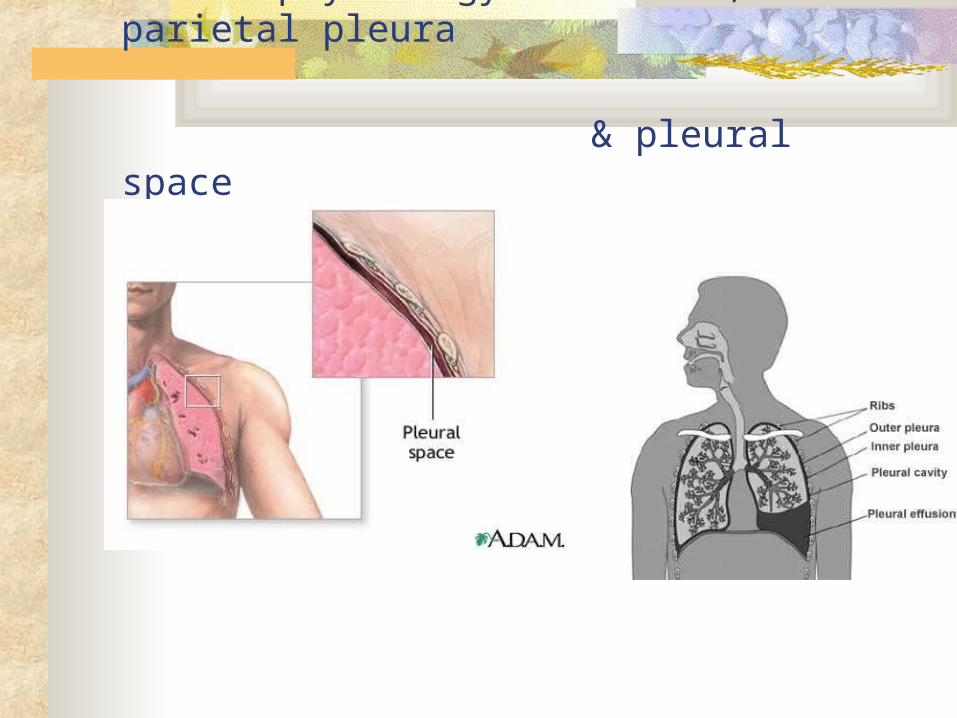
Pleural injury: Normal physiology- visceral, parietal pleura
& pleural space
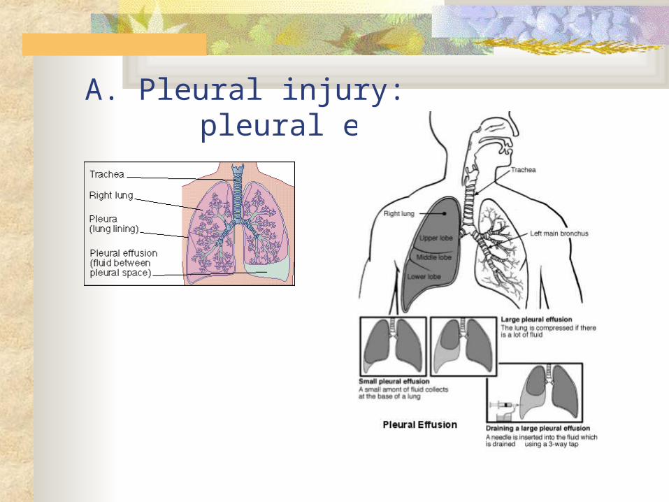
A. Pleural injury: pleural effusion
Pleural effusion
Etiology/Patho- excess fluid pleural space- may contain pus
(empyema) or blood Occurs with local disease- lung cancer, pneumonia,
trauma or systemic disease (heart failure/liver/renal disease)
Common manifestations/complications Dyspnea, pleuritic pain, dec/absent breath sounds,
limited chest wall movement
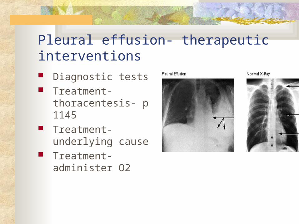
Pleural effusion- therapeutic interventions Diagnostic tests Treatment- thoracentesis-
p 1145 Treatment- underlying
cause Treatment- administer O2
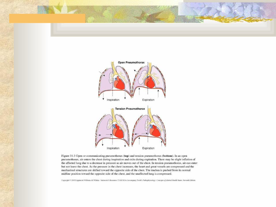
B. Pleural injury: pneumothorax
Etiology/Patho- air in pleural space- p. 1147 Spontaneous Traumatic Tension
Common manifestations/complications p. 1147 with illustrations
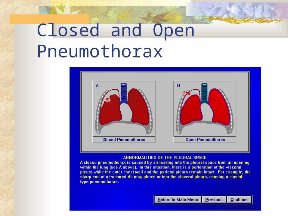
Closed and Open Pneumothorax
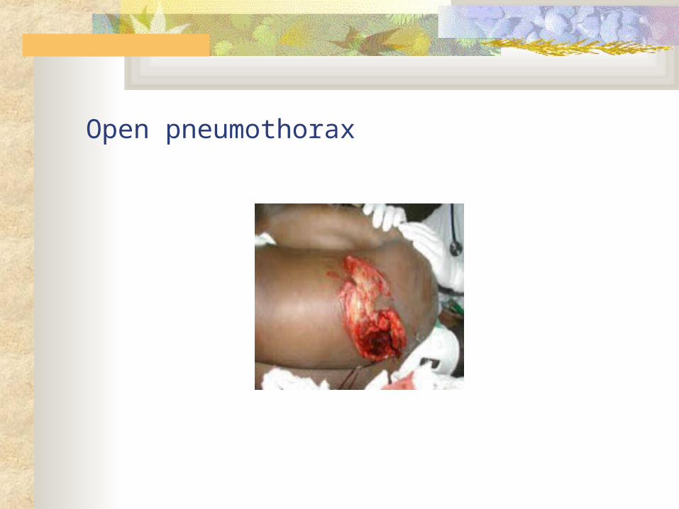
Open pneumothorax
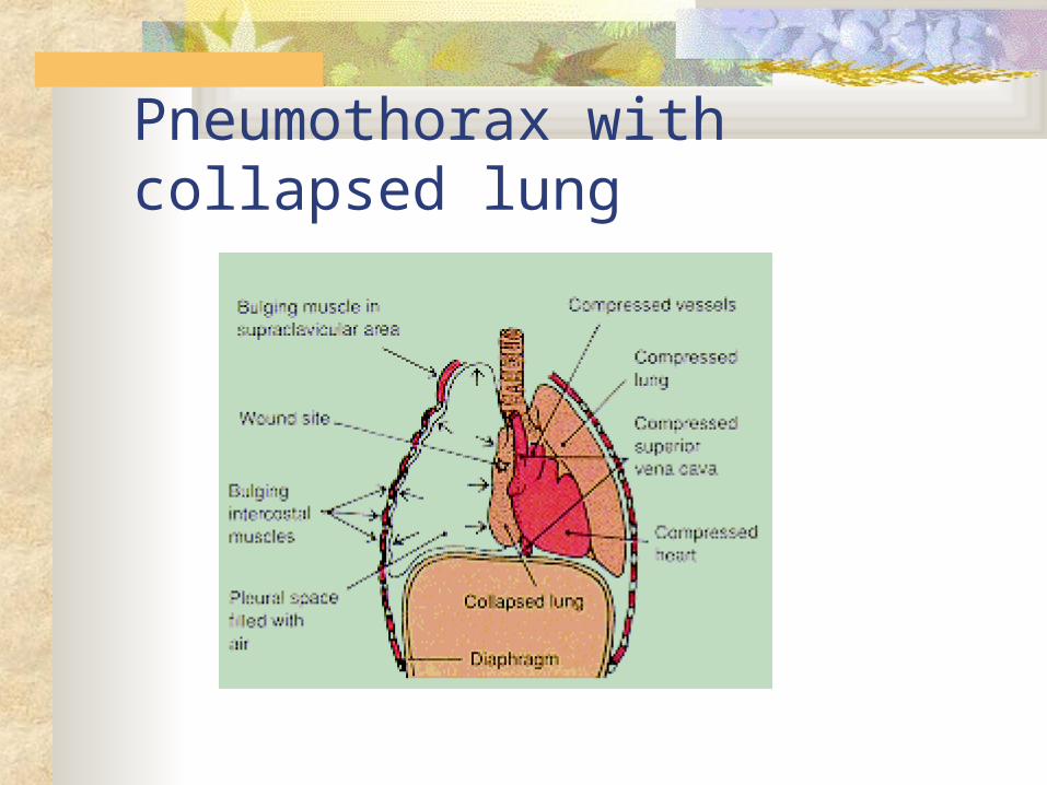
Pneumothorax with collapsed lung
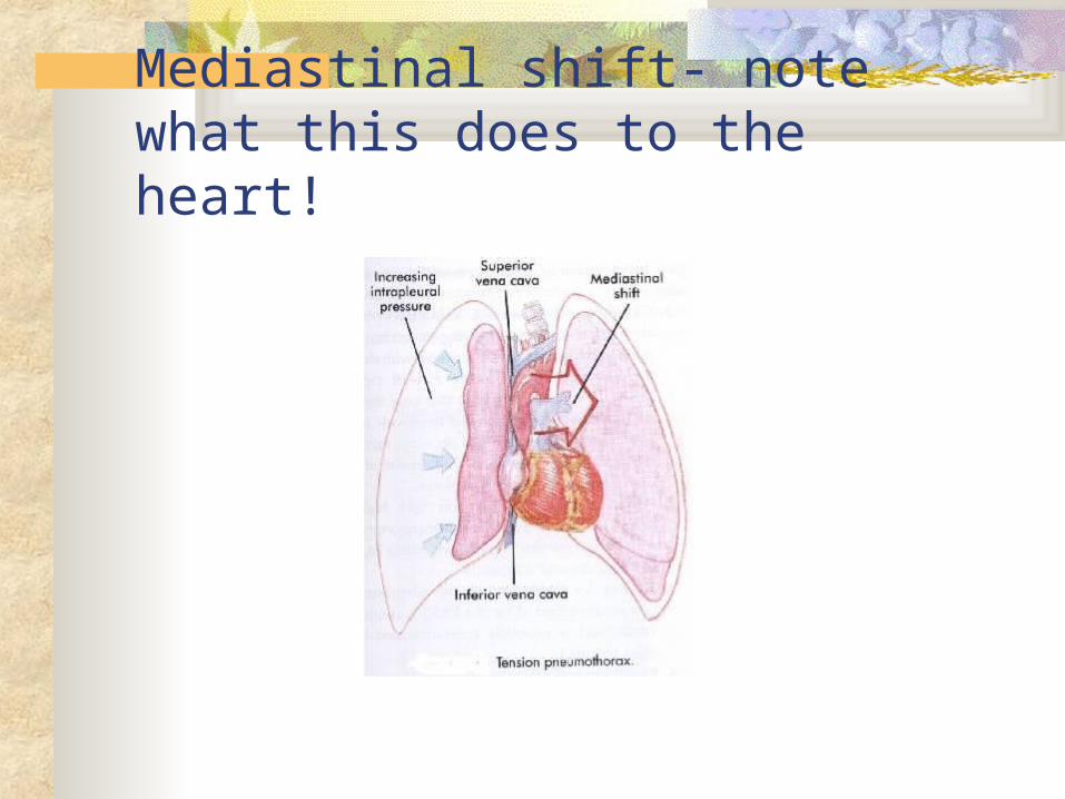
Mediastinal shift- note what this does to the heart!
Pleural injury: pneumothorax therapeutic interventions
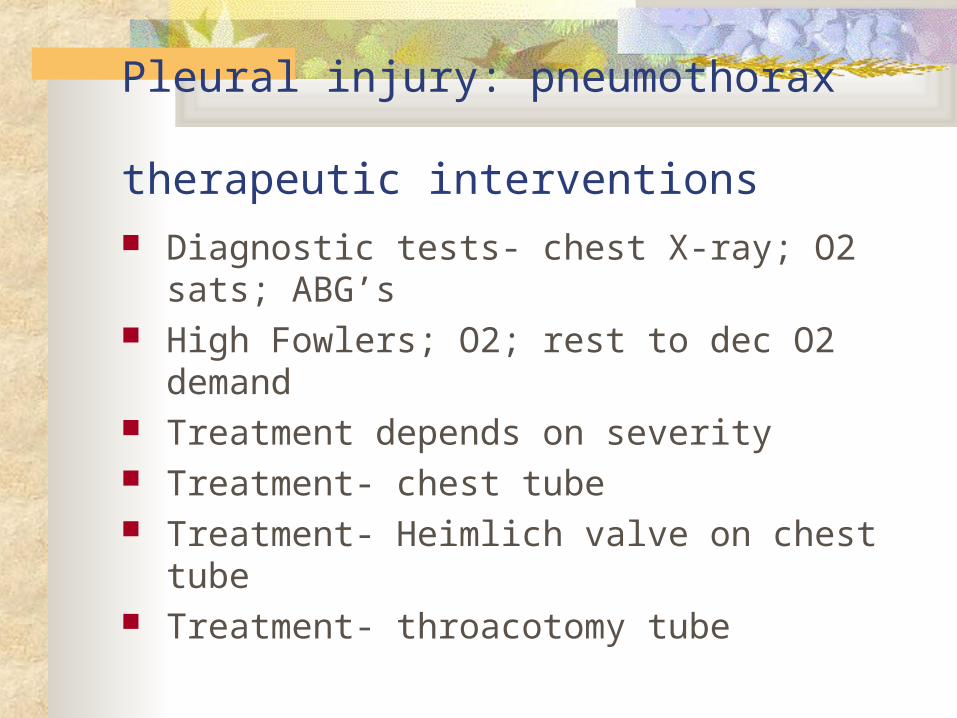
Diagnostic tests- chest X-ray; O2 sats; ABG’s High Fowlers; O2; rest to dec O2 demand Treatment depends on severity Treatment- chest tube Treatment- Heimlich valve on chest tube Treatment- throacotomy tube
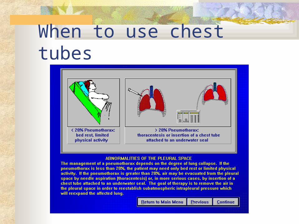
When to use chest tubes
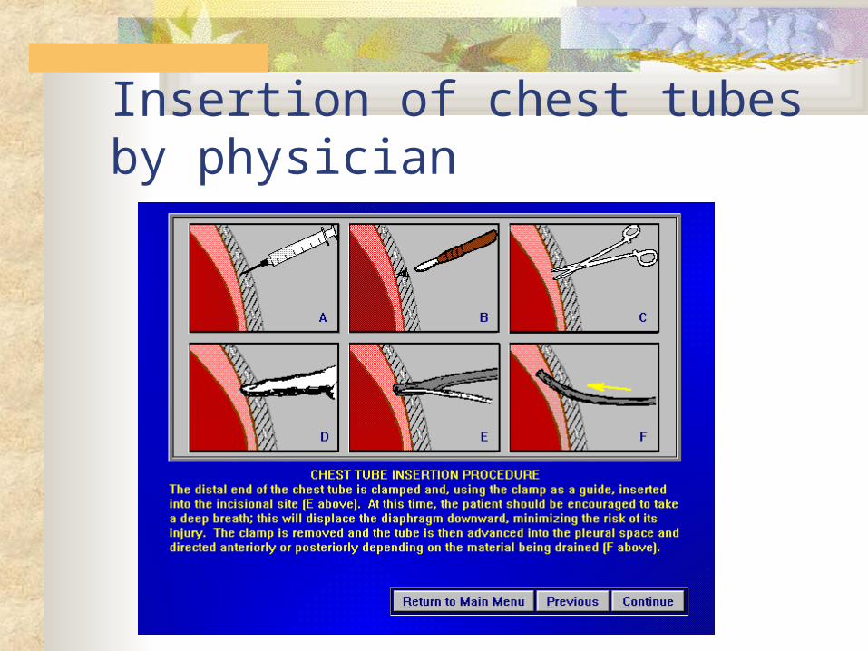
Insertion of chest tubes by physician
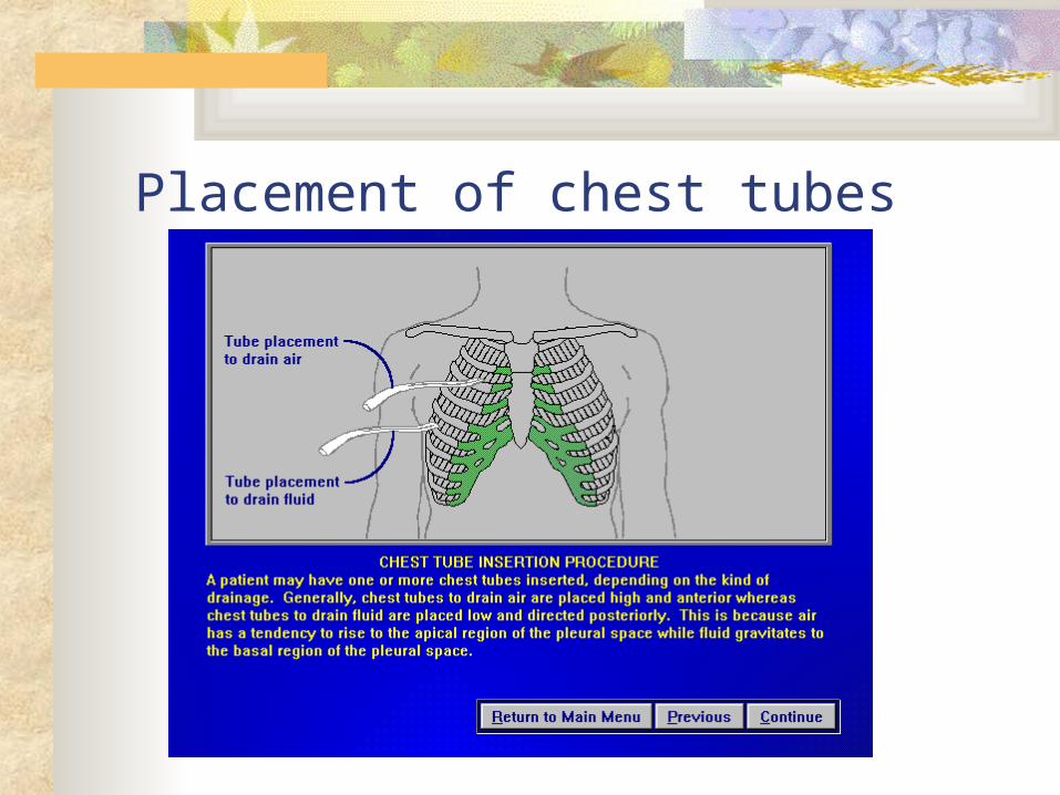
Placement of chest tubes
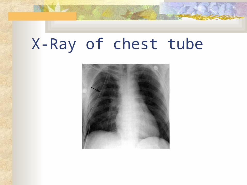
X-Ray of chest tube
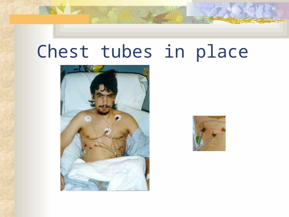
Chest tubes in place
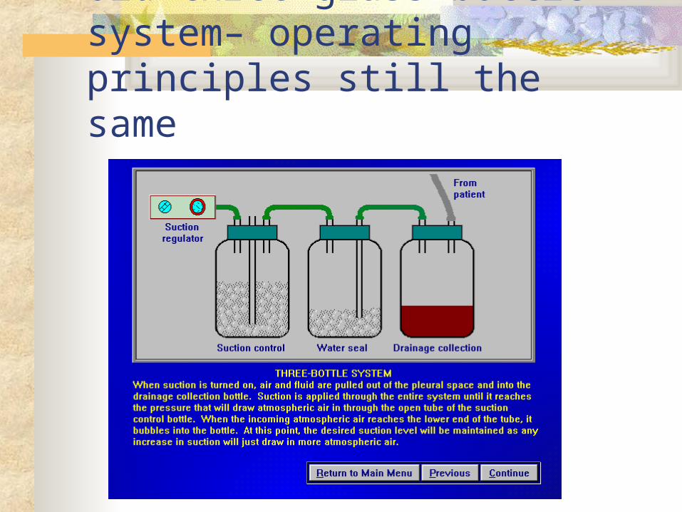
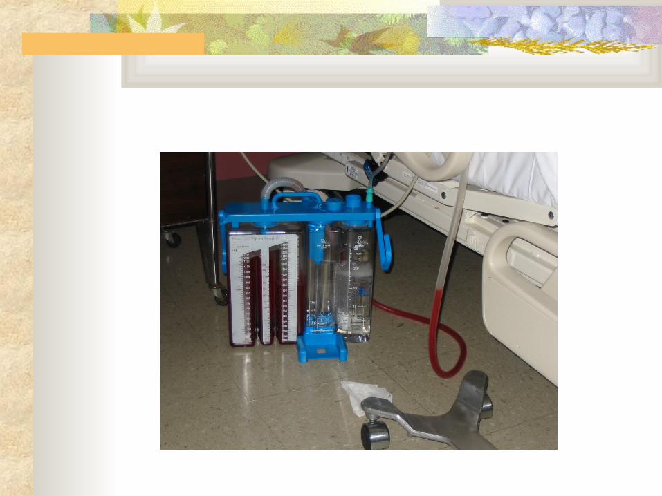
Old three glass bottle system– operating principles still the same
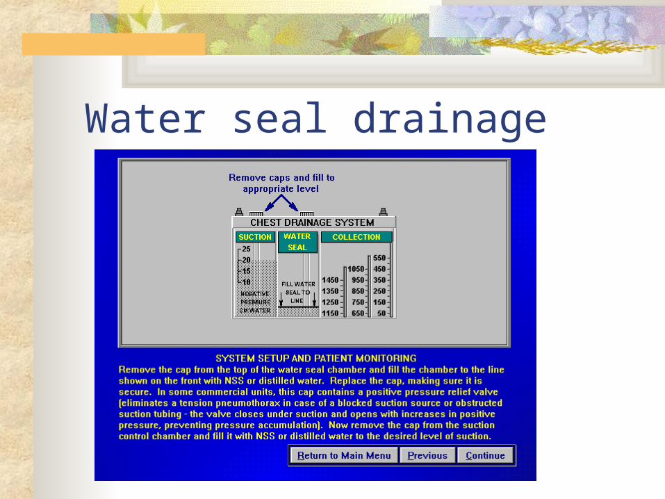
Water seal drainage
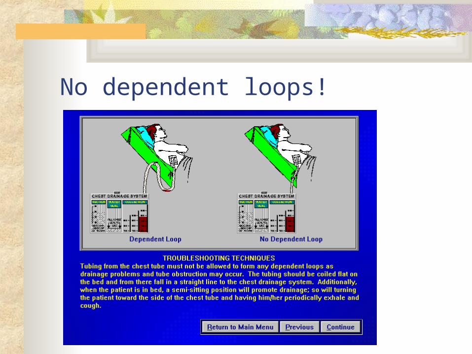
No dependent loops!
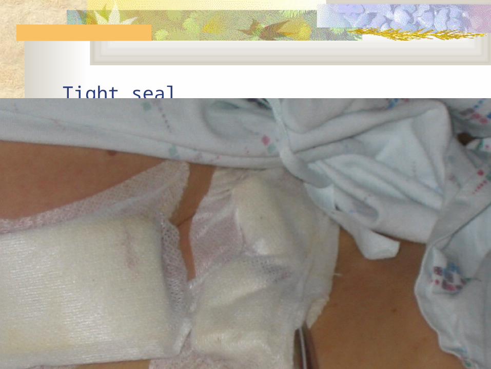
Tight seal
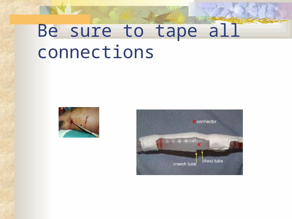
Be sure to tape all connections
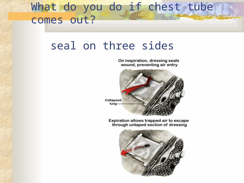
What do you do if chest tube comes out? seal on three sides
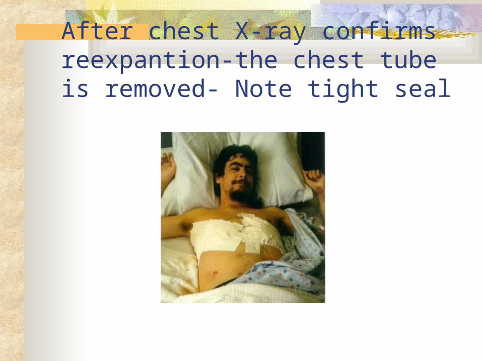
After chest X-ray confirms reexpantion-the chest tube is removed- Note tight seal
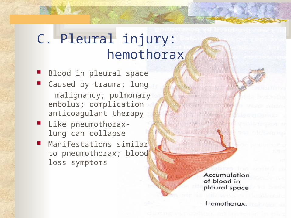
C. Pleural injury: hemothorax Blood in pleural space Caused by trauma; lung
malignancy; pulmonary embolus; complication anticoagulant therapy
Like pneumothorax- lung can collapse
Manifestations similar to pneumothorax; blood loss symptoms
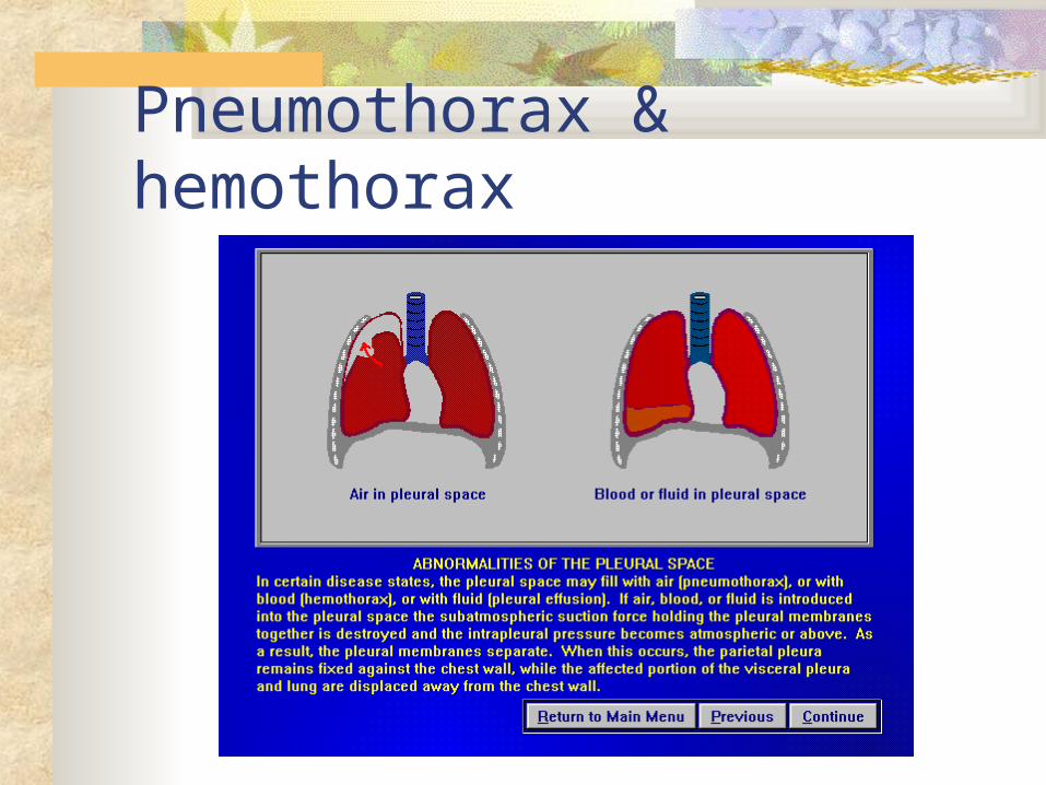
Pneumothorax & hemothorax
Pleural injury:A. pleural effusion; B. pneumothorax & C. hemothorax
Nursing assessment specific to pleural injury Health history- resp disease, injury, smoking,
progression of symptoms Physical exam- degree of apparent resp distress, lung
sounds, O2 sat, VS, LOC, neck vein distention, position of trachea
Pertinent nursing problems and interventions Impaired gas exchange Risk for injury Home care
Thoracic Injury Etiology/path
Rib fractures- most common; flail chest- 2 or more ribs fractured; pulmonary contusion- alveoli arterioles rupture
Common manifestations Rib fractures- pain on inspiration, shallow breathing Flail chest- severe dyspnea, cyanosis, tachypnea, paradoxial
chest, crepitus Pulmonary contusion- may not see 12-24 hrs post injury, inc
resp diff, restless, chest pain, coughing up sputum
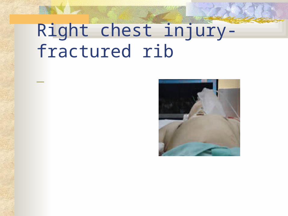
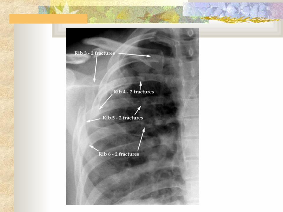
Right chest injury- fractured rib
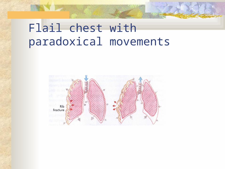
Flail chest with paradoxical movements
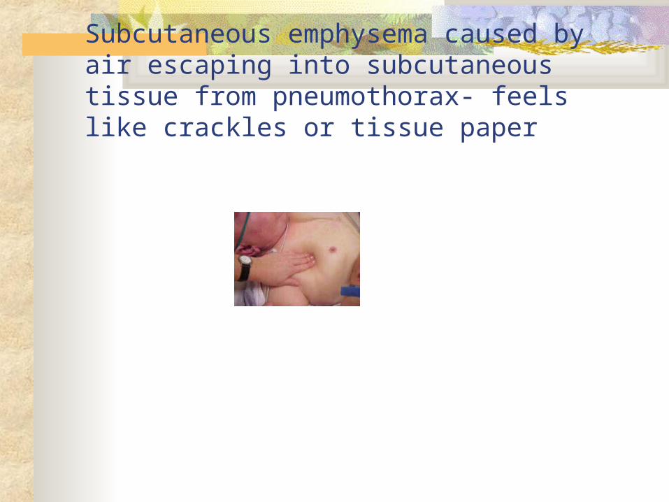
Subcutaneous emphysema caused by air escaping into subcutaneous tissue from pneumothorax- feels like crackles or tissue paper
Thoracic Injury: Therapeutic interventions Diagnostic test- all require chest X-ray; ABG’s Rib fracture- analgesics; do not restrict chest movement Flail chest-
Mild- deep breathing, pain management intercostal nerve blocks
Resp distress- intubation and mechanical ventilation- positive pressure to stabilize flail chest; external fixation
Pulmonary contusion- endotracheal tube and mechanical ventilation; bronchoscopy to remove secretions to prevent atelectasis
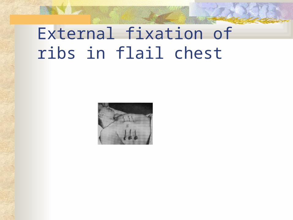
External fixation of ribs in flail chest
Pleural effusion: nursing assessment and pertinent nursing problems/interventions
Health history Physical exam All require observation for lung symptoms Pertinent nursing problems
Acute pain Ineffective airway clearance Impaired gas exchange Home care