Embed Size (px)
Citation preview

Respiratory SystemDr. Muhammad Atif Qureshi
Associate ProfessorDepartment of Medicine

• A 45 years old male presented with one year history of:
• Recurrent chest infections• Fever• Cough and expectoration

• 17 year old female presented with 3 months history of:
• Weight loss • Cough with expectoration• Fever • Night sweats

• 60 years old male• Chronic chain smoker• Weight loss• Cough • Hemoptysis

Chest Pain
1. Onset2. Severity3. Site4. Character5. Nature6. Aggravating factors7. Relieving factor8. Radiation9. Referred pain10. Associated complaints
Cough
1. Onset2. Severity3. Character4. Aggravating factors5. Relieving factor6. Associated complaints7. Hemoptysis
• Other presenting symptoms– Apnea– Hoarseness– Stridor– Snoring – Fever– Night sweating– Weight loss

General physical examination
Related to
Respiratory system

• Vital signs• General appearance• Hands
– Tremers– Nicotine stains– Clubbing– Koilonychia– Pallor– Cyanosis– Palmar erythema
• Use of accessory muscles of respiration• Lymph nodes

Hands examination
• Central Cyanosis– COPD, Asthma– Pulmonary fibrosis– Pneumonia, PE– A/V malformation– Cardiac Rt to Lt shunts
• Peripheral cyanosis– Cold weather– Low COP

Clubbing
– Bronchiectasis– Ca lung– Lung abscess– Pulmonary fibrosis– Asbestosis– Cystic fibrosis


• Hypertrophic pulmonary osteoarthropathy
– Carcinoma lung

• Tremors– Fine B2 agonist– Flapping CO2 retention

CHEST EXAMINATION:
Inspection:
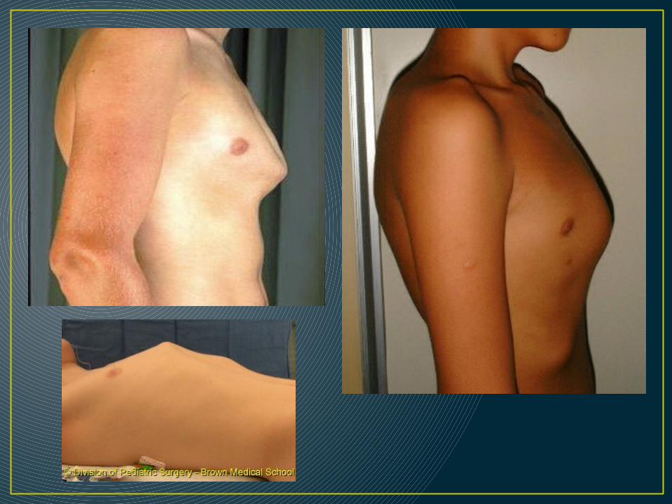
1. Shape: AP diameter compared to transverse (barrel-chest), pectus excavatum, pectus carinatum, kyphoscoliosis,…. others
2. Symmetry: assessment of upper & lower lobes should be done posteriorly looking for ↓ or delayed chest movement during moderate respirat’n.
3. Scars: from previous operat’n or chest drains or cautery marks or radiotherapy markings.
4. Prominent veins: in case of SVC obstruct’n


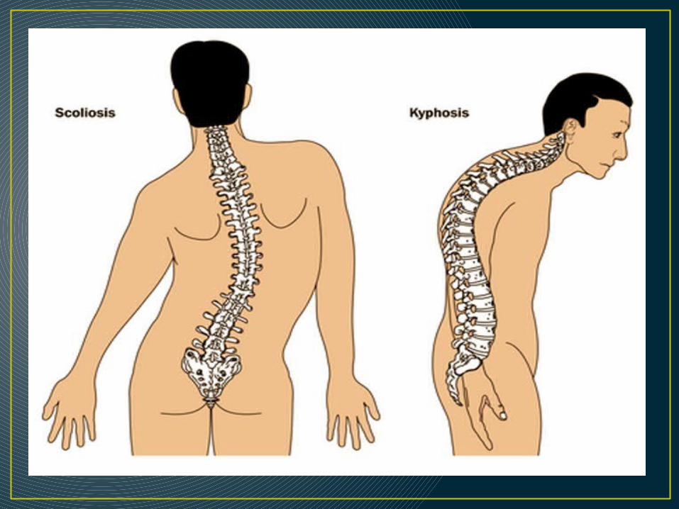


KYPHOSCOLIOSIS

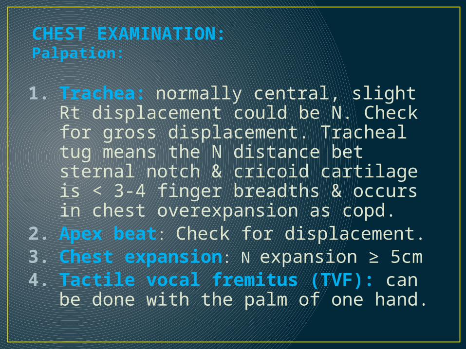
CHEST EXAMINATION:Palpation:
1. Trachea: normally central, slight Rt displacement could be N. Check for gross displacement. Tracheal tug means the N distance bet sternal notch & cricoid cartilage is < 3-4 finger breadths & occurs in chest overexpansion as copd.
2. Apex beat: Check for displacement.3. Chest expansion: N expansion ≥ 5cm4. Tactile vocal fremitus (TVF): can be done with
the palm of one hand.


TACTILE VOCAL FREMITUS
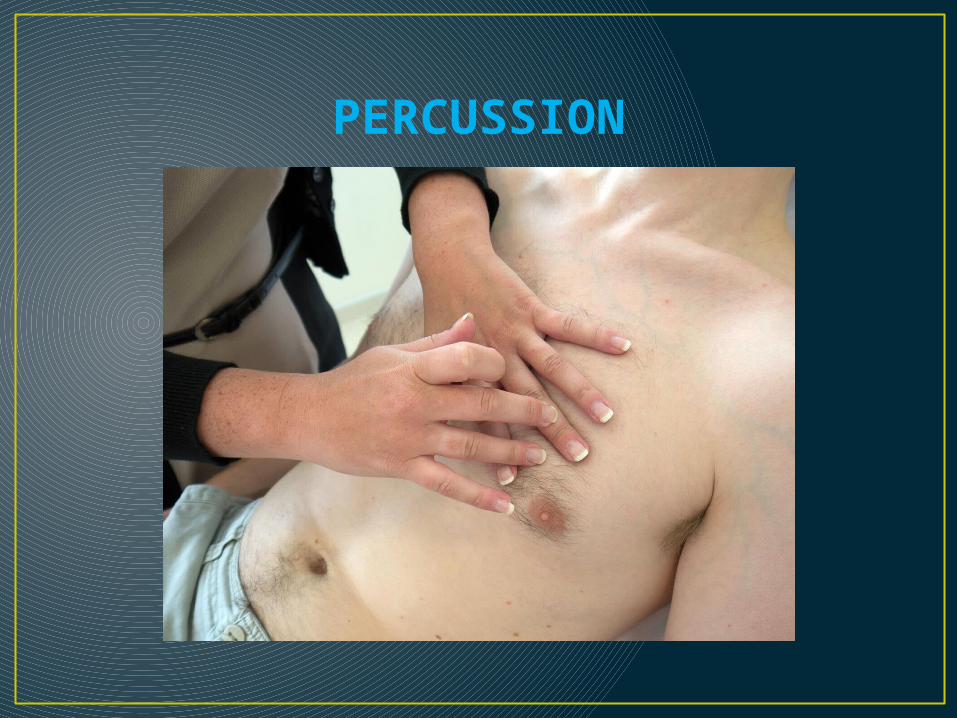
PERCUSSION

CHEST EXAMINATION:PERCUSSION:
• Should be done symmetrically (Lt compared with the Rt), posteriorly (the back), anteriorly (the front) & laterally (the sides).
• Supraclavicular area, then clavicles should be percussed directly to evaluate the upper lobes.
• Liver dullness: of the upper edge starting at the 5th rib MCL, resonant note below this area indicates hyper-inflation (copd, severe asthma)
• Cardiac dullness: may be ↓ in hyperinfated chest.
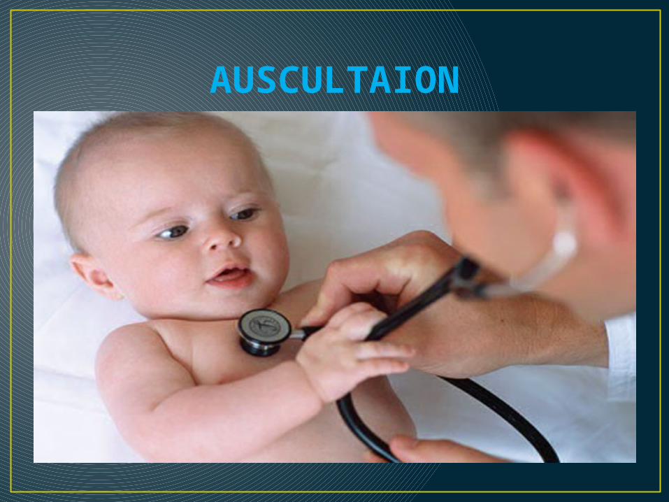
AUSCULTAION

CHEST EXAMINATION:AUSCULTATION: Using the diaphragm of a stethoscope & comment
on the following:1. Breath sounds (BS): • Intensity: N or ↓ as in (consolidation, collapse, pl effusion,
pneumothorax, lung fibrosis)• Quality: Vesicular or bronchial in consolidation• Differentiation between vesicular & bronchial BS: Vesicular: louder &longer on inspiration than expiratory phase &
has no gap between the 2 phasesBronchial: louder &longer on exp phase & has a gap between the
2 phases

ADDED SOUNDS:• Type: Wheezes or Crackles or friction rub• Timing: inspiratory or expiratory• WHEEZES: are continuous musical polyphonic sound,
heard louder on expiration & can be heard on inspiration which may imply severe AW narrowing. High pitched- wheezes are found in BA due to acute/chronic airflow limitation & low pitched in COPD. Localized monophonic wheeze due to fixed AW obstruct’n in CA bronchus.
• CRACKLES: interrupted non-musical inspiratory sound • Crackles may be early, late or pan-inspiratory & fine,
medium or coarse. Ex: late/pan-insp coarse crackles in bronchiectasis, late/pan-insp medium crackles in pul edema , late/pan-insp fine crackles in pul fibrosis

FRICTION RUB:
It’s due to thickened or roughened pleural surfaces rub together as lungs expand & contract & give off a continuous or intermittent grating sound. It indicates pleurisy & may be heard in pneumonia or pulmonary infarction.
VOCAL RESONANCE:
• It’s the ability to transmit sounds.• Ask patients to say 123 (Urdu) or 99 (English) &
listen for the transmitted sound which may be ↓ or ↑ or N (low pitched component of speech heard with booming & high pitched become attenuated).

4. EGOPHONY:
When the patient with consolidation is asked to say ‘e’ it sounds like ‘a’
5. WHISPERING PECTORILOQUY: The whispered speech is heard very loudly over the
consolidated area.

Thank You