Embed Size (px)
Citation preview
CASE REPORT Open Access
Restorative treatment in a case ofamelogenesis imperfecta and 9-year follow-up: a case reportMartin M. I. Sabandal1* , Till Dammaschke2 and Edgar Schäfer1
Abstract
Background: Amelogenesis imperfecta is a hereditary malformation showing various manifestations regardingenamel dysplasia. This case report shows a 9-year follow-up after restorative treatment of a 16-year old femalepatient affected by a hypoplastic type of amelogenesis imperfecta. The caries-free, hypersensitive teeth of the patientwere restored by direct dentin adhesive composite restorations performed in total etch technique.
Case presentation: After rehabilitation the patient reported a marked improvement of the mastication ability andquality of life especially during food intake. Accumulation of plaque was reduced and the ability to performadequate oral hygiene was improved. During follow-up of 9 years recurring secondary caries and debonding offillings were recognized and retreated.
Conclusions: The retrospective assessment exhibits that the performed restorative treatment prolonged the timeuntil further treatment has to be considered, such as prosthetic treatment.
Keywords: Amelogenesis imperfecta, Direct restoration, Follow-up, Restorative treatment
BackgroundAmelogenesis imperfecta (AI) is a genetic derived devel-opment disorder of the ameloblasts during the formationof enamel. The disease is characterized by visible malfor-mation of the enamel in a varying degree of the wholepermanent and deciduous dentition, respectively [1, 2].The prevalence of AI ranges from 1:700 to 1:14,000
[1]. Because of the various manifestations of AI Wein-mann et al. suggested in 1945 a classification [3] that hasbeen modified and adapted during the following yearsdepending on the diagnostic possibilities. A useful classi-fication was proposed by Witkop [2], which is based ona division of the manifestation of AI into 4 main groupswith further subdivisions [2].
Type I -hypoplastic.IA -hypoplastic, pitted autosomal dominant.IB -hypoplastic, local autosomal dominant.IC -hypoplastic, local autosomal recessive.ID -hypoplastic, smooth autosomal dominant.IE -hypoplastic, smooth X-linked dominant.IF -hypoplastic, rough autosomal dominant.IG -enamel agenesis, autosomal recessive.Type II -hypomaturation.IIA -hypomaturation, pigmented autosomal recessive.IIB -hypomaturation, X-linked recessive.IIC snowcapped teeth, X-linked.IID autosomal dominant?Type III -hypocalcified.IIIA -autosomal dominant.IIIB -autosomal recessive.Type IV -hypomaturation-hypoplastic with taurodontism.IVA -hypomaturation-hypoplastic with taurodontism,
autosomal dominant.
© The Author(s). 2020 Open Access This article is licensed under a Creative Commons Attribution 4.0 International License,which permits use, sharing, adaptation, distribution and reproduction in any medium or format, as long as you giveappropriate credit to the original author(s) and the source, provide a link to the Creative Commons licence, and indicate ifchanges were made. The images or other third party material in this article are included in the article's Creative Commonslicence, unless indicated otherwise in a credit line to the material. If material is not included in the article's Creative Commonslicence and your intended use is not permitted by statutory regulation or exceeds the permitted use, you will need to obtainpermission directly from the copyright holder. To view a copy of this licence, visit http://creativecommons.org/licenses/by/4.0/.The Creative Commons Public Domain Dedication waiver (http://creativecommons.org/publicdomain/zero/1.0/) applies to thedata made available in this article, unless otherwise stated in a credit line to the data.
* Correspondence: [email protected] Interdisciplinary Ambulance in the School of Dentistry, University ofMünster, Albert-Schweitzer-Campus 1, Gebäude W30, Waldeyerstr. 30, 48149Münster, GermanyFull list of author information is available at the end of the article
Sabandal et al. Head & Face Medicine (2020) 16:28 https://doi.org/10.1186/s13005-020-00243-1
IVB -hypoplastic-hypomaturation with taurodontism,autosomal dominant.
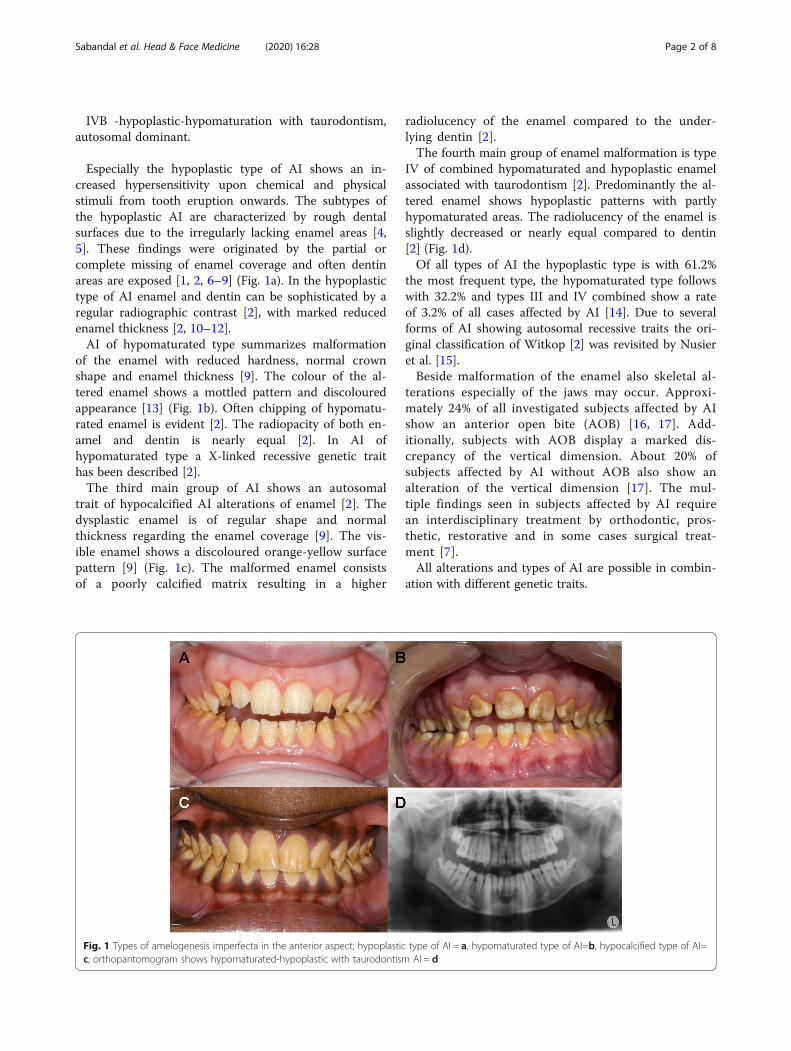
Especially the hypoplastic type of AI shows an in-creased hypersensitivity upon chemical and physicalstimuli from tooth eruption onwards. The subtypes ofthe hypoplastic AI are characterized by rough dentalsurfaces due to the irregularly lacking enamel areas [4,5]. These findings were originated by the partial orcomplete missing of enamel coverage and often dentinareas are exposed [1, 2, 6–9] (Fig. 1a). In the hypoplastictype of AI enamel and dentin can be sophisticated by aregular radiographic contrast [2], with marked reducedenamel thickness [2, 10–12].AI of hypomaturated type summarizes malformation
of the enamel with reduced hardness, normal crownshape and enamel thickness [9]. The colour of the al-tered enamel shows a mottled pattern and discolouredappearance [13] (Fig. 1b). Often chipping of hypomatu-rated enamel is evident [2]. The radiopacity of both en-amel and dentin is nearly equal [2]. In AI ofhypomaturated type a X-linked recessive genetic traithas been described [2].The third main group of AI shows an autosomal
trait of hypocalcified AI alterations of enamel [2]. Thedysplastic enamel is of regular shape and normalthickness regarding the enamel coverage [9]. The vis-ible enamel shows a discoloured orange-yellow surfacepattern [9] (Fig. 1c). The malformed enamel consistsof a poorly calcified matrix resulting in a higher
radiolucency of the enamel compared to the under-lying dentin [2].The fourth main group of enamel malformation is type
IV of combined hypomaturated and hypoplastic enamelassociated with taurodontism [2]. Predominantly the al-tered enamel shows hypoplastic patterns with partlyhypomaturated areas. The radiolucency of the enamel isslightly decreased or nearly equal compared to dentin[2] (Fig. 1d).Of all types of AI the hypoplastic type is with 61.2%
the most frequent type, the hypomaturated type followswith 32.2% and types III and IV combined show a rateof 3.2% of all cases affected by AI [14]. Due to severalforms of AI showing autosomal recessive traits the ori-ginal classification of Witkop [2] was revisited by Nusieret al. [15].Beside malformation of the enamel also skeletal al-
terations especially of the jaws may occur. Approxi-mately 24% of all investigated subjects affected by AIshow an anterior open bite (AOB) [16, 17]. Add-itionally, subjects with AOB display a marked dis-crepancy of the vertical dimension. About 20% ofsubjects affected by AI without AOB also show analteration of the vertical dimension [17]. The mul-tiple findings seen in subjects affected by AI requirean interdisciplinary treatment by orthodontic, pros-thetic, restorative and in some cases surgical treat-ment [7].All alterations and types of AI are possible in combin-
ation with different genetic traits.
Fig. 1 Types of amelogenesis imperfecta in the anterior aspect; hypoplastic type of AI = a, hypomaturated type of AI=b, hypocalcified type of AI=c; orthopantomogram shows hypomaturated-hypoplastic with taurodontism AI = d
Sabandal et al. Head & Face Medicine (2020) 16:28 Page 2 of 8
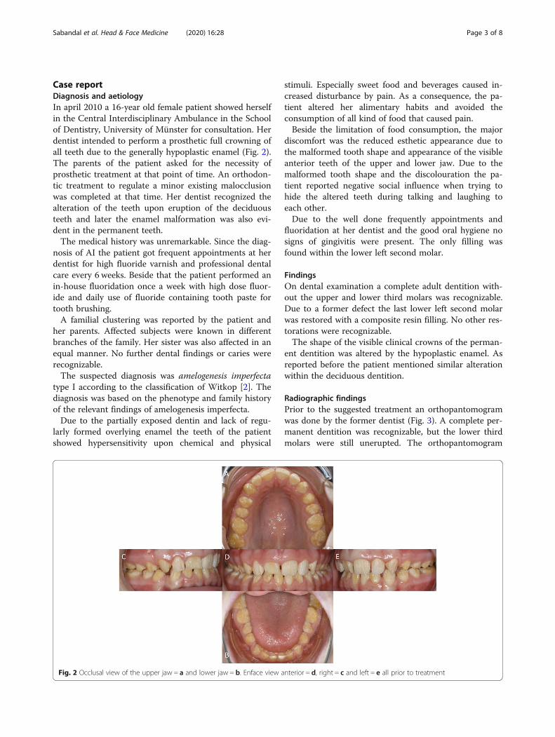
Case reportDiagnosis and aetiologyIn april 2010 a 16-year old female patient showed herselfin the Central Interdisciplinary Ambulance in the Schoolof Dentistry, University of Münster for consultation. Herdentist intended to perform a prosthetic full crowning ofall teeth due to the generally hypoplastic enamel (Fig. 2).The parents of the patient asked for the necessity ofprosthetic treatment at that point of time. An orthodon-tic treatment to regulate a minor existing malocclusionwas completed at that time. Her dentist recognized thealteration of the teeth upon eruption of the deciduousteeth and later the enamel malformation was also evi-dent in the permanent teeth.The medical history was unremarkable. Since the diag-
nosis of AI the patient got frequent appointments at herdentist for high fluoride varnish and professional dentalcare every 6 weeks. Beside that the patient performed anin-house fluoridation once a week with high dose fluor-ide and daily use of fluoride containing tooth paste fortooth brushing.A familial clustering was reported by the patient and
her parents. Affected subjects were known in differentbranches of the family. Her sister was also affected in anequal manner. No further dental findings or caries wererecognizable.The suspected diagnosis was amelogenesis imperfecta
type I according to the classification of Witkop [2]. Thediagnosis was based on the phenotype and family historyof the relevant findings of amelogenesis imperfecta.Due to the partially exposed dentin and lack of regu-
larly formed overlying enamel the teeth of the patientshowed hypersensitivity upon chemical and physical
stimuli. Especially sweet food and beverages caused in-creased disturbance by pain. As a consequence, the pa-tient altered her alimentary habits and avoided theconsumption of all kind of food that caused pain.Beside the limitation of food consumption, the major
discomfort was the reduced esthetic appearance due tothe malformed tooth shape and appearance of the visibleanterior teeth of the upper and lower jaw. Due to themalformed tooth shape and the discolouration the pa-tient reported negative social influence when trying tohide the altered teeth during talking and laughing toeach other.Due to the well done frequently appointments and
fluoridation at her dentist and the good oral hygiene nosigns of gingivitis were present. The only filling wasfound within the lower left second molar.
FindingsOn dental examination a complete adult dentition with-out the upper and lower third molars was recognizable.Due to a former defect the last lower left second molarwas restored with a composite resin filling. No other res-torations were recognizable.The shape of the visible clinical crowns of the perman-
ent dentition was altered by the hypoplastic enamel. Asreported before the patient mentioned similar alterationwithin the deciduous dentition.
Radiographic findingsPrior to the suggested treatment an orthopantomogramwas done by the former dentist (Fig. 3). A complete per-manent dentition was recognizable, but the lower thirdmolars were still unerupted. The orthopantomogram
Fig. 2 Occlusal view of the upper jaw = a and lower jaw = b. Enface view anterior = d, right = c and left = e all prior to treatment
Sabandal et al. Head & Face Medicine (2020) 16:28 Page 3 of 8
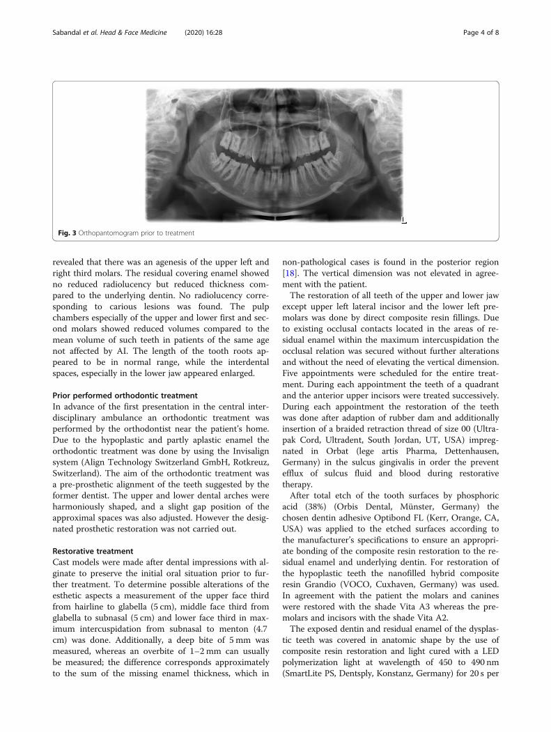
revealed that there was an agenesis of the upper left andright third molars. The residual covering enamel showedno reduced radiolucency but reduced thickness com-pared to the underlying dentin. No radiolucency corre-sponding to carious lesions was found. The pulpchambers especially of the upper and lower first and sec-ond molars showed reduced volumes compared to themean volume of such teeth in patients of the same agenot affected by AI. The length of the tooth roots ap-peared to be in normal range, while the interdentalspaces, especially in the lower jaw appeared enlarged.
Prior performed orthodontic treatmentIn advance of the first presentation in the central inter-disciplinary ambulance an orthodontic treatment wasperformed by the orthodontist near the patient’s home.Due to the hypoplastic and partly aplastic enamel theorthodontic treatment was done by using the Invisalignsystem (Align Technology Switzerland GmbH, Rotkreuz,Switzerland). The aim of the orthodontic treatment wasa pre-prosthetic alignment of the teeth suggested by theformer dentist. The upper and lower dental arches wereharmoniously shaped, and a slight gap position of theapproximal spaces was also adjusted. However the desig-nated prosthetic restoration was not carried out.
Restorative treatmentCast models were made after dental impressions with al-ginate to preserve the initial oral situation prior to fur-ther treatment. To determine possible alterations of theesthetic aspects a measurement of the upper face thirdfrom hairline to glabella (5 cm), middle face third fromglabella to subnasal (5 cm) and lower face third in max-imum intercuspidation from subnasal to menton (4.7cm) was done. Additionally, a deep bite of 5 mm wasmeasured, whereas an overbite of 1–2 mm can usuallybe measured; the difference corresponds approximatelyto the sum of the missing enamel thickness, which in
non-pathological cases is found in the posterior region[18]. The vertical dimension was not elevated in agree-ment with the patient.The restoration of all teeth of the upper and lower jaw
except upper left lateral incisor and the lower left pre-molars was done by direct composite resin fillings. Dueto existing occlusal contacts located in the areas of re-sidual enamel within the maximum intercuspidation theocclusal relation was secured without further alterationsand without the need of elevating the vertical dimension.Five appointments were scheduled for the entire treat-ment. During each appointment the teeth of a quadrantand the anterior upper incisors were treated successively.During each appointment the restoration of the teethwas done after adaption of rubber dam and additionallyinsertion of a braided retraction thread of size 00 (Ultra-pak Cord, Ultradent, South Jordan, UT, USA) impreg-nated in Orbat (lege artis Pharma, Dettenhausen,Germany) in the sulcus gingivalis in order the preventefflux of sulcus fluid and blood during restorativetherapy.After total etch of the tooth surfaces by phosphoric
acid (38%) (Orbis Dental, Münster, Germany) thechosen dentin adhesive Optibond FL (Kerr, Orange, CA,USA) was applied to the etched surfaces according tothe manufacturer’s specifications to ensure an appropri-ate bonding of the composite resin restoration to the re-sidual enamel and underlying dentin. For restoration ofthe hypoplastic teeth the nanofilled hybrid compositeresin Grandio (VOCO, Cuxhaven, Germany) was used.In agreement with the patient the molars and canineswere restored with the shade Vita A3 whereas the pre-molars and incisors with the shade Vita A2.The exposed dentin and residual enamel of the dysplas-
tic teeth was covered in anatomic shape by the use ofcomposite resin restoration and light cured with a LEDpolymerization light at wavelength of 450 to 490 nm(SmartLite PS, Dentsply, Konstanz, Germany) for 20 s per
Fig. 3 Orthopantomogram prior to treatment
Sabandal et al. Head & Face Medicine (2020) 16:28 Page 4 of 8
increment. The restoration of the occlusal, buccal and oralsurfaces was done by free-hand modelling whereas theproximal parts of the teeth were modelled with matrices(Orbis Dental, Münster, Germany) and interdental wedges(Kerr Hawe, Bioggio, Switzerland).After completion of the restorations all restored sur-
faces were contoured, the occlusion was corrected byyellow coded diamond burs (Brasseler, Lemgo,Germany) and polished with Identoflex Composite Pol-ishers and Occlubrush (both KerrHawe). The treatmentof the upper jaw required 3 appointments whereas thetreatment of the lower jaw required 2 appointments.In agreement with the patient, some teeth (upper left
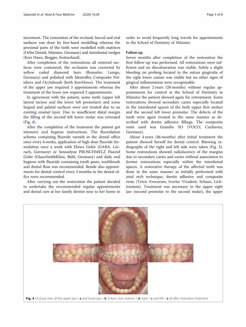
lateral incisor and the lower left premolars) and somelingual and palatal surfaces were not treated due to anexisting enamel layer. Due to insufficient distal marginthe filling of the second left lower molar was retreated(Fig. 4).After the completion of the treatment the patient got
intensive oral hygiene instructions. The fluoridationschema containing fluoride varnish in the dental officeonce every 6 weeks, application of high dose fluoride for-mulation once a week with Elmex Gelée (GABA, Lör-rach, Germany) or Sensodyne PROSCHMELZ FluoridGelée (GlaxoSmithKline, Bühl, Germany) and daily oralhygiene with fluoride containing tooth paste, toothbrushand dental floss was recommended. Beside also appoint-ments for dental control every 3 months in the dental of-fice were recommended.After carrying out the restoration the patient decided
to undertake the recommended regular appointmentsand dental care at her family dentist near to her home in
order to avoid frequently long travels for appointmentsto the School of Dentistry of Münster.
Follow-upSeven months after completion of the restoration thefirst follow-up was performed. All restorations were suf-ficient and no discolouration was visible. Solely a slightbleeding on probing located in the sulcus gingivalis ofthe right lower canine was visible but no other signs ofgingival inflammation were recognizable.After about 2 years (28 months) without regular ap-
pointments for control at the School of Dentistry inMünster the patient showed again for retreatment. Somerestorations showed secondary caries especially locatedin the interdental spaces of the both upper first molarsand the second left lower premolar. The defects of theteeth were again treated in the same manner as de-scribed with dentin adhesive fillings. The compositeresin used was Grandio SO (VOCO, Cuxhaven,Germany).About 4 years (46 months) after initial treatment the
patient showed herself for dental control. Bitewing ra-diographs of the right and left side were taken (Fig. 5).Some restorations showed radiolucency of the marginsdue to secondary caries and caries without association toformer restorations especially within the interdentalspaces. A restorative therapy of the affected teeth wasdone in the same manner as initially performed withtotal etch technique, dentin adhesive and compositeresin (Tetric Evoceram, Ivoclar Vivadent, Schaan, Lich-tenstein). Treatment was necessary to the upper rightjaw (second premolar to the second molar), the upper
Fig. 4 Occlusal view of the upper jaw = a and lower jaw = b. Enface view anterior = d, right = c and left = e all after restorative treatment
Sabandal et al. Head & Face Medicine (2020) 16:28 Page 5 of 8
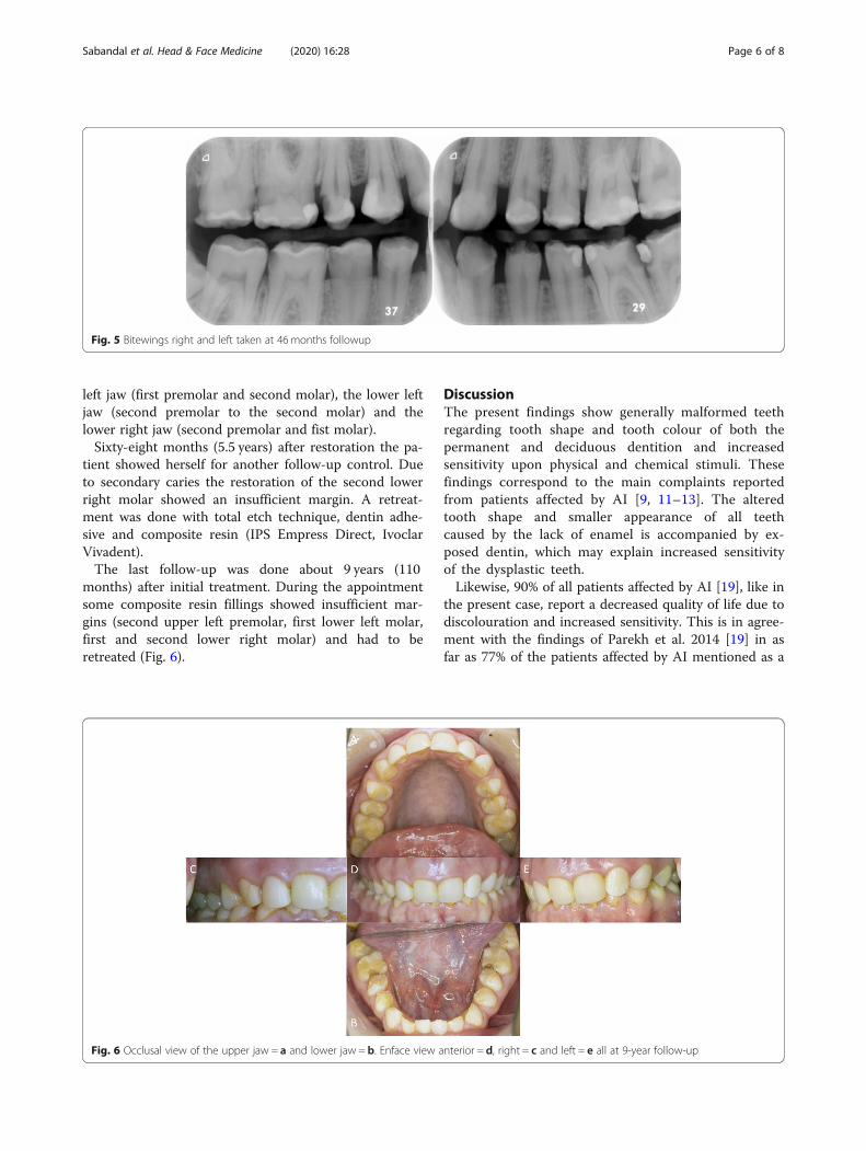
left jaw (first premolar and second molar), the lower leftjaw (second premolar to the second molar) and thelower right jaw (second premolar and fist molar).Sixty-eight months (5.5 years) after restoration the pa-
tient showed herself for another follow-up control. Dueto secondary caries the restoration of the second lowerright molar showed an insufficient margin. A retreat-ment was done with total etch technique, dentin adhe-sive and composite resin (IPS Empress Direct, IvoclarVivadent).The last follow-up was done about 9 years (110
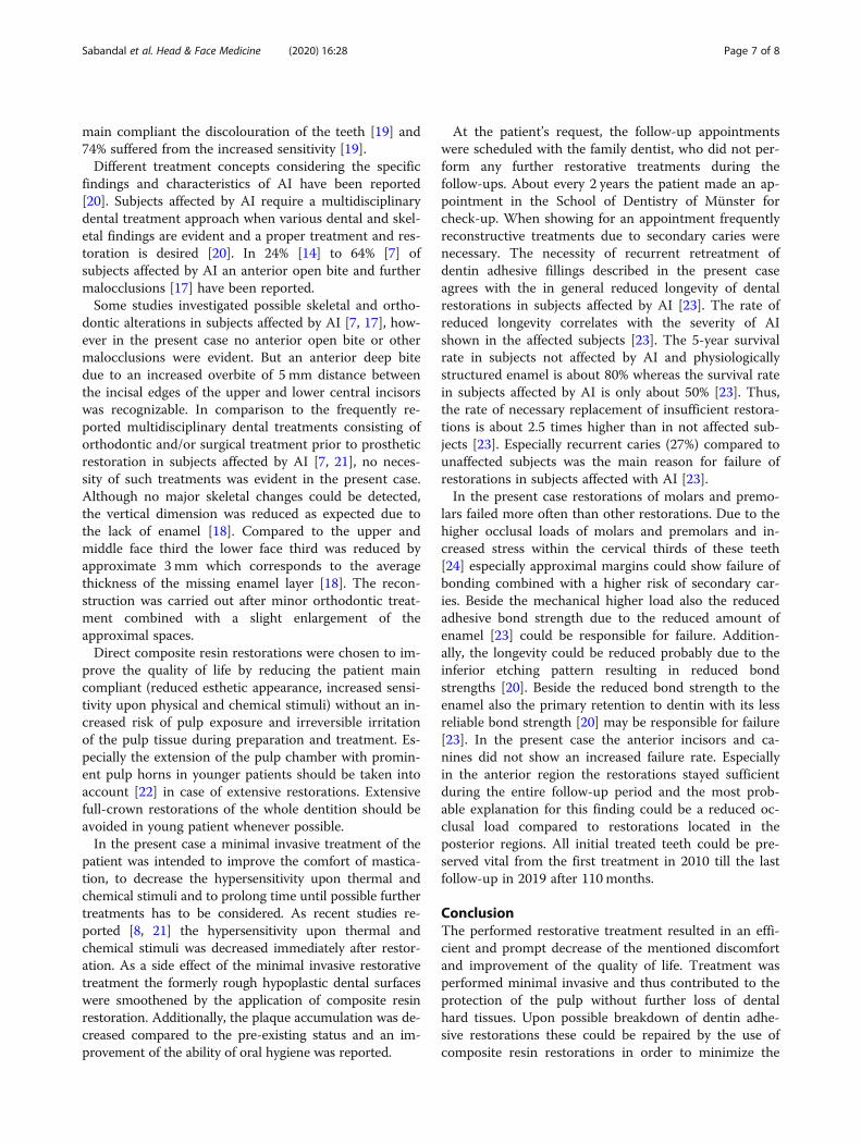
months) after initial treatment. During the appointmentsome composite resin fillings showed insufficient mar-gins (second upper left premolar, first lower left molar,first and second lower right molar) and had to beretreated (Fig. 6).
DiscussionThe present findings show generally malformed teethregarding tooth shape and tooth colour of both thepermanent and deciduous dentition and increasedsensitivity upon physical and chemical stimuli. Thesefindings correspond to the main complaints reportedfrom patients affected by AI [9, 11–13]. The alteredtooth shape and smaller appearance of all teethcaused by the lack of enamel is accompanied by ex-posed dentin, which may explain increased sensitivityof the dysplastic teeth.Likewise, 90% of all patients affected by AI [19], like in
the present case, report a decreased quality of life due todiscolouration and increased sensitivity. This is in agree-ment with the findings of Parekh et al. 2014 [19] in asfar as 77% of the patients affected by AI mentioned as a
Fig. 5 Bitewings right and left taken at 46 months followup
Fig. 6 Occlusal view of the upper jaw = a and lower jaw = b. Enface view anterior = d, right = c and left = e all at 9-year follow-up
Sabandal et al. Head & Face Medicine (2020) 16:28 Page 6 of 8
main compliant the discolouration of the teeth [19] and74% suffered from the increased sensitivity [19].Different treatment concepts considering the specific
findings and characteristics of AI have been reported[20]. Subjects affected by AI require a multidisciplinarydental treatment approach when various dental and skel-etal findings are evident and a proper treatment and res-toration is desired [20]. In 24% [14] to 64% [7] ofsubjects affected by AI an anterior open bite and furthermalocclusions [17] have been reported.Some studies investigated possible skeletal and ortho-
dontic alterations in subjects affected by AI [7, 17], how-ever in the present case no anterior open bite or othermalocclusions were evident. But an anterior deep bitedue to an increased overbite of 5 mm distance betweenthe incisal edges of the upper and lower central incisorswas recognizable. In comparison to the frequently re-ported multidisciplinary dental treatments consisting oforthodontic and/or surgical treatment prior to prostheticrestoration in subjects affected by AI [7, 21], no neces-sity of such treatments was evident in the present case.Although no major skeletal changes could be detected,the vertical dimension was reduced as expected due tothe lack of enamel [18]. Compared to the upper andmiddle face third the lower face third was reduced byapproximate 3 mm which corresponds to the averagethickness of the missing enamel layer [18]. The recon-struction was carried out after minor orthodontic treat-ment combined with a slight enlargement of theapproximal spaces.Direct composite resin restorations were chosen to im-
prove the quality of life by reducing the patient maincompliant (reduced esthetic appearance, increased sensi-tivity upon physical and chemical stimuli) without an in-creased risk of pulp exposure and irreversible irritationof the pulp tissue during preparation and treatment. Es-pecially the extension of the pulp chamber with promin-ent pulp horns in younger patients should be taken intoaccount [22] in case of extensive restorations. Extensivefull-crown restorations of the whole dentition should beavoided in young patient whenever possible.In the present case a minimal invasive treatment of the
patient was intended to improve the comfort of mastica-tion, to decrease the hypersensitivity upon thermal andchemical stimuli and to prolong time until possible furthertreatments has to be considered. As recent studies re-ported [8, 21] the hypersensitivity upon thermal andchemical stimuli was decreased immediately after restor-ation. As a side effect of the minimal invasive restorativetreatment the formerly rough hypoplastic dental surfaceswere smoothened by the application of composite resinrestoration. Additionally, the plaque accumulation was de-creased compared to the pre-existing status and an im-provement of the ability of oral hygiene was reported.
At the patient’s request, the follow-up appointmentswere scheduled with the family dentist, who did not per-form any further restorative treatments during thefollow-ups. About every 2 years the patient made an ap-pointment in the School of Dentistry of Münster forcheck-up. When showing for an appointment frequentlyreconstructive treatments due to secondary caries werenecessary. The necessity of recurrent retreatment ofdentin adhesive fillings described in the present caseagrees with the in general reduced longevity of dentalrestorations in subjects affected by AI [23]. The rate ofreduced longevity correlates with the severity of AIshown in the affected subjects [23]. The 5-year survivalrate in subjects not affected by AI and physiologicallystructured enamel is about 80% whereas the survival ratein subjects affected by AI is only about 50% [23]. Thus,the rate of necessary replacement of insufficient restora-tions is about 2.5 times higher than in not affected sub-jects [23]. Especially recurrent caries (27%) compared tounaffected subjects was the main reason for failure ofrestorations in subjects affected with AI [23].In the present case restorations of molars and premo-
lars failed more often than other restorations. Due to thehigher occlusal loads of molars and premolars and in-creased stress within the cervical thirds of these teeth[24] especially approximal margins could show failure ofbonding combined with a higher risk of secondary car-ies. Beside the mechanical higher load also the reducedadhesive bond strength due to the reduced amount ofenamel [23] could be responsible for failure. Addition-ally, the longevity could be reduced probably due to theinferior etching pattern resulting in reduced bondstrengths [20]. Beside the reduced bond strength to theenamel also the primary retention to dentin with its lessreliable bond strength [20] may be responsible for failure[23]. In the present case the anterior incisors and ca-nines did not show an increased failure rate. Especiallyin the anterior region the restorations stayed sufficientduring the entire follow-up period and the most prob-able explanation for this finding could be a reduced oc-clusal load compared to restorations located in theposterior regions. All initial treated teeth could be pre-served vital from the first treatment in 2010 till the lastfollow-up in 2019 after 110 months.
ConclusionThe performed restorative treatment resulted in an effi-cient and prompt decrease of the mentioned discomfortand improvement of the quality of life. Treatment wasperformed minimal invasive and thus contributed to theprotection of the pulp without further loss of dentalhard tissues. Upon possible breakdown of dentin adhe-sive restorations these could be repaired by the use ofcomposite resin restorations in order to minimize the
Sabandal et al. Head & Face Medicine (2020) 16:28 Page 7 of 8
risk of pulp exposure upon extensive treatments. Due tothe combination of minimal invasive treatment approachwith the possibility of repair of composite resin restora-tions pulp vitality was maintained over the entire obser-vation period and the necessity of more invasivetreatment options could be prolonged for nearly 10years.
AbbreviationsAI: Amelogenesis imperfecta; AOB: Anterior open bite
AcknowledgementsThis research did not receive any specific grant from funding agencies in thepublic, commercial, or not-for-profit sectors.
Authors’ contributionsConceptualization: MMI.S., E.S.; Methodology: MMI.S., T.D.; Writing-OriginalDraft Preparation: MMI.S.; Writing-Review & Editing: T.D., E.S.; All authors haveapproved the final version of the manuscript.
FundingThis research received no external funding. Open Access funding enabledand organized by Projekt DEAL.
Availability of data and materialsNot applicable.
Ethics approval and consent to participateNot applicable.
Consent for publicationNot applicable.
Competing interestsThe authors declare that they have no competing interests.
Author details1Central Interdisciplinary Ambulance in the School of Dentistry, University ofMünster, Albert-Schweitzer-Campus 1, Gebäude W30, Waldeyerstr. 30, 48149Münster, Germany. 2Department of Periodontology and Operative Dentistry,University of Münster, Münster, Germany.
Received: 22 April 2020 Accepted: 9 November 2020
References1. Crawford PJM, Aldred M, Bloch-Zupan A. Amelogenesis imperfecta.
Orphanet J Rare Dis. 2007;2:17.2. Witkop CJ Jr. Amelogenesis imperfecta, dentinogenesis imperfecta and
dentin dysplasia revisited: problems in classification. J Oral Pathol. 1988;17:547–53.
3. Weinmann JP, Svoboda JF, Woods RW. Hereditary disturbances of enamelformation and calcification. J Am Dent Assoc. 1945;32:397–418.
4. Kumar S, Gupta S. The restoration of function and esthetics of a patientwith amelogenesis imperfecta using a combination of orthodontic andprosthodontic treatment: a case report. J Contemp Dent Pract. 2009;10:E079–85.
5. Malone W, Bazola FN. Early treatment of amelogenesis imperfecta. JProsthet Dent. 1966;16:540–4.
6. Canger EM, Celenk P, Yenisey M, Odyakmaz SZ. Amelogenesis Imperfecta,hypoplastic type associated with some dental abnormalities: a case report.Braz Dent J. 2010;21:170–4.
7. Gisler V, Enkling N, Zix J, Kim K, Kellerhoff NM, Mericske-Stern R. Amultidisciplinary approach to the functional and esthetic rehabilitation ofamelogenesis imperfecta and open bite deformity: a case report. J EsthetRestor Dent. 2010;22:282–93.
8. Sabatini C, Guzmán-Armstrong S. A conservative treatment foramelogenesis imperfecta with direct resin composite restorations: a casereport. J Esthet Restor Dent. 2009;21:161–9.
9. Seow WK. Developmental defects of enamel and dentine: challenges forbasic science research and clinical management. Aust Dent J. 2014;59(Suppl1):143–54.
10. Pavlič A, Battelino T, Podkrajšek KT, Ovsenik M. Craniofacial characteristicsand genotypes of amelogenesis imperfecta patients. Eur J Orthod. 2011;33:325–31.
11. Hunter L, Stone D. Supraoccluding cobalt-chrome onlays in themanagement of amelogenesis imperfecta in children: a 2-year case report.Quintessence Int. 1997;28:15–9.
12. Chan KHC, Ho EHT, Botelho MG, Pow EHN. Rehabilitation of amelogenesisimperfecta using a reorganized approach: a case report. Quintessence Int.2011;42:385–91.
13. Horowitz RA, Gautam DK, Karol S, Kumari B. Periodontal management andrestoration of an amelogenesis imperfecta patient: a case report. CompendContin Educ Dent. 2014;35:e6–11.
14. Koruyucu M, Bayram M, Tuna EB, Gencay K, Seymen F. Clinical findings andlong-term managements of patients with amelogenesis imperfecta. Eur JDent. 2014;8:546–52.
15. Nusier M, Yassin O, Hart TC, Samimi A, Wright JT. Phenotypic diversity andrevision of the nomenclature for autosomal recessive amelogenesisimperfecta. Oral Surg Oral Med Oral Pathol Oral Radiol Endod. 2004;97:220–30.
16. Robinson FG, Haubenreich JE. Oral rehabilitation of a young adult withhypoplastic amelogenesis imperfecta: a clinical report. J Prosthet Dent. 2006;95:10–3.
17. Rowley R, Hill FJ, Winter GB. An investigation of the association betweenanterior open-bite and amelogenesis imperfecta. Am J Orthod. 1982;81:229–35.
18. Gillings B, Buonocore M. Thickness of enamel at the base of pits andfissures in human molars and bicuspids. J Dent Res. 1961;40:119–33.
19. Parekh S, Almehateb M, Cunningham SJ. How do children withamelogenesis imperfecta feel about their teeth? Int J Paediatr Dent. 2014;24:326–35.
20. Sabandal MMI, Schäfer E. Amelogenesis imperfecta: review of diagnosticfindings and treatment concepts. Odontology. 2016;104:245–56.
21. Akin H, Tasveren S, Yeler DY. Interdisciplinary approach to treating a patientwith amelogenesis imperfecta: a clinical report. J Esthet Restor Dent. 2007;19:131–5.
22. Turkün LS. Conservative restoration with resin composites of a case ofamelogenesis imperfecta. Int Dent J. 2005;55:38–41.
23. Lundgren GP, Dahllöf G. Outcome of restorative treatment in youngpatients with amelogenesis imperfecta. A cross-sectional, retrospectivestudy. J Dent. 2014;42:1382–9.
24. Musani I, Prabhakar AR. Biomechanical stress analysis of mandibular firstpermanent molar; restored with amalgam and composite resin: acomputerized finite element study. Int J Clin Pediatr Dent. 2010;3:5–14.
Publisher’s NoteSpringer Nature remains neutral with regard to jurisdictional claims inpublished maps and institutional affiliations.
Sabandal et al. Head & Face Medicine (2020) 16:28 Page 8 of 8